TECHNICAL REPORT: UPDATE ON LYME DISEASE PREVENTION AND CONTROL - DOUG SIDER MD MSC FRCPC SAMIR PATEL PHD FCCM CURTIS RUSSELL PHD NINA ...
←
→
Page content transcription
If your browser does not render page correctly, please read the page content below
Technical Report:
Update on Lyme Disease
Prevention and Control

Doug Sider MD MSc FRCPC
Samir Patel PhD FCCM
Curtis Russell PhD
Nina Jain-Sheehan BSc
Stephen Moore MPH

February 2012

Introduction                                                                    1
Tick Surveillance                                                               2
Human Surveillance                                                              6
Clinical Signs and Symptoms                                                     6
Diagnosis and Testing                                                           8
Testing Algorithm                                                               9
Controversies in Laboratory Testing                                             10
Treatment Issues                                                                11
Moving Forward                                                                  15
References                                                                      16
Appendix A: Provincial Case Definitions for Reportable Diseases: Lyme Disease   18

Introduction
This technical report was created for the field, further to a number of requests from Ontario public health
units and the Office of the Chief Medical Officer of Health. Public Health Ontario staff within the Infectious
Diseases Prevention and Control team and the Public Health Laboratory developed the technical report to
address issues raised and summarize the available science and evidence. Peer review by external
stakeholders from the Public Health Agency of Canada’s National Microbiology Laboratory and selected public
health units led to a first full draft. In October 2011, this initial draft was sent out for open consultation.
Comments were closely reviewed and considered in the development of this report.

Lyme disease (LD) is a spirochete infection caused by species within the Borrelia burgdorferi complex which is
transmitted to humans through the bite of an infected tick. In Ontario, the tick, Ixodes scapularis or
blacklegged tick (sometimes called the deer tick) is the primary vector of LD. LD is the most common vector-
borne disease in North America.1,2 LD was first recognized as a distinct disease in the late 1970s.3 It became a
reportable disease in Ontario in 1988 and a nationally notifiable disease in 2010. The distribution of
populations of infected blacklegged ticks is limited to defined geographical locations in Ontario and elsewhere
in Canada, although isolated cases of LD can occur outside of these tick endemic areas. Since 1988, the
majority of Ontario-acquired human cases originate from the southern parts of the province. Generally, this
area has more favourable climatic conditions for the tick vector. Recent studies have reported that established
populations of blacklegged ticks are increasing in Canada, likely aided by the effects of climate change and
other factors.4 In the 1990s, only one region in Ontario was home to one endemic area. By 2009, six additional
sites in the southern parts of the province had been identified through research studies and public health
surveillance.

A number of factors may be responsible for the relatively recent increase in the range of blacklegged ticks in
Ontario including: natural range expansion aided in part by climate warming, lengthening summer and fall
seasons, and possible changes in the range of key hosts for ticks, such as the white-tailed deer. Most of the
recently established tick populations are in the southern Ontario and areas further north may lack suitable
climate conditions, habitat and/or hosts to support tick establishment. Similarly these more northern areas
may not be “seeded” with as many bird-borne ticks thus lowering the probability of eventual tick
establishment. All tick surveillance indicators suggest that the current range of blacklegged tick populations is
expanding and will likely continue to do so in the future.

Blacklegged ticks occur sporadically over a wide geographic range in Canada and this is because larvae and
nymphs of the blacklegged tick readily attach to migratory birds. Thus birds serve to transport blacklegged
ticks from endemic areas in the United States and Canada to widely separated localities across Canada. As a
result, small numbers of “bird-borne” ticks are found throughout the Ontario; however, only certain areas in
the province have the appropriate climate, habitat and available hosts to support establishment of new
blacklegged tick populations. Bird-borne ticks create the theoretical possibility of persons being bitten by an
infected tick almost anywhere in Ontario. Human cases of LD have been reported in Ontario from outside of
tick endemic areas but the risk of exposure is much less than in endemic areas. Risk of LD is usually much
greater in tick endemic areas because the probability of bites from infected ticks (including nymphs) are much
greater and the infection rate in host-seeking ticks is typically much higher than in non-endemic localities.

PHO Technical Report: Update on Lyme Disease Prevention and Control February 2012                          1
Tick Surveillance
Tick surveillance can determine the level of establishment of blacklegged tick populations within an area and
assess the possible risk of LD infection to humans. Tick surveillance may be active (collecting ticks from their
natural habitat), or it may be passive (examining ticks brought into health unit offices by members of the
public). While quite different in their methods, both are useful in determining the level of community risk
from LD.
The established populations of vector ticks and the prevalence of infection with B. burgdorferi over two
consecutive seasons determine whether a location is endemic for LD. This provides an evidence-based
estimate of the risk of human exposure to infected ticks in a given jurisdiction. There are three categories used
to describe the presence/absence of blacklegged tick populations: established, adventitious or not present.5
Established - populations of blacklegged ticks must exhibit all three active stages (larva, nymph, adult) in a
contiguous sampling area on resident animals, or in the environment, for at least two years.
Adventitious - ticks are found only sporadically, both temporally and spatially, and usually only a single stage
of tick (e.g., adult females) is present.
Not Present - ticks have not been found in an area after studies have been conducted to assess the level of
establishment.
See Figure 1 for a decision tree on determining if an area is endemic for LD. For an area to be considered
endemic for LD the following must exist:
1. there is an established blacklegged tick population
2. this tick population and host mammals have tested positive for B. burgdorferi the agent of LD over two
consecutive years.
In Ontario, blacklegged ticks are more commonly found in rural areas along the north shores of Lake Erie, Lake
Ontario, and the St. Lawrence River. These areas have the most suitable habitat and climate for tick
populations to become established. The endemic areas for LD are Long Point Provincial Park, Turkey Point
Provincial Park, Rondeau Provincial Park, Point Pelee National Park, Prince Edward Point National Wildlife
Area, Wainfleet Bog Conservation Area, and the St. Lawrence Islands National Park. The precise boundaries of
these established tick populations are difficult to define and some of these populations continue to expand
into neighbouring areas. The risk of LD increases in areas where infected blacklegged tick populations are
established because contact with infected ticks (especially nymphs) is greater in these areas compared to
areas where blacklegged ticks are not established.
The objective of passive tick surveillance is to understand the risk of LD infection. The information can also
help identify new areas where active surveillance for tick populations would be required. Passive surveillance
should guide active surveillance. An important aspect of passive surveillance is documenting location to
identify specific areas having multiple tick submissions over multiple years. Finding multiple ticks from a single
location may be indicative of an established or establishing population and should be monitored. It should be
noted that passive ticks submitted by the public and/or physicians is to assist in determining areas of risk, and
not for the purpose of providing information for a physician’s diagnosis of a patient. Active surveillance, in the
form of drag sampling, should also be considered. It is important to note that American dog ticks
(Dermacentor variabilis) and groundhog ticks (Ixodes cookeii) frequently bite people in Ontario but these tick
species are not competent vectors of LD.
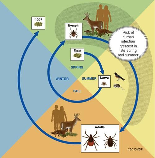
PHO Technical Report: Update on Lyme Disease Prevention and Control February 2012 2The most widely used methods for sampling ticks as part of active surveillance are i) drag sampling and ii) capture and examination of small mammals.i Drag sampling is the single most reliable method for quantitatively sampling immature populations of blacklegged ticks, however, drag sampling may be less sensitive than capture and examination of hosts for detecting tick populations when their abundance is low.6 While adult ticks are more commonly infected with B. burgdorferi because they have two chances to become infected (once as a larva and once as a nymph), nymphs are believed to be responsible for almost all LD cases in northeastern USA.3,7,8 Nymphs primarily seek hosts in the spring and summer which is when humans are at the highest risk of contracting LD (Figure 2).9 Adult ticks are active in the spring and fall months (Figure 2).10 i Capture and examination requires that the host animals be trapped, euthanized, and carefully examined for the presence of Ixodes scapularis, followed by testing of larval I. scapularis and/or tissues from the host animals. PHO Technical Report: Update on Lyme Disease Prevention and Control February 2012 3
Figure 1: A decision tree to assist the Medical Officer of Health in determining how to proceed with different levels of LD
tick surveillance (R. Lindsay PHAC). Note this process can take over two years to determine if an area is endemic. The
first year involves tick surveillance while the second year involves small mammal surveillance.
BLTs = blacklegged ticks Bb = Borrelia burgdorferi

PHO Technical Report: Update on Lyme Disease Prevention and Control February 2012                                    4
Figure 2: The life cycle of the blacklegged tick, I. scapularis. While an adult tick has had two chances to feed and is
therefore more likely to test positive for B. burgdorferi than a nymph; it is the nymphs that are most responsible for
human infections. This is due to their small size, which hinders detection. While blacklegged ticks will also feed on dogs in
Ontario the majority of ticks found on dogs will most likely be dog ticks which cannot transmit B. burgdorferi.

PHO Technical Report: Update on Lyme Disease Prevention and Control February 2012                                     5
Human Surveillance
LD is a reportable disease in Ontario. Ontario has a surveillance case definition (see Appendix A) which is
similar to the national surveillance case definition as well as the Center for Disease Control and Prevention’s
(CDC) case definition. As stated by the CDC “these case definitions are intended to establish uniform criteria
for disease reporting; they should not be used as sole criteria for establishing clinical diagnoses, determining
the standard of care necessary for a particular patient, setting guidelines for quality assurance, providing
standards for reimbursement, or initiating public health actions. Use of additional clinical, epidemiologic and
laboratory data may enable a physician to diagnose a disease even though the surveillance case definition may
not be met.”11 Physicians would make the diagnosis of LD based on symptoms and signs of the infection,
potential exposures in known endemic areas, the presence or absence of confirmatory lab results and, to a
certain extent, the response to treatment. Physician diagnosis would not necessarily be dependent on the
patient having visited or resided in an endemic area, given the potential for bird-transported infectious tick
distribution throughout Ontario. Similar to other reportable diseases, there is likely under reporting of LD.
This could be due to physicians making a clinical diagnosis and not reporting the case to their local health unit.

In 2010 Ontario’s incidence rate was 0.7 cases per 100,000 population (Figure 3). In the United States, twelve
states in the north-east accounted for 95% of cases reported in the country with an average incidence rate of
37.1 per 100,000 population.12 It should be noted that neighbouring states which report a high incidence of LD
usually do not have intense foci of LD immediately adjacent to the Ontario border but rather the tick
populations are established, in some instances, hundreds of kilometres from the Canadian border. In most
instances, this is likely due to the availability of suitable habitat, climate and human population size.

Clinical Signs and Symptoms
Symptoms usually begin within three days to one month after being bitten by an infected tick. An infected tick
must attach and feed on a human for 24 to 36 hours before the agent of LD is transmitted. This is the amount
of time required for the bacteria to migrate from the tick’s gut to its salivary glands where the bacteria are
injected into the host. Therefore, if people conduct a thorough check of themselves after being outdoors and
promptly remove any attached ticks, even bites from infected ticks will not result in an infection. The first sign
of infection is usually a circular rash called erythema migrans (EM), commonly known as the “bull’s-eye” rash.
This rash typically occurs in 70 to 80 percent of those infected and it varies in shape and size.

During the initial stage of infection, symptoms may include: fatigue, chills, fever, headache, muscle and joint
pain, and swollen lymph nodes. If left untreated, the patient may progress to the second stage that can last
several months. The symptoms for the second stage may include: multiple skin rashes, heart palpitations,
arthritis and arthritic symptoms, extreme fatigue and general weakness, and central and peripheral nervous
system disorders. The third stage may last for months or years with recurring neurological problems and
arthritis. For more information about the clinical signs and symptoms of LD, see Wormser.2

PHO Technical Report: Update on Lyme Disease Prevention and Control February 2012                           6
Figure 3: Incidence of LD in Ontario: 2001-2010

Source: Ontario Ministry of Health and Long-Term Care, integrated Public Health Information System (iPHIS) database, extracted by
Public Health Ontario [2011/08/30].
Ontario Population: Ontario Ministry of Health and Long-Term Care, intelliHEALTH Ontario.

Note: Since 2009, probable cases of LD have been included in annual totals to ensure valid comparisons to annual counts prior to the
2009 change in the provincial surveillance case definition for LD. Prior to the 2009 case definition change, some probable cases were
reported as confirmed. Since 2009, roughly 20% of total LD cases are reported as probable. The yearly totals include travel-related
cases.

PHO Technical Report: Update on Lyme Disease Prevention and Control February 2012                                             7
Diagnosis and Testing
The diagnosis of LD, particularly the early stage of LD, is primarily based on clinical symptoms
associated with LD and epidemiological risk factors. Within this context, laboratory testing plays a
supporting role in diagnosis of LD. Currently, based on surveillance data, the province of Ontario,
except for a few regions, is considered low or non-endemic for LD.

Despite the low risk, physicians appear to be considering LD in the differential diagnosis and the
number of requests for serological testing submitted by physicians has increased. In 2010, over
13,000 specimens were submitted to Public Health Ontario Laboratories (PHL) for LD antibody
testing, which is significantly higher than 4000 or so specimens submitted to PHL in 2003. Blood tests
are the most commonly used laboratory tests to supplement clinical information about possible LD
and most labs use either Indirect immunofluorescent-antibody assays (IFA), Enzyme-linked
immunosorbant assay (ELISA), or western blot (WB) as their front-line serological assays. Other
laboratory-based detection methods are available such as bacterial culture and polymerase chain
reaction (PCR) assays but these tests are much less frequently used. Each of these assays has
advantages and disadvantages that are influenced by factors such as the duration of disease,
specimen type, and prevalence of diseases.

B. burgdorferi sensu lato can be recovered from various tissues and body fluids of patients
with LD, including biopsy from EM skin lesions, cerebrospinal fluid, and blood specimens.1 The
sensitivity of culture method from skin biopsy of EM lesion from untreated patients is shown to be
from 57% to 86%.13 The bacterium usually cannot be recovered from EM lesions of patients who
have already received appropriate antibiotic treatment.14 Culture method is highly labour intensive
requiring up to 12 weeks of incubation and lacks readily available culture media. In addition, it is
generally useful only for untreated patients as culture positivity rapidly decreases in treated patients.
Furthermore, it is highly insensitive in patients with extra cutaneous manifestation of LD, thus it is
not commonly used in routine clinical diagnostic settings. Culture method is generally used in
research settings as it allows researchers to better understand pathogenesis of LD as well as biology
of the bacterium.

Similarly, various PCR-based protocols have been developed over the years to detect B. burgdorferi
DNA from clinical specimens. The median sensitivity of PCR varies from 18% in blood specimens, 64%
in biopsy specimen from EM lesions to 73% from CSF specimens.1 Similar to culture method, PCR
method has not been widely used in routine clinical settings as the sensitivity is low in blood and CSF
specimens. The use of PCR method in patients with EM is rarely used as physicians usually make a
diagnosis of LD based on clinical presentations with the presence of characteristics EM lesions.

As mentioned above, many clinical laboratories throughout the world uses serological methods to
detect antibodies developed in patients infected with B. burgdorferi. The immunoglobulin M (IgM)
and immunoglobulin G (IgG) antibodies appear two to four weeks and four to six weeks, respectively,
after the onset of EM. It has been shown that IgM may be elevated for more than six months after
the infection, whereas IgG antibodies persist for years. The sensitivity of serology assays is reported
to be only between 33 – 49% during acute stage of disease.1 Therefore, patients with early stage LD
are primarily diagnosed based on clinical presentations compatible with LD and epidemiological risk
factors, as serological testing at this stage of the disease is often negative. The laboratory testing
becomes particularly useful during late stages where clinical symptoms are non-specific and there
has been adequate time for antibodies to develop. The sensitivity increases significantly as the
disease progresses from acute to convalescent to late-stage Lyme arthritis (Table 1).1 In addition, it is

PHO Technical Report: Update on Lyme Disease Prevention and Control February 2012                           8
important to note that approximately 15% of patients treated with antibiotics early in the infection
will have either delayed or no antibody response. It has been reported that negative serological
testing in patients with prolonged non-specific symptoms essentially rules out LD, and physicians
should pursue other clinical and laboratory investigations to establish cause of these symptoms.15

Testing Algorithm
Currently, B. burgdorferi sensu stricto, B. garinii, and B. afzelii are the only species known to infect humans. All
three species are found in Europe, whereas B. burgdorferi is the only species identified in North America that
is known to cause disease in humans. The PHL perform serological testing to detect antibodies against B.
burgdorferi. If a patient was exposed to other species of Borrelia such as those that occur in Europe, the
physician can state travel history to Europe and request testing for European LD. The specimens from these
patients are sent to the National Microbiology Laboratory for antibody testing.

Table 1                 PERFORMANCE CHARACTERISTICS OF SEROLOGICAL ASSAY IN PATIENTS WITH LD.
                                                        1
                        (ADAPTED FROM AGUERO-ROSENFELD )

                                                        % Reactivity in patients with

                                                                         Neurological
Test                    EM, acute               EM, convalescent *       involvement             Arthritis

 Whole-cell ELISA               33-49                   75-86                79 (IgG only)           100 (IgG only)

       IgM WB                   43-44                   75-84                      80                        16

       IgG WB                    0-13                   15-21                    64-72                   96-100

  Two-tier testing              29-40                   29-78                      87                        97

* Sera obtained after antibiotic treatment

% Reactivity in the above table refers to the frequency that the different serological assays will be positive
depending of the stage of the LD infection

PHO Technical Report: Update on Lyme Disease Prevention and Control February 2012                                     9
PHL follows guidelines published by the Canadian Public Health Laboratory Network (CPHLN).16 These guidelines are consistent with guidelines published by other organizations including Centre for Disease Control (CDC), Infectious Disease Society of America (IDSA), British Infection Association, and German Association of Hygiene and Medical Microbiology.2,17,18 The PHL testing algorithm follows two-tier serological testing: initially, the patient sera are tested using ELISA to detect total IgM and IgG antibodies against B. burgdorferi. If results from ELISA are either positive or indeterminate, 2nd tier (i.e. supplemental testing) using WB is performed. This test is comprised of separate IgM and IgG immunoblots to detect antibodies against B. burgdorferi. The test result is interpreted as per manufacturer’s instructions. Both ELISA and WB assays in two-tier system are considered complementary rather than independent tests to improve accuracy of the laboratory results. ELISA testing is highly sensitive but not specific, and therefore serves as an ideal screening test to detect antibodies to B. burgdorferi. Since it has lower specificity, it can produce false-positive results and may cross react with antibodies that are produced as a result of other infections including those caused by other spirochetes. In addition, patients with autoimmune disorders and inflammatory conditions may also yield positive ELISA results. The WB, especially IgM, can also yield false-positive results if not interpreted correctly in the context of clinical disease presentation. An IgM only reactive result in a patient with persisting non- specific symptoms for more than two months will most likely represent a false-positive result. Many studies have shown that patients with other infections either acute or past infections caused by spirochetes (syphilis), viruses (cytomegalovirus, Epstein–Barrvirus, Hepatitis B virus, Hepatitis C virus, Parvovirus) or bacteria may have circulating antibodies that cross-react with B. burgdorferi antigens on WB assay, thus producing positive results. Both ELISA and WB assays used at PHL in Ontario are approved by the Medical Devices Branch of Health Canada. To ensure that accurate laboratory results are reported to physicians, PHL has established an internal quality assurance (QA) system. In addition, the laboratory participates in an external QA program and uses a proficiency panel obtained from the College of American Pathology (CAP) to ensure that test kits and laboratory procedures are providing accurate results. Controversies in Laboratory Testing A number of private laboratories in the United States offer testing for LD that does not follow the same testing protocols and recommendations used by accredited Canadian or American laboratories. Such private testing facilities have been known to use testing methods which have not been validated, and results from these labs must be interpreted with caution. Results of serological tests provide supportive evidence, not the sole evidence, for a diagnosis of LD.19 In 2005 the CDC placed a notice in their Morbidity and Mortality Weekly Report (MMWR)20 cautioning about using these private laboratories. The results from laboratories that are not using validated tests can lead to misdiagnoses that can be harmful to patients, to the extent that appropriate diagnoses and treatment can be delayed or precluded. The WB interpretations used by these laboratories, based on their own internal validation studies, are much more liberal than interpretations recommended based on CDC guidelines. As has been noted in the CDC advisory referenced above, labs using unvalidated LD IgG and IgM WB testing will use a more limited number of bands in their PHO Technical Report: Update on Lyme Disease Prevention and Control February 2012 10
determination of what constitutes a positive test. Given that these tests are most usually undertaken in
individuals who purport to have chronic LD (see below), the important consideration is the WB IgG
interpretation. CDC has made recommendations about the number of bands as well as which bands to use to
interpret WB results. These recommendations are based on validated scientific studies that are peer-reviewed
and accepted by the scientific community. On the other hand, private labs’ interpretation of WB results may
place additional weight on specific bands that are not validated and peer-reviewed by the scientific
community.

Treatment Issues
As stated earlier, LD is mainly diagnosed through clinical symptoms and signs along with a history of
appropriate exposure to ticks, which happens most frequently via residence in/travel to established areas
endemic for LD. A clinical diagnosis of LD can be made regardless of the outcomes of diagnostic testing.
Currently Canadian specialty bodies such as the Association of Medical Microbiology and Infectious Disease
Canada and the Canadian Pediatrics Society recommend use of the IDSA’s clinical practice guidelines that
cover assessment, treatment, and prevention of LD, which can be accessed via:

http://www.journals.uchicago.edu/doi/abs/10.1086/508667

As noted in the IDSA guidelines, there are controlled clinical trials that provide the basis for the treatment of
early LD, especially erythema migrans and acute disseminated non-neurologic infection, but limited if no
controlled clinical trials for acute neurological and cardiac presentations or for the late manifestations of LD,
especially neurologic (encephalomyelitis, encephalopathy, neuropathies) and arthritic presentations. It is also
clear from the clinical studies and case series that, while recommended treatments are highly effective in early
LD, treatment of late forms of LD can be associated with persistence of a wide variety of symptoms beyond
the treatment period, especially arthralgia, pain, fatigue, weakness, malaise and cognitive disturbances (e.g.
memory, concentration). While this may infrequently be due to concurrent infection with other tick-borne
pathogens, especially Babesia, in areas where both are endemic, there are other, more probable explanations.
As noted in the 2006 IDSA Guidelines:

        “…it can be expected that a minority of patients with LD will be symptomatic following a
        recommended course of antibiotic treatment as a result of the slow resolution of symptoms over the
        course of weeks to months, or as the result of a variety of other factors, such as the high frequency of
        identical complaints in the general population.”

The IDSA 2006 Guidelines noted that there was general confusion as to the reality of post LD syndromes and a
broader conception of “chronic LD” held by a number of patients, LD advocacy groups and physicians who
considered themselves LD-literate (Lyme-literate MDs, or LLMDs). The IDSA 2006 Guidelines, in an attempt to
address these issues, proposed a case definition of post-LD syndrome (PLDS) that defined the nature and
timing of persistent non-specific symptoms, included exclusion criteria to address other potential or proven
causes of PLDS and required objective evidence of previous LD diagnosis, through either clinical or preferably
laboratory results as well as access to and compliance with recommended treatment regimens. On the basis
of this proposed case definition, the IDSA reviewed and summarized in its 2006 Guidelines the clinical trial
evidence addressing longer-term antibiotic therapy for PLDS, and concluded that it should not be
recommended, given the absence of evidence of benefit and the clear evidence of harms (especially related to
infectious complications from intravenous catheters).

PHO Technical Report: Update on Lyme Disease Prevention and Control February 2012                         11
The development and dissemination of the IDSA 2006 Guidelines was not without controversy, given the
existence of a variety of LD advocacy organizations and LLMDs in disagreement with the IDSA Guidelines. This
led to an anti-trust action instituted by the then Connecticut Attorney-General against the IDSA, alleging
conflict-of-interest in the development of the IDSA 2006 Guidelines. Through an agreed-upon resolution, the
IDSA convened in 2009 a review panel, carefully assessed by an independent adjudicator for conflicts-of-
interest, which reviewed the evolving science and clinical studies relevant to the prevention, assessment and
treatment of all forms of LD. The IDSA 2009 review panel was not constituted to update the IDSA 2006
Guidelines but to assess the status of the recommendations contained therein. Following extensive
consultations and review of evidence the review panel supported all of the Guideline recommendations,
including those related to the contentious issues involved in the case definition and treatment of PLDS. The
following section of this technical report is taken verbatim from the IDSA 2010 Review Panel Report21:
POST LYME SYNDROMES
2006 Recommendation
There is no well-accepted definition of post–LD syndrome. This has contributed to confusion
and controversy and to a lack of firm data on its incidence, prevalence, and pathogenesis. In
an attempt to provide a framework for future research on this subject and to reduce
diagnostic ambiguity in study populations, a definition for post–LD syndrome is proposed in
these guidelines. Whatever definition is eventually adopted, having once had objective
evidence of B. burgdorferi infection must be a condition sine qua non. Furthermore, when
laboratory testing is done to support the original diagnosis of LD, it is essential that it be
performed by well-qualified and reputable laboratories that use recommended and
appropriately validated testing methods and interpretive criteria. Unvalidated test methods
(such as urine antigen tests or blood microscopy for Borrelia species) should not be used.
2006 Recommendation
To date, there is no convincing biologic evidence for the existence of symptomatic chronic B.
burgdorferi infection among patients after receipt of recommended treatment regimens for
LD.
When the 2006 Lyme Guidelines are next updated, the Review Panel suggests that
consideration be given to changing the phrase “no convincing biologic evidence” to
something more specific, such as “Reports purporting to show the persistence of
viable B. burgdorferi organisms after treatment with recommended regimens for LD
have not been conclusive or corroborated by controlled studies.” It has been
proposed by some that there are hardy, drug-tolerant reservoirs of B. burgdorferi,
including intracellular cystic forms. To date, this has not been shown to correlate
with symptom persistence, nor has eradication of these forms been shown to
correlate with symptom improvement.
PHO Technical Report: Update on Lyme Disease Prevention and Control February 2012 122006 Recommendation
Antibiotic therapy has not proven to be useful and is not recommended for patients with
chronic (>6 months) subjective symptoms after recommended treatment regimens for LD
(E-I).
The Review Panel reviewed numerous sources of evidence for this contentious
topic. These included but were not limited to: 1) a large volume of case reports and
case series submitted by representatives of the International Lyme and Associated
Diseases Society(ILADS) and referenced by that society’s published guidelines; 2)
case reports cited by representatives of ILADS and patient representatives in oral
presentations to the Panel during the Hearing on July 30, 2009; 3) journal
correspondence published in response to several LD practice guidelines, editorials,
and clinical trials; 4) patient testimony; and 5) the available placebo-controlled
randomized clinical trials of long term antibiotic therapy for symptoms attributed to
LD.
Upon reviewing this abundance of material, and after lengthy discussions among the
Review Panel members, the Review Panel reached the following conclusions:
1. The prospective, controlled clinical trials for extended antibiotic treatment of
LD have demonstrated considerable risk of harm, including potentially life-
threatening adverse events. Such events include intravenouscatheter infection,
including septicemia (line sepsis), venous thromboembolism, drug
hypersensitivity reactions, and drug-induced cholecystitis. Minor adverse
events, such as diarrhea and candidiasis, were also more common in antibiotic
treated patients. In a recent cohort of 200 patients, catheter-associated adverse
events such as thrombosis and infection occurred on average 81 days into
therapy, underscoring the cumulative risk of adverse events with increasing
time.
In clinical trials evaluating prolonged IV antibiotics for LD, there has been a
lower rate of line sepsis in patients receiving IV ceftriaxone than those receiving
IV placebo. It must be emphasized however, this adverse event is directly
related to the intravenous access device. As ceftriaxone is intrinsically inactive
against many common causes of line sepsis, including Enterococcus, Candida,
methicillin resistant Staphylococcus aureus (MRSA), and coagulase-negative
Staphylococci, it should not be seen as mitigating the potential risk of
septicemia due to long term intravenous lines.
2. Prospective, controlled clinical trials have demonstrated little benefit from
prolonged antibiotic therapy. Nearly all primary outcome measures have failed
to demonstrate a benefit to prolonged antibiotic therapy. Statistically significant
improvements in treatment groups were not demonstrated across studies, were
nonspecific, were of unclear clinical importance, and in one case, not sustained
at the end of the trial.
PHO Technical Report: Update on Lyme Disease Prevention and Control February 2012 133. The risk/benefit ratio from prolonged antibiotic therapy strongly discourages
prolonged antibiotic courses for LD. Several presenters in the July 30th hearing
argued that patients with symptoms attributed to chronic LD confer
considerable societal cost. This argument, however, was not accompanied by
quantitative evidence from controlled trials that prolonged antibiotic therapy
could even partly reduce this cost. The Panel concluded that a societal benefit
was at best hypothetical based on current evidence.
It has been argued that prolonged parenteral antibiotics are considered
sufficiently safe for their routine use in such infections as osteomyelitis and
endocarditis. The Panel does not agree with this comparison, however, because
in these conditions clinical trials have decisively shown a clinical and mortality
benefit. On the other hand, in the case of LD, there has yet to be a single high
quality clinical study that demonstrates comparable benefit to prolonging
antibiotic therapy beyond one month. Therefore, the Review Panel concluded
that in the case of LD inherent risks of long-term antibiotic therapy were not
justified by clinical benefit.
This conclusion was reached despite the large volume of case reports, case
series, anecdotes, and patient testimonials reviewed that attested to perceived
clinical improvement during antibiotic therapy. Such evidence is by its nature
uncontrolled and highly subject to selection and reporting biases. In many
published case reports patients did not receive initial LD therapy consistent with
the current standard of care, so it was impossible to be sure that shorter
duration therapy had failed. In some cases the diagnosis of LD was doubtful
based on clinical presentations consistent with other illnesses. Some patients
were abnormal hosts and not representative of the general population. Many
reports included patients whose diagnosis was made before the implementation
of the CDC recommendation for 2-tier serological testing, and were therefore
based on less stringent criteria. Finally, caution should be used in extrapolating
results from European studies to North American patients, due to the well-
established microbiological and clinical distinctions in Lyme borreliosis on the
two continents.
In the end, such sources of evidence were felt to be fertile material for
hypothesis generation, but intrinsically incapable of hypothesis-testing. By
contrast, the prospective, randomized, controlled trials were formal hypothesis
tests with strict recruitment criteria, prospectively defined outcome measures,
and independent oversight. The Panel’s conclusions, which are consistent with
those reached by guidelines panels from the IDSA as well as other societies,
represent the state of medical science at the time of writing. Only high-quality,
prospective, controlled clinical trial data demonstrating both benefit and safety
will be sufficient to change the current recommendations.”
Additional information on these two issues can be found at:
http://www.niaid.nih.gov/topics/lymeDisease/understanding/Pages/chronic.aspx
http://www.niaid.nih.gov/topics/lymeDisease/research/Pages/antibiotic.aspx
PHO Technical Report: Update on Lyme Disease Prevention and Control February 2012 14Moving Forward
The public health management of LD will continue to require public health staff to be aware of emerging
science and current controversies.22Surveillance for LD in humans and tick populations will continue to
contribute new information about the geographical distribution and risk of LD. Local health units should
continue to conduct risk assessments with support from Public Health Ontario.
There has been, and will continue to be, controversy around a number of LD-related issues, especially as these
relate to:
Incidence of the infection;
Extent of endemic areas;
Diagnostic approaches/methods; and
Treatment issues, especially related to PLDS.
This technical report has sought to inform these issues with current, evidence-based information.
PHO Technical Report: Update on Lyme Disease Prevention and Control February 2012 15References
1. Aguero-Rosenfeld ME, Wang G, Schwartz I, Wormser GP. Diagnosis of Lyme Borreliosis. ClinAguero-
Rosenfeld ME, Wang G, Schwartz I, Wormser GP. Diagnosis of lymeborreliosis. ClinMicrobiol Rev. 2005
[cited 2011 Oct 19]; 18(3):484-509. Available from:
http://cmr.asm.org/cgi/content/full/18/3/484?view=long&pmid=16020686.
2. Wormser GP, Dattwyler RJ, Shapiro ED, Halperin JJ, Steere AC, Klempner MS, Krause PJ, Bakken JS, Strle F,
Stanek G, Bockenstedt L, Fish D, Dumler JS, Nadelman RB. The clinical assessment, treatment, and
prevention of lyme disease, human granulocytic anaplasmosis, and babesiosis: clinical practice guidelines
by the Infectious Disease Society of America. Clin Infect Dis. 2006 [cited 2011 Oct 19];43(9):1089-1134.
Available from: http://cid.oxfordjournals.org/content/43/9/1089.long.
3. Stafford KC. Tick management handbook. New Haven, CT: The Connecticut Agricultural Experiment
Station, Connecticut General Assembly; 2004.
4. Ogden NH, Lindsay LR, Morshed M, Sockett PN, Artsob H. The rising challenge of Lyme borreliosis in
Canada. Can Commun Dis Rep. 2008 [cited 2011 Oct 19];34(1):1-26. Available from: http://www.phac-
aspc.gc.ca/publicat/ccdr-rmtc/08vol34/dr-rm3401a-eng.php.
5. Consensus conference on Lyme disease. CMAJ. 1991 [cited 2011 Oct 19]; 144(12):1627-1632. Available
from: http://www.ncbi.nlm.nih.gov/pmc/articles/PMC1335531/pdf/cmaj00241-0037.pdf.
6. Falco RC, Fish D. A comparison of methods for sampling the deer tick, Ixodes dammini, in a Lyme disease
endemic area. ExpApplAcarol. 1992;14(2):165-173.
7. Daniels TJ, Boccia TM, Varde S, Marcus J, Le J, Bucher DJ, Falco RC, Schwartz I. Geographic risk of lyme
disease and human granulocytic ehrlichiosis in southern New York State. Appl Environ Microbiol. 1998
[cited 2011 Oct 19];64(12):4663-4669. Available from:
http://aem.asm.org/cgi/content/full/64/12/4663?view=long&pmid=9835546.
8. Hanrahan, JP, Benach JL, Coleman JL, Bosler EM, Grabau JC, Morse DL. Epidemiologic features of Lyme
disease in New York. Yale J Biol Med. 1984 [cited 2011 Oct 19];57(4):643-650. Available from:
http://www.ncbi.nlm.nih.gov/pmc/articles/PMC2590009/pdf/yjbm00100-0190.pdf.
9. Oraze MJ, Charland SJ. Lyme disease surveillance and prevention plan. Indian Head, MD: Naval Surface
Warfare Center, Naval Sea Systems Command, United States Navy; 1995.
10. Lindsay LR, Dibernardo A, Artsob H. Field studies on the establishment of the lyme disease vector tick,
ixodes scapularis, and associated zoonotic agents, in St. Lawrence Islands National Park, Ontario. Interim
report. Winnipeg, MB: National Microbiology Laboratory, 2006.
11. Centers for Disease Control and Prevention. Case definitions for public health surveillance. MMWR 1990
[cited 2011 Oct 20];39(RR-13):3 Available from:ftp://ftp.cdc.gov/pub/Publications/mmwr/rr/rr3913.pdf
12. Centres for Disease Control and Prevention. Lyme disease incidence rates by state, 2005-2010 [homepage
on the Internet]. Atlanta, GA: Centers for Disease Control and Prevention; 2010 [cited 2011 Oct 15].
Available from: http://www.cdc.gov/lyme/stats/.
PHO Technical Report: Update on Lyme Disease Prevention and Control February 2012 1613. Schwartz I, Wormser GP, Schwartz JJ, Cooper D, Weissensee P, Gazumyan A, Zimmermann E, Goldberg
NS, Bittker S, Campbell GL, Pavia CS. Diagnosis of early lyme disease by polymerase chain reaction
amplification and culture of skin biopsies from erythema migrans lesions. J Clinical Microbiology. 1992
Dec;30(12):3082-8.
14. Wormser GP, Nadelman RB, Dattwyler RJ, Dennis DT, Shapiro ED, Steere AC, Rush TJ, Rahn DW, Coyle
PK, Persing DH, Fish D, Luft BJ. 2000. Practice guidelines for the treatment of Lyme disease. Infectious
Disease Society America Clinical Infectious. Disease 31(Suppl. 1):1–14.
15. Stanek G, Fingerle V, Hunfeld KP, Jaulhac B, Kaiser R, Krause A, Kristoferitsch W, O’Connell S, Ornstein K,
Strle F, and Gray J. Lyme borreliosis: clinical case definitions for diagnosis and management in Europe.
2011. ClinMicrobiol Infect. 17: 69-79.
16. Canadian Public Health Laboratory Network. The laboratory diagnosis of Lyme borreliosis: guidelines from
the Canadian Public Health Laboratory Network. Can J Infect Dis Med Microbiol. 2007 [cited 2011 Oct
19];18(2):145-148. Available from:
http://www.ncbi.nlm.nih.gov/pmc/articles/PMC2533539/?tool=pubmed.
17. British Infection Association. The epidemiology, prevention, investigation and treatment of Lyme
borreliosis in United Kingdom patients: a position statement by the British Infection Association. 2011. J
Infect. 62: 329-338.
18. German Society for Hygiene and Microbiology 2000. Quality standards for the microbiological diagnosis of
infectious diseases. MiQ 12-2000 [homepage on the Internet]. Munich: Medical Iectorate, Urban & Fisher
Verlag; 2000 [cited 2011 May 15]. Available from: http://nrz-borrelien.lmu.de/miq-lyme/frame-miq-
lyme.html.
19. Public Health Agency of Canada. Lyme disease fact sheet [homepage on the Internet]. Ottawa, ON: Her
Majesty the Queen in Right of Canada; 2010 [cited 2011 Feb 4]. Available from: http://www.phac-
aspc.gc.ca/id-mi/lyme-fs-eng.php.
20. Centers for Disease Control and Prevention. Notice to readers: caution regarding testing for Lyme disease.
MMWR 2005;54(05):125
Availablefrom:http://www.cdc.gov/mmwr/preview/mmwrhtml/mm5405a6.htm
21. Infectious Diseases Society of America. Final report of the Lyme disease review panel. Infectious Disease
Society of America. 2010 [cited 2011 October 17]. Available from:
http://www.idsociety.org/uploadedFiles/IDSA/Topics_of_Interest/Lyme_Disease/IDSALymeDiseaseFin
alReport.pdf.
22. Auwaerter Paul G, Bakken Johan S, Dattwyler Raymond J, Dumler J Stephen, J Halperin John, McSweegan
Edward, Nadelman Robert B, O'Connell Susan, Shapiro Eugene D, Sood Sunil K, Steere Allen C, Weinstein
Arthur, Wormser Gary P. Antiscience and ethical concerns associated with advocacy of Lyme disease.
Lancet Infect Dis. 2011;11:713-719.
PHO Technical Report: Update on Lyme Disease Prevention and Control February 2012 17Appendix A: Provincial Case Definitions
for Reportable Diseases: Lyme Disease
The following appendix is from the Ontario Provincial Case Definition for Lyme disease, available at:
http://www.health.gov.on.ca/english/providers/program/pubhealth/oph_standards/ophs/progst
ds/idprotocol/appendixb/lyme_disease_cd.pdf
1.0 Provincial Reporting
Confirmed and probable cases of disease
2.0 Type of Surveillance
Case-by-case
3.0 Case Classification
3.1 Confirmed case
Erythema migrans (EM)1 with laboratory confirmation by polymerase chain reaction (PCR)2 or
culture3 OR
EM with laboratory support by serological methods2, and a history of residence in, or visit to, an
endemic area4 OR
Objective symptoms of disseminated Lyme disease5 with laboratory confirmation by PCR or
culture OR
Objective symptoms of disseminated Lyme disease with laboratory support by serological
methods, and a history of residence in, or visit to, an endemic area
3.2 Probable case
EM with laboratory support by serological methods but with no history of residence in, or visit to,
an endemic area OR
Objective symptoms of disseminated Lyme disease with laboratory support by serological
methods, but with no history of residence in, or visit to an endemic area OR
EM without laboratory confirmation, but with history of residence in, or visit to, an endemic area
4.0 Laboratory Evidence
4.1 Laboratory Confirmation
Any of the following will constitute a confirmed case of Lyme disease:
Isolation of B. burgdorferi from an appropriate clinical specimen
Positive nucleic acid amplification test (NAT) for B. burgdorferi
Serological evidence using the two-tier enzyme-linked immuno-sorbent assay (ELISA) and Western
Blot criteria
(Serological evidence alone is not confirmatory: positive predictive value is greater provided that the
patient has EM or objective symptoms of disseminated Lyme disease, and has had contact with a
region endemic for Lyme disease.)
PHO Technical Report: Update on Lyme Disease Prevention and Control February 2012 184.2 Approved/Validated Tests
Standard culture for B. burgdorferi
Commercial B. burgdorferi Immunoglobulin M (IgM) and Immunoglobulin G (IgG) tests (ELISA and
Western Blot)
NAT for B. burgdorferi
4.3 Indications and Limitations
Only serum samples are acceptable for serology
Initial negative serological tests in patients with skin lesions suggestive of EM should have testing
repeated after four weeks
Sera that are screened negative for antibodies using an EIA should not be subjected to Western
blot testing
EIA tests presently in use lack the specificity necessary to base a diagnosis of Lyme disease on an
unconfirmed result
The possibility of false-positive Western blot results should not be ignored
When patients are treated very early in the course of illness, antibodies may not develop
5.0 Clinical Evidence
A systemic, tick-borne disease with protean manifestations, including dermatologic, rheumatologic,
neurologic, and cardiac abnormalities. The best clinical marker for the disease is erythema migrans
(EM), the initial skin lesion that occurs in 60%-80% of patients. Secondary lesions may also occur.
For most patients, the expanding EM lesion is accompanied by other acute symptoms, particularly
fatigue, fever, headache, mildly stiff neck, arthralgia, or myalgia. These symptoms are typically
intermittent. The diagnosis of EM must be made by a physician. Laboratory confirmation is
recommended for persons with no known exposure6
For purposes of surveillance, late manifestations include any of the following when an alternate
explanation is not found:
Nervous system: Any of the following, alone or in combination: lymphocytic meningitis; cranial
neuritis, particularly facial palsy (may be bilateral); radiculoneuropathy; or, rarely,
encephalomyelitis. Headache, fatigue, paresthesia, or mildly stiff neck alone are not criteria
for neurologic involvement.
Musculoskeletal system: Recurrent, brief attacks (weeks or months) of objective joint swelling
in one or a few joints, sometimes followed by chronic arthritis in one or a few joints.
Manifestations not considered as criteria for diagnosis include chronic progressive arthritis not
preceded by brief attacks and chronic symmetrical polyarthritis. Additionally, arthralgia,
myalgia, or fibromyalgia syndromes alone are not criteria for musculoskeletal involvement.
Cardiovascular system: Acute onset of high-grade (2nd-degree or 3rd-degree) atrioventricular
conduction defects that resolve in days to weeks and are sometimes associated with
myocarditis. Palpitations, bradycardia, bundle branch block, or myocarditis alone are not
criteria for cardiovascular involvement.
6.0 ICD Code(s)
ICD 10 Code A69.2
PHO Technical Report: Update on Lyme Disease Prevention and Control February 2012 197.0 Comments
1 Erythema migrans is a pathognomonic sign of Lyme disease. It is defined as a skin lesion that typically
begins as a red macule or papule and expands over a period of days to weeks to form a round or oval
expanding erythematous area. Some lesions are homogeneously erythematous, whereas others have
prominent central clearing or a distinctive target-like appearance. A single primary lesion must reach ≥ 5
cm in size across its largest diameter. On the lower extremities, the lesion may be partially purpuric. EM
represents a response to the bacterium as it spreads intradermally from the site of the infecting tick bite.
It appears 1-2 weeks (range 3-30 days) after infection and persists for up to 8 weeks, by which time the
bacterium leaves the skin and disseminates haematogenously. An erythematous skin lesion that presents
while a tick vector is still attached or which has developed within 48 hours of detachment is most likely a
tick bite hypersensitivity reaction (i.e., a non-infectious process), rather than erythema migrans. Tick bite
hypersensitivity reactions are usually < 5 cm in largest diameter, sometimes have an urticarial
appearance, and typically begin to disappear within 24–48 hours. Signs of acute or chronic inflammation
are not prominent. There is usually little pain, itching, swelling, scaling, exudation or crusting, erosion or
ulceration, except that some inflammation associated with the tick bite itself may be present at the very
centre of the lesion.
2 PCR and serological methods on cerebrospinal fluid (CSF) are investigational only. The role of PCR (or
more appropriately NAT) testing should be limited to CSF or tissue samples as there is limited data to
support its use on blood and/or urine samples.
3 Culturing for B. burgdorferi is a low-yield procedure and is not encouraged; if performed, it should be
done only on biopsies from EM lesions and synovial or spinal fluid.
4 An endemic area is defined here as a census subdivision in which a reproducing population of Ixodes
scapularis or Ixodes pacificus tick vectors is known to occur, which has been demonstrated by molecular
methods to support transmission of B. burgdorferi at that site.
5 Symptoms of disseminated Lyme disease are those objective symptoms as described in the 2006 clinical
practice guidelines of the Infectious Diseases Society of America. Other symptoms that are, or have been
suggested to be associated with Lyme disease (including those of so-called ‘chronic’ Lyme disease and
post Lyme disease syndromes) are considered too non-specific to define cases for surveillance purposes,
whether or not they may be caused by B. burgdorferi infection.
6 Because available serological screening tests have limitations to their specificity, screening of patients
with non-specific subjective symptoms is strongly discouraged. Patients should be made aware that
antibody testing is subject to false-positive results, and that a positive test in the absence of objective
findings and credible exposure histories usually represent false-positive results.
PHO Technical Report: Update on Lyme Disease Prevention and Control February 2012 208.0 References
Canadian Public Health Laboratory Network. The laboratory diagnosis of Lyme borreliosis: Guidelines
from the Canadian Public Health Laboratory Network. Can J Infect Dis Med Microbiol.2007;18(2):145-8.
Ministry of Health and Long-Term Care, Public Health Division. iPHIS manual. Toronto, ON: Queen’s
Printer for Ontario; 2005.
Nationally Notifiable Diseases Case Definitions with Canadian Public Health Laboratory Network
(CPHLN) and Epidemiologic Group Draft Edits. March 2007. Based on case definitions as written in the:
Health Canada. Case definitions for diseases under national surveillance. Can Commun Dis Rep. 2000;
26 Suppl 3:i-iv 1-122. Available from http://www.phac-aspc.gc.ca/publicat/ccdr-
rmtc/00pdf/cdr26s3e.pdf.
Wormser GP, Dattwyler RJ, Shapiro ED, Halperin JJ, Steere AC, Klempner MS, et al. The clinical
assessment, treatment, and prevention of Lyme disease, human granulocytic anaplasmosis, and
babesiosis: clinical practice guidelines by the Infectious Diseases Society of America. Clin Infect Dis.
2006 ;43(9):1089-134. Erratum in: Clin Infect Dis. 2007 ;45(7):941.
PHO Technical Report: Update on Lyme Disease Prevention and Control February 2012 21PHO Technical Report: Update on Lyme Disease Prevention and Control February 2012 22
480 University Avenue, Suite 300, Toronto, Ontario M5G 1V2
647 260 7246 www.oahpp.caYou can also read