Please provide feedback Please support the ScholarWorks@UMBC repository by emailing and telling us what having access ...

Page created by Rick Franklin
 
CONTINUE READING
Please provide feedback Please support the ScholarWorks@UMBC repository by emailing and telling us what having access ...
This work was written as part of one of the author's official duties as an Employee of the United
States Government and is therefore a work of the United States Government. In accordance
with 17 U.S.C. 105, no copyright protection is available for such works under U.S. Law. Access to
this work was provided by the University of Maryland, Baltimore County (UMBC)
ScholarWorks@UMBC digital repository on the Maryland Shared Open Access (MD-SOAR)
platform.

Please provide feedback
Please support the ScholarWorks@UMBC repository by
emailing scholarworks-group@umbc.edu and telling us
what having access to this work means to you and why
it’s important to you. Thank you.
Please provide feedback Please support the ScholarWorks@UMBC repository by emailing and telling us what having access ...
Strange eyes, stranger brains: exceptional
                                                                       royalsocietypublishing.org/journal/rspb
                                                                                                                        diversity of optic lobe organization in
                                                                                                                        midwater crustaceans
                                                                                                                        Chan Lin1, Henk-Jan T. Hoving2, Thomas W. Cronin3 and Karen J. Osborn1,4
                                                                       Research                                         1
                                                                                                                          Department of Invertebrate Zoology, Smithsonian National Museum of Natural History, Washington,
                                                                       Cite this article: Lin C, Hoving H-JT, Cronin    DC 20013, USA
                                                                                                                        2
                                                                                                                          GEOMAR, Helmholtz Centre for Ocean Research Kiel, Düsternbrooker Weg 20, 24105 Kiel, Germany
                                                                       TW, Osborn KJ. 2021 Strange eyes, stranger       3
                                                                                                                          Department of Biological Sciences, University of Maryland Baltimore County, Baltimore, MD 21250, USA
                                                                       brains: exceptional diversity of optic lobe      4
                                                                                                                          Monterey Bay Aquarium Research Institute, Moss Landing, CA 95039, USA
                                                                       organization in midwater crustaceans.                  CL, 0000-0003-2527-8810; H-JTH, 0000-0002-4330-6507; TWC, 0000-0001-7375-9382;
                                                                       Proc. R. Soc. B 288: 20210216.                         KJO, 0000-0002-4226-9257
                                                                       https://doi.org/10.1098/rspb.2021.0216
Downloaded from https://royalsocietypublishing.org/ on 28 April 2021

                                                                                                                            Nervous systems across Animalia not only share a common blueprint at the
                                                                                                                            biophysical and molecular level, but even between diverse groups of ani-
                                                                                                                            mals the structure and neuronal organization of several brain regions are
                                                                       Received: 26 January 2021                            strikingly conserved. Despite variation in the morphology and complexity
                                                                       Accepted: 12 March 2021                              of eyes across malacostracan crustaceans, many studies have shown that
                                                                                                                            the organization of malacostracan optic lobes is highly conserved. Here,
                                                                                                                            we report results of divergent evolution to this ‘neural ground pattern’ dis-
                                                                                                                            covered in hyperiid amphipods, a relatively small group of holopelagic
                                                                                                                            malacostracan crustaceans that possess an unusually wide diversity of com-
                                                                       Subject Category:                                    pound eyes. We show that the structure and organization of hyperiid optic
                                                                       Neuroscience and cognition                           lobes has not only diverged from the malacostracan ground pattern, but is
                                                                                                                            also highly variable between closely related genera. Our findings demon-
                                                                       Subject Areas:                                       strate a variety of trade-offs between sensory systems of hyperiids and
                                                                                                                            even within the visual system alone, thus providing evidence that selection
                                                                       neuroscience, evolution
                                                                                                                            has modified individual components of the central nervous system to gener-
                                                                                                                            ate distinct combinations of visual centres in the hyperiid optic lobes. Our
                                                                       Keywords:                                            results provide new insights into the patterns of brain evolution among
                                                                       hyperiid amphipods, neuroanatomy, compound           animals that live under extreme conditions.
                                                                       eyes, optic lobes, brain evolution

                                                                                                                        1. Introduction
                                                                       Author for correspondence:
                                                                                                                        The vast column of water below the ocean’s surface and above the deep-sea
                                                                       Chan Lin                                         floor, the midwater, harbours a diverse community of poorly documented ani-
                                                                       e-mail: linch@si.edu                             mals that display numerous adaptations to survival in this habitat like no
                                                                                                                        other [1]. In the upper reaches of the midwater (100–1000 m), limited solar
                                                                                                                        light penetration, an abundance of bioluminescence and the need to see without
                                                                                                                        being seen have pushed the evolution of visual systems to the extreme [2].
                                                                                                                        Members of the amphipod suborder Hyperiidea (Arthropoda: Crustacea: Mala-
                                                                                                                        costraca) live exclusively in the midwater and exhibit a particularly impressive
                                                                                                                        diversity of eye designs. These include reduced or absent eyes (figure 1b) [3],
                                                                                                                        reflective eye cups [4], dorsally directed eyes covering the entire head (figure 1a)
                                                                                                                        [5], eyes with dorsally and laterally directed zones (figure 1c) [5–7], replicate eye
                                                                                                                        pairs (figure 1d) [5–7], eyes with 360° fields of view [8] and eyes with numerous
                                                                                                                        retinas [9]. Despite the broad variation seen in hyperiid external visual struc-
                                                                                                                        tures, visual circuits and neural organization behind these eyes are largely
                                                                                                                        under-investigated.
                                                                                                                             In arthropods, visual information is relayed from photoreceptor cells in the
                                                                                                                        eye to the central brain through a series of visual processing neuropils in the
                                                                       Electronic supplementary material is available   optic lobes. In Malacostraca (the largest class of crustaceans with approx.
                                                                       online at https://doi.org/10.6084/m9.figshare.   40 000 extant species including shrimps, crabs, lobsters, krill, isopods and amphi-
                                                                                                                        pods), the organization of the optic lobes is typified by a distinct ground pattern
                                                                       c.5359411.
                                                                                                                        of three nested optic neuropils connected with two successive optic chiasmata

                                                                                                                            © 2021 The Author(s) Published by the Royal Society. All rights reserved.
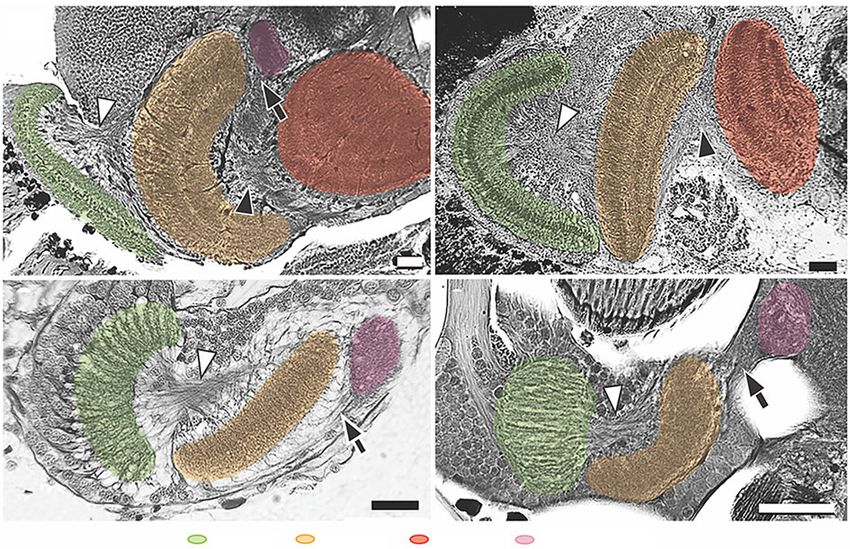
Please provide feedback Please support the ScholarWorks@UMBC repository by emailing and telling us what having access ...
(a)                                                                             (b)                                                          2

                                                                                                                                                                                                                                               royalsocietypublishing.org/journal/rspb
                                                                                                    (c)                                     (d)

                                                                                                                                                                                                                                               Proc. R. Soc. B 288: 20210216
                                                                       Figure 1. Four hyperiid amphipod eye morphologies. (a) Cystisoma magna, huge dorsally directed compound eyes with a diffuse retinal sheet. Ventral view of the
Downloaded from https://royalsocietypublishing.org/ on 28 April 2021

                                                                       brain and the retinal sheet (left) and whole animal (right). (b) Lanceola sayana, tiny compound eyes (white arrowhead). (c) Hyperia galba, one pair, large, dome-like
                                                                       compound eyes with dorsally and laterally directed regions. (d ) Phronima sedentaria, two pairs compound eyes (dashed line indicates dorsal eye, dotted indicates
                                                                       the lateral eye). Body lengths approximately: 8 cm C. magna (a), 1 cm L. sayana (b), 0.8 cm H. galba (c) and 1.5 cm P. sedentaria (d ). (Online version in colour.)

                                                                       (crossed axons) [10–12]. These optic neuropils are, from distal
                                                                       to proximal (from the eyes in), the lamina, medulla and lobula.
                                                                                                                                                              2. Methods
                                                                       A fourth optic neuropil, the satellite lobula plate, which is                          (a) Animals
                                                                       linked through uncrossed axons from the medulla and                                    Specimens of Cystisoma magna and Lanceola sayana were collected
                                                                       lobula, has also been identified in various groups of malacos-                         with the Monterey Bay Aquarium Research Institute’s remotely
                                                                       tracans [11–13]. Three putative optic lobe neuropils have also                         operated vehicle Doc Ricketts operated from the Research Vessel
                                                                       been identified in several stem-group arthropod fossils from                           Western Flyer between December 2016 and September 2018.
                                                                       the lower and middle Cambrian [14–16], suggesting that this                            Specimens of Hyperia galba and Phronima sedentaria were collected
                                                                       ground pattern arrangement may have been evolutionarily                                over the same time period, from the R/V Western Flyer using a
                                                                                                                                                              modified midwater tucker trawl (1.5 m × 1.5 m opening, 1000–
                                                                       stable for more than 500 Myr [14–16]. It is worth noting that
                                                                                                                                                              200 µm mesh). ROV dives and trawls were completed over the
                                                                       the names used here for the malacostracan optic neuropils
                                                                                                                                                              Monterey Submarine Canyon between the surface and 1500 m
                                                                       are adopted from, but may not be homologous to, those                                  depth (36° 320 N, 122° 300 W) from 06.00 to 00.00 h. Additional
                                                                       neuropils with the same names in insects.                                              specimens of C. magna, P. sedentaria and H. galba were collected
                                                                           The functions of malacostracan optic neuropils have been                           in February 2018 (POS520) and 2019 (POS532) from the submers-
                                                                       studied with electrophysiology and optical recording in the                            ible Jago and multinet hauls using a Hydrobios Maxi multinet
                                                                       brachyuran crabs Neohelice granulata and Carcinus maenas. It                           (0.5 m2 in aperture, 2 mm mesh size, nine nets) aboard the
                                                                       was shown that neurons in the lobula are essential for com-                            R/V Poseidon operated by GEOMAR Helmholtz-Zentrum für
                                                                       puting object features [17], object motion [18–22] and                                 Ozeanforschung. Specimens of Procambarus clarkii were obtained
                                                                       encoding certain flow field information [23], while those in                           from Carolina Biological Supply, Burlington, NC. Specimens of
                                                                       the lobula plate are implicated in computing wide-field                                Alima pacifica were collected at Lizard Island Research Station
                                                                                                                                                              near Australia’s Great Barrier Reef (Great Barrier Reef Marine
                                                                       motion and processing optic flow information that mediates
                                                                                                                                                              Park Authority Permit no. G12/35005.1, Fisheries Act no.
                                                                       optomotor responses [12,13]. Although the experimental evi-
                                                                                                                                                              140763), Neogonodactylus oerstedii in the Florida Keys, USA and
                                                                       dence came only from a few model species, those functional                             Gammarus mucronatus, in Gloucester Point, VA, USA.
                                                                       attributes are generally assumed to apply across malacostra-
                                                                       cans based on the overwhelming structural conservation
                                                                       and anatomical similarity of those constituent neurons in                              (b) Osmium-ethyl gallate staining
                                                                       the optic lobes [12,24].                                                               The staining was described in a previous study [25]. In brief,
                                                                           Given the broad diversity of eye morphologies within                               heads from live animals were detached and fixed in cacodylate
                                                                       hyperiids, how are their optic lobes organized to serve                                fixative (2% glutaraldehyde, 1% paraformaldehyde in 0.16 M
                                                                       those unique eyes? Because the central nervous system is                               sodium cacodylate buffer) with 10% sucrose at 4°C overnight.
                                                                       one of the most energetically expensive tissues, is an expan-                          After several washes in cacodylate buffer, brain tissue was
                                                                       sion in the optic lobes accompanied by a reduction in other                            dissected and immersed in 1% osmium tetroxide in the dark
                                                                       sensory processing centres, such as the olfactory lobes? In                            with continuous agitation for 2.5 h at 4°C and an additional
                                                                                                                                                              1 h at room temperature. After several washes in buffer, tissue
                                                                       this study, we investigate the brain organization and neural
                                                                                                                                                              was put in a second immersion with supersaturated ethyl gallate
                                                                       circuits that lie beneath the various hyperiid eye types.
                                                                                                                                                              (approx. 1% in distilled water) in the dark with continuous
                                                                       Specifically, we address whether or not the malacostracan                              agitation for 1.5 h at 4°C and an additional 30 min at room
                                                                       optic lobe ground pattern remains conserved across hyperiids                           temperature. After several washes in distilled water, tissue
                                                                       with different eye forms, and how the various sensory adap-                            was dehydrated, transferred into Durcupan plastic (Sigma,
                                                                       tations in the eyes relate to the structure and complexity of                          St. Louis, MO) via propylene-oxide and polymerized at 65°C.
                                                                       the brain.                                                                             Blocks were serially sectioned at 12–16 µm, mounted with
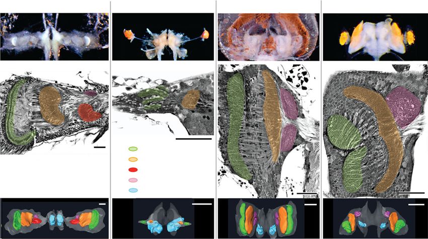
Please provide feedback Please support the ScholarWorks@UMBC repository by emailing and telling us what having access ...
(b)                                      (c)                                                    3
                                                                                      (a)

                                                                                                                                                                                                                                               royalsocietypublishing.org/journal/rspb
                                                                                     (d)                                          (e)                                              (f)

                                                                                                                                                                                                                                               Proc. R. Soc. B 288: 20210216
Downloaded from https://royalsocietypublishing.org/ on 28 April 2021

                                                                                                          lamina             medulla              lobula             lobula plate               olfactory lobe
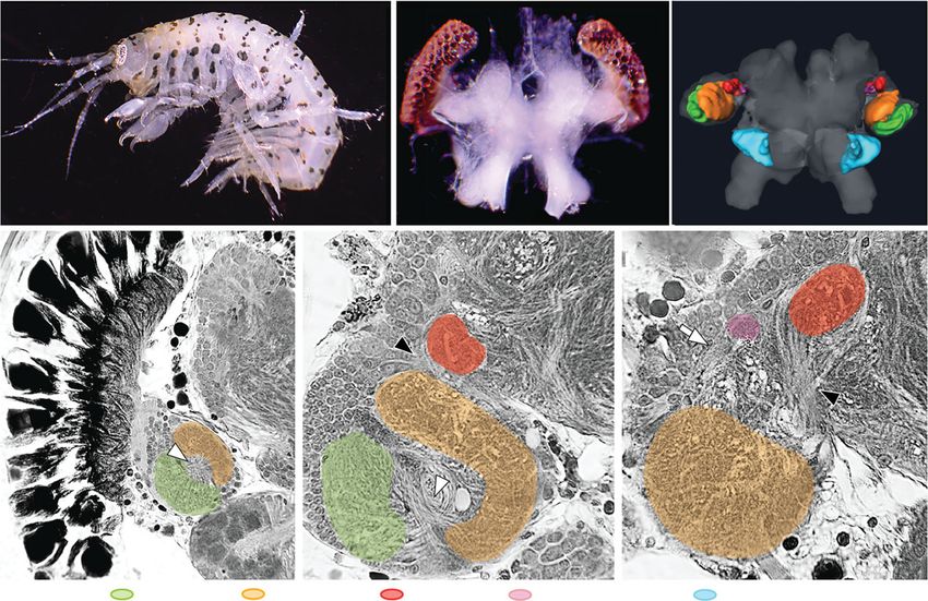
                                                                       Figure 2. The brain and optic lobe organization of the near shore, non-hyperiid amphipod, Gammarus mucronatus. (a–c) The animal, its brain and eyes, and three-
                                                                       dimensional reconstruction of the brain with highlighted optic and olfactory lobes. (d–f ) Osmium-ethyl gallate-stained optic lobe sections in different planes show-
                                                                       ing the characteristic optic lobe first and second chiasmata (white and black arrowheads, respectively), uncrossed neural connections between medulla and lobula
                                                                       plate (white arrow in f ), and all four optic neuropils. (Online version in colour.)

                                                                       Permount (Electron Microscopy Science, Hatfield, PA) and                               obtained by the ‘material statistics’ modules. Confocal reconstruc-
                                                                       covered with a cover slip for light microscopy.                                        tions of immunolabeled optic lobes were made with a Leica SP5
                                                                                                                                                              laser scanning confocal microscope (Leica Microsystems, Buffalo
                                                                                                                                                              Grove, IL). Images of 1024 × 1024 pixel resolution at 12-bit
                                                                       (c) Immunohistochemistry                                                               colour depth were scanned using a 10×/0.4 Plan Apochromat
                                                                       Five specimens of each of the following species, the hyperiids                         objective or a 20×/0.75 PL APO CS2 objective. Selected images
                                                                       H. galba and P. sedentaria, the crayfish P. clarkii and the mantis                     were digitally assembled and adjusted for brightness and contrast,
                                                                       shrimp N. oerstedii, were used for comparative immunolabelling                         and had a high pass filter uniformly applied using Adobe
                                                                       of their optic lobes, following the procedures described previously                    Photoshop CC 2019 (Adobe Systems, San Jose, CA).
                                                                       [26]. In brief, brains were fixed overnight in 4% paraformaldehyde
                                                                       in phosphate buffer ( pH 7.4) with 10% sucrose, and then washed
                                                                       in phosphate-buffered saline (PBS), embedded in albumin gelatin
                                                                       and sectioned at 60 µm with a vibratome. After being washed                            3. Results
                                                                       with PBS-TX (0.5% Triton X-100 in PBS), sections were blocked
                                                                       in 5% normal goat serum (Vector Laboratories, Burlingame, CA)                          (a) Conserved organization in a non-hyperiid
                                                                       for 1 h and then incubated overnight in monoclonal anti-allatosta-
                                                                       tin antibody (Developmental Studies Hybridoma Bank,                                        amphipod’s optic lobes
                                                                       University of Iowa, IA) and anti-FMRFamide antibody (Immu-                             Using histology and three-dimensional brain reconstructions,
                                                                       nostar, Hudson, WI) on a shaker at room temperature. The                               we first identified the existence of the four optic neuropils
                                                                       following day, sections were washed with PBS-TX and incubated                          with their respective characteristic optic chiasmata and
                                                                       overnight in the secondary goat anti-mouse immunoglobulins                             uncrossed neural connections in G. mucronatus, a non-hyperiid
                                                                       conjugated to Alexa Fluor 555 (3 : 1000) and goat anti-rabbit                          amphipod found in intertidal marine habitats such as
                                                                       immunoglobulins conjugated to Alexa Fluor 633 (3 : 1000;                               bays, estuaries and mangroves (figure 2). This amphipod is
                                                                       Thermo Fisher Scientific, Waltham, MA). The following day, sec-                        equipped with a typical, modest pair of compound eyes
                                                                       tions were washed with PBS, mounted with elvanol (25%
                                                                                                                                                              (figure 2a) and a small pair of optic lobes (figure 2c) consisting
                                                                       polyvinyl alcohol, 25% glycerol and 50% PBS) and covered with
                                                                                                                                                              of all four expected optic neuropils (figure 2d–f ), comparable
                                                                       a cover slip for confocal microscopy.
                                                                                                                                                              to other malacostracans studied thus far. Fundamental
                                                                                                                                                              visual processing pathways are, therefore, expected to be
                                                                       (d) Imaging and three-dimensional reconstructions                                      conserved within Amphipoda as well.
                                                                       Osmium-ethyl gallate-stained preparations were serially imaged
                                                                       using an Olympus BX 63 microscope with camera, imported
                                                                       into Amira (6.5) and aligned in the z-plane with ‘automatic align-                     (b) Highly variable eyes and optic lobe organization
                                                                       ment’ module in Amira. Three-dimensional reconstructions were
                                                                       made by manually tracing the outline of each neuropil at each                              among hyperiids
                                                                       depth, followed by volume rendering using the ‘generate surface’                       In stark contrast to Gammarus and other malacostracans, we
                                                                       module. The size of brain, optic lobe and each optic neuropil was                      found that hyperiid amphipods exhibit extreme variability
Please provide feedback Please support the ScholarWorks@UMBC repository by emailing and telling us what having access ...
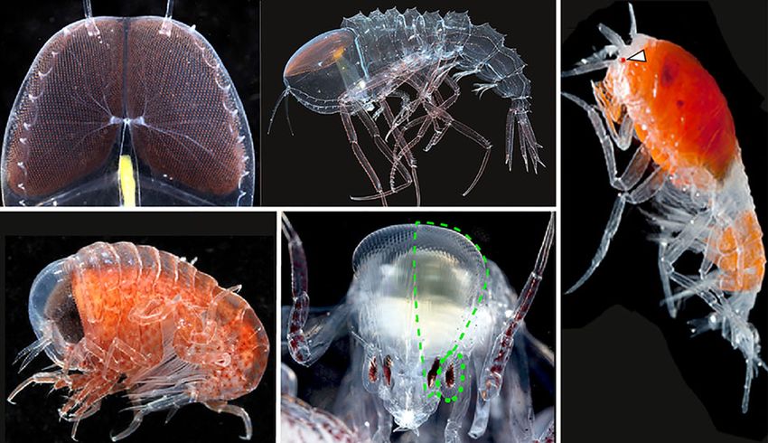
Cystisoma magna                  Lanceola sayana                 Hyperia galba              Phronima sedentaria                              4

                                                                                                                                                                                                                                         royalsocietypublishing.org/journal/rspb
                                                                                                                                  lamina
                                                                                                                                  medulla
                                                                                                                                  lobula
                                                                                                                                  lobula plate

                                                                                                                                                                                                                                         Proc. R. Soc. B 288: 20210216
                                                                                                                                  olfactory lobe
Downloaded from https://royalsocietypublishing.org/ on 28 April 2021

                                                                       Figure 3. Hyperiid optic lobe arrangements and brain morphologies. Cystisoma magna, enlarged optic lobe with all four optic neuropils as seen in other malacos-
                                                                       tracans. Lanceola sayana, reduced optic lobe with lamina and medulla only and enlarged olfactory lobes. Hyperia galba, enlarged optic lobe comprise lamina,
                                                                       medulla and dorsal and ventral lobula plates. Phronima sedentaria, enlarged optic lobe comprise dorsal and ventral laminas, a fused medulla and dorsal
                                                                       lobula plate receiving inputs solely from the dorsal half of the medulla. Scale bars, 100 µm (black) and 200 µm (white). (Online version in colour.)

                                                                       in brain morphology, brain-to-body size and especially in the                      Axon output from the dorsal- and lateral-eye laminas project
                                                                       number of optic neuropils in their optic lobes. The early                          to a single medulla on each side (figure 3). A small neuropil
                                                                       branching hyperiid C. magna [27–29] possesses a gigantic                           lies beneath the medulla and receives uncrossed projections
                                                                       pair of dorsally directed eyes whose retinas consist of a thin,                    from the dorsal, but not the lateral region of the medulla
                                                                       mesh-like sheet of suspended rhabdoms (figure 1a). Within                          (figures 3 and 4d). The lobula is absent. Thus, like H. galba,
                                                                       an unusually large optic lobe (73% total brain volume,                             P. sedentaria also lacks both the second optic chiasma and
                                                                       figure 3), photoreceptor axons of C. magna project to an optic                     the lobula (n = 24), yet the remaining optic lobes still account
                                                                       lobe consisting of the same four retinotopic optic neuropils                       for 61% of total brain volume (figure 3).
                                                                       that are also found in other malacostracans (figure 3).
                                                                            The deep-living L. sayana, on the other end of the
                                                                       spectrum, possesses a tiny pair of eyes (figure 1b). Photo-                        (c) Additional optic neuropil simplifications
                                                                       receptor axons project to a minute optic lobe (6% total brain                      In addition to the lost optic neuropils described above, the
                                                                       volume) that consists of only the lamina and medulla, lacking                      medulla also appeared to be structurally simplified in hyperiids
                                                                       both the lobula and lobula plate (n = 15, figure 3) that are                       compared to that of other malacostracans. In all four representa-
                                                                       typical of other malacostracans. The reduction of optic neuro-                     tive hyperiids we observed a lack of neuronal stratification
                                                                       pils in the L. sayana brain is offset by the size of the L. sayana                 within optic neuropils (figures 3 and 4), an anatomical feature
                                                                       olfactory lobe (24% total brain volume) compared to the other                      typifying distinct neuronal layers for serial and parallel visual
                                                                       hyperiids examined here, whose olfactory lobes account for                         processing [34]. To further demonstrate the lack of neuronal
                                                                       just 2% of total brain volume (figure 3).                                          stratification in hyperiid optic neuropils, we employed
                                                                            Hyperia galba possesses a single pair of large dome-like eyes                 immunohistochemistry with antisera against allatostatin and
                                                                       (figure 1c). Photoreceptor axons from each retina project to a                     FMRFamide, two neuropeptides that are highly expressed in
                                                                       single planar lamina. There, visual information is relayed reti-                   distinctive cell types of the optic lobes of malacostracans [35].
                                                                       notopically with second-order neurons, through the first                           The representative stomatopod (N. oerstedii, a mantis shrimp)
                                                                       chiasma, to a single medulla. Third-order medullary neurons                        and decapod (P. clarkii, a crayfish) showed distinct allatostatin-
                                                                       then project uncrossed axons to a pair of lobula plates                            and FMRFamide-like stratified immunoreactive layers in both
                                                                       that are partially connected to each other (figures 3 and 4c).                     the medulla and lobula following the expected malacostracan
                                                                       No second optic chiasma or lobula is present in H. galba (n =                      optic lobe ground pattern (figure 5a,b). However, in H. galba
                                                                       18). Despite the absence of the lobula, H. galba optic lobes                       and P. sedentaria, FMRFamide-like immunoreactivity dispersed
                                                                       still account for 62% of total brain volume (figure 3), a                          throughout the entire medulla and lobula plate without stratifi-
                                                                       number that is drastically larger than any other non-hyperiid                      cations. In addition, no allatostatin immunoreactivity was
                                                                       malacostracans [30,31].                                                            found in the optic lobes of either hyperiid (figure 5c,d), although
                                                                            Phronima sedentaria possesses two pairs of eyes                               positive labelling was found in their central brains in both
                                                                       (figure 1d)— a dorsally directed pair with greatly enlarged                        species. Immunolabelling data in C. magna and L. sayana were
                                                                       facets attached to a specialized array of long light guides                        not possible because fresh specimens were not available. How-
                                                                       [32], and a smaller pair of laterally directed eyes. Each of                       ever, based on histology, their medullas also appear to be
                                                                       the four eyes has a highly condensed, darkly pigmented                             reduced in thickness and without clear neuronal stratifications
                                                                       retina [33] (figure 1d), with photoreceptor axons connecting                       (compare figure 3 with figure 4a,b). The absence of several mor-
                                                                       to one of the four lenticular-shaped laminas (figure 3).                           phological distinct allatostatinergic medulla neurons typically
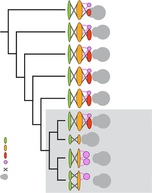
Please provide feedback Please support the ScholarWorks@UMBC repository by emailing and telling us what having access ...
5
                                                                                                    (a)                                                      (b)

                                                                                                                                                                                                                                                royalsocietypublishing.org/journal/rspb
                                                                                                    (c)                                                      (d)

                                                                                                                                                                                                                                                Proc. R. Soc. B 288: 20210216
Downloaded from https://royalsocietypublishing.org/ on 28 April 2021

                                                                                                                              lamina        medulla         lobula       lobula plate
                                                                       Figure 4. Osmium-ethyl gallate-stained optic lobe sections of various crustaceans at the antero-posterior plane showing the characteristic optic lobe first and second
                                                                       chiasmata (white and black arrowheads, respectively) and optic neuropils, including the structurally simplified medulla found in hyperiids. (a) Alima pacifica (mantis
                                                                       shrimp), (b) Procambarus clarkii (crayfish), (c) Hyperia galba (hyperiid) and (d ) Phronima sedentaria (hyperiid). Black arrows indicate the uncrossed neuronal con-
                                                                       nections between medulla and lobula plate. Scale bars, 50 µm. (Online version in colour.)
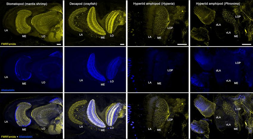
                                                                           (a)                                     (b)                                    (c)                                    (d)

                                                                       Figure 5. FMRFamide-like (yellow, top row) and allatostatin-like (blue, middle row) immunoreactivity reveals distinct layers of neuronal organization in (a) a
                                                                       stomatopod (mantis shrimp, Neogonodactylus oerstedii) lamina (LA), medulla (ME) and lobula (LO) and in (b) a decapod (crayfish Procambarus clarkii). However,
                                                                       in (c) Hyperia galba and (d ) Phronima sedentaria FMRFamide-like immunoreactivity is scattered throughout the entire medulla and lobula plate (LOP).
                                                                       No allatostatin-like immunoreactivity is detected in the hyperiid optic lobes. Scale bars, 100 µm. (Online version in colour.)

                                                                       found in malacostracans [35] demonstrates a further reduction                            given animal group are typically highly conserved [36,37].
                                                                       in neuronal complexity and diversity of cell types in the hyperiid                       This is most evident when comparing the arthropod brains,
                                                                       optic lobes.                                                                             where three nested optic neuropils connected with two axonal
                                                                                                                                                                chiasmata typify the optic lobe organization of malacostracan
                                                                                                                                                                crustaceans [10–12]. Our study nevertheless demonstrates an
                                                                                                                                                                unusual nervous system diversification among sister taxa.
                                                                       4. Discussion                                                                            Figure 6 illustrates the unusual variation found in the hyperiid
                                                                       Because nervous tissue is one of the most energetically expens-                          optic lobes compared to all other malacostracan lineages. Our
                                                                       ive tissues to build and maintain, neural arrangements in a                              examined hyperiids, with the exception of C. magna, the earliest
limited food availability, has likely driven the adaptive radi-        6
                                                                                                                           Phyllocarida               ation that resulted in the extreme diversity of lifestyles and

                                                                                                                                                                                                                           royalsocietypublishing.org/journal/rspb
                                                                                                                                                      eye forms among close hyperiid relatives (electronic sup-
                                                                                                                           Hoplocarida                plementary material, table S1). Observed differences in
                                                                                                                           (stomatopod)
                                                                                                                                                      depth of occurrence, swimming ability, free-living or host-
                                                                                                                           Decapoda                   associated lifestyles, body size and crypsis create different
                                                                                                                                                      visual needs for each hyperiid group, and these differences
                                                                                                                                                      may have driven the evolution of dramatically different eyes
                                                                                                                           Isopoda
                                                                                                                                                      and subsequently the visual circuits in the brains that support
                                                                                                                           Amphipoda                  those eyes. Given the large proportion of energy dedicated to
                                                                                                                           (gammarid)                 the development and maintenance of their greatly enlarged
                                                                                                                                                      eyes, it is not surprising to find that a similarly disproportion-
                                                                                                                           Cystisoma                  ate amount of central brain tissue is also dedicated to vision.
                                                                                                                                                      In addition, finding that there is a clear trade-off between the

                                                                                                                                                                                                                           Proc. R. Soc. B 288: 20210216
                                                                                                                                      Hyperiidea
                                                                                   lamina                                  Lanceola                   allocation of peripheral and central nervous system tissue to
                                                                                   medulla                                                            one sensory modality over another is not unexpected. What
                                                                                   lobula                                  Hyperia
                                                                                   lobula plate                                                       is unexpected is the degree to which the central brain tissues
                                                                                   chiasm                                                             have been selectively modified within different hyperiids
Downloaded from https://royalsocietypublishing.org/ on 28 April 2021

                                                                                   central brain                           Phronima                   to focus on specific visual needs. These modifications are
                                                                                                                                                      seen as the reduced complexity of the medulla in all hyperiids
                                                                       Figure 6. Optic lobe organization in malacostracan crustaceans with com-       examined here (figures 3–5), the loss of the lobula in L. sayana,
                                                                       pound eyes. Hyperiid amphipods exhibit dramatic variation in the number        H. galba and P. sedentaria that may compromise their ability to
                                                                       of optic neuropils unlike all other known malacostracans, including all pre-   distinguish object features and object motion, and the loss of
                                                                       viously known amphipods [10–12]. Phylogeny modified from [27,38].              the lobula plate in L. sayana and the ventral lobula plate in P.
                                                                       (Online version in colour.)                                                    sedentaria that may limit their ability to process wide-field
                                                                                                                                                      motion. The loss of the ability to distinguish an object’s fea-
                                                                                                                                                      tures and motion would seem detrimental to finding prey or
                                                                       branching hyperiid available for this study, show losses of optic              a host, nevertheless, all three species lacking the lobula in
                                                                       neuropils in multiple ways. Based on the finding that other                    this study are often observed free-swimming—an indication
                                                                       malacostracans and the basal hyperiid Cystisoma are equipped                   of prey searching. Do they simply attack anything they can
                                                                       with both lobula and lobula plate, and the second optic chiasma                catch? Given that the principal prey of H. galba and L. sayana
                                                                       (figure 3), the most parsimonious explanation would be a loss of               are gelatinous zooplankton including slow-moving jellies
                                                                       the lobula in H. galba, a loss of the lobula and the ventral lobula            and pelagic tunicates, this may not be a bad strategy. Phronima
                                                                       plate in P. sedentaria and a loss of both the lobula and the lobula            sedentaria, on the other hand, feeds additionally on other
                                                                       plate in L. sayana (figures 3 and 4). In addition, given that neur-            hyperiids and invertebrates. Behavioural work is needed to
                                                                       ons in the lobula are used for computing object features [17],                 determine how they recognize and track their prey. One possi-
                                                                       object motion [18–22] and certain flow field information [23],                 bility would be the exploitation of the lobula plate, which is
                                                                       the complete absence of the lobula in H. galba, P. sedentaria                  peculiarly enlarged in association with the enlarged eyes of
                                                                       and L. sayana are significant secondary reductions of the optic                H. galba and the dorsal eyes of P. sedentaria (figures 3 and 4).
                                                                       lobes (figures 3 and 4). Likewise, the loss of ventral or entire               While the abundant marine snow, other plankton and biolu-
                                                                       lobula plate in P. sedentaria and L. sayana, respectively, are                 minescent point sources in the surrounding packet of water
                                                                       additional significant secondary reductions of the optic lobes                 would provide the needed visual flow field feedback for navi-
                                                                       (figures 3 and 4). Despite the loss of these optic neuropils,                  gation, moving objects against this flow-field background can
                                                                       H. galba and P. sedentaria have substantially enlarged optic                   potentially be detected using the same neural circuitry, as
                                                                       lobes (62% and 61% total brain volume, respectively, figure 3),                shown in the wide-field motion-sensitive neurons found in
                                                                       which is similar to C. magna (73% total brain volume, figure 3)                the modified lobula plates of several predatory larval insects
                                                                       but contrasts with L. sayana (6% total brain volume, figure 3),                in support of their prey hunting [39,40].
                                                                       and G. mucronatus (representative non-hyperiid amphipod,
                                                                       15% total brain volume, figure 2). These findings indicate that
                                                                       in H. galba and P. sedentaria the remaining optic neuropils                    (b) Brain evolution in the midwater
                                                                       (lamina, medulla) are enlarged in size to compensate for the                   Examples of sensory modality compensation have been
                                                                       loss of the lobula (and partial lobula plate in P. sedentaria).                observed in animals living in light-limited environments,
                                                                       In L. sayana, on the other hand, the dramatic reduction of                     such as in the deep sea or caves. For example, the loss of
                                                                       optic lobes is compensated for by other sensory modalities, as                 eyes in many cave-dwelling animals is compensated by
                                                                       discussed below.                                                               enlarged tactile and/or olfactory organs. In the brain, likewise,
                                                                                                                                                      the eyeless amphipod Niphargus puteanus and other blind per-
                                                                                                                                                      acarids from cave habitats show reduced optic lobes, with
                                                                       (a) Ecological implications                                                    complete losses of lamina and medulla, but extensively elabo-
                                                                       The unexpected variability in hyperiid optic lobes may be due                  rated olfactory and mechanosensory neuropils [41,42]. The
                                                                       to the unique set of selective pressures that act on organisms                 hyperiid brains examined in this study show a similar com-
                                                                       living in the midwater. The high predatory pressure, limited                   pensatory pattern between optic and olfactory lobes. While
                                                                       illumination and a highly structured mesopelagic light                         the huge eyes and enlarged optic lobes in C. magna, H. galba
                                                                       regime, absence of substrate and hence hiding places and                       and P. sedentaria are accompanied by much smaller olfactory
lobes, the reduction of optic neuropils in L. sayana is offset by                           These changes should yield increased sensitivity, higher con-                           7
                                                                       the enlargement of their olfactory lobes (figure 3). These                                  trast and, in H. galba, better wide-field motion vision, but

                                                                                                                                                                                                                                                         royalsocietypublishing.org/journal/rspb
                                                                       results provide the first support for sensory modality compen-                              reduced object recognition. Our findings thus provide new
                                                                       sation in deep-sea hyperiids.                                                               insights into the patterns of brain evolution and sensory
                                                                           In addition to the sensory system trade-offs between                                    adaptation among animals that live in extreme habitats
                                                                       vision and olfaction, our findings indicate a variety of                                    such as the largest living space on the planet, the deep pelagic
                                                                       trade-offs even within the hyperiid visual system. While                                    realm or the midwater.
                                                                       their eyes and optic lobes are greatly enlarged, they have
                                                                       eliminated selected visual processing centres (lobula and                                   Data accessibility. All data are included in the manuscript.
                                                                       lobula plate) and reduced neuronal complexity in the remain-                                Authors’ contributions. C.L. and K.J.O. conceptualized and designed the
                                                                                                                                                                   study. C.L. carried out the study. C.L. and K.J.O. analysed the results
                                                                       ing visual centres (figures 3–6). Two prominent hypotheses                                  and wrote the manuscript. H.-J.T.H. provided access to live speci-
                                                                       have been proposed to explain the variation in brain struc-                                 mens and edited the manuscript. T.W.C. provided research space,
                                                                       tures seen among mammalian lineages. The ‘concerted                                         equipment and edited the manuscript. All authors approved the
                                                                       brain evolution’ hypothesis states that the brain evolves as                                final manuscript.

                                                                                                                                                                                                                                                         Proc. R. Soc. B 288: 20210216
                                                                       a single unit and correlated changes between major brain                                    Competing interests. The authors declare no competing interests.
                                                                       regions exist due to developmental constraints, suggesting                                  Funding. C.L. was supported by a Smithsonian Peter Buck Postdoctoral
                                                                                                                                                                   Fellowship and SI-NMNH Fenner Chase and Mary Jane Rathbun
                                                                       that natural selection cannot act independently on individual
                                                                                                                                                                   Crustacean Endowments. H.-J.T.H. was funded by the German
                                                                       brain regions [43]. Our observation of the changes to the size
Downloaded from https://royalsocietypublishing.org/ on 28 April 2021

                                                                                                                                                                   Research Foundation’s Emmy Noether Junior Research Group grant
                                                                       and complexity of the hyperiid visual systems directly contra-                              (HO 5569/2-1) and GEOMAR’s POF III.
                                                                       dict the ‘concerted brain evolution’ hypothesis. Alternatively,                             Acknowledgements. We thank Bruce Robison and Kakani Katija for the
                                                                       our findings support the ‘mosaic brain evolution’ hypothesis,                               invitations to participate in MBARI field expeditions, the crew of
                                                                       which postulates that different brain regions can evolve inde-                              MBARI’s R/V Western Flyer and GEOMAR’s Poseidon, the pilots of
                                                                                                                                                                   the ROV Doc Ricketts and the Jago Team and Stephanie Bush, Kyra
                                                                       pendently of each other [44]. In hyperiids, we see that strong
                                                                                                                                                                   Schlining, Kate Thomas, Kim Reisenbichler and Robert Sherlock for
                                                                       selective pressure has individually increased eye and selected                              providing assistance with specimen collection. We also thank Freya
                                                                       optic neuropil sizes but decreased neuronal complexity                                      Goetz, Scott Whittaker and Tagide DeCarvalho for microscopy and
                                                                       within neuropils and eliminated other optic neuropils.                                      imaging support.

                                                                       References
                                                                       1.  Robison BH. 2004 Deep pelagic biology. J. Exp. Mar.     11. Sinakevitch I, Douglass JK, Scholtz G, Loesel R,       18. de Astrada MB, Tomsic D. 2002 Physiology and
                                                                           Biol. Ecol. 300, 253–272. (doi:10.1016/j.jembe.             Strausfeld NJ. 2003 Conserved and convergent               morphology of visual movement detector neurons
                                                                           2004.01.012)                                                organization in the optic lobes of insects and             in a crab (Decapoda: Brachyura). J. Comp. Physiol. A
                                                                       2. Warrant EJ, Locket NA. 2004 Vision in the deep sea.          isopods, with reference to other crustacean                188, 539–551. (doi:10.1007/s00359-002-0328-4)
                                                                           Biol. Rev. 79, 671–712. (doi:10.1017/                       taxa. J. Comp. Neurol. 467, 150–172. (doi:10.1002/     19. de Astrada MB, Bengochea M, Sztarker J, Delorenzi
                                                                           S1464793103006420)                                          cne.10925)                                                 A, Tomsic D. 2013 Behaviorally related neural
                                                                       3. Zeidler W. 2012 A review of the hyperiidean amphipod     12. Strausfeld NJ. 2005 The evolution of crustacean and        plasticity in the arthropod optic lobes. Curr. Biol.
                                                                           families Mimonectidae and Proscinidae (Crustacea:           insect optic lobes and the origins of chiasmata.           23, 1389–1398. (doi:10.1016/j.cub.2013.05.061)
                                                                           Amphipoda: Hyperiidea: Scinoidea). Zootaxa 3533,            Arthropod Struct. Dev. 34, 235–256. (doi:10.1016/j.    20. Medan V, de Astrada MB, Scarano F, Tomsic D. 2015
                                                                           1–74. (doi:10.11646/zootaxa.3533.1.1)                       asd.2005.04.001)                                           A network of visual motion-sensitive neurons for
                                                                       4. Land MF. 2000 Eyes with mirror optics. J. Opt. A:        13. Bengochea M, Beron de Astrada M, Tomsic D,                 computing object position in an arthropod.
                                                                           Pure Appl. Opt. 2, R44–R50. (doi:10.1088/1464-              Sztarker J. 2018 A crustacean lobula plate:                J. Neurosci. 35, 6654–6666. (doi:10.1523/
                                                                           4258/2/6/204)                                               morphology, connections, and retinotopic                   JNEUROSCI.4667-14.2015)
                                                                       5. Land MF, Nilsson D-E. 2012 Animal eyes, 2nd ed.              organization. J. Comp. Neurol. 526, 109–119.           21. Scarano F, Sztarker J, Medan V, de Astrada MB,
                                                                           New York, NY: Oxford University Press.                      (doi:10.1002/cne.24322)                                    Tomsic D. 2018 Binocular neuronal processing of
                                                                       6. Land MF. 1981 Optics and vision in invertebrates. In     14. Ma X, Hou X, Edgecombe GD, Strausfeld NJ. 2012             object motion in an arthropod. J. Neurosci. 38,
                                                                           Handbook of sensory physiology (ed. H Autrum),              Complex brain and optic lobes in an early Cambrian         6933–6948. (doi:10.1523/JNEUROSCI.3641-17.2018)
                                                                           pp. 471–592. Berlin, Germany: Springer.                     arthropod. Nature 490, 258–261. (doi:10.1038/          22. Scarano F, Tomsic D, Sztarker J. 2020 Direction
                                                                       7. Land MF. 1989 The eyes of hyperiid amphipods:                nature11495)                                               selective neurons responsive to horizontal motion in
                                                                           relations of optical structure to depth. J. Comp.       15. Strausfeld NJ, Ma X, Edgecombe GD. 2016                    a crab reflect an adaptation to prevailing
                                                                           Physiol. A 164, 751–762. (doi:10.1007/BF00616747)           Fossils and the evolution of the arthropod brain.          movements in flat environments. J. Neurosci. 40,
                                                                       8. Meyer-Rochow VB. 1978 The eyes of mesopelagic                Curr. Biol. 26, R989–R1000. (doi:10.1016/j.cub.            5561–5571. (doi:10.1523/JNEUROSCI.0372-20.2020)
                                                                           crustaceans, II. Streetsia challenger (Amphipoda).          2016.09.012)                                           23. Horseman BF, Macauley MWS, Barnes WJP. 2011
                                                                           Cell Tissue Res. 186, 337–349. (doi:10.1007/            16. Aria C, Caron JA. 2019 A middle Cambrian                   Neuronal processing of translational optic flow in
                                                                           bf00225542)                                                 arthropod with chelicerae and proto-book gills.            the visual system of the shore crab Carcinus
                                                                       9. Baldwin Fergus JL, Johnsen S, Osborn KJ. 2015 A              Nature 573, 586–589. (doi:10.1038/s41586-019-              maenas. J. Exp. Biol. 214, 1586–1598. (doi:10.1242/
                                                                           unique apposition compound eye in the mesopelagic           1525-4)                                                    jeb.050955)
                                                                           hyperiid amphipod Paraphronima gracilis. Curr. Biol.    17. Medan V, Oliva D, Tomsic D. 2007 Characterization      24. Tomsic D. 2016 Visual motion processing subserving
                                                                           25, 473–478. (doi:10.1016/j.cub.2014.12.010)                of lobula giant neurons responsive to visual stimuli       behavior in crabs. Curr. Opin. Neurobiol. 41,
                                                                       10. Elofsson R, Dahl E. 1970 The optic neuropiles and           that elicit escape behaviors in the crab                   113–121. (doi:10.1016/j.conb.2016.09.003)
                                                                           chiasmata of Crustacea. Z. Zellforsch. Mikrosk. Anat.       Chasmagnathus. J. Neurophysiol. 98, 2414–2428.         25. Lin C, Cronin TW. 2018 Two visual systems in one
                                                                           107, 343–360. (doi:10.1007/BF00336672)                      (doi:10.1152/jn.00803.2007)                                eyestalk: the unusual optic lobe metamorphosis in
the stomatopod Alima pacifica. Dev. Neurobiol. 78,      32. Ball EE. 1977 Fine structure of the compound eyes            Malacostraca and Branchiopoda: highlighting the           8
                                                                             3–14. (doi:10.1002/dneu.22550)                              of the midwater amphipod Phronima in relation to             strength of taxon-specific matrices in

                                                                                                                                                                                                                                                              royalsocietypublishing.org/journal/rspb
                                                                       26.   Lin C, Strausfeld NJ. 2013 A precocious adult visual        behavior and habitat. Tissue Cell 9, 521–536.                phylogenomics. Proc. R. Soc. B 285, 20181524.
                                                                             center in the larva defines the unique optic lobe of        (doi:10.1016/0040-8166(77)90010-6)                           (doi:10.1098/rspb.2018.1524)
                                                                             the split-eyed whirligig beetle Dineutus sublineatus.   33. Land MF. 1981 Optics of the eyes of Phronima and       39.   Mizutani A, Toh Y. 1995 Optical and physiological
                                                                             Front. Zool. 10, 7. (doi:10.1186/1742-9994-10-7)            other deep sea amphipods. J. Comp. Physiol. 145,             properties of the larval visual system of the tiger
                                                                       27.   Hurt C, Haddock SHD, Browne WE. 2013 Molecular              209–226. (doi:10.1007/BF00605034)                            beetle, Cicindela chinensis. J. Comp. Physiol. A 177,
                                                                             phylogenetic evidence for the reorganization of the     34. Millard SS, Pecot MY. 2018 Strategies for                    591–599. (doi:10.1007/BF00207188)
                                                                             hyperiid amphipods, a diverse group of pelagic              assembling columns and layers in the Drosophila        40.   Okamura JY, Toh Y. 2004 Morphological and
                                                                             crustaceans. Mol. Phylogenet. Evol. 67, 28–37.              visual system. Neural. Dev. 13, 11. (doi:10.1186/            physiological identification of medulla interneurons
                                                                             (doi:10.1016/j.ympev.2012.12.021)                           s13064-018-0106-9)                                           in the visual system of the tiger beetle larva.
                                                                       28.   Copilaş-Ciocianu D, Borko Š, Fišer C. 2020 The late     35. Meth R, Wittfoth C, Harzsch S. 2017 Brain                    J. Comp. Physiol. A 190, 449–468. (doi:10.1007/
                                                                             blooming amphipods: global change promoted                  architecture of the Pacific white shrimp Penaeus             s00359-004-0509-4)
                                                                             post-Jurassic ecological radiation despite Palaeozoic       vannamei Boone, 1931 (Malacostraca,                    41.   Stegner ME, Stemme T, Iliffe TM, Richter S, Wirkner

                                                                                                                                                                                                                                                              Proc. R. Soc. B 288: 20210216
                                                                             origin. Mol. Phylogenet. Evol. 143, 106664. (doi:10.        Dendrobranchiata): correspondence of brain                   CS. 2015 The brain in three crustaceans from
                                                                             1016/j.ympev.2019.106664)                                   structure and sensory input? Cell Tissue Res. 369,           cavernous darkness. BMC Neurosci. 16, 19. (doi:10.
                                                                       29.   Biancani LM. 2019 Multi-locus phylogenetic analysis         255–271. (doi:10.1007/s00441-017-2607-y)                     1186/s12868-015-0138-6)
                                                                             of Amphipoda indicates a single origin of the pelagic   36. Karten HJ, Shimizu T. 1989 The origins of neocortex:   42.   Ramm T, Scholtz G. 2017 No sight, no
Downloaded from https://royalsocietypublishing.org/ on 28 April 2021

                                                                             suborder Hyperiidea. College Park, MD: University of        connections and lamination as distinct events in             smell? — Brain anatomy of two amphipod
                                                                             Maryland.                                                   evolution. J. Cogn. Neurosci. 1, 291–301. (doi:10.           crustaceans with different lifestyles. Arthropod
                                                                       30.   Sandeman DC, Scholtz G, Sandeman RE. 1993 Brain             1162/jocn.1989.1.4.291)                                      Struct. Dev. 46, 537–551. (doi:10.1016/j.asd.2017.
                                                                             evolution in decapod Crustacea. J. Exp. Zool. 265,      37. Shanahan M, Bingman VP, Shimizu T, Wild M,                   03.003)
                                                                             112–133. (doi:10.1002/jez.1402650204)                       Güntürkün O. 2013 Large-scale network                  43.   Finlay BL, Darlington RB. 1995 Linked regularities in
                                                                       31.   Krieger J, Hornig MK, Sandeman RE, Sandeman DC,             organization in the avian forebrain: a                       the development and evolution of mammalian
                                                                             Harzsch S. 2020 Masters of communication: the               connectivity matrix and theoretical analysis.                brains. Science 268, 1578–1584. (doi:10.1126/
                                                                             brain of the banded cleaner shrimp Stenopus                 Front. Comput. Neurosci. 7, 89. (doi:10.3389/fncom.          science.7777856)
                                                                             hispidus (Olivier, 1811) with an emphasis on                2013.00089)                                            44.   Barton RA, Harvey PH. 2000 Mosaic evolution of
                                                                             sensory processing areas. J. Comp. Neurol. 528,         38. Schwentner M, Richter S, Rogers DC, Giribet G. 2018          brain structure in mammals. Nature 405,
                                                                             1561–1587. (doi:10.1002/cne.24831)                          Tetraconatan phylogeny with special focus on                 1055–1058. (doi:10.1038/35016580)
You can also read