Structural basis for RNA recognition by the N-terminal tandem RRM domains of human RBM45
←
→
Page content transcription
If your browser does not render page correctly, please read the page content below
2946–2958 Nucleic Acids Research, 2021, Vol. 49, No. 5 Published online 12 February 2021
doi: 10.1093/nar/gkab075
Structural basis for RNA recognition by the N-terminal
tandem RRM domains of human RBM45
Xiaolei Chen1,2,† , Zhongmei Yang1,2,† , Wenfeng Wang1,2 , Kaiyue Qian1,2,3 , Mingjie Liu1,2,3 ,
Junchao Wang1,2,3 and Mingzhu Wang 1,2,3,*
1
Institutes of Physical Science and Information Technology, Anhui University, 111 Jiulong Road, Hefei 230601, Anhui,
China, 2 School of Life Sciences, Anhui University, 111 Jiulong Road, Hefei 230601, Anhui, China and 3 Key
Laboratory of Human Microenvironment and Precision Medicine of Anhui Higher Education Institutes, Anhui
University, 111 Jiulong Road, Hefei 230601, Anhui, China
Downloaded from https://academic.oup.com/nar/article/49/5/2946/6134178 by guest on 30 March 2021
Received August 24, 2020; Revised January 16, 2021; Editorial Decision January 25, 2021; Accepted January 28, 2021
ABSTRACT 43 (TDP-43) (7) and fused-in sarcoma (FUS) (8,9), is a
common feature of amyotrophic lateral sclerosis (ALS) and
RBM45 is an RNA-binding protein involved in neural frontotemporal lobar dementia (FTLD) (10–14). LLPS
development, whose aggregation is associated with is driven by both multivalent protein−RNA interactions
neurodegenerative diseases, such as amyotrophic and multivalent protein−protein interactions (15,16). The
lateral sclerosis (ALS) and frontotemporal lobar de- RNA-binding motif protein 45 (RBM45), also known
mentia (FTLD). However, the mechanisms of RNA- as developmentally regulated RNA-binding protein 1
binding and aggregation of RBM45 remain uneluci- (Drb1), is an RBP that plays an important role in neural
dated. Here, we report the crystal structure of the development (17,18). RBM45 is predominantly localized
N-terminal tandem RRM domains of human RBM45 in the nucleus and can shuttle between the nucleus and
in complex with single-stranded DNA (ssDNA). Our cytoplasm (19,20). RBM45 interacts with >100 proteins
structural and biochemical results revealed that both in vivo, with most of them being RBPs (21), and it is
hypothesized to regulate RNA splicing and processing
the RRM1 and RRM2 of RBM45 recognized the GAC
in the nucleus (22). RBM45 aggregates and co-localizes
sequence of RNA/ssDNA. Two aromatic residues and with the well-understood ALS-linked RBP TDP-43 in the
an arginine residue in each RRM were critical for cytoplasmic inclusions in neurons and glia of ALS and
RNA-binding, and the interdomain linker was also in- FTLD patients (20,23). Investigators have recently found
volved in RNA-binding. Two RRMs formed a pair of that RBM45 also formed nuclear inclusions in neurons
antiparallel RNA-binding sites, indicating that the N- and glia in ALS, FTLD and Alzheimer’s disease (AD) (22).
terminal tandem RRM domains of RBM45 bound sep- These results suggested that the aggregation of RBM45
arate GAC motifs in one RNA strand or GAC motifs was associated with neurodegenerative diseases (24).
in different RNA strands. Our findings will be helpful RBM45 contains three RNA recognition motif (RRM)
in the identification of physiologic targets of RBM45 domains: the RRM1 and RRM2 at the N-terminus, and
and provide evidence for understanding the physio- the RRM3 near the C-terminus. RRM is a common RNA-
binding module characterized by a typical ␣␣ topol-
logic and pathologic functions of RBM45.
ogy and two conserved aromatic residues (25,26). All the
three RRMs in RBM45 contain the characteristic aro-
INTRODUCTION matic residues, suggesting that they may all manifest RNA-
binding capability. RBM45 contains a nuclear localization
RNA-binding proteins (RBPs) interact with RNAs sequence (NLS) at the C-terminus, and mutations in NLS
sequence-specifically and/or structure-specifically and lead to the cytoplasmic aggregation of RBM45 (19,20). The
regulate RNA metabolism, including splicing, maturation, linker region between RRM2 and RRM3, which was pre-
localization, translation and degradation (1–4). Aberrant dicted to be a pseudo-RRM domain, was reported to pro-
expression and dysfunction of RBPs correlate with several mote the homo-oligomerization of RBM45 and was there-
serious human diseases, such as cancers, infertility and neu- fore termed the homo-oligomer assembly (HOA) domain
rodegenerative diseases (3–6). The cytoplasmic inclusions, (20). The aggregation of RBM45 also involves interactions
or liquid-liquid phase separation (LLPS), formed by the between RBM45 and other ALS-linked proteins. The inter-
aggregation of RBPs, such as TAR DNA-binding protein
* To whom correspondence should be addressed. Mingzhu Wang. Email: wangmzh@ahu.edu.cn
†
The authors wish it to be known that, in their opinion, the first two authors should be regarded as Joint First Authors.
C The Author(s) 2021. Published by Oxford University Press on behalf of Nucleic Acids Research.
This is an Open Access article distributed under the terms of the Creative Commons Attribution-NonCommercial License
(http://creativecommons.org/licenses/by-nc/4.0/), which permits non-commercial re-use, distribution, and reproduction in any medium, provided the original work
is properly cited. For commercial re-use, please contact journals.permissions@oup.com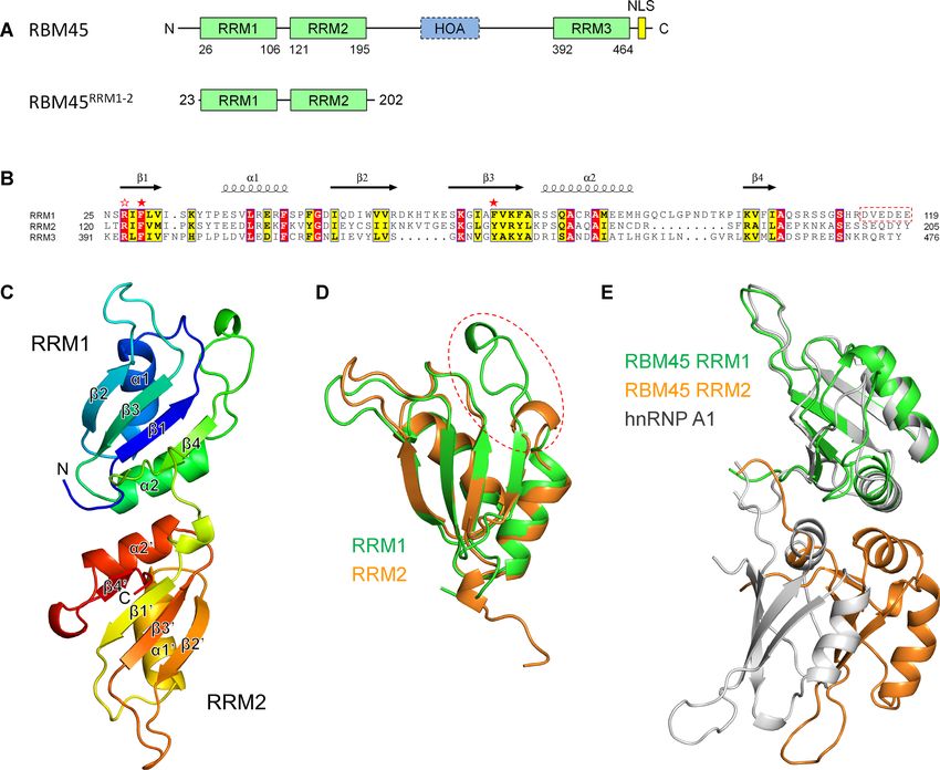
Nucleic Acids Research, 2021, Vol. 49, No. 5 2947
action between RBM45 and TDP-43 is not well defined. mM imidazole. The proteins were further purified by a pre-
Although RBM45 can bind to TDP-43 without RNA in equilibrated HiLoad 16/600 Superdex 75 PG column (GE
vitro (19), the interaction becomes weaker when RNA is Healthcare) in a buffer containing 20 mM Tris–HCl pH 7.5,
absent (20), suggesting that the interaction is most likely 300 mM NaCl and 1 mM DTT. The peak fractions contain-
RNA-dependent. In contrast, RBM45 binds FUS, an- ing highly purified RBM45RRM1–2 or its mutants were col-
other well-understood ALS-linked RBP, in an RNA/DNA- lected for later use in crystallization and biochemical assays.
independent manner (20,27). RBM45 was also reported to
compete with histone deacetylase 1 (HDAC1) for binding
RNA and DNA oligonucleotides
to FUS in the DNA-damage response (DDR), suggesting
that RBM45 may regulate the FUS-related DDR signaling The RNA oligonucleotides used for gel filtration binding
(27). assays and SEC-MALS assays, the 5 -FAM RNA and 5 -
Although the relationship between RBM45 and neurode- FAM DNA oligonucleotides used for fluorescent polariza-
generative diseases has been well established, the mecha- tion assays, and the DNA oligonucleotides used for crystal-
nisms underlying RBM45’s roles in these diseases remain lization were purchased from General Biosystems (China).
Downloaded from https://academic.oup.com/nar/article/49/5/2946/6134178 by guest on 30 March 2021
unclear. The RNA-binding property of RBM45 is criti-
cal in understanding its physiologic and pathologic func-
Crystallization, data collection and structure determination
tions. It was reported that the full-length RBM45 bound
the GACGAC (28) and ACGC (29) sequences of RNA. Re- The crystals were grown using the sitting-drop vapor dif-
cent work also showed that the full-length RBM45 bound fusing method at 16◦ C. For the crystallization of apo
GGGACGGU RNA with a dissociation constant (KD ) of RBM45RRM1–2 , the C-terminal His-tagged RBM45RRM1–2
18.2 ± 9.3 nM and that the RRM1-truncated RBM45 protein was concentrated to 3 mg/ml in a buffer containing
bound an 18-mer GGGACGGU-containing RNA with 20 mM Tris–HCl pH 7.5 and 95 mM NaCl. The crystals
higher affinity (21.8 ± 1.5 nM) than the full-length pro- were obtained in the condition containing 0.2 M ammo-
tein (36.9 ± 6.3 nM) (30). Although these results suggested nium phosphate dibasic and 20% (w/v) polyethylene gly-
that RBM45 might bind ACG-containing RNA, the RNA- col (PEG) 3350. For co-crystallization with single-stranded
binding preference of each RRM module remains unclear. DNA (ssDNA), the N-terminal His-tagged protein was
In the present work, we demonstrated that both the RRM1 concentrated to 7 mg/ml in a buffer containing 20 mM
and RRM2 of RBM45 recognized the GAC sequence of Tris–HCl pH 7.5 and 300 mM NaCl. The RBM45RRM1–2 –
RNA or ssDNA, and we determined the crystal structure ssDNA complex was prepared by mixing the protein with
of the tandem RRM1 and RRM2 of human RBM45 in 11-nt 5 -CGACGGGACGC-3 ssDNA at a molar ratio of
complex with ssDNA. Our structural and biochemical re- 1:1.2. The complex crystals were obtained in a reservoir so-
sults uncovered the mechanism of the recognition of RNA lution consisting of 0.2 M sodium formate and 20% (w/v)
sequences by the N-terminal tandem RRMs of RBM45. PEG 3350. The crystals were soaked in reservoir solution
Moreover, the three-dimensional arrangement of the two with 10% (v/v) glycerol and flash-cooled in liquid nitrogen
N-terminal RRMs provided additional information for un- before data collection.
derstanding the physiologic and pathologic functions of The X-ray diffraction data of the RBM45RRM1–2 crys-
RBM45. tal were collected at beamline BL17U of the Shanghai
Synchrotron Radiation Facility (SSRF) at a wavelength
of 0.9792 Å using a Dectris Eiger 16 M detector (31).
MATERIALS AND METHODS The data were processed with the HKL2000 package (32).
The structure was solved by the molecular replacement
Cloning, protein expression and purification
method with PHASER (33) in the CCP4 suite (34), us-
The DNA encoding the RRM1 and RRM2 domains of ing the crystal structure of human RBM38 (PDB code:
human RBM45 (RBM45RRM1–2 , residues 23–202) was ob- 6JVX) (35) as the search model. The model was rebuilt with
tained from full-length RBM45 gene synthesized from San- PHENIX (36) and COOT (37), and refined with PHENIX.
gon Biosystems (China) and cloned into pET-28a (+) and The RBM45RRM1–2 −ssDNA complex data were collected
pET-22b (+) with an N-terminal or C-terminal His-tag, re- at beamline BL18U of SSRF at a wavelength of 0.9793 Å us-
spectively, by using restriction enzymes NdeI and XhoI. ing a Dectris Pilatus 6 M detector. The data were processed
Mutant plasmids were produced from the wild-type pET- with the HKL3000 package (32). The structure was solved
28a (+) construction via site-directed mutant PCR. The by the molecular replacement method with PHASER, us-
recombinant plasmids were verified by DNA sequencing. ing the protein alone structure as the search model. The
All recombinant proteins were expressed in Escherichia coli electron density of DNA was clear after several rounds of
BL21 (DE3) cells in LB medium. After the growth of bacte- refinement with REFMAC5 (38) and COOT, allowing an
rial cells up to an OD600 of 0.6–0.8, protein expression was unambiguous DNA model building with COOT. The final
induced by 0.3 mM isopropyl--D-thiogalactoside (IPTG) model was refined with PHENIX. The statistics of data col-
and continued at 16◦ C for 20 h. For protein purification, lection and structure refinement are shown in Table 1.
the cells were suspended in a buffer containing 20 mM
Tris–HCl, pH 7.5 and 300 mM NaCl and lysed by sonica-
Fluorescence polarization analysis
tion. RBM45RRM1–2 or its mutants were initially purified by
Ni-NTA (GE) affinity column and eluted in a buffer con- Fluorescence polarization (FP) analysis was used to detect
taining 20 mM Tris–HCl pH 7.5, 300 mM NaCl and 200 the RNA/DNA-binding affinities of RBM45RRM1–2 and its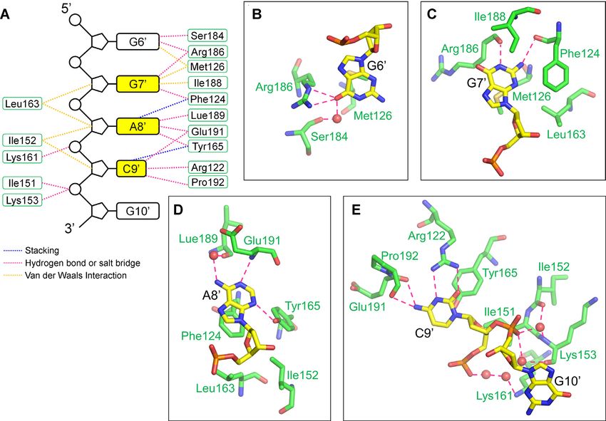
2948 Nucleic Acids Research, 2021, Vol. 49, No. 5
Table 1. Data collection and structure refinement statistics Size exclusion chromatography with multi-angle light scatter-
ing (SEC-MALS)
RBM45 RRM1–2
RBM45 RRM1–2
−ssDNA
A DAWN HELEOS-II (Wyatt Technology, Santa Barbara,
Data collection
CA, USA) multi-angle bright scattering detector and an
Wavelength (Å) 0.9792 0.9793
Space group P43 P43
Optilab T-rEX differential refractometer (Wyatt Technol-
Cell dimensions ogy) were used in-line with a Superdex 200 Increase 10/300
a, b, c (Å) 62.48, 62.48, 52.02 86.55, 86.55, 27.34 GL (GE Healthcare) column. RBM45RRM1–2 (3 mg/ml)
α, β, γ (◦ ) 90, 90, 90 90, 90, 90 and RNA containing two GAC motifs with different spaces
Resolution (Å) 50.00−2.50 50.00−1.80 (1.86−1.80) were mixed at a molar ratio of 1:1.1 in the SEC-MALS
(2.59−2.50)* buffer (20 mM Tris–HCl, pH 7.5, 150 mM NaCl and 1 mM
Rmerge 0.160 (0.950) 0.091(1.391)
DTT) and incubated for 30 min on ice before each experi-
I/I 16.4 (3.0) 21.3 (1.7)
CC1/2 0.982 (0.810) 0.999 (0.764) ment. The samples were run at a flow rate of 0.4 ml/min in
Completeness (%) 99.7 (99.3) 100.0 (100.0) the SEC-MALS buffer at room temperature. The data were
Downloaded from https://academic.oup.com/nar/article/49/5/2946/6134178 by guest on 30 March 2021
Redundancy 10.9 (9.2) 7.3 (7.4) analyzed using ASTRA 6.1 software.
Total/Unique 76 581/7056 141 140/19410
reflections
Refinement RESULTS
Resolution (Å) 44.18−2.50 21.64−1.80
No. reflections 7029 19273 Structure of the N-terminal tandem RRM domains of human
Rwork /Rfree 0.201/0.255 0.185/0.234 RBM45
No. atoms
RBM45 contains two N-terminal tandem RRM do-
Protein 1366 1413
DNA / 212 mains (RRM1 and RRM2), a C-terminal RRM domain
Water 86 169 (RRM3) and an HOA domain between RRM2 and RRM3
B-factors (Å2 ) (Figure 1A and B). In this study, we expressed, purified and
Protein 38.0 43.9 crystallized the N-terminal tandem RRM domains (23–
DNA / 63.4 202, hereafter referred to as RBM45RRM1–2 ), which was free
Water 37.3 50.1 of nucleic acid contamination (Supplementary Figure S1),
R.m.s. deviations
Bond lengths (Å) 0.002 0.004
and solved the structure at a resolution of 2.5 Å (Figure
Bond angles (◦ ) 0.514 0.596 1C). The final model contained amino acid residues from
MolProbity score 1.27 0.79 Pro23 to Lys193, whereas the residues 109–111 in the in-
Ramachandran plot terdomain linker region were disordered. Both RRM do-
Favored 98.2% 98.3% mains bore the typical RRM topology of 1–␣1–2–3–
Allowed 1.8% 1.7% ␣2–4, as seen in other RRM structures. For clarification,
Outliers 0 0 the four -strands and two helices in RRM2 will be named
*
1 –4 and ␣1 –␣2 , respectively. In each domain, the four
Values in parentheses are for the highest-resolution shell.
-strands formed an antiparallel four-stranded -sheet in
the order 4–1–3–2, with the two helices packed on
one side of the sheet. Two RRM domains could be eas-
mutants. The FAM-labeled RNA or DNA at 100 nM was ily superimposed, with a C␣ root-mean-square deviation
incubated with increasing amounts of RBM45RRM1–2 or its (RMSD) of 0.82 Å for 53 residues, except for the ␣2–4 loop
mutants in the binding buffer containing 10 mM Tris-HCl, regions, where the RRM1 was much longer than RRM2
200 mM NaCl, 1 mM DTT, 1 mM EDTA and 5% (v/v) glyc- (Figure 1B and D). The two domains were pseudo two-fold
erol for 30 min at room temperature. The 535 nm fluores- symmetric and interacted primarily through their ␣2 and
cence polarization measurements were carried out at 25◦ C ␣2 helices and the interdomain loop, resulting in a pair
on a SpectraMax Paradigm Multi-Mode detection platform of antiparallel potential RNA-binding sites on the same
(Molecular Devices, USA), with an excitation wavelength of side (Figure 1C and Supplementary Figure S2A). The mu-
485 nm. tation of Ala175 in the inter-domain interface to arginine
resulted in significant aggregation of RBM45RRM1–2 (Sup-
plementary Figure S2B), suggesting that this interface was
not just a result of crystal packing. The interaction between
Gel filtration assays for binding of RBM45RRM1-2 with two-
the two RRM domains was similar to those of hnRNP A1
GAC-motif RNA
(39,40) and hnRNP A2/B1 (41); however, they differed in
RBM45RRM1–2 (3 mg/ml) and RNA molecules that con- detail. In hnRNP A1 and hnRNP A2/B1, the two RRM
tained two GAC motifs at increasing distances (Supplemen- domains were not pseudo twofold symmetric; rather, the ␣2
tary Table S2) were mixed at a molar ratio of 1:1.2 in the GF of RRM2 interacted with ␣2 and 4 of RRM1, resulting
buffer (20 mM Tris–HCl, pH 7.5, 150 mM NaCl and 1 mM in a 10−20 Å shift in RRM2 relative to RBM45 (Figure
DTT) and incubated for 30 min on ice, and then loaded to a 1E and Supplementary Figure S3). The residues involved
Superdex 200 Increase 10/300 GL (GE Healthcare) column in domain−domain interaction are not conserved between
equilibrated in the GF buffer. A 6-mer RNA (CGACGG) RBM45 and hnRNP A1 or hnRNP A2/B1 (Supplementary
was used as a control. Figure S4).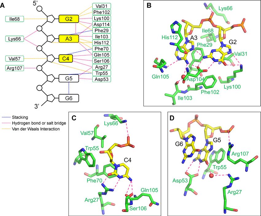
Nucleic Acids Research, 2021, Vol. 49, No. 5 2949
Downloaded from https://academic.oup.com/nar/article/49/5/2946/6134178 by guest on 30 March 2021
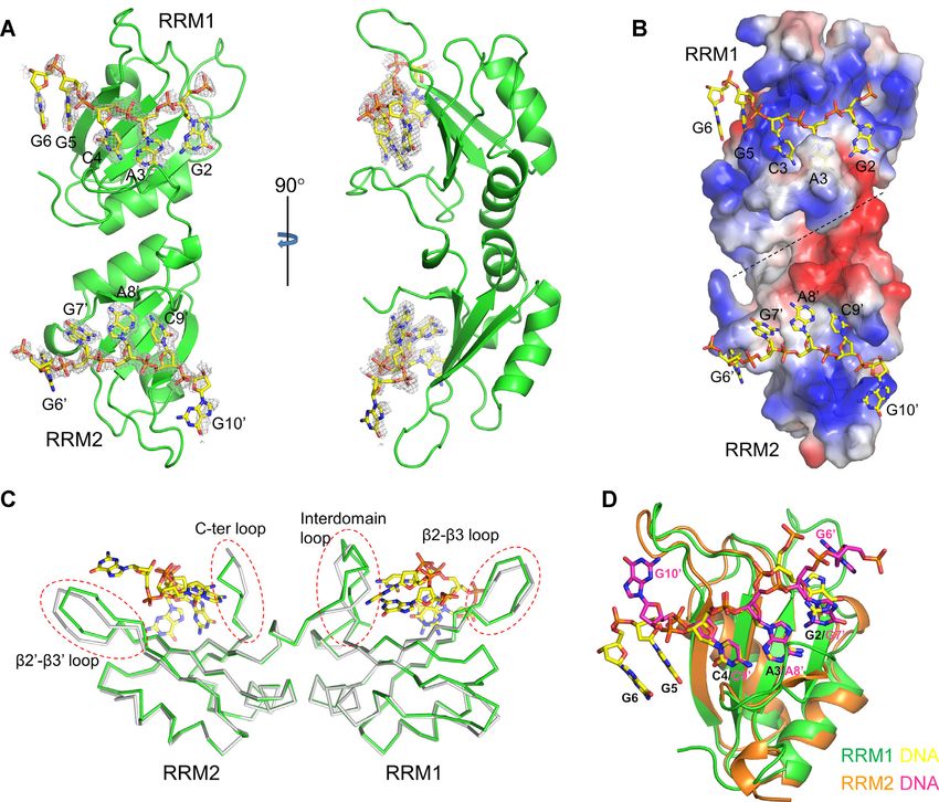
Figure 1. The structure of RBM45RRM1–2 . (A) The domains of human RBM45. (B) Sequence alignment of RRM1, RRM2 and RRM3. The identical
residues and conserved residues are highlighted in the red and yellow background, respectively. The two conserved aromatic residues are denoted by red
stars, the conserved arginine residues are denoted by a red blank star. (C) The overall structure of RBM45RRM1–2 . (D) Superimposition of RRM1 and
RRM2. The RRM1 and RRM2 are shown as green and orange cartoons, respectively. The ␣2−4 loop is enclosed in a red dashed circle. (E) Superimposi-
tion of RBM45RRM1–2 and hnRNP A1. The RRM1 of RBM45 is superposed with the RRM1 of hnRNP A1 (PDB code: 1HA1). The RRM1 and RRM2
of RBM45 are colored green and orange, respectively, the hnRNP A1 is colored gray.
Both RRM1 and RRM2 recognize GAC-containing RNA ure S5), slightly weaker relative to RNA, suggesting that
RBM45RRM1–2 can also bind ssDNA.
Previous studies have shown that the full-length RBM45
Next, although we attempted to identify the recogni-
bound GACGAC (28), ACGC (29) and GGGACGGU
tion sequence of each single RRM domain, we failed
(30) of RNA. To identify the RNA recognition sequences
to achieve an adequate amount of soluble RRM1 and
of RRM1 and RRM2 of RBM45, we synthesized a
RRM2 for the RNA-binding assay. Thus, we generated
5 -FAM-labeled 5 -GGACGG-3 6-mer RNA and ana-
a double mutant where the two key aromatic amino acid
lyzed its RBM45RRM1–2 -binding affinity using the fluores-
residues in RRM2 (i.e. Phe124 and Tyr165) were mutated
cence polarization (FP) method. RBM45RRM1–2 bound the
to alanine to mimic the RRM1; and a double mutant
GGACGG RNA with a dissociation constant (KD ) of 0.457
that the two key aromatic amino acid residues in RRM1
± 0.053 M, whereas the KD of the negative control––a
(Phe29 and Phe70) were mutated to alanine to mimic the
5 -FAM-labeled polyU 6-mer RNA––was 57.2 ± 10.2 M
RRM2. The F29A/F70A/F124A/Y165A quadruple mu-
(Figure 2A), suggesting that GGACGG was a preferred
tant decreased the RNA-binding affinity approximately
binding sequence for RBM45RRM1–2 . We additionally an-
150-fold (Figure 2B), indicating that mutations in these key
alyzed the RBM45RRM1–2 -binding affinity of a 5 -FAM-
residues virtually completely destroyed the RNA-binding
labeled 6-mer 5 -GGACGG-3 single-stranded DNA (ss-
ability of RBM45RRM1–2 . This result allowed us to assess
DNA) and discerned that RBM45RRM1–2 bound this ss-
the RNA/DNA-binding affinities of the two single RRMs
DNA with a KD of 0.805 ± 0.108 M (Supplementary Fig-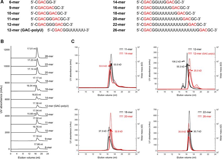
2950 Nucleic Acids Research, 2021, Vol. 49, No. 5
Downloaded from https://academic.oup.com/nar/article/49/5/2946/6134178 by guest on 30 March 2021
Figure 2. The RNA-binding affinities of RBM45RRM1–2 and its mutants. (A) The fluorescence polarization (FP) measures of binding affinities of
RBM45RRM1–2 to GGACGG and UUUUUU RNA. (B) The FP measures of RNA-binding affinities of F124A/Y165A (RRM1 analog), F29/F70A
(RRM2 analog) and F29/F70A/F124A/Y165A mutants of RBM45RRM1–2 . The data shown here are the averages of three independent measurements
with the same protein batch. The error bars indicate the standard deviations of three replicates.
separately by using the above two double mutants. The binding affinities, while substitutions at other positions
RRM1 analog F124A/Y165A bound the GGACGG RNA did not significantly influence affinities (Supplementary
6-mer and ssDNA 6-mer with KD s of 0.788 ± 0.092 and Table S1 and Figure S8). These results indicated that both
1.21 ± 0.09 M, respectively, slightly weaker than the RRM1 and RRM2 recognized the GAC sequence.
wild type (WT) RBM45RRM1–2 , whereas the RRM2 ana- As the GAC motif is a major N6 -methyladenosine (m6 A)
log F29A/F70A bound the GGACGG RNA 6-mer and RNA modification site (42,43), we next analyzed the bind-
ssDNA 6-mer with KD s of 1.48 ± 0.11 and 2.31 ± 0.19 ing affinity of RBM45RRM1–2 with a methylated RNA, 5 -
M, respectively, ∼3 times weaker than WT RBM45RRM1–2 GG(m6 A)CGG-3 . The FP assays showed that the WT
(Figure 2B and Supplementary Figure S5). These results in- RBM45RRM1–2 , as well as the F124A/Y165A and the
dicated that GGACGG was a preferred sequence binding F29A/F70A mutants, bound methylated RNA with sim-
both RRM1 and RRM2. ilar affinities of unmethylated RNA (Supplementary Fig-
To determine the exact recognition sequence of ure S9), which suggested that neither RRM1 nor RRM2 of
RBM45RRM1–2 and each single RRM domain, we changed RBM45 possesses selectivity for methylation of RNA.
each residue of the 6-mer RNA to other bases and
tested their RBM45RRM1–2 -binding affinities. For WT
Structure of RBM45RRM1-2 in complex with ssDNA
RBM45RRM1–2 , changing the positions 2 (G), 3 (A) or 4
(C) to any other bases resulted in 4−15-fold diminutions To investigate the mechanism underlying the RNA
in RBM45RRM1–2 -binding affinities, whereas changing recognition of RBM45, we attempted to co-crystallize
the other positions did not significantly influence their RBM45RRM1–2 with RNA or ssDNA in different se-
affinities (Supplementary Table S1 and Figure S6), indi- quences and lengths. Ultimately, we obtained crys-
cating that GAC was the critical recognition sequence of tals of RBM45RRM1–2 in complex with an 11-nt 5 -
RBM45RRM1–2 . For the F124A/Y165A mutant (RRM1 CGACGGGACGC-3 (the GAC motif residues are
analog), the substitutions at positions 2, 3 or 4 resulted in underlined) ssDNA, which contained two GAC motifs,
12−19-fold decreases in binding affinities, while the sub- and solved the structure at a resolution of 1.8 Å. Clear
stitutions at other positions did not significantly influence electron densities of the GAC motifs were observed at the
the affinities (Supplementary Table S1 and Figure S7). For potential RNA-binding sites of RRM1 and RRM2. The
the F29A/F70A mutant (RRM2 analog), the substitutions final model contained RBM45 residues Pro23–Asn194 and
at positions 2, 3 or 4 resulted in 13−20-fold decreases in two ssDNA fragments, with GACGG bound to RRM1 and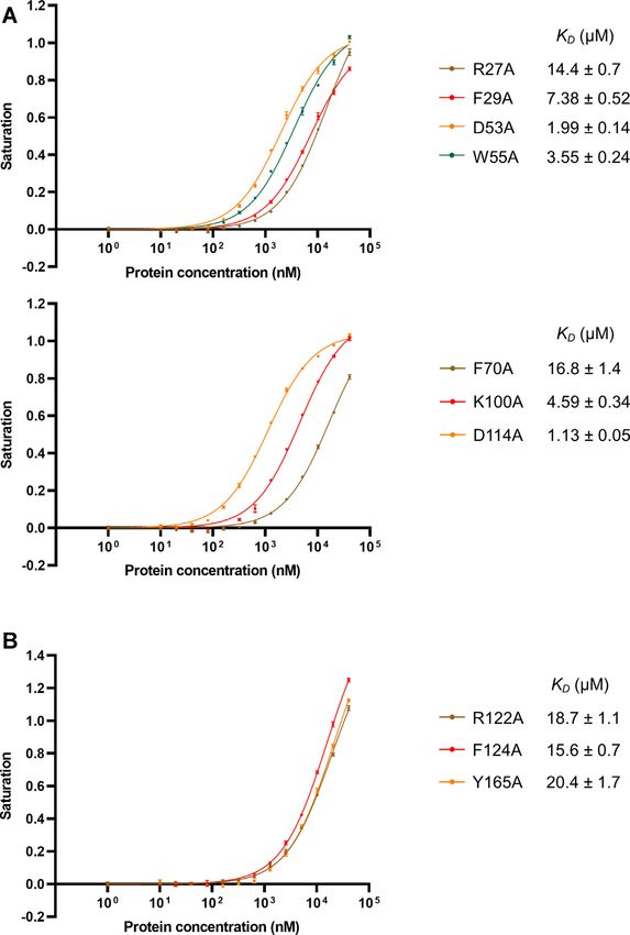
Nucleic Acids Research, 2021, Vol. 49, No. 5 2951
GGACG bound to RRM2 (Figure 3A). For clarification, In RRM2, the base moiety of G6 contacted the side
we numbered the nucleotides according to the sequence of chain of Met126, whereas the O6 hydrogen-bonded with
the 11-nt ssDNA, the RRM1-bound DNA from G2 to G6, the side chain of Arg186 and with the main-chain carbonyl
and the RRM2-bound DNA from G6 to G10 . Although of Ser184 through a water molecule. The N7 hydrogen-
an 11-nt ssDNA containing two GAC motifs was used for bonded with the side chain of Arg186 through a water
co-crystallization, only two 5-nt ssDNA segments, each of molecule (Figure 5A and B). The purine ring of G7 in the
which contained one GAC motif, were found in the struc- GAC motif was sandwiched between two hydrophobic side
ture. As expected, the two ssDNAs bound in an antiparallel chains of Met126 and Ile188, and its N1 and N2 formed
manner to the positively charged patches of RBM45RRM1–2 hydrogen bonds with the main-chain carbonyl groups of
(Figure 3B). The distance between each end of the two Arg186 and Phe124, respectively (Figure 5A and C). The
DNAs did not allow a direct linking, suggesting that the purine ring of A8 was stacked with the conserved aromatic
two short DNA strands were not a result of a disorder. residue Phe124, whereas its N1 and N3 formed hydrogen
The two RRM domains of the same molecule most likely bonds with the main-chain amino group of Glu191 and the
bound different DNA molecules. Interestingly, the 5 ends hydroxyl group of Tyr165, respectively. Its N6 hydrogen-
Downloaded from https://academic.oup.com/nar/article/49/5/2946/6134178 by guest on 30 March 2021
of the RRM1- and RRM2-bound DNAs were close to the bonded with the main-chain carbonyl of Leu189 through
3 ends of the RRM1- and RRM2-bound DNAs in the a water molecule (Figure 5A and D). The pyrimidine ring
symmetrical molecule, respectively (Supplementary Figure of C9 was stacked with the conserved aromatic residue
S10), suggesting that the ssDNA that contained two GAC Tyr165. The O2 and N3 formed two hydrogen bonds with
motifs might mediate crystal packing. the guanidine group of Arg122, and N4 formed hydrogen
The interaction between the two RRM domains in the bonds with the main-chain carbonyl groups of Glu191 and
complexed structure was identical to that in the protein Pro192 (Figure 5A and E).
alone structure. RBM45RRM1–2 in two structures could be
superimposed with a C␣ RMSD of 0.33 Å for 123 residues.
Binding assays of the interaction surface
Two structures differed principally in some loop regions, in-
cluding the 2−3 loop, the interdomain loop, the 2 −3 To validate our structural findings, we generated several
loop and the C-terminal loop, which were all involved in mutants and analyzed their binding to the GGACGG 6-
ssDNA binding (Figure 3C). The interdomain loop, which mer RNA using the FP method. As we failed to obtain
was partially disordered in the protein alone structure, in- an adequate amount of soluble single RRM1 or RRM2,
teracted with the RRM1-bound DNA and was entirely built the F124A/Y165A and F29A/F70A mutants were used to
into the complexed structure. Both the ssDNA strands in mimic the single RRM1 domain and single RRM2 domain,
the structure adopted extended conformations. The GAC respectively. The point mutants of key residues in RRM1
motifs bound to two RRM domains could be superimposed and RRM2 were generated based on the F124A/Y165A
well; in particular, the adenines and cytosines that bound to and F29/F70A mutants, respectively. For RRM1, the FP
two domains bore exactly the same conformation (Figure assays showed that the mutants of the residues interacted
3D). with G2, K100A and D114A, bound RNA with KD values
of 4.59 and 1.13 M, respectively. The former resulted in
an approximately 5-fold reduction in RNA-binding affin-
Structural details of nucleic acid recognition
ity, while the latter did not significantly affect the affin-
Both the backbone atoms and the base moieties of the ss- ity. Mutation of the aromatic residue that stacked with A3
DNA were involved in the protein−DNA interaction (Fig- (F29A) bound RNA with a KD of 7.38 M, attenuating the
ures 4A and 5A). Specifically, in RRM1, the N1 and N2 RNA-binding affinity approximately 9-fold. Mutation of
of G2 formed two hydrogen bonds with the side chain the aromatic residue stacked with C4 (F70A) bound RNA
of Asp114, whereas the O6 and N7 formed two hydrogen with a KD of 16.8 M, reducing the RNA-binding affin-
bonds with the side chain of Lys100. The purine ring of ity more than 20-fold. Mutation of the arginine that inter-
A3 was sandwiched between the conserved aromatic residue acted with C4 by two hydrogen bonds (R27A) reduced the
Phe29 in 1 and His112 in the interdomain loop. The N1 RNA-binding affinity approximately 18-fold, with a KD of
formed a hydrogen bond with the main-chain amino of 14.4 M. For residues that interacted with G5, the W55A
Gln105, and the N6 hydrogen-bonded with the side chain and D53A mutants bound RNA with KD values of 3.54
of Asp114 directly and with the main-chain carbonyl of and 1.99 M, reducing the binding affinities 4.5- and 2.5-
Ile103 through a water molecule (Figure 4A and B). The fold, respectively (Figure 6A). These results indicated that
pyrimidine ring of C4 was stacked with the conserved aro- Phe29, Phe70 and Arg27 were critical for the RNA-binding
matic residue Phe70 in 3; its O2 and N3 formed two of RRM1 and that Lys100 and Trp55 also played important
hydrogen bonds with the guanidine group of Arg27, and roles in RNA-binding.
its N4 formed hydrogen bonds with the main-chain car- For RRM2, the FP results showed that mutation of the
bonyl groups of Gln105 and Ser106 (Figure 4A and C). aromatic residue stacked with A8 (F124A) bound RNA
The purine ring of G5 was stacked with Trp55, and the N1 with a KD of 15.6 M, which was approximately 10-fold
and N2 formed two hydrogen bonds with the side chain of weaker than the RRM2 analog. Mutation of the aromatic
Asp53; its O6 formed a hydrogen bond with the side-chain residue stacked with C9 (Y165A) reduced the RNA-binding
amino of Arg27 through a water molecule. G6 did not di- affinity approximately 14-fold, with a KD of 20.4 M. Mu-
rectly contact protein but was stacked with G5 (Figure 4A tation of Arg122, which formed two hydrogen bonds with
and D). C9, to alanine reduced the RNA-binding affinity approxi-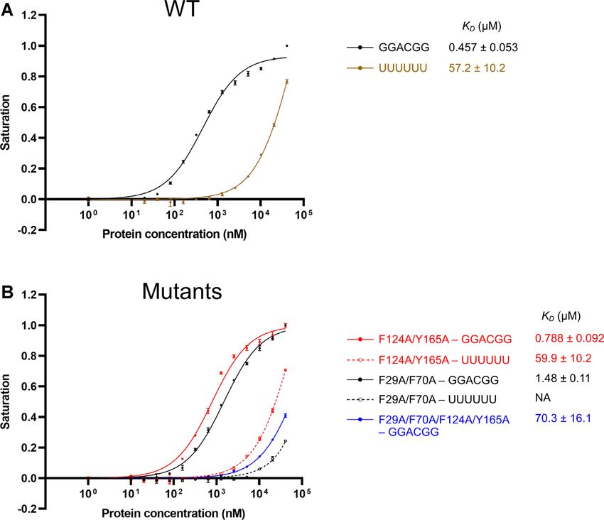
2952 Nucleic Acids Research, 2021, Vol. 49, No. 5
Downloaded from https://academic.oup.com/nar/article/49/5/2946/6134178 by guest on 30 March 2021
Figure 3. Structure of RBM45RRM1–2 −ssDNA complex. (A) The overall structure of the RBM45RRM1–2 −ssDNA complex. RBM45 is shown as green
cartoon, the ssDNAs are shown as yellow sticks. The simulated annealing omit map of ssDNA (Fo − Fc contoured at 2.5 ) are shown as gray meshes.
(B) The surface electrostatic potential of RBM45RRM1–2 . The ssDNA bind to the positively charged regions. (C) Structural changes of RBM45RRM1–2 by
DNA-binding. The RBM45RRM1–2 in complex and apo structures are shown as green and gray ribbons, respectively. The DNA is shown as yellow sticks.
The changed regions are enclosed in red dashed circles. (D) Superimposition of RRM1 and RRM2. The RRM1 and RRM2 are shown as green and orange
cartoons, respectively. The ssDNA bound RRM1 and RRM2 are shown as yellow and magenta sticks.
mately 12-fold, with a KD of 18.7 M (Figure 6B). These RBM45RRM1–2 . The SEC-MALS results showed that the
results indicated that Phe124, Tyr165 and Arg122 were crit- molecular weight of the RBM45RRM1–2 −11-mer RNA was
ical for the RNA-binding of RRM2. about 53.5 kDa, close to the theoretical molecular weight
of two proteins and two RNAs (53.2 kDa), suggesting that
they most likely formed a 2P−2R complex. Interestingly,
Binding of RBM45RRM1-2 with RNA containing two GAC when the distance was increased to 3 nt (12-mer RNA), in
motifs addition to the main peak with a retention volume close to
To further study the RNA-binding property of that of the RBM45RRM1–2 −11-mer RNA complex, there
RBM45RRM1–2 , we analyzed the binding of RBM45RRM1–2 was a small peak with a significantly smaller retention
with a set of longer RNA molecules containing two GAC volume. The SEC-MALS result showed that the molecular
motifs at increasing distances (Figure 7A) using the gel weights of the main and small peaks were approximately
filtration method (Figure 7B). Some of them were selected 55.3 and 106.2 kDa, respectively, consistent with the
for further analysis by the size exclusion chromatography theoretical molecular weights of 2P−2R (53.9 kDa) and
with multi-angle light scattering (SEC-MALS) method 4P−4R (107.8 kDa) complexes, respectively. These results
to determine the accurate molecular weights (Figure 7C). suggested that the 12-mer RNA can form a higher-order
The gel filtration retention volumes of the protein−RNA assembly with RBM45RRM1–2 in addition to the 2P−2R
complexes with 2-GAC-motif RNA with 0–2 nt spaces complex. To verify whether this binding property was
(9-mer, 10-mer and 11-mer RNA) were significantly smaller sequence-specific, we performed the same experiments
than that of the 6-mer one-GAC-motif RNA, which could with a 12-mer RNA containing one GAC motif and a 3
only form one protein−one RNA (1P−1R) or 1P−2R com- polyU sequence and found that the high molecular weight
plex, implying these RNAs formed larger complexes with peak was disappeared, and the main peak was moved to aNucleic Acids Research, 2021, Vol. 49, No. 5 2953
Downloaded from https://academic.oup.com/nar/article/49/5/2946/6134178 by guest on 30 March 2021
Figure 4. The interactions between RRM1 and ssDNA. (A) A schematic drawing of RRM1−ssDNA interaction. The DNA bases, deoxyriboses, and
phosphates are represented as black boxes, pentagons and circles, respectively. The recognition bases are highlighted in yellow. The amino acid residues are
represented as green boxes. The magenta, blue, and orange dashed lines denote hydrogen bonds or salt bridges, stacking and Van der Waals interactions,
respectively. (B−D) Detail views of interactions between DNA and RRM1 of RBM45. The involved RBM45 and DNA residues are shown as green and
yellow sticks, respectively. The hydrogen bonds and salt bridges are indicated by magenta dashed lines.
larger retention volume with a molecular weight of about These results implied that RBM45RRM1–2 can form 1P-1R,
25.0 kDa, close to the theoretical molecular weights of 2P-2R or 4–4R complexes with two-GAC-motifs RNA
a 1P−1R complex (26.8 kDa). These results suggested dependent on the distances between the two motifs.
that the assembly of RBM45RRM1–2 −12-mer RNA was
sequence-specific. However, when the distance between the DISCUSSION
two GAC motifs was further increased, the peak of the
higher-order assembly was not observed. The gel filtration In the present study, we identified the RRM1 and RRM2
and the SEC-MALS results suggested that the RNA domains of RBM45 as both recognizing the GAC sequence
containing two GAC motif with 5 nt or 7 nt spaces (14-mer of RNA or ssDNA, which suggested that RBM45 prefers to
or 16-mer RNA) formed a 2P−2R complex as the 11-mer bind RNA/ssDNA with multiple GAC motifs. The recog-
RNA did. The binding of a 9-nt-spaced 2-GAC-motif nition sequence of RBM45 has been investigated by both
RNA (18-mer RNA) resulted in a broader gel filtration high-throughput (28,29) and biochemical (30) methods. An
peak with a larger retention volume than that of 16-mer RNAcompete (44) study showed that full-length RBM45
RNA and a molecular weight of approximately 37.3 kDa, recognized GACGAC sequence (28), a duplex of GAC mo-
which suggested a mixture of 2P−2R (theoretical molec- tif, whereas an RNA Bind-n-Seq (45) study reported an-
ular weight 57.6 kDa) and 1P−1R (theoretical molecular other sequence recognized by full-length RBM45, ACGC
weight 28.8 kDa) complexes. The retention volumes of (29). Our finding agreed with the former result and differ
gel filtration peaks of 11-nt-, 13-nt- and 17-nt-spaced slightly from the latter. The RNA-binding of RBM45 was
2-GAC-motif RNA−RBM45RRM1–2 samples were all also analyzed by the biolayer interferometry (BLI) method,
larger than that of 16-mer RNA, with molecular weights of which showed that RBM45 bound to a GGGACGGU se-
approximately 32.8, 30.7, and 30.8 kDa, respectively, close quence (30). This result was consistent with our finding be-
to the theoretical molecular weights of 1P−1R complexes cause the RNA they used contained a GAC motif. In this
of RBM45RRM1–2 with 20-mer (29.4 kDa), 22-mer (30.0 work, we identified the accurate and condensed recognition
kDa) and 26-mer (31.2 kDa) RNA, suggesting that these sequence of RRM1 and RRM2 of RBM45, which will help
RNAs all formed 1P−1R complexes with RBM45RRM1–2 . identify the physiologic targets of RBM45. In addition, as2954 Nucleic Acids Research, 2021, Vol. 49, No. 5
Downloaded from https://academic.oup.com/nar/article/49/5/2946/6134178 by guest on 30 March 2021
Figure 5. The interactions between RRM2 and ssDNA. (A) A schematic drawing of RRM2−ssDNA interaction. The DNA bases, deoxyriboses and
phosphates are represented as black boxes, pentagons and circles, respectively. The recognition bases are highlighted in yellow. The amino acid residues are
represented as green boxes. The magenta, blue and orange dashed lines denote hydrogen bonds or salt bridges, stacking and Van der Waals interactions,
respectively. (B−E) Detail views of interactions between DNA and RRM2 of RBM45. The involved RBM45 and DNA residues are shown as green and
yellow sticks, respectively. The hydrogen bonds and salt bridges are indicated by magenta dashed lines.
it was reported that RBM45 was involved in DNA damage two arginine residues were critical for RNA-binding, con-
response by interacting with FUS (27), the ssDNA-binding firming our structural results.
ability of RBM45 suggested that RBM45 might also di- In addition to the dinucleotide that binds at these two
rectly interact with ssDNA during the FUS-related DNA positions, the RRM domain usually binds to additional nu-
damage response. cleotides (25,46). In RRM1 of RBM45, the bases of two
Our structural results revealed that the recognition pri- guanines, G2 and G5, were found to directly bind RRM1.
marily involved two aromatic residues and an arginine Our binding assays showed that the binding of G2 was
residue in each domain. The two aromatic residues were sequence-specific. Mutation of Lys100, which formed two
conserved in RRMs and played critical roles in RNA- hydrogen bonds with the base of G2, significantly reduced
binding (25). The aromatic residue in 1 (Phe29 in RRM1 the RNA-binding affinity, supporting the specificity of G2.
and Phe124 in RRM2) was stacked with the adenine of G2 also formed two hydrogen bonds with Asp114 in the
the GAC motif, while the aromatic residue in 2 (Phe70 interdomain linker; however, the mutation of Asp114 did
in RRM1 and Tyr165 in RRM2) was stacked with the cy- not significantly affect binding. One possible explanation
tosine of the GAC motif. Whereas the stacking interac- for this apparent contradiction might be that the interdo-
tions between the conserved aromatic residues and bases, main linker was rich in acidic residues (Figure 1B) and
as well as the electrostatic interactions between basic amino that the other acidic residues might therefore have res-
acids and the backbone phosphates, provide the fundamen- cued the effect of the Asp114 mutation. The binding of
tal binding power for nucleotides, the hydrogen-bond in- G5 was not sequence-specific, and the mutation of Asp53,
teractions between the atoms of the bases and the side- which formed hydrogen bonds with G5, did not signifi-
chain or main-chain atoms of the protein determine the cantly affect the binding. In RRM2, the bases of G6 and
RNA/DNA-binding specificity. In both RRM1 and RRM2 G7 directly bound protein, although the binding of G6
of RBM45, the hydrogen bonds of the two adenine bases was not sequence-specific. In contradistinction, the bind-
principally involved the main-chain atoms, and the guani- ing of G7 was sequence-specific, with the specificity deter-
dine group of the arginine (Arg27 in RRM1 and Arg122 in mined by hydrogen bonds between the base and the two
RRM2) formed two hydrogen bonds with the cytosine. Our main-chain atoms in 1 and 4 . The three key residues
binding assays showed that the four aromatic residues and in each RRM and most of the residues that are involvedNucleic Acids Research, 2021, Vol. 49, No. 5 2955
Downloaded from https://academic.oup.com/nar/article/49/5/2946/6134178 by guest on 30 March 2021
Figure 6. RNA-binding affinities of RBM45RRM1–2 mutants. (A) The FP assays of mutants of key residues in RRM1. The mutants were generated based
on the F124A/Y165A mutant (RRM1 analog). Only mutated residues in the RRM1 domain are indicated in each plot. (B) The FP assays of mutants of
key residues in RRM2. The mutants were generated based on the F29A/F70A mutant (RRM2 analog). Only mutated residues in the RRM2 domain are
indicated in the plot. The data shown here are the averages of three independent measurements with the same protein batch. The error bars indicate the
standard deviations of three replicates.
in human RBM45RRM1–2 −DNA interaction are conserved (54), hnRNP A2/B1 (41) and PTB (55) form separate and
among RBM45s from human to Drosophila (Supplemen- approximately antiparallel RNA-binding sites (Supplemen-
tary Figure S11), suggesting that the recognition mecha- tary Figure S12B). Also, there are many RBPs, such as nu-
nisms of RBM45 might be conserved across a wide range cleolin (56) and CUGBP1 (57), containing tandem RRM
of organisms. domains without stable interaction. Although the arrange-
As the RNA-binding specificity of a single RRM do- ment of RRM1 and RRM2 of RBM45 differs from those of
main is usually limited, RBPs often contain multiple RRM any known tandem RRM structures, it is similar to hnRNP
domains for higher specificity as well as higher affinity A1 and hnRNP A2/B1, which result in a pair of antiparallel
(47). However, the three-dimensional (3D) arrangements of RNA-binding sites. While the RBPs that contain multiple
domains are diverse in multiple-RRM RBPs with known RRMs with continuous RNA-binding surfaces usually bind
structures. The two tandem RRM domains in TDP-43 (48), adjacent recognition elements in one RNA strand, RBPs
PABP1 (49), HuD (50), HuR (51), Sxl (52) and Hrp1 (53) containing separate RNA-binding sites can bind separated
create continuous RNA-binding surfaces, although their recognition elements in both the same RNA strand (Supple-
3D arrangements are not identical (Supplementary Figure mentary Figure S13A) and different RNA strands (Supple-
S12A). Contrarily, the two tandem RRMs in hnRNP A1 mentary Figure S13B). In this work, we tested the binding2956 Nucleic Acids Research, 2021, Vol. 49, No. 5
Downloaded from https://academic.oup.com/nar/article/49/5/2946/6134178 by guest on 30 March 2021
Figure 7. The binding assays of RBM45RRM1–2 with RNA containing two GAC motifs with different spaces. (A) The RNA sequences used for gel filtration
and SEC-MALS assays. The GAC motifs are highlighted in red. (B) The gel filtration results of RBM45RRM1–2 interacted with RNA containing two GAC
motif at increasing distances. The assays were performed using a Superdex 200 Increase 10/30 GL column. The 280 nm absorbance of each experiment
is shown. The RNA used for each assay is denoted on the right side of each curve. The retention volume of each peak is denoted near the peak. The
6-mer RNA containing one GAC motif was used as a control. (C) The SEC-MALS results of RBM45RRM1–2 −RNA complexes. Curves in different colors
represent different samples. The RNA used in experiments are denoted on the upper right of each plot. The 280 nm and 260 nm absorbances are shown as
solid and dashed curves, respectively. The molecular weight values measured by SEC-MALS are denoted.
of RBM45RRM1–2 with RNA molecules that contained two icantly different from their distance in the sequence, there-
GAC motifs with increasing distances and found that when fore, our in vitro results with short RNA can not be simply
the distance between the two GAC motifs was longer than 9 applied to transcripts in vivo.
nt, RBM45RRM1–2 formed a 1:1 complex with RNA. When Although the association between the cytoplasmic ag-
the distance was short (0−7 nt), RBM45RRM1–2 tended gregation of RBM45 and neurodegenerative diseases has
to form a 2:2 complex with RNA, implying that the two been well established, no physiological target of RBM45 has
RRM domains bound two GAC motifs from two RNA been reported to date. To date, the only reported RBM45
molecules respectively (Supplementary Figure S13C). We target is a viral RNA segment identified in vitro, which
also observed a 4:4 complex when the distance was 3 nt, contained a GAC sequence (30), consistent with our find-
which suggested that RBM45RRM1–2 could form a higher- ing. Our structural results and binding assays suggested
order assembly with this RNA through a protein−RNA that the targets of RBM45 should contain separate mul-
network (Supplementary Figure S13D). These results im- tiple GAC motifs. Given that the recognition sequence of
plied that the binding of one RBM45RRM1–2 with two GAC the RRM1 or RRM2 contains only three bases, the RNA-
motifs of the same RNA molecule will happen only when binding specificity of a single domain should be relatively
the distance between two motifs is long enough. While the low. Multiple domains usually significantly improve the
distance is short, the two GAC motifs will bind RRM do- binding specificity (47), however, our binding assays showed
mains from different RRM45RRM1–2 molecules, leaving two that RBM45RRM1–2 can bind to RNA with two GAC mo-
unbound RRM domains to recruit other RNA molecules tifs spaced over a wide range of distances, therefore, the
mediating RNA-RNA interactions. It should be noted that improvement in binding specificity by dual domains of
because RNA molecules generally form secondary struc- RBM45 should be limited. RNA sequences that match the
tures, the spatial distance between two motifs may be signif- recognition feature of RBM45RRM1–2 may occur every fewNucleic Acids Research, 2021, Vol. 49, No. 5 2957
hundred nucleotide residues, therefore most transcripts are Conflict of interest statement. None declared.
expected to carry such sequences. However, this does not
mean that RBM45 can bind to most transcripts, because
the accessibility of RBP is significantly affected by the sec- REFERENCES
ondary structure of RNA (58,59). Our structural results 1. Moore,M.J. (2005) From birth to death: the complex lives of
suggested that the RRM1 and RRM2 of RBM45 could only eukaryotic mRNAs. Science, 309, 1514–1518.
bind to a GAC motif in extended conformation. The pre- 2. Hentze,M.W., Castello,A., Schwarzl,T. and Preiss,T. (2018) A brave
diction of the targets of RBM45 requires not only the se- new world of RNA-binding proteins. Nat. Rev. Mol. Cell Biol., 19,
quence information but also the secondary structure infor- 327–341.
3. Gerstberger,S., Hafner,M. and Tuschl,T. (2014) A census of human
mation, which is largely lacking and highly dependent on RNA-binding proteins. Nat. Rev. Genet., 15, 829–845.
bioinformatical predictions at present. Therefore, although 4. Glisovic,T., Bachorik,J.L., Yong,J. and Dreyfuss,G. (2008)
our study has figured out a sequence characteristic of the RNA-binding proteins and post-transcriptional gene regulation.
RNA recognized by RBM45RRM1–2 , the identification of FEBS Lett., 582, 1977–1986.
5. Singh,G., Pratt,G., Yeo,G.W. and Moore,M.J. (2015) The Clothes
RBM45 targets is still a challenging work.
Downloaded from https://academic.oup.com/nar/article/49/5/2946/6134178 by guest on 30 March 2021
Make the mRNA: past and present trends in mRNP fashion. Annu.
The C-terminal RRM of RBM45, RRM3, also con- Rev. Biochem., 84, 325–354.
tains the two conserved aromatic residues and the argi- 6. Nussbacher,J.K., Tabet,R., Yeo,G.W. and Lagier-Tourenne,C. (2019)
nine that interacts with cytosine (Figure 1B), implying that Disruption of RNA metabolism in neurological diseases and
it may also possess RNA-binding affinity. The HOA do- emerging therapeutic interventions. Neuron, 102, 294–320.
7. Neumann,M., Sampathu,D.M., Kwong,L.K., Truax,A.C.,
main between RRM2 and RRM3 was reported to medi- Micsenyi,M.C., Chou,T.T., Bruce,J., Schuck,T., Grossman,M.,
ate the oligomerization of RBM45 (20) and interact with Clark,C.M. et al. (2006) Ubiquitinated TDP-43 in frontotemporal
other ALS-related RBPs such as FUS (27). Therefore, the lobar degeneration and amyotrophic lateral sclerosis. Science, 314,
full-length RBM45, which might carry three RNA-binding 130–133.
8. Kwiatkowski,T.J. Jr., Bosco,D.A., Leclerc,A.L., Tamrazian,E.,
modules and a protein-protein interaction domain, may Vanderburg,C.R., Russ,C., Davis,A., Gilchrist,J., Kasarskis,E.J.,
form more complex RNA-protein networks. Our structural Munsat,T. et al. (2009) Mutations in the FUS/TLS gene on
and biochemical results suggest a model encompassing the chromosome 16 cause familial amyotrophic lateral sclerosis. Science,
physiologic and pathologic functions of RBM45. Under 323, 1205–1208.
normal conditions, RBM45 functions as an RNA-binding 9. Vance,C., Rogelj,B., Hortobagyi,T., De Vos,K.J., Nishimura,A.L.,
Sreedharan,J., Hu,X., Smith,B., Ruddy,D., Wright,P. et al. (2009)
protein that binds RNA that contains separate multiple Mutations in FUS, an RNA processing protein, cause familial
GAC motifs. While under stress, RBM45 binds multiple amyotrophic lateral sclerosis type 6. Science, 323, 1208–1211.
RNA molecules and forms complex protein-RNA networks 10. Ling,S.C., Polymenidou,M. and Cleveland,D.W. (2013) Converging
with the participation of other proteins, such as TDP-43 mechanisms in ALS and FTD: disrupted RNA and protein
homeostasis. Neuron, 79, 416–438.
and FUS, thus leading to aggregation. 11. van Es,M.A., Hardiman,O., Chio,A., Al-Chalabi,A.,
Pasterkamp,R.J., Veldink,J.H. and van den Berg,L.H. (2017)
Amyotrophic lateral sclerosis. Lancet, 390, 2084–2098.
DATA AVAILABILITY 12. Shin,Y. and Brangwynne,C.P. (2017) Liquid phase condensation in
The atomic coordinates and structure factors of cell physiology and disease. Science, 357, eaaf4382.
13. Elbaum-Garfinkle,S. (2019) Matter over mind: liquid phase
RBM45RRM1–2 and RBM45RRM1–2 −ssDNA have been separation and neurodegeneration. J. Biol. Chem., 294, 7160–7168.
deposited to the Protein Data Bank (PDB) with accession 14. Peng,C., Trojanowski,J.Q. and Lee,V.M. (2020) Protein transmission
numbers 7CSX and 7CSZ, respectively. in neurodegenerative disease. Nat. Rev. Neurol., 16, 199–212.
15. Rhine,K., Vidaurre,V. and Myong,S. (2020) RNA Droplets. Annu.
Rev. Biophys., 49, 247–265.
SUPPLEMENTARY DATA 16. Zhang,H., Ji,X., Li,P., Liu,C., Lou,J., Wang,Z., Wen,W., Xiao,Y.,
Zhang,M. and Zhu,X. (2020) Liquid-liquid phase separation in
Supplementary Data are available at NAR Online. biology: mechanisms, physiological functions and human diseases.
Sci. China Life Sci., 63, 953–985.
17. Tamada,H., Sakashita,E., Shimazaki,K., Ueno,E., Hamamoto,T.,
ACKNOWLEDGEMENTS Kagawa,Y. and Endo,H. (2002) cDNA cloning and characterization
of Drb1, a new member of RRM-type neural RNA-binding protein.
We thank the staff at beamlines BL17U and BL18U of the Biochem. Biophys. Res. Commun., 297, 96–104.
Shanghai Synchrotron Radiation Facility (SSRF) for assis- 18. Cooper-Knock,J., Robins,H., Niedermoser,I., Wyles,M., Heath,P.R.,
tance during data collection. We thank Xiuhai Wang, Xu Higginbottom,A., Walsh,T., Kazoka,M., Ince,P.G.,
Hautbergue,G.M. et al. (2017) Targeted genetic screen in amyotrophic
Li and the staff of the National Training Center for Labo- lateral sclerosis reveals novel genetic variants with synergistic effect
ratory Techniques of Life Science, University of Science and on clinical phenotype. Front. Mol. Neurosci., 10, 370.
Technology of China, for their help during SEC-MALS ex- 19. Mashiko,T., Sakashita,E., Kasashima,K., Tominaga,K., Kuroiwa,K.,
periments. We thank the staff for providing technical sup- Nozaki,Y., Matsuura,T., Hamamoto,T. and Endo,H. (2016)
port with using the facility of the Institute of Health Sci- Developmentally regulated RNA-binding protein 1
(Drb1)/RNA-binding motif protein 45 (RBM45), a
ences & Technology, Anhui University. nuclear-cytoplasmic trafficking protein, forms TAR DNA-binding
protein 43 (TDP-43)-mediated cytoplasmic aggregates. J. Biol. Chem.,
291, 14996–15007.
FUNDING 20. Li,Y., Collins,M., Geiser,R., Bakkar,N., Riascos,D. and Bowser,R.
(2015) RBM45 homo-oligomerization mediates association with
Natural Science Foundation of China [31470719 to M.W.]. ALS-linked proteins and stress granules. Sci. Rep., 5, 14262.
Funding for open access charge: Natural Science Founda- 21. Li,Y., Collins,M., An,J., Geiser,R., Tegeler,T., Tsantilas,K.,
tion of China [31470719 to M.W.]. Garcia,K., Pirrotte,P. and Bowser,R. (2016) Immunoprecipitation2958 Nucleic Acids Research, 2021, Vol. 49, No. 5
and mass spectrometry defines an extensive RBM45 protein-protein 40. Xu,R.M., Jokhan,L., Cheng,X., Mayeda,A. and Krainer,A.R. (1997)
interaction network. Brain Res., 1647, 79–93. Crystal structure of human UP1, the domain of hnRNP A1 that
22. Collins,M., Li,Y. and Bowser,R. (2020) RBM45 associates with contains two RNA-recognition motifs. Structure, 5, 559–570.
nuclear stress bodies and forms nuclear inclusions during chronic 41. Wu,B., Su,S., Patil,D.P., Liu,H., Gan,J., Jaffrey,S.R. and Ma,J. (2018)
cellular stress and in neurodegenerative diseases. Acta Neuropathol. Molecular basis for the specific and multivariant recognitions of
Commun., 8, 91. RNA substrates by human hnRNP A2/B1. Nat. Commun., 9, 420.
23. Collins,M., Riascos,D., Kovalik,T., An,J., Krupa,K., Krupa,K., 42. Wei,C.M. and Moss,B. (1977) Nucleotide sequences at the
Hood,B.L., Conrads,T.P., Renton,A.E., Traynor,B.J. et al. (2012) The N6-methyladenosine sites of HeLa cell messenger ribonucleic acid.
RNA-binding motif 45 (RBM45) protein accumulates in inclusion Biochemistry, 16, 1672–1676.
bodies in amyotrophic lateral sclerosis (ALS) and frontotemporal 43. Fu,Y., Dominissini,D., Rechavi,G. and He,C. (2014) Gene expression
lobar degeneration with TDP-43 inclusions (FTLD-TDP) patients. regulation mediated through reversible m6A RNA methylation. Nat.
Acta Neuropathol., 124, 717–732. Rev. Genet., 15, 293–306.
24. Uversky,V.N. (2017) The roles of intrinsic disorder-based 44. Ray,D., Kazan,H., Chan,E.T., Pena Castillo,L., Chaudhry,S.,
liquid-liquid phase transitions in the “Dr. Jekyll-Mr. Hyde” behavior Talukder,S., Blencowe,B.J., Morris,Q. and Hughes,T.R. (2009) Rapid
of proteins involved in amyotrophic lateral sclerosis and and systematic analysis of the RNA recognition specificities of
frontotemporal lobar degeneration. Autophagy, 13, 2115–2162. RNA-binding proteins. Nat. Biotechnol., 27, 667–670.
Downloaded from https://academic.oup.com/nar/article/49/5/2946/6134178 by guest on 30 March 2021
25. Maris,C., Dominguez,C. and Allain,F.H. (2005) The RNA 45. Lambert,N., Robertson,A., Jangi,M., McGeary,S., Sharp,P.A. and
recognition motif, a plastic RNA-binding platform to regulate Burge,C.B. (2014) RNA Bind-n-Seq: quantitative assessment of the
post-transcriptional gene expression. FEBS J., 272, 2118–2131. sequence and structural binding specificity of RNA binding proteins.
26. Clery,A., Blatter,M. and Allain,F.H. (2008) RNA recognition motifs: Mol. Cell, 54, 887–900.
boring? Not quite. Curr. Opin. Struct. Biol., 18, 290–298. 46. Auweter,S.D., Oberstrass,F.C. and Allain,F.H. (2006)
27. Gong,J., Huang,M., Wang,F., Ma,X., Liu,H., Tu,Y., Xing,L., Sequence-specific binding of single-stranded RNA: is there a code for
Zhu,X., Zheng,H., Fang,J. et al. (2017) RBM45 competes with recognition? Nucleic Acids Res., 34, 4943–4959.
HDAC1 for binding to FUS in response to DNA damage. Nucleic 47. Lunde,B.M., Moore,C. and Varani,G. (2007) RNA-binding proteins:
Acids Res., 45, 12862–12876. modular design for efficient function. Nat. Rev. Mol. Cell Biol., 8,
28. Ray,D., Kazan,H., Cook,K.B., Weirauch,M.T., Najafabadi,H.S., 479–490.
Li,X., Gueroussov,S., Albu,M., Zheng,H., Yang,A. et al. (2013) A 48. Lukavsky,P.J., Daujotyte,D., Tollervey,J.R., Ule,J., Stuani,C.,
compendium of RNA-binding motifs for decoding gene regulation. Buratti,E., Baralle,F.E., Damberger,F.F. and Allain,F.H. (2013)
Nature, 499, 172–177. Molecular basis of UG-rich RNA recognition by the human splicing
29. Dominguez,D., Freese,P., Alexis,M.S., Su,A., Hochman,M., factor TDP-43. Nat. Struct. Mol. Biol., 20, 1443–1449.
Palden,T., Bazile,C., Lambert,N.J., Van Nostrand,E.L., Pratt,G.A. 49. Deo,R.C., Bonanno,J.B., Sonenberg,N. and Burley,S.K. (1999)
et al. (2018) Sequence, structure, and context preferences of human Recognition of polyadenylate RNA by the poly(A)-binding protein.
RNA binding proteins. Mol. Cell, 70, 854–867. Cell, 98, 835–845.
30. Wang,J., Ganaie,S.S., Cheng,F., Xu,P., Ning,K., Wang,X., 50. Wang,X. and Tanaka Hall,T.M. (2001) Structural basis for
Kleiboeker,S., Cheng,S. and Qiu,J. (2020) RNA binding motif protein recognition of AU-rich element RNA by the HuD protein. Nat.
RBM45 regulates expression of the 11-kilodalton protein of Struct. Biol., 8, 141–145.
parvovirus B19 through binding to novel intron splicing enhancers. 51. Wang,H., Zeng,F., Liu,Q., Liu,H., Liu,Z., Niu,L., Teng,M. and Li,X.
mBio, 11, e00192-20. (2013) The structure of the ARE-binding domains of Hu antigen R
31. Wang,Q.-S., Zhang,K.-H., Cui,Y., Wang,Z.-J., Pan,Q.-Y., Liu,K., (HuR) undergoes conformational changes during RNA binding. Acta
Sun,B., Zhou,H., Li,M.-J., Xu,Q. et al. (2018) Upgrade of Crystallogr. D. Biol. Crystallogr., 69, 373–380.
macromolecular crystallography beamline BL17U1 at SSRF. Nucl. 52. Handa,N., Nureki,O., Kurimoto,K., Kim,I., Sakamoto,H.,
Sci. Tech., 29, 68. Shimura,Y., Muto,Y. and Yokoyama,S. (1999) Structural basis for
32. Otwinowski,Z. and Minor,W. (1997) Processing of X-ray diffraction recognition of the tra mRNA precursor by the Sex-lethal protein.
data collected in oscillation mode. Methods Enzymol., 276, 307–326. Nature, 398, 579–585.
33. McCoy,A.J., Grosse-Kunstleve,R.W., Adams,P.D., Winn,M.D., 53. Perez-Canadillas,J.M. (2006) Grabbing the message: structural basis
Storoni,L.C. and Read,R.J. (2007) Phaser crystallographic software. of mRNA 3 UTR recognition by Hrp1. EMBO J., 25, 3167–3178.
J. Appl. Crystallogr., 40, 658–674. 54. Ding,J., Hayashi,M.K., Zhang,Y., Manche,L., Krainer,A.R. and
34. Winn,M.D., Ballard,C.C., Cowtan,K.D., Dodson,E.J., Emsley,P., Xu,R.M. (1999) Crystal structure of the two-RRM domain of
Evans,P.R., Keegan,R.M., Krissinel,E.B., Leslie,A.G., McCoy,A. hnRNP A1 (UP1) complexed with single-stranded telomeric DNA.
et al. (2011) Overview of the CCP4 suite and current developments. Genes Dev., 13, 1102–1115.
Acta Crystallogr. D. Biol. Crystallogr., 67, 235–242. 55. Oberstrass,F.C., Auweter,S.D., Erat,M., Hargous,Y., Henning,A.,
35. Qian,K., Li,M., Wang,J., Zhang,M. and Wang,M. (2020) Structural Wenter,P., Reymond,L., Amir-Ahmady,B., Pitsch,S., Black,D.L. et al.
basis for mRNA recognition by human RBM38. Biochem. J., 477, (2005) Structure of PTB bound to RNA: specific binding and
161–172. implications for splicing regulation. Science, 309, 2054–2057.
36. Adams,P.D., Afonine,P.V., Bunkoczi,G., Chen,V.B., Davis,I.W., 56. Allain,F.H., Bouvet,P., Dieckmann,T. and Feigon,J. (2000) Molecular
Echols,N., Headd,J.J., Hung,L.W., Kapral,G.J., basis of sequence-specific recognition of pre-ribosomal RNA by
Grosse-Kunstleve,R.W. et al. (2010) PHENIX: a comprehensive nucleolin. EMBO J., 19, 6870–6881.
Python-based system for macromolecular structure solution. Acta 57. Teplova,M., Song,J., Gaw,H.Y., Teplov,A. and Patel,D.J. (2010)
Crystallogr. D. Biol. Crystallogr., 66, 213–221. Structural insights into RNA recognition by the alternate-splicing
37. Emsley,P. and Cowtan,K. (2004) Coot: model-building tools for regulator CUG-binding protein 1. Structure, 18, 1364–1377.
molecular graphics. Acta Crystallogr. D. Biol. Crystallogr., 60, 58. Taliaferro,J.M., Lambert,N.J., Sudmant,P.H., Dominguez,D.,
2126–2132. Merkin,J.J., Alexis,M.S., Bazile,C.A. and Burge,C.B. (2016) RNA
38. Murshudov,G.N., Skubak,P., Lebedev,A.A., Pannu,N.S., sequence context effects measured in vitro predict in vivo protein
Steiner,R.A., Nicholls,R.A., Winn,M.D., Long,F. and Vagin,A.A. binding and regulation. Mol. Cell, 64, 294–306.
(2011) REFMAC5 for the refinement of macromolecular crystal 59. Bevilacqua,P.C., Ritchey,L.E., Su,Z. and Assmann,S.M. (2016)
structures. Acta Crystallogr. D. Biol. Crystallogr., 67, 355–367. Genome-wide analysis of RNA secondary structure. Annu. Rev.
39. Shamoo,Y., Krueger,U., Rice,L.M., Williams,K.R. and Steitz,T.A. Genet., 50, 235–266.
(1997) Crystal structure of the two RNA binding domains of human
hnRNP A1 at 1.75 A resolution. Nat. Struct. Biol., 4, 215–222.You can also read