Penetration mechanics of elongated female and male genitalia of earwigs - Nature
←
→
Page content transcription
If your browser does not render page correctly, please read the page content below
www.nature.com/scientificreports
OPEN Penetration mechanics
of elongated female and male
genitalia of earwigs
Yoko Matsumura1*, Yoshitaka Kamimura2, Chow‑Yang Lee3,4, Stanislav N. Gorb1,5 &
Hamed Rajabi1,5
We unveiled the penile penetration mechanics of two earwig species, Echinosoma horridum, whose
intromittent organ, termed virga, is extraordinarily long, and E. denticulatum, whose virga is
conversely short. We characterised configuration, geometry, material and bending stiffness for both
virga and spermatheca. The short virga of E. denticulatum has a material gradient with the stiffer
base, whereas the long virga of E. horridum and the spermathecae of both species are homogeneously
sclerotised. The long virga of E. horridum has a lower bending stiffness than the spermatheca. The
virga of E. denticulatum is overall less flexible than the spermatheca. We compared our results
to a previous study on the penetration mechanics of elongated beetle genitalia. Based on the
comparison, we hypothesised that the lower stiffness of the male intromittent organ comparing to
the corresponding female structure is a universal prerequisite for the penetration mechanics of the
elongated intromittent organ in insects.
The penetration of slender structures without buckling failure is a challenging task from the mechanical point
of view, but organisms have overcome this challenge1,2. Well-studied examples in insects are steerable probes in
variable substrates, such as elongated rostra (snout) of chestnut weevils3–6 and ovipositors of wasps7–9. In these
cases, the penetration process occurs while excavating channels in substrates. Due to the analogy of percutane-
ous instruments in medicine, unveiling the principles of probes in nature has a potential to be applied to such
man-made medical t ools1,2.
In contrast, penetration of elongated intromittent organs in insects lacks an excavation process. However,
these organs seem to be an analogue to rostra and ovipositors. It was, for instance, demonstrated that the intro-
mittent organ of the leaf beetle Cassida rubiginosa Müller, 1776 is an elaborate probe, and a potential contribution
of the knowledge about its mechanics was previously pointed out for medical tool developments, i.e. c atheters10–12.
There is a common functional prerequisite between those biological probes: It is essential to avoid buckling failure
during the penetration process, while flexibility remains mandatory to fit in corresponding structures or to be
able to steer in s ubstrates6,8,9,12. Nevertheless, in contrast with rostra and ovipositors, the penetration mechanics
of the elongated intromittent organ remains mostly unclear.
There are hitherto only a few biomechanical studies on penile penetration of aforementioned cassidine beetles
despite the prevalence of the elongated intromittent organ in animals13,14. In Cassidinae, males have an elongated
intromittent organ, called flagellum, and females also have an elongated spermathecal duct15,16. The male flagel-
lum is slender and in some species much longer than body length. During copulation, it is inserted into the
spermathecal duct to transfer sperm15. The spermathecal duct is often highly coiled, and both the degree of coil
curvatures and the shape vary among species16,17. Numerical simulations demonstrated that the curvatures of the
coils and the presence of reversal turns affect penetration v elocities10. Moreover, geometry, material distributions
and bending stiffnesses of the male and female structures play indispensable roles in the penile penetration11,12.
The penetration force is applied at its base due to the muscle contraction surrounding the entire fl agellum18. The
cylindrical geometry and the gradient of bending stiffness along the flagellum enable it to penetrate complex-
shaped female ducts by fitting the coil s hape12. In C. rubiginosa, a material gradient at the flagellum tip might
help to avoid mechanical damage since the spermathecal duct seems to be well-sclerotised and less flexible than
1
Department of Functional Morphology and Biomechanics, Zoological Institute, Kiel University, Am Botanischen
Garten 1–9, 24118 Kiel, Germany. 2Department of Biology, Keio University, 4‑1‑1 Hiyoshi, Yokohama 223‑8521,
Japan. 3Urban Entomology Laboratory, Vector Control Research Unit, School of Biological Sciences, Universiti
Sains Malaysia, 11800 Minden, Penang, Malaysia. 4Present address: Department of Entomology, University
of California, Riverside, CA, USA. 5These authors jointly supervised this work: Stanislav N. Gorb and Hamed
Rajabi. *email: yoko.matumura.hamupeni@gmail.com
Scientific Reports | (2021) 11:7920 | https://doi.org/10.1038/s41598-021-86864-1 1
Vol.:(0123456789)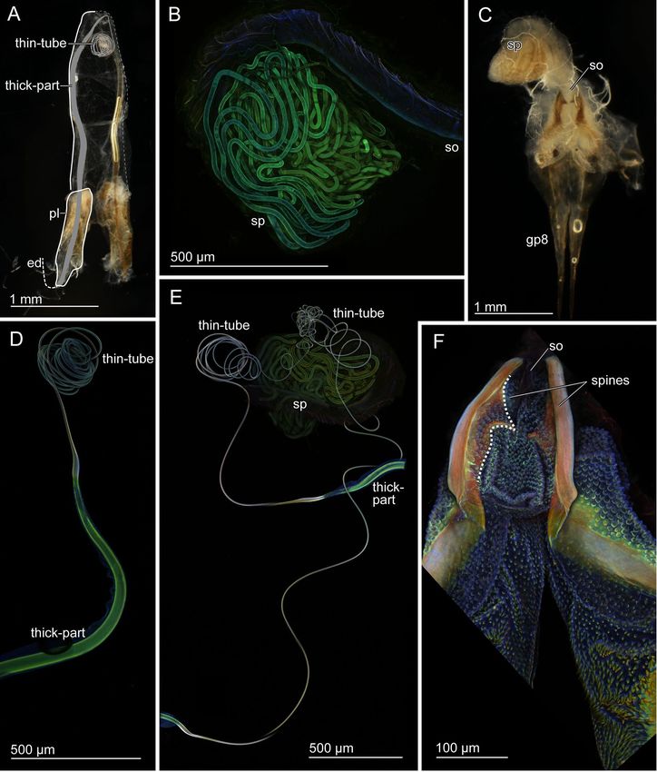
www.nature.com/scientificreports/
Figure 1. Schemes of the male genitalia of Pygidicranidae. The left half is drawn. (A) The virga and the
posterior half of the penis lobe are invaginated in its anterior half, and the entire penis lobe orients cranially in
repose. At the initiation of mating, the penis lobe is unfolded and orients caudally in the female genital chamber
(B), and the invaginated part of the penis lobe and the virga are evaginated and penetrate in the female genital
chamber (C). Abbreviations: ed, ejaculatory duct; pl, penis lobe; pm, paramere; vir, virga.
the flagellum11,12.. The generality of these functional principles, i.e. the cylindrical geometry, bending stiffness
gradient of the flagellum, and higher bending stiffness of the flagellum comparing to the spermatheca, has not
been clarified yet.
In the present study, we aimed to test the generality of the abovementioned functional principles demon-
strated and hypothesised based on the cassidine beetle genitalia and addressed the following questions: (1) Are
the functional principles limited to the beetles? (2) Are they correlated to the genital length? (3) Do these func-
tional principles depend on the mechanical properties of corresponding female structures? For this purpose, here
we chose a distantly related insect taxon, order D ermaptera19 and compared their genitalia with the cassidine
beetle genitalia. We focused on two pygidicranid earwig species Echinosoma horridum Dohrn, 1863, whose
intromittent organ, termed virga, is long and narrow20, and E. denticulatum Hincks, 1959, whose homologous
structure is conversely s hort21,22. We characterised their geometry and analysed material distributions within
male and female interacting genital structures using imaging and microscopy techniques, e.g. scanning electron
microscopy and confocal laser scanning microscopy. The material stiffness (i.e., Young’s modulus) was estimated
based on micrographs, and resulting data were used to calculate the bending stiffness (i.e., flexural rigidity) of
the virga and spermatheca. Based on results, we determine the general principles of the penetration mechanics
of the genitalia in earwigs and compare them with the previous studies on the beetle genital.
Results
Morphology of genitalia. As a plesiomorphic characteristic of the extant earwigs, males of Echinosoma
species have paired penis lobes, each of which encloses a rod- or filament-like virga that orients cranially in repose
(Fig. 1A)20,23. Females of Echinosoma have a single, tube-shaped sperm storage organ, termed spermatheca21,24.
In most earwig species, one of the paired penis lobes is unfolded and inserted into the female vagina at the initia-
tion of mating, and the virga is inserted into the spermatheca of a female and functions as a sperm-transferring
tube during copulation (Fig. 1B,C)25.
In E. horridum, males have extremely long paired virgae, which are almost as long as their body length
(Table 1). The virga is composed of a thicker base, which tapers towards the apex and a long thin tube, which
coils in the genital chamber when the virga is not in use (thick-part and thin-tube, Fig. 2A). Their transition zone
tappers sharply and appears to be structured (Fig. 4A,B), as if the gonopore was situated, but our observation
revealed that the virga, from the thicker base to the distal end of the thin-tube, is an entire tube with no opening
in this zone (Fig. 4C–I). The penis lobe of E. horridum males does not carry any spiny auxiliary sclerites, unlike
that of E. denticulatum. In the female, the spermatheca is situated on the dorsal side of the vagina (Fig. 2C) and
Scientific Reports | (2021) 11:7920 | https://doi.org/10.1038/s41598-021-86864-1 2
Vol:.(1234567890)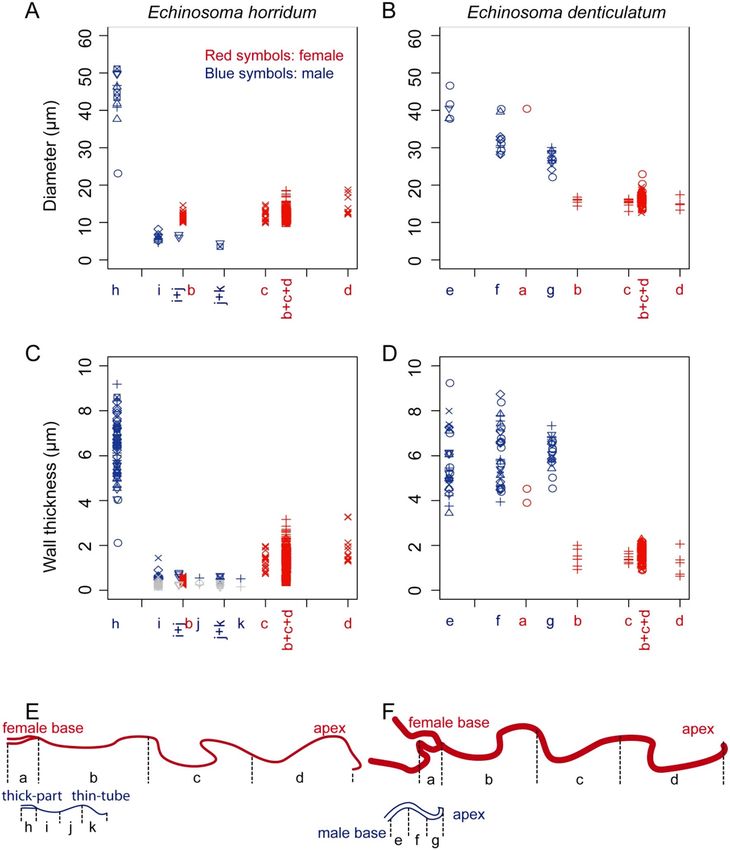
www.nature.com/scientificreports/
Male Female
Body length (mm) Virga length (mm) Body length (mm) Spermatheca length (mm)
Echinosoma horridum 12.66 ± 0.19 (4) 10.77 ± 1.02 (5) 11.89 ± 0.47 (3) 45.84 ± 2.20 (2)
Echinosoma denticulatum 12.71 ± 0.28 (3) 0.58 ± 0.018 (4) 12.79 ± 2.02 (3) 8.46 (1)
Table 1. Measurements of the body length and genitalia of Echinosoma horridum and E. denticulatum,
mean ± sd. Forceps lengths were included in the body size measurements. The numbers in the parentheses
represent the number of individuals used for measuring the lengths.
is a convoluted tube (Fig. 2B). The spermatheca of E. horridum is more than four times longer than the insect
body length (Table 1). Paired spines are present and lay adjacent to the spermathecal opening (Fig. 2F).
Males of E. denticulatum possess very short paired virgae (Fig. 3A, Table 1), carrying a radially expand-
ing brim at the tip (Fig. 6A,B). The invaginated surfaces of the penis lobes are covered with dozens of spines
(Fig. 3G,H). The penis lobe encloses a helicoidal virga (Fig. 3D) and an associated sclerite, termed spring herein,
due to the resilin richness as described below (see material distributions). The spring is also helicoidal and turns
in the opposite direction, forming a seemingly double-helix structure with the virga (see vir and spr in Fig. 3G,H).
Females carry hundreds of long setae at the spermathecal opening (Fig. 3F). The length of the spermatheca
of E. denticulatum is two-thirds of the body length (Table 1), and the intact spermatheca is a convoluted tube
(Fig. 3B,C).
The geometry of virgae and spermathecae. To examine the geometry, we cut specimens into either
three (base, middle, apex) or four (thick-part, thin base, thin middle, thin apex in the E. horridum male) regions,
observed their cross-sections and measured the diameter and wall thickness. Due to the difficulty in sample
preparation, we measured these parameters as many as we could from every individual. We calculated the
measurements described below based on all measured data and performed statistical tests using means of the
measurements for each individual to avoid pseudoreplication. Detailed measurement data is available in sup-
plementary Tables 1–6.
The cross-sections of the virga in E. horridum was circular in the thick-part (Fig. 5E,F). The virga abruptly
flattened in the transition zone between the thick-part and the thin-tube (Fig. 4A,B). The internal surface of
the thick-part partly carried microtrichia (Fig. 5E). The entire thin-tube was hollow and was flattened semi-
circular to crescent-like in cross-section (Fig. 5A–D). The diameter of the thick-part was 43.20 ± 7.87 µm (N = 11)
[mean ± sd], and the wall thickness of the same location was 6.28 ± 1.30 µm (N = 61) (Fig. 7A,C,E). The thin-
tube had a diameter of 5.74 ± 1.23 µm (N = 12) (Fig. 7A), which was calculated from circumferences with an
assumption that the cross-section is annular. The wall thickness was not constant within a circumference; the
outer side of the circumference had a thicker wall comparing with that of the inner side [0.60 ± 0.20 µm (N = 27)
vs 0.26 ± 0.10 µm (N = 24)] (Figs. 5A–D, 7C). Among the measurement positions of the thin-tube, the diameter
(Kendall’s rank correlation coefficient τ = − 0.467, N = 7, P = 0.1742), the wall thicknesses of the outer (τ = − 0.281,
N = 11, P = 0.264) and the inner side (τ = − 0.185, N = 11, P = 0.457) were statistically consistent with neither
increasing nor decreasing trend.
The cross-section of the virga in E. denticulatum was approximately circular (Fig. 6). The virga tapered from
the base to the apex [40.83 ± 3.62 µm (N = 5) in the base, 32.23 ± 3.92 µm (N = 12) in the middle, 26.91 ± 2.36 µm
(N = 11) in the apex] (τ = − 0.779, N = 14, P < 0.001) (Fig. 7B,F). The wall thickness of the virga was constant along
the measurement locations (τ = 0.233, N = 18, P = 0.225) and was 5.94 ± 1.19 µm (N = 81) (Fig. 7D,F).
The spermatheca geometry of both species was simpler than the male ones. In both species, it showed a
circular cross-section (Figs. 5G,H, 6G,H). In E. horridum, the diameter was constant among the measurement
locations [12.66 ± 1.72 µm (N = 158)] (τ = 0.258, N = 6, P = 0.499) (Fig. 7A,E), and the wall thickness slightly
increased from the base to the apex [0.44 ± 0.094 µm (N = 45) in the base, 1.25 ± 0.39 µm (N = 19) in the middle,
1.91 ± 0.66 µm (N = 13) in the apex] (τ = 0.775, N = 6, P = 0.042) (Fig. 7C,E). In E. denticulatum, the diameter
was constant among the measurement locations except for the widened basal part [16.20 ± 1.68 µm (N = 70)]
(τ = − 0.430, N = 6, P = 0.260) (Fig. 7A,B). The wall thickness of E. denticulatum was also constant along the
spermatheca [1.71 ± 1.49 µm (N = 122)] (τ = − 0.120, N = 5, P = 0.782) (Fig. 7D,F).
Material distributions. To analyse material distributions within the virga and spermatheca in the study
species, we applied the method established by Michels and G orb26 using confocal laser scanning microscopy
(CLSM). We visualised autofluorescence of materials comprising the cuticle with four sets of lasers and filters
and assigned blue, green, red (50% saturation) or red (50% saturation) to each image produced with each set of
lasers and filters, respectively. We interpreted resulting overlaid images as follows: (1) red-coloured structures
are well-sclerotised and stiff, (2) green coloured ones are less sclerotised and (3) blue-coloured ones are com-
posed of the larger amount of resilin and are likely softer than the other parts.
Heterogeneity of a material distribution along the virga was detected in the virga of E. horridum, but it was
not remarkable (Fig. 2D, supplementary Fig. 1). It tended that the thick-part of the virga was visualised with
green and that the thin-part was whitish-blue. The transition zone between the thick-part and thin-tube was
visualised with white, i.e. a mixture of three primary colours. The thick-part is likely moderately sclerotised,
while the thin-part is less sclerotised comparing to the thick-part of the virga within each individual. The tran-
sition zone may be more sclerotised than the rest. Among the four virgae used for the CLSM analysis, whose
Scientific Reports | (2021) 11:7920 | https://doi.org/10.1038/s41598-021-86864-1 3
Vol.:(0123456789)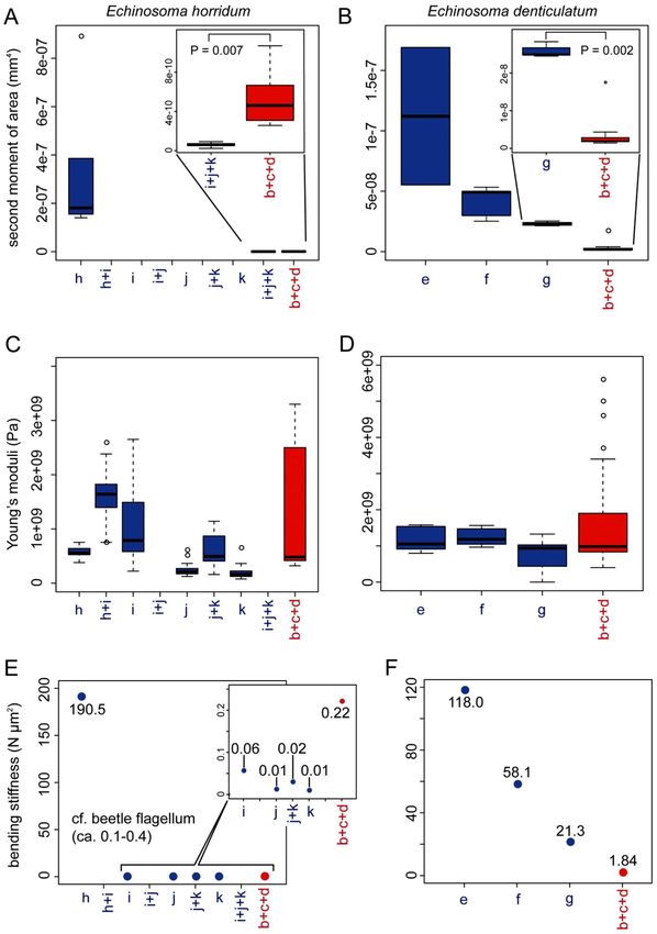
www.nature.com/scientificreports/

                                            Figure 2.  Male and female genitalia of Echinosoma horridum. (A, C) Stereomicrographs, (B, D–F) confocal
                                            laser scanning micrographs. (A) Male genitalia. Paired virgae are situated in the genitalia chamber and directed
                                            cranially in repose. Half of the paired structures is highlighted. (B) Spermatheca. (C) Female genitalia. (D)
                                            Virga. (E) Virgae and spermatheca. (F) A pair of spines in the vicinity of a spermatheca opening. Abbreviations:
                                            ed, ejaculatory duct; gp8, gonapophysis VIII; pl, penis lobe; so, spermatheca opening; sp, spermatheca; vir-thick,
                                            thick-part of virga; vir-thin, thin-tube of virga.

                                            autofluorescence intensities were individually adjusted, there was one exceptional case where the thin-tube was
                                            visualised with reddish colouration. However, the thick-part and transition zone were comparable to the results
                                            mentioned above (supplementary Fig. 1).
                                               The spermatheca of E. horridum seems to be moderately sclerotised (green to reddish-green), and no gra-
                                            dients were observed except for the proximal part, plausibly membranous (dark blue) (Fig. 2B, supplementary
                                            Fig. 2). Comparisons between the sexes of E. horridum (Fig. 2E, supplementary Fig. 3) showed that the virga

          Scientific Reports |   (2021) 11:7920 |                https://doi.org/10.1038/s41598-021-86864-1                                                  4

Vol:.(1234567890)
www.nature.com/scientificreports/
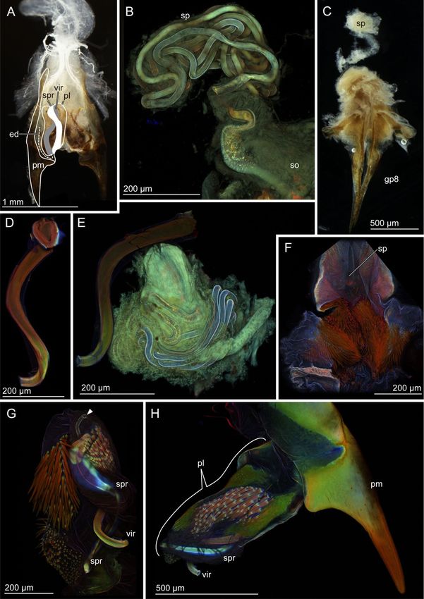
Figure 3. Male and female genitalia of Echinosoma denticulatum. (A, C) Stereomicrographs, (B, D–H) confocal
laser scanning micrographs. (A) Male genitalia. Paired penises are situated in the genitalia chamber and direct
cranially in repose as seen here. Half of the paired structures is highlighted. (B) Spermatheca. (C) Female
genitalia. (D) Virga. (E) Virga and spermatheca. (F) Numerous setae surrounding a spermatheca opening. (G)
Virga and its surrounding penis lobe carrying a resilin-enriched spring and numerous spines. The arrowhead
denotes the opening of the ejaculatory duct. (H) Paramere, virga and penis lobe. Abbreviations: ed, ejaculatory
duct; gp8, gonapophysis VIII; pl, penis lobe; so, spermatheca opening; sp, spermatheca; spr, spring; vir, virga.
Scientific Reports | (2021) 11:7920 | https://doi.org/10.1038/s41598-021-86864-1 5
Vol.:(0123456789)www.nature.com/scientificreports/
Figure 4. Scanning electron micrographs of the transition zone of the virga of Echinosoma horridum. (A)
Overview. The inner side. (B) Overview. The outer side. (D–F) Magnified surfaces marked in images (A) and
(B). (G–I) Caudal views of the transition area between the thick-part and thin-tube of the virga.
and spermatheca were green to white, indicating that sclerotised levels of these structures are comparable to
each other.
Scientific Reports | (2021) 11:7920 | https://doi.org/10.1038/s41598-021-86864-1 6
Vol:.(1234567890)www.nature.com/scientificreports/
Figure 5. Geometry of the virga and spermatheca of Echinosoma horridum. Confocal laser scanning
micrographs (A–C,F) and scanning electron micrographs (D, E, G, H). (A–C) Optical slices of the thin-tube of
the virga taken using CLSM. Cross-sections are enclosed with broken lines. (D) A cross-section of the thin-tube
of the virga. The cross-section was visualised by cutting the thin-tube with micro scissors that caused slight
deformation (cf. images A–C). (E) A cross-section of the thick-part of the virga. Microtrichia in the lumen are
visible. (F) The thick-part of the virga. A maximum intensity projection. A distal part is optically cut so that the
hollow inside is also visible. (G, H) Cross-sections of the spermatheca. Spermatozoa are visible (H).
Scientific Reports | (2021) 11:7920 | https://doi.org/10.1038/s41598-021-86864-1 7
Vol.:(0123456789)www.nature.com/scientificreports/
Figure 6. Geometry of the virga and spermatheca of Echinosom denticulatum. Scanning electron micrographs
of the virga (A–F) and the spermatheca (G, H). (A) The very tip of the virga. (B) A magnified image of the
tip of the virga. (C–F) Cross-sections of the virga at the apex (C), at the middle (D) and at the base (E). (F)
A magnified surface images of the inner surface of the virga. (G, H) Cross-sections of the spermatheca.
Spermatozoa are visible (H).
A material gradient was observed along the virga of E. denticulatum (Fig. 3D, supplementary Fig. 4). The
virga has a well sclerotised base and middle, which were visualized in red, and possesses a less sclerotized apical
part (Fig. 3D, supplementary Fig. 4).
The spermatheca of E. denticulatum was visualised homogeneously with bluish-green, indicating that it is
likely less sclerotised (Fig. 3B, supplementary Fig. 5). Comparative results were also obtained from micrographs
of male and female structures recorded together (Fig. 3E, supplementary Fig. 6). These images suggest that the
spermatheca is likely less sclerotised in comparison to the virga but the very tip of the virga.
We analysed autofluorescence of some other genital structures as well, i.e. genital spines and thick setae
(Figs. 2F, 3F–H). The hundreds of penile spines and long female setae of E. denticulatum and the paired spines
of the E. horridum female were red in all the analysed conditions (Figs. 2F, 3F–H). It seems that those long setae
and spines are well-sclerotised. Another remarkable result is that the sclerite situated at the penis lobe of E.
denticulatum is seemingly resilin dominated, which here is termed spring (Fig. 3G,H).
Bending stiffness (flexural rigidity, EI). Bending stiffness is defined as the product of Young’s modulus
(E) and the second moment of area (I). Bending stiffness of the virga and spermatheca was estimated using the
Scientific Reports | (2021) 11:7920 | https://doi.org/10.1038/s41598-021-86864-1 8
Vol:.(1234567890)www.nature.com/scientificreports/
Figure 7. Geometry of the virga and spermatheca of Echinosoma horridum (A, C, E) and E. denticulatum (B, D,
F). (A, B) Outer diameters of the virga and spermatheca. (C, D) Wall thicknesses of the virga and spermatheca.
These were not constant along circumferences in the thin-tube of the virga of E. horridum, and larger wall
thicknesses and smaller ones are shown respectively with blue and grey colouration in (C). Each symbol in
(A–D) represents data from one individual. The letters correspond to locations, whose diameters were measured
and shown in schemes of the virga and spermatheca (E, F). Since it was impossible to measure wall thicknesses
and diameters for each area separately, we needed to categorise some areas together, i.e. i + j, j + k and b + c + d.
Physically interacting areas between the virga and spermatheca during mating are depicted as overlapped each
other in every graph.
calculated second moment of area based on either SEM or CLSM images using the BoneJ plugin27 for Fiji and the
estimated Young’s modulus based on the CLSM images using the program developed by Eshghi et al.28.
The second moment of area, i.e. a measurement of structural stiffness results from a cross-sectional shape, was
calculated based on the SEM or CLSM images, in which cross-sections were neither deformed nor tilted. Dried
samples that were prepared for the SEM were brittle and could simply be broken (i.e. no plastic deformation),
except for the thin-tube of the virga of E. horridum, which was impossible to fracture. Hence, we had to cut them
using a blade and micro-scissors, which could have resulted in permanent deformation of cross-sections (see
Scientific Reports | (2021) 11:7920 | https://doi.org/10.1038/s41598-021-86864-1 9
Vol.:(0123456789)www.nature.com/scientificreports/
Fig. 5A–C vs Fig. 5D, which shows the least deformation among SEM images we have taken). Therefore, for the
thin-tube of the virga of E. horridum, we used optical slices of CLSM images (Fig. 5A–C), but it was impossible
to recognise the base, middle and apex of the thin-tube with this method (Fig. 8A).
In E. horridum, the second moment of area was more than three orders of magnitude higher in the thick-part
of the virga (1.81 × 10⁻⁷ mm4, hereafter we present medians and used them for calculation of the bending stiff-
ness, due to high variations in estimated Young’s moduli presented below) than that of the thin part (6.02 × 10⁻¹¹
mm4). The second moment of area of the spermatheca (4.61 × 10⁻¹⁰ mm4) was statistically higher than that of the
thin-part of the virga (Mood’s median test: N male = 6, N female = 15, P = 0.0070) (Fig. 8A). This result corresponded
to the abovementioned geometric data showing that the diameter and the wall thickness of the spermatheca of E.
horridum were smaller than those of the thick-part of the virga and larger than those of its thin-tube (Fig. 7A,C).
To check the validity of the data, we calculated the second moment of area manually based on the abovemen-
tioned geometric measurements (see the formula in materials and methods), and the manually calculated values
were comparable to these ones.
The second moment of area of the virga in E. denticulatum decreased from the base to the apex (1.12 × 10⁻⁷
mm4 in the base, 4.91 × 10⁻⁸ mm4 in the middle and 2.27 × 10⁻⁸ mm4 in the apex; τ = − 0.744, N = 11, P = 0.0039;
Fig. 8B). The corresponding value of the spermatheca except for the widened basal region was statistically
smaller (1.88 × 10⁻⁹ mm4) than that of the virga in the apical region (Wilcoxon rank-sum test: W = 40, N male = 4,
N female = 10, P = 0.0020) (Fig. 8B). This difference was also visible in the geometric data (Fig. 7B). The manually
calculated second moment of area was comparable to the calculations.
Estimated Young’s moduli varied much within each measurement location (Fig. 8C,D). The estimated Young’s
moduli showed neither increasing nor decreasing tendency along the measurement locations in the male (E.
horridum: the thick-part: 0.56 GPa; the transition zone between the thick-part and thin-tube: 1.64 GPa, the thin-
tube: 0.16–0.79 GPa; τ = − 0.744, N = 7 , P = 0.244; Fig. 8C) (E. denticulatum, the base: 1.05 GPa; the middle: 1.18
GPa, the apex: 0.94 GPa; τ = − 0.415, N = 6, P = 0.164; Fig. 8D). However, the sclerotisation level of the transition
zone of the virga in E. horridum may be slightly higher than the rest of the virga [τ = − 0.415, N = 8, P = 0.164 (the
thick-part was removed from the statistical test); Fig. 8C], and this corresponded to the CLSM results described
above. Although it was not statistically significant, in E. denticulatum, the apical part of the virga showed a slightly
lower Young’s modulus than that of the rest, as expected based on the CLSM results described above. The Young’s
moduli in the female except for the widened basal region were estimated without any information on measure-
ment locations, and all data were pooled for each species (E. horridum: 0.48 GPa, E. denticulatum: 0.98 GPa).
Based on those results (the Young’s modulus and second moment of area), we calculated bending stiffnesses of
the virga and spermatheca (Fig. 8 E, F). Since there was no significant difference in the estimated Young’s moduli
among the measurement locations, the results resembled those of the second moment of area. In E. horridum, the
thick-part of the virga showed the highest bending stiffness (190.5 N µm2) followed by the spermatheca (0.22 N
µm2) and the thin-tube (0.01–0.06 N µm2) (Fig. 8E). Since it was not possible to calculate the second moment
of area at each measurement location with our method (Fig. 8A), the sole I value calculated in the study was
applied to all the estimated Young’s moduli of the thin-tube of E. horridum (Fig. 8C,E). The bending stiffness of
E. denticulatum was overall higher in the male than that in the female (Fig. 8F). In the basal and middle regions
of the virga (118.0 N µm2 in the base and 58.1 N µm2 in the middle), the bending stiffnesses were higher than
that of the apical region (21.3 N µm2) and that of the spermatheca except for the basal widened region (1.84 N
µm2) (Fig. 8F).
Discussion
The present study shows that males and females of the pygidicranid earwig E. horridum have extremely long
genitalia (85.1% of body length in the male and 386% in the female). In contrast, in the other earwig species
E. denticulatum, the genitalia are much shorter (4.5% of body length in the male and 66.1% in the female).
Despite the tremendous difference in genital size, the genital configuration is common between the study spe-
cies: Male external genitalia are composed of paired penis lobes with virgae situated in the genital chamber with
the position directing cranially (Fig. 1A), and females have one spermatheca. This configuration of male and
female genitalia is commonly known in Pygidicranidae21,25. Male genitalia with paired penis lobes are plausibly
plesiomorphic conditions in Dermaptera29,30, although a plesiomorphic state of the orientation of the penises
in repose remained u nclear30.
Kamimura and L ee21 examined genital coupling of E. denticulatum and reported that one of the paired penis
lobes is extended caudally and evaginates during mating (Fig. 1B,C). The virga is then inserted into the female
vagina and the wide proximal region of the spermatheca for transferring sperm. The penis lobe functions as
a storage space of the virga and spiny area in repose21. In the present study, we found a resilin-enriched and
helicoidal spring forming a double-helix structure with the helicoidal virga (Fig. 3G,H). The spring likely assists
the virga to quickly move back and forth while the penis lobe is evaginated by increased haemolymph pressure
and is pulled back.
Moreover, except for the basal widened region where is plausibly membranous, the spermatheca is narrower
than the virga (Fig. 7B). This implies that the tip of the virga, carrying a radially expanding brim, can fit the
entrance of the thin spermathecal duct and avoid flowing sperm backward during insemination. At the same
time, the tip of the short virga may encounter the highest risk of breakage during the insertion process since the
tip has frequent physical contacts while exposed to the female genital chamber. Therefore, we expect that the
bending stiffness gradients found in the virga of E. denticulatum, particularly the less sclerotised and flexible
apical part, reduce the contact stresses at this critical zone.
Similar bending stiffness and material gradient were previously reported for the intromittent organ of Cassida
rubiginosa (Table 2)11,12. In this species, the flexibility of the elongated intromittent organ is possibly essential to
Scientific Reports | (2021) 11:7920 | https://doi.org/10.1038/s41598-021-86864-1 10
Vol:.(1234567890)www.nature.com/scientificreports/
Figure 8. The second moment of area, Young’s moduli and bending stiffnesses along the virga and spermatheca
of Echinosoma horridum (A, C, E) and E. denticulatum. (B, D, F). (A, B) The calculated second moment of
area using Fiji equipped with the plugin BoneJ. (C, D) Estimated Young’s moduli. (E, F) Resulting bending
stiffnesses. The alphabets correspond to locations whose second moment of area and Young’s modulus were
measured and shown in schemes of the virga and spermatheca (E) and (F) in Fig. 7.
fit into coils of the spermathecal duct. It is worthwhile to mention that the similar bending stiffness gradients
Scientific Reports | (2021) 11:7920 | https://doi.org/10.1038/s41598-021-86864-1 11
Vol.:(0123456789)www.nature.com/scientificreports/
Male intromittent organ Interaction between the sexes Spermatheca
Relative length to body Bending stiffness Material distribution Penile penetration Flexibility, comparing
Species length gradient heterogeneity mechanism to the spermatheca Shape
Echinosoma denticu- a Present, apical region Present, but not a,e a
4.56% (0.58 mm) Pushed from the base Smaller in the male Convoluteda,e
latum more flexiblea prominenta
The thin-tube gradually
Larger in the male (the
No gradient, constant penetrates a folded site,
Echinosoma horridum 85.1% (10.8 mm)a Absent in the thin-tubea thin-tube of the virga vs Convoluteda
along the thin-tubea not from the tipa (a
spermatheca)a
hypothesis, Fig. 9)
157% of body length Present, apical region
Cassida rubiginosa Present at the tipc,d Pushed from the basef Larger in the maleb,g Regularly coiledf,h
(10.2 mm)b more flexiblec
Not mandatory for pen-
etration of the elongated Universal prerequisite
Current explanations Plausibly reduces the risk
- penis, and plausibly - for penetration of the -
(hypothesis) of penile breakage
reduces the risk of penile elongated penis
breakage
Table 2. A comparison of the penetration mechanics between Echinosoma genitalia and Cassida rubiginosa
genitalia documented and hypothesised in the present and previous studies. a The present study, bMatsumura
et al.36, cMatsumura et al.12, dFilippov et al.11, e Kamimura and Lee21, fMatsumura et al.18, g Matsumura et al.
(submitted), h Filippov et al.10.
plausibly function as assistances of the variable penetration processes in distantly related insect taxa. The mate-
rial gradient at the tip of C. rubiginosa presumably avoids its breakage12, as it is also hypothesised in the present
study for E. denticulatum. Although the length of the virga of E. denticulatum is much shorter than that of E.
horridum and C. rubiginosa, the penile penetration mechanism is fundamentally the same between E. denticu-
latum and C. rubiginosa, i.e. the elongated intromittent organ is merely pushed from its base into the sclerotised
spermatheca (Table 2)18. This commonality between the distantly related species suggests that the presence of
material heterogeneity at the apical or tip region is likely related to its penetration mechanism, and this working
hypothesis has to be further tested.
In contrast to E. denticulatum, information on the reproductive biology in E. horridum is hitherto limited.
However, since the configuration of the male and female genitalia are congruent between the studied species
except for the absence of a spring on the penis lobe in E. horridum, the penetration mechanism of the virga may
be very alike. In other words, increased haemolymph pressure in one of two penis lobes, carrying the slender
virga, causes posterior extension of the penis lobe, evaginating and pushing the virga into the spermatheca
(Fig. 1). Contrary to E. denticulatum, our geometric data showed that the diameter of the thin-tube in E. horridum
is small enough to deeply penetrate the spermatheca (Fig. 7A), which implies that the thin-tube of the virga of E.
horridum can be fully inserted into the spermatheca. The present study also documented that the slender virga
is an entire tube continuing from the gonopore, indicating that the male uses the virga to transfer sperm, as in
E. denticulatum. Moreover, the spermatheca of E. horridum is much longer than that of E. denticulatum. Such a
positive correlation between the virga and spermatheca lengths connotes that the long virga has physical interac-
tions with the spermatheca since such a correlation between elongated intromittent organs and corresponding
spermathecae are well-known in i nsects15,31–33.
It appears to be a challenge for males to insert and withdraw the extremely slender virga (aspect ratio, 1:2117)
into the spermatheca since the high aspect ratio can easily cause buckling of the structure. Although it is known
that indeed such long virgae in other earwigs penetrate the s permatheca34,35, the insertion and withdrawal mecha-
nisms remain unclear. The geometry and bending stiffness reported here on E. horridum suggest the following
penetration mechanism (Fig. 9). (1) While either of two penis lobes is unfolded from cranially to caudally during
mating (Fig. 9A), (2) the thin-tube base of the virga is folded due to a limited space of the female genital chamber
and the spermatheca (Fig. 9B), and the fold is inserted to the spermatheca (Fig. 9C). (3) At the same time, the
thin-tube of the virga, which is plausibly filled with sperm due to capillary action36, and the fold move deeply in
the spermatheca by the capillary force (Fig. 9C). The possible withdrawal mechanism of the virga is that the male
pulls the entire virga out by moving apart from a female mate, while the penis lobe is moved cranially to store the
whole virga back to the male genital chamber probably by a combination of the decrease of haemolymph pres-
sure and protractor muscles. Analogies of these hypothetical insertion and withdrawal mechanisms are known
in the fruit fly Ceratitis capitata37 and the rove beetle Aleochara tristis38. Still, our explanation for E. horridum is
merely a hypothesis, and the reproductive biology of this species has to be investigated.
This hypothetical insertion and withdrawal mechanisms can explain why neither geometric measurements
(Fig. 7A) nor calculated bending stiffnesses (Fig. 8E) of the virga of E. horridum shows gradients or heterogene-
ity of material distributions, unlike the short virga of E. denticulatum. With this penetration mechanism, the
prerequisite is high pliability of the virga. The bending stiffness of the thin-tube of the virga is markedly lower
than the virga of E. denticulatum (Fig. 8E,F), and due to the semi-circular cross-section, the thin-tube tends to
bend in one direction (Fig. 9A,C). Because of this feature, we could not break even the dried thin-tubes, which
were just bent, unlike our other specimens.
Another noteworthy point we found is that the bending stiffness of the thin-tube of the virga is much lower
than that of the spermatheca of E. horridum, but it is reverse in E. denticulatum. Filippov et al.11 numerically
demonstrated that a stiff intromittent organ of males deforms a less stiff spermatheca during penetration and
this penile behaviour leads to a slower penetration. By contrast, a stiff intromittent organ with a flexible apical
Scientific Reports | (2021) 11:7920 | https://doi.org/10.1038/s41598-021-86864-1 12
Vol:.(1234567890)www.nature.com/scientificreports/
Figure 9. Schemes of the male genitalia of Echinosoma horridum and the hypothetical penetration mechanism
of the virga. The left half is drawn. (A) The virga and the posterior half of the penis lobe is invaginated in its
anterior half, and the entire penis lobe orients cranially in repose. At the initiation of mating, the penis lobe
is unfolded and orients caudally in the female genital chamber (B), and then the invaginated part of the penis
lobe and the virga are evaginated (C). At the same time, the thin-tube of the virga is folded and inserted into the
spermatheca, and due to ejaculate pressure generated by the capillary action, the fold moves deeper and deeper
into the spermatheca (C). Abbreviations: pl, penis lobe; pm, paramere; sp, spermatheca; sp-wide, the proximal
and wide region of the spermatheca.
region results in a comparatively quick p enetration11, suggesting a smooth penetration. The combination of a
flexible male and comparatively stiff female genitalia is also experimentally demonstrated in Cassida rubigi-
nosa12 (see Table 2) . Although to date, only two exemplary cases showed this relative stiffness combination, we
consider this can be one of the universal principles of the penetration mechanics of slender and hyper-elongated
intromittent organs.
In Table 2, we summarised findings and insights regarding the penetration mechanics of earwig and beetle
genitalia. In this way, we may partially address the three questions posed in the introduction: (1) Are the func-
tional principles limited to the beetle species? (2) Are they correlated to the genital length? (3) Do the functional
principles depend on the mechanical properties of corresponding female structures? As discussed above, the
functional principles found in the beetle were only partially applicable to the earwig species, and not all of them
are associated with the genital length. The male genitalia are more flexible than the corresponding female genitalia
in both species with the hyper-elongated penis. Interestingly, the bending stiffness gradient, i.e. the apical part is
more flexible than the rest in the male, was found in E. denticulatum with the short virga and C. rubiginosa with a
hyper-elongated penis. Irrespective of a huge difference in the length of the male genitalia of E. denticulatum and
C. rubiginosa, it is conceivable that the bending stiffness gradient plays a vital role in the penetration mechanics
due to their similar penetration mechanism.
The present and previous studies unveiled that a number of variable factors affect penetration mechan-
ics, and we hope that further research will shed more light on the penetration mechanics. In addition to this,
functions of female genital spines, setae and microtrichia, observed on the inner surface of the virga, are still
entirely unclear. Although these do not seem to be directly related to the topic of the present study, these details
Scientific Reports | (2021) 11:7920 | https://doi.org/10.1038/s41598-021-86864-1 13
Vol.:(0123456789)www.nature.com/scientificreports/
could be important to understand the penetration process more comprehensively. The reproductive biology
and morphology of earwigs have to be further examined. Earwigs would be a great candidate group to unveil
the penetration mechanics from a comparative perspective due to their remarkable diversity of the virga and
spermatheca morphology and function25. In Dermaptera, for example, it is known that the virga of Euborellia
plebeja (Dohrn, 1863) (Anisolabididae) is used for sperm removal and that its tip is specialised to rake s perm34,
whereas the virga tip of Marava arachidis (Yersin, 1860) (Spongiphoridae) is curved and slightly swollen in shape,
and the virga may not be used for physical removal of rival sperm35. In future studies, they may be candidate
species for comparison to Echinosoma species.
Material and methods
Specimens. Echinosoma denticulatum specimens were collected in forested areas of Bukit Jambul and Bukit
Kukus, Penang Island, Malaysia and were kept in laboratory conditions at 26 ± 1 °C (12 h photoperiod)21. Sam-
ples of E. horridum were collected in forested areas in Perak and Selangor states, Malaysia and kept in similar
condition as above. This field study was conducted in 2012 and 2013 with the approval of the Economic Planning
Unit, Malaysia (Reference No. UPE: 40/200/19/2844). In total, we examined five males and five females of E.
denticulatum and eight males and four females of E. horridum. We mainly used either anaesthetised animals or
specimens preserved in a conventional freezer and also investigated some specimens preserved in 70% ethanol,
as described below in detail. Before dissection, entire bodies of earwigs were photographed at different focus
planes using a Leica M205A stereomicroscope equipped with a Leica DFC420 camera, and the photographs
were stacked with the aid of the software LAS V3.8 (Leica Microscopy GmbH, Wetzlar, Germany). Based on
the images, the body length, i.e. the length from the anterior end of the head to the distal tip of forceps (cerci),
was measured using the software Fiji 1.53c39. All specimens were dissected in 0.1 M phosphate-buffered saline
(PBS; pH = 7.4; Carl Roth GmbH & Co. KG, Karlsruhe, Germany) under an Olympus SZX12 stereomicroscope
(Olympus Corporation, Tokyo, Japan).
Genital length measurement. The genital length was measured based on micrographs using the software
Fiji. The male virga lengths of E. denticulatum were measured based on CLSM images. By contrast, virgae of E.
horridum and spermathecae of both species were complex, and it was impossible to measure the lengths based
on CLSM images. Therefore, after CLSM imaging, five specimens of E. horridum virgae were uncurled and cov-
ered with cover glasses. Micrographs of the virgae were taken using a Zeiss Axioplan light microscope equipped
with a Zeiss AxioCam MRc camera (Carl Zeiss Microscopy GmbH, Jena, Germany), and based on the micro-
graphs, the lengths of the virgae were measured using the software Fiji. To measure the spermatheca lengths of
both species, we submerged one sample of E. denticulatum and two spermathecae of E. horridum in 10% KOH
to unfold folded and convoluted ducts.
Geometrical characterisation. Some of the male and female samples used for the CLSM analyses were
rinsed with distilled water and dehydrated with ascending ethanol series (10% to 99.5% ethanol, with each step
20 min, except for 99.5% ethanol, where the samples were kept for a night). Some samples were immersed in
Hexamethyldisilazane (HMDS) and were kept at room temperature, ca. 20 °C, in a fume hood to evaporate the
chemical. In contrast, other samples were dried at critical point using critical point driers (E3100, Quorum Tech-
nologies LTD, Kent, UK and Leica EM CPD 300, Leica Microscopy GmbH). Then, when possible, they were cut
into either three (base, middle, apex) or four (thick-part, thin base, thin middle, thin apex in E. horridum males)
regions using micro-scissors and blades. It was practically not possible to cut them into three or four segments
with exactly same lengths, and segments closer to the base are defined as base and ones closer to the apex were
defined as apex. Each segment was broken into small pieces and perpendicularly placed on SEM stubs to observe
their cross-sections using a Hitachi S-4800 scanning electron microscope (Hitachi High-Tech Corp., Tokyo,
Japan) at an accelerating voltage of 3 kV. All samples were sputter-coated with gold–palladium (ca. 10 nm in
thickness) using a Leica EM SCD500 high vacuum sputter coater (Leica Microscopy GmbH) before observation.
Based on resulting micrographs, we measured diameters and wall-thicknesses of male and female ducts using
the software Fiji. The measurements were analysed using the software R.
The abovementioned SEM-based observations showed that cross-sections of the thin-part of the E. horridum
virga are semi-circular. However, this shape could be an artefact resulting from the cutting of the virga using the
micro-scissors. To confirm this observation, we also applied a Zeiss LSM 700 confocal laser scanning microscope
(Carl Zeiss Microscopy GmbH, Jena, Germany) to other samples. For this purpose, two samples preserved in 70%
ethanol were dissected in distilled water. The virgae were hydrated in distilled water for a while and submerged
in glycerine (≥ 99.5%, free of water, Carl Roth GmbH & Co. KG) on glass slides. Between a cover glass and a
glass slide, we glued transparent reinforcement rings to make a small gap and to avoid contacts between samples
and the glasses following Michels and B untzow40. A stable solid-state laser with a wavelength of either 488 nm
or 555 nm was used for excitation. For the detection of selective emitted autofluorescence, a longpass emission
filter, transmitting light with either wavelength ≥ 490 nm or ≥ 560 nm, was applied, respectively. An object lens
with 20 × magnification (Zeiss Plan-Apochromat, air immersion, NA = 0.8) was used.
Material distribution analyses. Virgae and spermathecae were dissected out in 0.1 M phosphate-buff-
ered saline (PBS; pH = 7.4; Carl Roth GmbH & Co. KG) under an Olympus SZX12 stereomicroscope (Olympus
Corporation) from male and female earwigs either anaesthetised with carbon dioxide or frozen. Then we sub-
merged the virgae and spermathecae in glycerine on glass slides, and as mentioned above, placed a gap holder
between the glasses, not to damage samples. To stabilise samples in glycerine, we kept the samples in a conven-
tional refrigerator (4 ºC) at least for one night. Four stable solid-state lasers with wavelengths of 405 nm, 488 nm,
Scientific Reports | (2021) 11:7920 | https://doi.org/10.1038/s41598-021-86864-1 14
Vol:.(1234567890)www.nature.com/scientificreports/
555 nm and 639 nm were used for excitation of autofluorescence. For detection of selective emitted autofluores-
cence, a bandpass emission filter, transmitting light with wavelengths 420–480 nm or longpass emission filters,
transmitting light with wavelengths ≥ 490 nm, ≥ 560 nm or ≥ 640 nm were applied, respectively. An objective lens
with either 10 × magnification (Zeiss Plan-Apochromat, numerical aperture 0.45) or 20 × magnification (Zeiss
Plan-Apochromat, air immersion, NA = 0.8) was applied. Following Michels & G orb26, we assigned blue, green,
red (50% saturation) or red (50% saturation) to each image produced with the abovementioned four sets of lasers
and filters. Then the resulting images were overlaid with maximal intensity projection using the software Zeiss
Efficient Navigation (ZEN) (Carl Zeiss MicroImaging GmbH). We described the results of material stiffnesses
following the colour code established by Michels & Gorb26 as mentioned above.
Estimation of bending stiffness. We first estimated Young’s modulus of male and female elongated geni-
talia based on their CLSM images using the programme developed by Eshghi et al.28. The programme establishes
a quantitative link between the autofluorescence properties of the constituent components of insect cuticle and
their Young’s modulus. Briefly, the programme detects the primary colour that has the maximum intensity in
each pixel in the CLSM image. The value of the intensity is multiplied by the corresponding Young’s modulus of
each primary (6.8 GPa, 1.5 GPa and 1.95 MPa for the red, green and blue primaries, respectively)28. The validity
of the assigned Young’s moduli has been tested by comparing experimentally measured Young’s moduli of adhe-
sive feet setae of a ladybird beetle41 to the estimated Young’s moduli obtained by the programme28.
To estimate the Young’s moduli in the male, using the programme, we first drew 30 uniformly distributed
lines, each of which is composed of 20 points. And every 20 points were distributed only along the male genitalia.
The Young’s moduli were extracted for every pixel, where the points were located. The obtained Young’s moduli
were averaged once for each line, and then the data that were situated in each measurement location, i.e., base,
middle and apex, were separately pooled. For the spermatheca, we extracted results of arbitrary 20–25 points
on spermathecal walls from each specimen. Since soft tissues surround the spermathecal walls, the standard
method applied to the male samples was not suitable for the female. For a quantitative comparison of the Young’s
moduli between the sexes, we used only CLSM images, in which male and female genitalia were scanned together.
The second moment of area for bending in the direction perpendicular to the longitudinal axis was calculated
using the BoneJ plugin27 for Fiji. For this purpose, cross-section images of male and female genitalia, obtained
with CLSM and SEM, were used. The cross-sections were traced manually with the software Illustrator CS5
(Adobe Systems, California, U.S.A.) to increase image contrast, and the resulting images were imported to Fiji
equipped with the plugin BoneJ. Taken into account that cross-sections often resembled an annulus except for
the thin-part of the virga of E. horridum, to ensure the accuracy of the results obtained by BoneJ, we calculated
the second moment of area using the following equation:
π 4
r − r14
I =
4 2
where I is the second moment of area, r2 is an outer radius and r1 is an inner radius.
The Young’s modulus of each measurement location in combination with their corresponding second moment
of area was used to calculate the bending stiffness of that location.
Statistics. All statistical analyses of the data were conducted using the ’R’ software (ver. 4.0.2)42. Due to dif-
ficulty in sample preparation, we obtained measurements as many as we can from every individual. However,
to avoid pseudoreplication, we calculated means for each individual, except for the data on the second moment
of area where we pooled all measurements. With the data, we tested increasing or decreasing trends of meas-
urements along the male and female genitalia using a Kendall’s rank correlation coefficient. To compare data
between the sexes, we used a Mood’s median test when both the assumptions of normality (tested by a Shapiro–
Wilk test) and homogeneity of variance (tested by a F-test) were rejected. When only the latter assumption was
rejected, we compared the data using a Wilcoxon rank-sum test.
Data availability
Supporting data are available as supporting information.
Received: 28 November 2020; Accepted: 19 March 2021
References
1. Sakes, A., Dodou, D. & Breedveld, P. Buckling prevention strategies in nature as inspiration for improving percutaneous instru-
ments: a review. Bioinspir. Biomim. 11, 21001 (2016).
2. Cerkvenik, U., Dodou, D., van Leeuwen, J. L. & Gussekloo, S. W. S. Functional principles of steerable multi-element probes in
insects. Biol. Rev. 94, 555–574 (2019).
3. Jansen, M. A., Singh, S. S., Chawla, N. & Franz, N. M. A multilayer micromechanical model of the cuticle of Curculio longinasus
Chittenden, 1927 (Coleoptera: Curculionidae). J. Struct. Biol. 195, 139–158 (2016).
4. Jansen, M. A., Williams, J., Chawla, N. & Franz, N. M. Avoidance of catastrophic structural failure as an evolutionary constraint:
biomechanics of the acorn weevil rostrum. Adv. Mater. 31, e1903526 (2019).
5. Singh, S. S., Jansen, M. A., Franz, N. M. & Chawla, N. Microstructure and nanoindentation of the rostrum of Curculio longinasus
Chittenden, 1927 (Coleoptera: Curculionidae). Mater. Charact. 118, 206–211 (2016).
6. Matsumura, Y. et al. Excavation mechanics of the elongated female rostrum of the acorn weevil Curculio glandium (Coleoptera;
Curculionidae). Appl. Phys. A (accepted).
7. Kundanati, L. & Gundiah, N. Biomechanics of substrate boring by fig wasps. J. Exp. Biol. 217, 1946–1954 (2014).
Scientific Reports | (2021) 11:7920 | https://doi.org/10.1038/s41598-021-86864-1 15
Vol.:(0123456789)www.nature.com/scientificreports/
8. Cerkvenik, U., van de Straat, B., Gussekloo, S. W. S. & van Leeuwen, J. L. Mechanisms of ovipositor insertion and steering of a
parasitic wasp. Proc. Natl. Acad. Sci. U.S.A. 114, E7822–E7831 (2017).
9. Cerkvenik, U. et al. Stiffness gradients facilitate ovipositor bending and spatial probing control in a parasitic wasp. J Exp. Biol. 222,
jeb195628 (2019).
10. Filippov, A. E., Kovalev, A. E., Matsumura, Y. & Gorb, S. N. Male penile propulsion into spiraled spermathecal ducts of female
chrysomelid beetles: a numerical simulation approach. J. Theor. Biol. 384, 140–146 (2015).
11. Filippov, A. E., Matsumura, Y., Kovalev, A. E. & Gorb, S. N. Stiffness gradient of the beetle penis facilitates propulsion in the spiraled
female spermathecal duct. Sci. Rep. 6, 27608 (2016).
12. Matsumura, Y., Kovalev, A. E. & Gorb, S. N. Penetration mechanics of a beetle intromittent organ with bending stiffness gradient
and a soft tip. Sci. Adv. 3, eaao5469 (2017).
13. Neufeld, C. J. & Palmer, A. R. Precisely proportioned: intertidal barnacles alter penis form to suit coastal wave action. Proc. R. Soc.
Biol. Lond. B Biol. Sci. 275, 1081–1087 (2008).
14. Matsumura, Y. et al. Two intromittent organs in Zorotypus caudelli (Insecta, Zoraptera): the paradoxical coexistence of an extremely
long tube and a large spermatophore. Biol. J. Linn. Soc. 112, 40–54 (2014).
15. Rodriguez, V., Windsor, D. M. & Eberhard, W. G. Tortoise beetle genitalia and demonstrations of a sexually selected advantage for
flagellum length in Chelymorpha alternans (Chrysomelidae, Cassidini, Stolaini). In New Developments in the Biology of Chrysomeli-
dae (eds Jolivet, P. et al.) 739–748 (AcademicPublisher, the Hague, 2004).
16. Chaboo, C. S. Biology and phylogeny of the Cassidinae Gyllenhal Sensu Lato (Tortoise and leaf-mining Beetles) (Coleoptera:
Chrysomelidae). Bull. Am. Mus. Nat. Hist. 305, 1–250 (2007).
17. Borowiec, L. & Skuza, M. The structure of spermatheca in the genus Chelymorpha Chevrolat, 1837 (Coleoptera: Chrysomelidae:
Cassidinae) and its taxonomic significance. Ann. Zool. 54, 439–451 (2004).
18. Matsumura, Y., Michels, J., Appel, E. & Gorb, S. N. Functional morphology and evolution of the hyper-elongated intromittent
organ in Cassida leaf beetles (Coleoptera: Chrysomelidae: Cassidinae). Zoology 120, 1–14 (2017).
19. Misof, B. et al. Phylogenomics resolves the timing and pattern of insect evolution. Science 346, 763–767 (2014).
20. Hincks, W. D. A Systematic Monograph of the Dermaptera of the World. Part II. Pygidicranidae excluding Diplatyinae. (British
Museum (Natural History), London, 1959).
21. Kamimura, Y. & Lee, C.-Y. Mating and genital coupling in the primitive earwig species Echinosoma denticulatum (Pygidicranidae):
implications for genital evolution in dermapteran phylogeny. Arthropod Syst. Phylo. 72, 11–21 (2014).
22. Kamimura, Y., Nishikawa, M. & Lee, C. Y. The earwig fauna (Insecta: Dermaptera) of Penang Island, Malaysia, with descriptions
of two new species. Zootaxa 4084, 233–257 (2016).
23. Steinmann, H. Dermaptera. Catadermaptera I. Das Tierreich 102, 1–343 (1986).
24. Klass, K.-D. The female genitalic region in basal earwigs (Insecta: Dermaptera: Pygidicranidae s.l.). Entomol. Abh. 61, 173–225
(2003).
25. Kamimura, Y. Pre- and postcopulatory sexual selection and the evolution of sexually dimorphic traits in earwigs (Dermaptera).
Entomol. Sci. 17, 139–166 (2014).
26. Michels, J. & Gorb, S. N. Detailed three-dimensional visualization of resilin in the exoskeleton of arthropods using confocal laser
scanning microscopy. J. Microsc. 245, 1–16 (2012).
27. Doube, M. et al. BoneJ: free and extensible bone image analysis in ImageJ. Bone 47, 1076–1079 (2010).
28. Eshghi, S. H., Jafarpour, M., Darvizeh, A., Gorb, S. N. & Rajabi, H. A simple, high-resolution, non-destructive method for deter-
mining the spatial gradient of the elastic modulus of insect cuticle. J. R. Soc. Interface 15, 20180312 (2018).
29. Kamimura, Y. In search of the origin of twin penises: molecular phylogeny of earwigs (Dermaptera: Forficulina) based on mito-
chondrial and nuclear ribosomal RNA genes. Ann. Entomol. Soc. Am. 97, 903–912 (2004).
30. Wipfler, B. et al. Phylogenomics changes our understanding about earwig evolution. Syst. Entomol. 45, 516–526 (2020).
31. Ilango, K. & Lane, R. P. Coadaptation of male aedeagal filaments and female spermathecal ducts of the old world phlebotomine
sand flies (Diptera: Psychodidae). J. Med. Entomol. 37, 653–659 (2000).
32. Kotrba, M., Huber, J. & Feijen, H. R. Coevolution of male and female genitalia in stalk-eyed flies (Diptera: Diopsidae). Org. Divers.
Evol. 14, 187–201 (2013).
33. Matsumura, Y., Yao, I., Beutel, R. G. & Yoshizawa, K. Molecular phylogeny of the leaf beetle subfamily Criocerinae (Coleoptera:
Chrysomelidae) and the correlated evolution of reproductive organs. Arthropod. Syst. Phylo. 72, 95–110 (2014).
34. Kamimura, Y. Possible removal of rival sperm by the elongated genitalia of the earwig, Euborellia plebeja. Zool. Sci. 17, 667–672
(2000).
35. Kamimura, Y., Tee, H.-S. & Lee, C.-Y. Ovoviviparity and genital evolution: a lesson from an earwig species with coercive traumatic
mating and accidental breakage of elongated intromittent organs. Biol. J. Linn. Soc. 118, 443–456 (2016).
36. Matsumura, Y., Michels, J., Rajabi, H., Shimozawa, T. & Gorb, S. N. Sperm transfer through hyper-elongated beetle penises - mor-
phology and theoretical approaches. Sci. Rep. 9, 10238 (2019).
37. Eberhard, W. G. & Pereira, F. The process of intromission in the mediterranean fruit fly, Ceratitis capitata (Diptera: Tephritidiae).
Psyche 102, 99–120 (1995).
38. Gack, C. & Peschke, K. ’Shouldering’ exaggerated genitalia: a unique behavioural adaptation for the retraction of the elongate
intromittant organ by the male rove beetle (Aleochara tristis Gravenhorst). Biol. J. Linn. Soc. 84, 307–312 (2005).
39. Schindelin, J. et al. Fiji: an open-source platform for biological-image analysis. Nat. Methods 9, 676–682 (2012).
40. Michels, J. & Buntzow, M. Assessment of Congo red as a fluorescence marker for the exoskeleton of small crustaceans and the
cuticle of polychaetes. J. Microsc. 238, 95–101 (2010).
41. Peisker, H., Michels, J. & Gorb, S. N. Evidence for a material gradient in the adhesive tarsal setae of the ladybird beetle Coccinella
septempunctata. Nat. Commun. 4, 1661 (2013).
42. R Core Team. R: A Language and Environment for Statistical Computing (R Foundation for Statistical Computing, Vienna, 2020).
Acknowledgements
We are grateful to Shahab Eshghi and Mohsen Jafarpour (Kiel University, Germany) for their assistance to
estimate Young’s moduli and to calculate the second moment of area. We are grateful also to Jan Michels, Alex-
ander Kovalev and Esther Appel (Kiel University, Germany) for their technical support for drying samples at
the critical point and for sputter coating of the samples. This study was supported through funding from the
German Research Foundation (DFG grant no. MA 7400/1-1) to YM and Grants‐in‐Aid for Scientific Research
(KAKENHI #19K06746) from the Japan Society for the Promotion of Science to YK.
Author contributions
Y.M. conceptualised the study through discussion with H.R., Y.K. and S.N.G.; Y.K. chose suitable species for
our research, collected, and reared the animals with the assistance of C.Y.L.; Y.M. performed experiments; Y.M.
and H.R. did calculations with the assistance of Shahab Eshghi and Mohsen Jafarpour not appearing as authors;
Scientific Reports | (2021) 11:7920 | https://doi.org/10.1038/s41598-021-86864-1 16
Vol:.(1234567890)You can also read