The Complexity of the cGAS-STING Pathway in CNS Pathologies - Frontiers
←
→
Page content transcription
If your browser does not render page correctly, please read the page content below

REVIEW
                                                                                                                                     published: 09 February 2021
                                                                                                                                 doi: 10.3389/fnins.2021.621501

                                            The Complexity of the cGAS-STING
                                            Pathway in CNS Pathologies
                                            Amelia L. Fryer, Amar Abdullah, Juliet M. Taylor* and Peter J. Crack*
                                            Neuropharmacology Laboratory, Department of Pharmacology and Therapeutics, University of Melbourne, Melbourne, VIC,
                                            Australia

                                            Neuroinflammation driven by type-I interferons in the CNS is well established to
                                            exacerbate the progression of many CNS pathologies both acute and chronic. The role
                                            of adaptor protein Stimulator of Interferon Genes (STING) is increasingly appreciated
                                            to instigate type-I IFN-mediated neuroinflammation. As an upstream regulator of
                                            type-I IFNs, STING modulation presents a novel therapeutic opportunity to mediate
                                            inflammation in the CNS. This review will detail the current knowledge of protective
                                            and detrimental STING activity in acute and chronic CNS pathologies and the current
                                            therapeutic avenues being explored.
                         Edited by:
                                            Keywords: STING, neuroinflammation, interferon, central nervous system, cGAS
              Flavia Eugenia Saravia,
        Universidad de Buenos Aires,
                           Argentina
                                            INTRODUCTION
                       Reviewed by:
                         Pavel Katsel,      Type-I interferons (IFNs) have been strongly implicated in the progression of neuroinflammation
   Icahn School of Medicine at Mount
                                            in a host of central nervous system (CNS) pathologies including Alzheimer’s disease (Taylor et al.,
                  Sinai, United States
               Anindya Bhattacharya,
                                            2014; Minter et al., 2016; Roy et al., 2020), Parkinson’s disease (Main et al., 2016; Qin et al., 2016),
 Janssen Research and Development           traumatic brain injury (Karve et al., 2016; Barrett et al., 2020) and amyotrophic lateral sclerosis
        (United States), United States      (ALS) (Oakes et al., 2017; Shelkovnikova et al., 2019). However, the role of the type-I IFN upstream
                  *Correspondence:
                                            regulator, the stimulator of interferon genes (STING), in driving this response in the CNS remains
                       Juliet M. Taylor     largely unknown. Over the last 10 years, STING signalling has been identified as a therapeutic target
               juliett@unimelb.edu.au       in autoinflammatory disorders and cancer with its role in neuroinflammation being increasingly
                        Peter J. Crack      recognised. Therefore, a greater understanding of STING signalling in driving a neuroinflammatory
              pcrack@unimelb.edu.au         response in the diseased brain may also uncover similar therapeutic potential in treating acute and
                                            chronic CNS pathologies.
                   Specialty section:
         This article was submitted to
                  Neuropharmacology,        TYPE-I INTERFERON SIGNALLING
               a section of the journal
            Frontiers in Neuroscience
                                            The type-I IFN response is known to be a key in the innate immune response to viral infection.
        Received: 26 October 2020           However, this response has also been associated with a potent inflammatory response in the
        Accepted: 19 January 2021
                                            absence of pathogen invasion. In the context of viral infection, pathogen-associated molecular
       Published: 09 February 2021
                                            patterns (PAMPs) are produced by the invading pathogen and bind to pattern recognition receptors
                              Citation:     (PRRs) including toll-like receptors (TLR) and cyclic GMP-AMP synthase (cGAS) on the surface of
       Fryer AL, Abdullah A, Taylor JM
                                            resident immune cells such as microglia and astrocytes in the CNS (Bowman et al., 2003; Olson
 and Crack PJ (2021) The Complexity
 of the cGAS-STING Pathway in CNS
                                            and Miller, 2004; Jack et al., 2005). This elicits an array of innate anti-viral responses, notably
                           Pathologies.     the production of pleiotropic pro-inflammatory cytokines known collectively as the type-I IFNs
           Front. Neurosci. 15:621501.      (Koyama et al., 2008; Murira and Lamarre, 2016). PRRs on CNS immune cells are capable of
      doi: 10.3389/fnins.2021.621501        mounting a similar pro-inflammatory response upon detection of endogenous damage-associated

Frontiers in Neuroscience | www.frontiersin.org                                 1                                      February 2021 | Volume 15 | Article 621501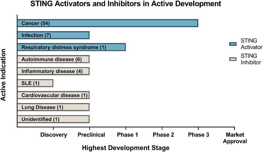
Fryer et al.                                                                                                          STING and Neuroinflammation

molecular patterns (DAMP) released during injury and stress                by TBK1 and IKK, NF-κB also translocates to the nucleus to
(Loane et al., 2014; Cox et al., 2015; Kumar, 2019).                       upregulate the production of proinflammatory cytokines and
   Following binding to their cognate receptor, IFNAR                      chemokines including TNF-α, IL-1β and IL-6, all implicated in
(composed of the IFNAR1 and IFNAR2 subunits), the type-I                   driving the neuroinflammatory response in the CNS (Barnes and
IFNs signal through the Janus kinase (JAK)-signal transducer               Karin, 1997) (Figure 1).
and activator of transcription (STAT) pathway to elicit an
anti-viral, anti-proliferative and immunostimulatory response
through interferon-stimulated gene (ISG) induction (Platanias,             STING ACTIVITY IN VIRAL INFECTIONS
2005; Schneider et al., 2014). This results in the secretion of
proinflammatory cytokines and chemokines, including tumour                 Much of our knowledge of STING signalling in the brain
necrosis factor-α (TNF-α), interleukin-6 (IL-6), interleukin-1β            originates from mouse models of viral infections. A protective
(IL-1β) and the type-I IFNs themselves (IFN-alpha [IFN-α] and              role of STING signalling in mice has been reported following
IFN-beta [IFN-β]) (Lousberg et al., 2010).                                 Herpes simplex virus (HSV) and West Nile virus (WNV)
                                                                           infection. Herpes simplex encephalitis (HSE) is a sporadic and
                                                                           fatal form of necrotising encephalitis caused by infection with
THE cGAS-STING PATHWAY                                                     herpes simplex virus 1 and 2 (Gnann and Whitley, 2017). STING
                                                                           knockout (STING−/− ) mice demonstrate a markedly increased
A DNA sensor known as cGAS was recently shown to be                        susceptibility and lethality to HSV-1 infection (Ishikawa et al.,
critical in type-I IFN induction (Sun et al., 2013). cGAS detects          2009). Furthermore, increased HSV-1 viral loads have been
circulating double-stranded DNA (dsDNA) in the cytosol and                 detected in the brains of STING−/− , STING loss of function
mounts a potent type-I IFN response through the adaptor protein            (STINGgt/gt ) and cGAS knockout (cGAS−/− ) mice compared
STING (Sun et al., 2013; Zhang et al., 2013). Exogenous DNA                to wild-type controls, indicating increased HSE susceptibility
introduced into the cells by invading pathogens is recognised as           (Ishikawa et al., 2009; Reinert et al., 2016). Microglia were the
a PAMP by cGAS, activating STING, a transmembrane adaptor                  primary producers of the type-I IFNs following HSV-1 infection,
protein located on the endoplasmic reticulum and eliciting a               and this IFN production was found to be STING dependent
potent type-I IFN response (Li et al., 2013; Watson et al., 2015).         (Reinert et al., 2016). STING-deficient mice also display increased
Endogenous DNA found outside of the nucleus, in the absence of             morbidity and mortality following WNV infection compared
pathogen invasion, is strongly immunogenic and prompts a pro-              to their wild-type counterparts (You et al., 2013; McGuckin
inflammatory response, termed sterile inflammation. Released               Wuertz et al., 2019). Infection with WNV can progress to West
from the nucleus and mitochondria, this DNA can be the result of           Nile Neuroinvasive Disease (WNND) resulting in meningitis,
cell death or genotoxic, mitochondrial or endoplasmic reticulum            encephalitis and Parkinsonian-like symptoms (Sejvar et al., 2003).
(ER) stress (Jahr et al., 2001; Kono and Rock, 2008; Petrasek              Taken together, these results support a neuroprotective role of
et al., 2013; West et al., 2015; Motwani and Fitzgerald, 2017). This       STING following HSV-1 and WNV infection.
cytosolic DNA is recognised by cGAS as a DAMP and initiates
the type-I IFN response through STING (Ishikawa and Barber,
2008; Ishikawa et al., 2009; Sun et al., 2013; Chen et al., 2016b).        STING ACTIVITY IN ACUTE CNS
Once bound to dsDNA, cGAS facilitates the production of a cyclic           PATHOLOGIES
dinucleotide, 20 5-cyclic adenosine monophosphate guanosine
monophosphate (20 50 -cGAMP) from adenosine triphosphate                   In direct contrast with acute viral infections, STING signalling
(ATP) and guanosine triphosphate (GTP) (Ablasser et al., 2013);            has recently been shown to be a key instigator of the detrimental
20 50 -cGAMP is the endogenous agonist of STING, inducing                  prolonged neuroinflammation that ensues following traumatic
STING phosphorylation and oligomerisation (Ablasser et al.,                brain injury (TBI), subarachnoid haemorrhage (SAH) and
2013; Shang et al., 2019). Alternatively, STING can be activated           hypoxia-ischemia (HI) (Table 1). Increased STING signalling was
by directly binding to bacterial cyclic dinucleotides (CDNs)               detected in post-mortem human TBI samples (Abdullah et al.,
(Burdette et al., 2011).                                                   2018) and 24 and 72 h post-CNS injury in mice in a controlled
    Once activated, the STING oligomer translocates to the                 cortical impact model of TBI (Abdullah et al., 2018; Barrett
Golgi apparatus where it recruits and phosphorylates kinases               et al., 2020). Additionally, STING−/− mice showed a significantly
tank binding kinase 1 (TBK1) and IκB kinase (IKK), forming                 smaller lesion size compared to wild-type mice suggesting that
multimeric dimers at the cytosolic domain of STING (Tanaka                 STING is a driver of TBI-induced neurodegeneration (Abdullah
and Chen, 2012; Liu et al., 2015; Haag et al., 2018; Zhang                 et al., 2018). Sen et al. (2020) identified a possible upstream
et al., 2019). Activated STING, TBK1 and IKK recruit and                   activator of STING in TBI, a protein produced in response to
phosphorylate interferon regulatory factor 3 (IRF3) and nuclear            endoplasmic reticulum stress known as protein kinase R-like
factor kappa-light-chain-enhancer of activated B cells (NF-κB) at          ER kinase (PERK). As STING is localised on the endoplasmic
the C-terminal tail of STING (Tanaka and Chen, 2012; Abe and               reticulum in its resting state, this supports a connection
Barber, 2014; Liu et al., 2015). IRF3 once activated migrates to the       between the ER stress response and STING. Significantly,
nucleus, binds to IFN promoter regions and potently upregulates            the TBI-induced activation of STING was attenuated in mice
type-I IFN production (Liu et al., 2015). Following activation             administered a PERK inhibitor (GSK2656157), with reduced

Frontiers in Neuroscience | www.frontiersin.org                        2                                  February 2021 | Volume 15 | Article 621501
Fryer et al. STING and Neuroinflammation FIGURE 1 | cGAS-STING pathway and type-I IFN signaling. Double-stranded DNA (dsDNA) released from damaged cells or following pathogen infection is taken up cells into the cytosol where it is detected by the enzyme cyclic GMP-AMP synthase (cGAS) which synthesises the cyclic di-nucleotide 20 30 -cGAMP from GTP and ATP. 20 30 -cGAMP is detected by stimulator of interferon genes (STING) residing on the endoplasmic reticulum, and once activated, STING oligomerises and translocates to the Golgi apparatus where it recruits kinases tank binding kinase 1 (TBK1) and IκB kinase (IKK). TBK1 recruits and phosphorylates interferon regulatory factor 3 (IRF3) and IKK recruits and phosphorylates nuclear factor kappa-light-chain-enhancer of activated B cells (NF-κB). IRF3 and NF-κB translocate to the nucleus and upregulate the production of type-I IFNs, which through their receptors interferon alpha and beta receptor subunits 1 and 2 (IFNAR1 and IFNAR2) activate Janus kinase 1 (JAK1) and tyrosine kinase 2 (TYK2). JAK1 and TYK2 activate signal transducer and activator of transcription 1 and 2 (STAT1 and STAT2), which phosphorylate IRF3, IRF7 and IRF9 to stimulate the transcription of interferon stimulated genes (ISG) in the nucleus. Image created with BioRender.com. lesion volume as well as improvements in anxiety and depression found to reduce infarct size and neurological impairments 48 h tests reported (Sen et al., 2020). SAH is a form of stroke after HI. The significant reduction in TBI lesion size, HI infarct often resulting from a ruptured aneurism or CNS injury size and neuronal damage through both direct and indirect (Tenny and Thorell, 2020). Recently, Peng et al. (2020) found inhibition of STING signalling suggests a critical role of the increased STING and p-TBK1 protein expression 12 h post- STING signalling pathway in perpetuating neurodegeneration injury in a mouse model of SAH. The administration of a with potential therapeutic opportunities to treat acute CNS STING agonist, CMA in SAH mice worsened the neuronal injuries such as TBI and stroke. damage and neurobehavioral deficits when compared to vehicle- treated mice. In contrast, administration of a small-molecule STING inhibitor C-176 shortly after SAH modelling conferred STING ACTIVITY IN CHRONIC CNS neuroprotection by reducing brain oedema, neuronal damage PATHOLOGIES and attenuated the upregulation of pro-inflammatory microglial markers including IL-1β, iNOS and caspase-1 (Peng et al., STING signalling has recently been associated with worsened 2020). Upregulation of STING signalling has also been reported disease progression in a number of chronic neurodegenerative in rats 48–72 h after neonatal HI (Gamdzyk et al., 2020). disease models (Table 2). The ME7 prion disease model Furthermore, silencing of STING signalling using siRNA was is a widely used mouse model for studying chronic Frontiers in Neuroscience | www.frontiersin.org 3 February 2021 | Volume 15 | Article 621501
Fryer et al.                                                                                                                                    STING and Neuroinflammation

TABLE 1 | STING activity in acute CNS pathologies.

Pathology                 Rodent models                 Human                       Genetic/pharmacological intervention                               References

Traumatic brain           Increased cortical            Increased STING             Smaller TBI-induced lesion size measured in STING−/− mice          Abdullah et al., 2018;
injury (TBI)              mRNA and protein              mRNA expression             Small-molecule inhibition of ER-stress protein PERK reduced        Barrett et al., 2020;
                          expression of STING 24        reported in                 white matter injury and improved mouse behavioural                 Sen et al., 2020
                          and 72 h post-TBI in          post-mortem brain           outcomes by attenuating STING dependent signalling
                          mice                          tissue of TBI patients
Subarachnoid              Increased STING and           N/A                         Administration of small-molecule STING inhibitor C-176 in          Peng et al., 2020
haemorrhage (SAH)         p-TBK1 protein                                            mice attenuated brain oedema, neuronal injury, and
                          expression 12–72 h                                        expression of microglial proinflammatory markers and
                          post-injury in mice                                       improved neurobehavioral outcomes
                                                                                    Administration of STING agonist CMA worsened
                                                                                    neurobehavioral performance, exacerbated neuronal
                                                                                    damage, and upregulated microglial proinflammatory
                                                                                    markers in mice
Hypoxia-ischemia (HI)     STING upregulated in          N/A                         Inhibition of STING signalling using siRNA attenuated the size     Gamdzyk et al., 2020
                          neonatal rats 24–48 h                                     of the infarct, neurodegeneration and neurological
                          post-HI                                                   impairments in rats

TABLE 2 | STING activity in chronic CNS pathologies.

Pathology               Rodent models                           Human                        Genetic/pharmacological intervention                    References

Parkinson’s             STING pathway implicated in the         N/A                          Genetic deletion of STING attenuates the loss of        Sliter et al., 2018
disease (PD)            onset of neuroinflammation,                                          dopaminergic neurons and motor deficits seen in
                        neurodegeneration and motor                                          Parkin−/− mice
                        deficits in Parkin−/− mice
Ataxia                  STING drives type-I IFN induction in    N/A                          Genetic deletion of STING reduced type-I IFN            Hartlova et al., 2015
telangiectasia (AT)     ATM−/− mice                                                          response caused by loss of ATM gene
Huntington’s            Increased cGAS, p-TBK1 and              Increased cGAS protein       N/A                                                     Sharma et al., 2020
disease (HD)            p-STING expression found in             expression found in HD
                        HdhQ111/Q111 mice                       striatal neurons
Multiple                STING-induced type-I IFN                cGAS and STING gene          Use of STING agonist c-di-GMP delayed disease           Lemos et al., 2014;
Sclerosis (MS)          production attenuates EAE               expression is                onset and severity in EAE mouse model                   Mathur et al., 2017;
                        pathology                               downregulated in             Activating STING using antiviral therapeutic            Masanneck et al., 2020
                        Mice lacking functional STING           relapsed MS patients         ganciclovir was able to attenuate disease
                        display attenuated EAE                                               progression in EAE mice
                        development
Systemic lupus          Loss of STING accelerates mortality     ISG inducing activity of     N/A                                                     Sharma et al., 2015;
erythematosus           and disease progression in Lupus        sera derived from SLE                                                                Kato et al., 2018
(SLE)                   prone mice (MRL-Faslpr )                patients is STING
                                                                dependent
Amyotrophic lateral     Increased cGAS and cGAMP                Elevated levels of           Administration of small-molecule STING inhibitor        Yu et al., 2020
sclerosis (ALS)         detected in the spinal cords of         cGAMP detected in the        H-151 reduced cortical and spinal cord
                        Prp-TDP-43Tg/+ mice                     spinal cords of ALS          proinflammatory cytokine gene expression and
                        Genetic deletion of STING in            patients                     reduced neurodegeneration in ALS mice.
                        Prp-TDP-43Tg/+ mice increased                                        Death of ALS patient iPSC -derived motor neurons
                        average lifespan by 40%                                              was prevented following H-151 administration

neurodegeneration. Nazmi et al. (2019) confirmed STING                                     mice (Sliter et al., 2018). This suggests an interplay between
is a critical driver of the type-I IFN mediated neurodegeneration                          mitochondrial stress and STING signalling in PD. Specifically,
in this model with mice deficient in STING or IFNAR1 displaying                            the inefficient clearing of damaged mitochondria by parkin
attenuated neuroinflammation (Nazmi et al., 2019). STING has                               leads to increased circulating cytosolic mtDNA which when
also been reported to exacerbate the neuropathology of a mouse                             recognised by cGAS initiates the STING signalling cascade.
model of Parkinson’s disease (PD). Mutations in PARKIN, a                                  Similarly, a detrimental role for STING in ataxia telangiectasia
ubiquitin ligase, are the most common cause of early-onset PD                              (AT), an autosomal recessive disorder caused by mutations
and have been linked in mouse models to the inefficient removal                            in the ataxia-telangiectasia (ATM) gene, has been reported.
or autophagy of dysfunctional mitochondria (Pickrell and                                   AT is clinically characterised by cerebellar degeneration,
Youle, 2015). In a model of PD, Parkin−/− mice lacking STING                               telangiectasia and immunodeficiency (Amirifar et al., 2019).
displayed attenuated neuroinflammation and neurodegeneration                               Mutations in the ATM gene in mice have been associated with
with improvements in motor function compared Parkin−/−                                     the accumulation of DNA in the cytoplasm, leading to increased

Frontiers in Neuroscience | www.frontiersin.org                                       4                                         February 2021 | Volume 15 | Article 621501
Fryer et al. STING and Neuroinflammation
type-I IFN production through a STING-mediated pathway shows promise in mouse models of MS, this same efficacy
(Hartlova et al., 2015). However, the implications of targeting may not translate to humans. Long-term IFN-β therapy has
this STING-mediated IFN production in terms of reducing the been associated with the increased incidence of adverse CNS
cerebellar degeneration and improving motor control in this effects including depression and ‘flu-like symptoms’ such as fever,
mouse model are still unknown. Recently, Sharma et al. (2020) muscle aches and headaches, which have been noted as a major
identified upregulated cGAS-STING signalling in Huntington’s factor for MS patients to discontinue IFN-β therapy in addition
disease (HD). Increased cGAS protein expression was found to poorly perceived efficacy (Neilley et al., 1996; O’Rourke and
in human HD striatal neurons and in neurons derived from Hutchinson, 2005; Fox et al., 2013).
HdhQ111/Q111 mice. Furthermore, increased expression of The role of STING in systemic lupus erythematosus (SLE)
p-TBK1 and p-STING, downstream of cGAS was detected is also less clear, with both detrimental and beneficial effects
in the striatal neurons of the HD mice (Sharma et al., 2020). in the disease progression reported. SLE is an autoimmune
Increased mRNA levels of Ccl5 and Cxcl10 was found to be disease that can present as neuropsychiatric lupus (NPSLE)
cGAS dependent in both human and mouse striatal HD tissue in approximately 20% of cases causing a range of syndromes
further implicating the cGAS-STING pathway in driving the including aseptic meningitis, cerebrovascular disease, seizure
neuroinflammatory response in HD (Sharma et al., 2020). disorders and cognitive dysfunction (Manson and Rahman,
A relationship between STING and TDP-43, a hallmark protein 2006). Lupus prone mice (MRL-Faslpr ) lacking STING display an
of ALS has recently been established with TDP-43 found to accelerated disease progression and mortality compared to lupus-
trigger mtDNA release into the cytoplasm, activating the cGAS- prone mice (Sharma et al., 2015). However, a later study with
STING pathway (Yu et al., 2020). Increased cGAS and cGAMP, apoptosis-derived vesicles (AdMVs) from the sera of patients
the STING activating molecule produced by cGAS was detected with SLE identified dampened ISG induction in STING−/−
in spinal cords and in the cortex of ALS mice overexpressing reporter cells compared to parental cells when challenged with
TDP-43 (Prp-TDP-43Tg/+ ). When STING was genetically these AdMVs (Kato et al., 2018). This suggests that ISG induction
deleted from these mice, the average lifespan increased by 40% in human SLE is amplified in the presence of STING. The
and the mice exhibited improved rotarod performance compared contrasting roles of STING in promoting disease susceptibility
to Prp-TDP-43Tg/+ mice with intact STING (Yu et al., 2020). and severity whilst amplifying the autoinflammatory response
Furthermore, elevated levels of the STING activator cGAMP in SLE warrant further investigation. In addition, these studies
were detected in spinal cord samples from ALS patients (Yu failed to study the role STING may play in the CNS and
et al., 2020). Together these findings implicate the cGAS-STING lupus progression.
pathway in driving the damaging inflammatory processes
present in ALS.
In contrast to other chronic neurodegenerative diseases, STING ACTIVITY IN THE AGEING BRAIN
the type-I IFNs have been implicated in a neuroprotective
role in Multiple Sclerosis (MS). Intramuscular IFN-β1 is used Ageing is a major risk factor for the development of
therapeutically in patients with relapsing and remitting MS to neurodegenerative diseases. Cell senescence is a hallmark
reduce the number and volume of brain lesions; however, its exact feature of ageing as senescent cells perpetuate chronic low-level
mechanism of action remains to be fully elucidated (Kieseier, inflammation by adopting the senescence-associated secretory
2011). More recently, a protective effect of STING activation has phenotype (SASP), resulting in the release of proinflammatory
been reported in experimental autoimmune encephalitis (EAE), cytokines, chemokines, growth factors and extracellular matrix
a mouse model of MS. The CDN STING activator c-di-GMP proteins (Coppé et al., 2010; McHugh and Gil, 2018). Increasing
and DNA nanoparticles (DNPs) were able to delay EAE and evidence has implicated cGAS-STING signalling in the chronic
reduce the overall disease severity (Lemos et al., 2014). A later inflammation associated with age-induced cell senescence.
study confirmed the therapeutic potential of activating STING Increased cytosolic DNA has been found in aged diploid
in EAE mice using a clinically approved antiviral, ganciclovir fibroblasts when compared to younger cells (Lan et al., 2019).
(GCV), which has been previously shown to attenuate EAE The inflammation observed in these cells was cGAS-STING
pathology in mice. The study found that STING was required dependent, suggesting aging triggers cGAS-STING mediated
for GCV to elicit its neuroprotective and anti-inflammatory inflammation through the accumulation of cytosolic DNA
activity in EAE mice (Mathur et al., 2017). This is supported (Lan et al., 2019). This idea was further supported using
by a gene expression analysis of peripheral blood mononuclear murine embryonic fibroblast cells, where genetic deletion
cells from relapsing remitting MS patients and healthy donors. of cGAS attenuated senesce processes in these cells (Glück
cGAS and STING gene expression was downregulated in relapsed et al., 2017). Furthermore, upregulation of proinflammatory
MS patients compared to both patients in remission and healthy markers IL-6 and CXCL-10 following irradiation in vitro and
donors, further suggesting a neuroprotective role of cGAS- in vivo was found to be dependent on intact cGAS-STING
STING signalling in MS (Masanneck et al., 2020). Together signalling (Glück et al., 2017). Further supporting cGAS-
these results support the therapeutic potential of upregulating STING signalling as a driver of age-induced inflammation, cells
STING mediated-type-I IFN production through the use of from AT and Hutchinson-Gilford progeria patients have been
STING agonists as an adjunct to antivirals such as GCV used shown to have increased cytosolic DNA compared to healthy
in MS. Although upregulating IFN production through STING donor cells (Lan et al., 2019). AT and Hutchinson-Gilford
Frontiers in Neuroscience | www.frontiersin.org 5 February 2021 | Volume 15 | Article 621501Fryer et al. STING and Neuroinflammation
progeria are genetically inherited disorders characterised by STING inhibitors in the form of competitive antagonists
premature ageing (Merideth et al., 2008; Rothblum-Oviatt et al., and covalent inhibitors have also been developed to treat
2016). Together these findings implicate cGAS-STING signalling autoinflammatory conditions such as Aicardi Goutières
as a driver of detrimental chronic inflammation associated syndrome (AGS) and STING-associated vasculopathy with
with ageing. onset in infancy (SAVI) (Haag et al., 2018; Hansen et al., 2018).
These compounds are nitrofuran and nitro-fatty acid derivatives
and have shown promising results in reducing serum type-I
THERAPEUTIC POTENTIAL OF IFN concentrations in a Trex1−/− mouse model of AGS and
TARGETING STING SAVI patient-derived fibroblasts (Haag et al., 2018; Hansen
et al., 2018). C-176 was shown by Haag et al. (2018) to ablate
IFN-α and IFN-β are abundantly expressed and highly implicated STING activity through the blockade of activation-induced
in normal and pathological conditions (González-Navajas et al., palmitoylation, impeding STING’s ability to translocate to the
2012; Malireddi and Kanneganti, 2013; Cho and Kelsall, 2014). Golgi in response to CDN binding. Administration of C-176
As a result, targeting type-I IFN signalling has shown to intraperitoneally in SAH induced mice has also been shown to
be promising in the treatment of infectious diseases, various improve neurobehavioural outcomes and reduced the expression
cancers, and autoimmune diseases including multiple sclerosis, pro-inflammatory microglial markers including IL-1β, TNF-α
SLE and psoriasis (Di Domizio and Cao, 2013; Aricò et al., and IL-6 (Peng et al., 2020). Peng et al. (2020) reported that
2019). However, their therapeutic success in clinical trials have inhibition of adenosine monophosphate-activated protein kinase
been variable and is severely limited due to side effects which (AMPK), a regulator of cellular energy homoeostasis (Hardie,
include fever, cognitive dysfunction, depression and in some 2011) reversed the anti-inflammatory activity of C-176 in vitro
cases death. With mounting evidence implicating the cGAS- and in vivo, suggesting that AMPK has a role in the inhibition of
STING pathway in driving neuroinflammation in both acute STING through C-176.
and chronic neurological diseases, modulation of type-I IFN Gain of function mutations in the STING gene, TMEM173,
signalling by targeting cGAS-STING pathways represents a viable is associated with autoinflammatory disorders including
therapeutic in the treatment of CNS disorders. familial chilblain lupus and STING-associated vasculopathy
The development of small-molecule agonists and antagonists with onset in infancy (SAVI). A heterozygous mutation in
to target STING signalling in mice and humans is a growing TMEM173 has been linked to familial chilblain lupus, a rare
area of research (Table 3). Small-molecule agonists and CDN autoinflammatory pathology characterised by early onset
analogues are currently being developed, with several in phase I arthralgia and lymphopenia (König et al., 2017). This gain of
and II clinical trials for use against viral infections such as human function mutation enhanced the ability of STING to dimerise in
papillomavirus (HPV) and in cancer immunotherapy with a the absence of cGAMP, resulting in constitutive IFN activation
focus on solid tumours (Figure 2) (Feng et al., 2020; Zhang et al., (König et al., 2017); de novo germline mutations in STING have
2020). The use of bacterial and synthetic CDNs such as c-di-GMP been identified in SAVI patients, causing a hypersensitivity to
and 30 30 cGMP as vaccine adjuvants have displayed promising ligand activation in STING, resulting in constitutive production
anti-tumour activity in mouse models of metastatic breast cancer, of type-I IFNs (Liu et al., 2014). This mutation manifested
melanoma and colon carcinoma (Chandra et al., 2014; Fu et al., clinically in the onset of systemic inflammation, acral necrosis
2015). STING activating nanoparticles is an increasingly active and interstitial lung in infants and children (Liu et al., 2014).
area of research in treating solid tumours as nanoparticles may Multiple STING allele variants have been detected in the
be able to overcome the translational challenges of using CDNs, human population, with one variant R293Q, known to impair
which include their negative charge and susceptibility to being the function of STING (Patel and Jin, 2019). A multicentre
rapidly enzymatically degraded (Wilson et al., 2018; Luo et al., study carried out in Polish Caucasians over 65 years of age,
2019; Su et al., 2019). found individuals carrying the R293Q STING allele were less
Currently, there is limited research on the use of STING susceptible to age-related chronic lung disease due to the lowered
agonists and antagonists in the CNS. Nonetheless, STING immune sensitivity associated with the dysfunctional STING
activation in astrocytes has been reported to promote the growth (Hamann et al., 2019). Given that mutations in the STING gene
of brain metastatic cancer cells in mice (Chen et al., 2016a). This correlate with disease prognosis, genetic screening for STING
study found that brain metastatic cells co-cultured with astrocytes mutants may serve as potential biomarker in diseases linked to
transported cGAMP into the neighbouring astrocytes via Cx43 impaired STING function. In addition, changes in expression
gap junctions to activate STING. Astrocytic production of IFN- levels of molecules downstream in the STING pathway have
α and TNF-α was correlated with the inhibition of apoptosis also been associated with various diseases. Exome sequencing
in the metastatic brain cells when exposed to chemotherapy, has identified TBK1 as a risk factor in ALS and fronto-temporal
suggesting that STING activation in astrocytes promotes survival dementia with mutations in the human TBK1 gene implicated in
of brain metastatic cancer cells in mice (Chen et al., 2016a). neuroinflammatory disorders (Cirulli et al., 2015; Ahmad et al.,
Further studies elucidating the impact of STING activation and 2016; Wilke et al., 2017). Unregulated levels of IRF3 and the
the development of brain cancers will be required to assess type-IFNs have been implicated in tumorigenesis and progression
the suitability of STING agonists for the treatment of these of autoimmune disorders including rheumatoid arthritis (RA),
CNS pathologies. SLE and primary Sjogren’s syndrome (Gottenberg et al., 2006;
Frontiers in Neuroscience | www.frontiersin.org 6 February 2021 | Volume 15 | Article 621501Fryer et al. STING and Neuroinflammation
TABLE 3 | STING modulators.
Compound Affinity Disease model Disease outcome References
STING activator mSTING; Kd∼110 nM, Viral infection+ ; Protective against viral infections; shows anti-tumour activity Burdette et al., 2011;
CDNs hSTING; ∼4.59 nM Cancer+ ; EAE+ in mouse models of various cancers; delays EAE and reduce Chandra et al., 2014;
the overall disease severity Lemos et al., 2014;
Li et al., 2014
STING activator mSTING; Kd∼130 nM Cancer+ Shows potent anti-tumour activity in mouse models of lung Conlon et al., 2013;
DMXAA cancer and mesothelioma Kim et al., 2013
STING activator CMA mSTING; Kd:3.5 µM SAH− Exacerbates neuronal damage and neurobehavioral deficits Zhang et al., 2015;
in a mouse model of SAH Peng et al., 2020
STING activator GCV m/hSTING; Kd:N/A EAE+ ; Viral infection+ Attenuates EAE pathology in mice; protective against Mathur et al., 2017
cytomegalovirus infections
STING inhibitor N/A TBI+ Reduces lesion volume and improves neurobehavioral Sen et al., 2020
GSK2656157 outcome
STING inhibitor mSTING; IC50 < 50 nM AGS+ ; SAH+ Ameliorates STING associated-inflammation in AGS mouse Haag et al., 2018;
C-176 model; reduces brain oedema, neuronal damage and Peng et al., 2020
neuroinflammatory response in SAH mouse model
Kd (dissociation constant) indicates the affinity of STING binding to the compound ligand.
Half-maximal inhibitory concentration (IC50 ) is the concentration of an inhibitor where the response (or binding) is reduced by half.
(+) denotes beneficial and (−) denotes detrimental effects of STING compound in disease outcome.
mSTING, mouse STNG; hSTING, human STING.
FIGURE 2 | Drugs targeting STING currently in development. Data obtained through Cortellis search (Clarivate Analytics). Data correct as of December 2020.
Crow, 2014; Muvaffak et al., 2014; Jiao et al., 2018; Barrat et al., found to exhibit a high degree of population stratification (Patel
2019; Petro, 2020). Together these results implicate genetic et al., 2017). Markedly different TMEM173 genotypes have
alterations in STING and its downstream mediators in the been detected in different ethnic groups and differential STING
progression of autoinflammatory disorders and highlight the protein expression has been found in cells of these different
importance of genetic characterisation of STING to gain a genotypes (Patel et al., 2017). Further characterisation of the
deeper insight into the mechanisms of STING dysregulation in variants of STING present in different populations is required
neurodegeneration. to ensure the accurate development of STING compounds.
Key challenges in targeting STING include the high The structural difference in mice (mSTING) and humans
heterogeneity of STING in the human population and the (hSTING) is also a challenge in STING targeted therapies
differences in structure and signalling between mouse and and has been attributed to the clinical failure of the small-
human STING. There are multiple alleles present for the gene molecule STING activator DMXAA. This initially displayed
that encodes STING (TMEM173) and these alleles have been promising anti-tumour capability in mice but failed to translate
Frontiers in Neuroscience | www.frontiersin.org 7 February 2021 | Volume 15 | Article 621501Fryer et al. STING and Neuroinflammation
to human studies due to the compound’s inability to bind to CONCLUSION
hSTING (Conlon et al., 2013). This highlights the importance
of developing animal and cell culture models that can accurately Recent studies on STING signalling in the brain have
mimic the activity of hSTING with the binding capabilities of increased our understanding of the role of this pathway
the compound with hSTING tested. The use of rats instead in neural innate immunity and inflammation-mediated
of mice has also been proposed to be a more accurate model neurodegeneration. STING activation occurs in response
to study compounds targeting STING as rat STING (rSTING) to a wide array of stressors, from viral infection to ER
has been found to mimic the substrate binding properties of and mitochondrial stress, suggesting it is a major player
hSTING more so than mSTING (Zhang et al., 2015). Moreover, in a number of neuropathologies. With both beneficial and
the double-edge sword of the immune system in suppressing and detrimental effects of STING reported, it appears there will
promoting tumour growth poses a challenge in targeting STING be complexity in targeting this pathway. However, with
as a cancer therapy. Prolonged activation of STING can result multiple small-molecule agonists and antagonists of STING
in a tolerogenic immune response, chronic neuroinflammation, emerging and the critical validation of findings from mouse
increased tumour growth and impaired T-lymphocyte function models in humans, we are gaining an increased understanding
all of which are detrimental in cancer treatment (Huang et al., of the therapeutic potential of targeting STING in specific
2013; Ahn et al., 2014; Larkin et al., 2017; Lemos et al., 2020). CNS disorders.
Conversely, prolonged suppression of the neuroinflammatory
response by STING inhibition may be detrimental in the
treatment of diseases that require an acute, beneficial initial AUTHOR CONTRIBUTIONS
neuroinflammatory response as seen in spinal cord injury, stroke,
and traumatic brain injury (DiSabato et al., 2016; Simon et al., ALF, AA, JMT, and PJC contributed to the writing of this
2017; Shields et al., 2020). Given the multifaceted role of STING manuscript. All authors contributed to the article and approved
and the magnitude of STING signalling pathways still remains the submitted version.
to be determined, caution should be taken in the development
and application of STING modulators. The optimal therapeutic
window for STING activation and inhibition will be essential FUNDING
in allowing STING modulators to exert their protective effects
whilst minimising toxicity in any disease treatment. NHMRC project grant funding to JMT and PJC.
REFERENCES traumatic brain injury by enhancing neuroinflammation that drives chronic
neurodegeneration. J. Neurosci. 40, 2357–2370. doi: 10.1523/JNEUROSCI.
Abdullah, A., Zhang, M., Frugier, T., Bedoui, S., Taylor, J. M., and Crack, 2516-19.2020
P. J. (2018). STING-mediated type-I interferons contribute to the Bowman, C. C., Rasley, A., Tranguch, S. L., and Marriott, I. (2003). Cultured
neuroinflammatory process and detrimental effects following traumatic astrocytes express toll-like receptors for bacterial products. Glia 43, 281–291.
brain injury. Neuroinflamm. J. 15:17. doi: 10.1186/s12974-018-1354-7 doi: 10.1002/glia.10256
Abe, T., and Barber, G. N. (2014). Cytosolic-DNA-mediated, STING-dependent Burdette, D. L., Monroe, K. M., Sotelo-Troha, K., Iwig, J. S., Eckert, B., Hyodo, M.,
proinflammatory gene induction necessitates canonical NF-kappaB activation et al. (2011). STING is a direct innate immune sensor of cyclic di-GMP. Nature
through TBK1. J. Virol. 88, 5328–5341. doi: 10.1128/JVI.00037-14 478, 515–518. doi: 10.1038/nature10429
Ablasser, A., Goldeck, M., Caviar, T., Deimling, T., Witte, G., Rohl, I., et al. Chandra, D., Quispe-Tintaya, W., Jahangir, A., Asafu-Adjei, D., Ramos, I., Sintim,
(2013). cGAS produces a 20 -50 -linked cyclic dinucleotide second messenger that H. O., et al. (2014). STING ligand c-di-GMP improves cancer vaccination
activates STING. Nature 498, 380–384. doi: 10.1038/naturel2306 against metastatic breast cancer. Cancer Immunol. Res. 2, 901–910. doi: 10.1158/
Ahmad, L., Zhang, S. Y., Casanova, J. L., and Sancho-Shimizu, V. (2016). Human 2326-6066.CIR-13-0123
TBK1: a gatekeeper of neuroinflammation. Trends Mol. Med. 22, 511–527. Chen, Q., Boire, A., Jin, X., Valiente, M., Er, E. E., Lopez-Soto, A., et al. (2016a).
doi: 10.1016/j.molmed.2016.04.006 Carcinoma-astrocyte gap junctions promote brain metastasis by cGAMP
Ahn, J., Xia, T., Konno, H., Konno, K., Ruiz, P., and Barber, G. N. transfer. Nature 533, 493–498. doi: 10.1038/nature18268
(2014). Inflammation-driven carcinogenesis is mediated through STING. Nat. Chen, Q., Sun, L., and Chen, Z. J. (2016b). Regulation and function of the cGAS-
Commun. 5:5166. doi: 10.1038/ncomms6166 STING pathway of cytosolic DNA sensing. Nat. Immunol. 17, 1142–1149. doi:
Amirifar, P., Ranjouri, M. R., Yazdani, R., Abolhassani, H., and Aghamohammadi, 10.1038/ni.3558
A. (2019). Ataxia-telangiectasia: a review of clinical features and molecular Cho, H., and Kelsall, B. L. (2014). The role of type I interferons in intestinal
pathology. Pediatr. Allergy Immunol. 30, 277–288. doi: 10.1111/pai.13020 infection, homeostasis, and inflammation. Immunol. Rev. 260, 145–167.
Aricò, E., Castiello, L., Capone, I., Gabriele, L., and Belardelli, F. (2019). Type I Cirulli, E. T., Lasseigne, B. N., Petrovski, S., Sapp, P. C., Dion, P. A., Leblond,
interferons and cancer: an evolving story demanding novel clinical applications. C. S., et al. (2015). Exome sequencing in amyotrophic lateral sclerosis identifies
Cancers 11:1943. doi: 10.3390/cancers11121943 risk genes and pathways. Science 347, 1436–1441. doi: 10.1126/science.
Barnes, P. J., and Karin, M. (1997). Nuclear factor-kappaB: a pivotal transcription aaa3650
factor in chronic inflammatory diseases. N. Engl. J. Med. 336, 1066–1071. doi: Conlon, J., Burdette, D. L., Sharma, S., Bhat, N., Thompson, M., Jiang, Z., et al.
10.1056/NEJM199704103361506 (2013). Mouse, but not human STING, binds and signals in response to the
Barrat, F. J., Crow, M. K., and Ivashkiv, L. B. (2019). Interferon target-gene vascular disrupting agent 5,6-dimethylxanthenone-4-acetic acid. J. Immunol.
expression and epigenomic signatures in health and disease. Nat. Immunol. 20, 190, 5216–5225. doi: 10.4049/jimmunol.1300097
1574–1583. doi: 10.1038/s41590-019-0466-2 Coppé, J.-P., Desprez, P.-Y., Krtolica, A., and Campisi, J. (2010). The senescence-
Barrett, J. P., Henry, R. J., Shirey, K. A., Doran, S. J., Makarevich, O. D., Ritzel, associated secretory phenotype: the dark side of tumor suppression. Annu. Rev.
R. M., et al. (2020). Interferon-beta plays a detrimental role in experimental Pathol. 5, 99–118. doi: 10.1146/annurev-pathol-121808-102144
Frontiers in Neuroscience | www.frontiersin.org 8 February 2021 | Volume 15 | Article 621501Fryer et al. STING and Neuroinflammation Cox, D. J., Field, R. H., Williams, D. G., Baran, M., Bowie, A. G., Cunningham, C., Jack, C. S., Arbour, N., Manusow, J., Montgrain, V., Blain, M., McCrea, E., et al. et al. (2015). DNA sensors are expressed in astrocytes and microglia in vitro and (2005). TLR signaling tailors innate immune responses in human microglia and are upregulated during gliosis in neurodegenerative disease. Glia 63, 812–825. astrocytes. J. Immunol. 175, 4320–4330. doi: 10.4049/jimmunol.175.7.4320 doi: 10.1002/glia.22786 Jahr, S., Hentze, H., Englisch, S., Hardt, D., Fackelmayer, F. O., Hesch, R. D., et al. Crow, M. K. (2014). Type I interferon in the pathogenesis of lupus. J. Immunol. (2001). DNA fragments in the blood plasma of cancer patients: quantitations 192, 5459–5468. doi: 10.4049/jimmunol.1002795 and evidence for their origin from apoptotic and necrotic cells. Cancer Res. 61, Di Domizio, J., and Cao, W. (2013). Fueling autoimmunity: type I interferon in 1659–1665. autoimmune diseases. Expert Rev. Clin. Immunol. 9, 201–210. doi: 10.1586/eci. Jiao, S., Guan, J., Chen, M., Wang, W., Li, C., Wang, Y., et al. (2018). Targeting 12.106 IRF3 as a YAP agonist therapy against gastric cancer. J. Exp. Med. 215, 699–718. DiSabato, D. J., Quan, N., and Godbout, J. P. (2016). Neuroinflammation: the doi: 10.1084/jem.20171116 devil is in the details. J. Neurochem. 139(Suppl. 2), 136–153. doi: 10.1111/jnc. Karve, I. P., Zhang, M., Habgood, M., Frugier, T., Brody, K. M., Sashindranath, 13607 M., et al. (2016). Ablation of Type-1 IFN signaling in hematopoietic cells Feng, X., Liu, D., Li, Z., and Bian, J. (2020). Bioactive modulators targeting STING confers protection following traumatic brain injury. eNeuro 3:ENEURO.128- adaptor in cGAS-STING pathway. Drug Discov. Today 25, 230–237. doi: 10. ENEURO.115. doi: 10.1523/ENEURO.0128-15.2016 1016/j.drudis.2019.11.007 Kato, Y., Park, J., Takamatsu, H., Konaka, H., Aoki, W., Aburaya, S., et al. (2018). Fox, R. J., Salter, A. R., Tyry, T., Sun, J., You, X., Laforet, G., et al. (2013). Apoptosis-derived membrane vesicles drive the cGAS-STING pathway and Treatment discontinuation and disease progression with injectable disease- enhance type I IFN production in systemic lupus erythematosus. Ann. Rheum. modifying therapies: findings from the north american research committee on Dis. 77, 1507–1515. doi: 10.1136/annrheumdis-2018-212988 multiple sclerosis database. Int. J. MS Care 15, 194–201. doi: 10.7224/1537- Kieseier, B. C. (2011). The mechanism of action of interferon-beta in relapsing 2073.2012-034 multiple sclerosis. CNS Drugs 25, 491–502. doi: 10.2165/11591110-000000000- Fu, J., Kanne, D. B., Leong, M., Glickman, L. H., McWhirter, S. M., Lemmens, E., 00000 et al. (2015). STING agonist formulated cancer vaccines can cure established Kim, S., Li, L., Maliga, Z., Yin, Q., Wu, H., and Mitchison, T. J. (2013). Anticancer tumors resistant to PD-1 blockade. Sci. Transl. Med. 7:283ra52. doi: 10.1126/ flavonoids are mouse-selective STING agonists. ACS Chem. Biol. 8, 1396–1401. scitranslmed.aaa4306 doi: 10.1021/cb400264n Gamdzyk, M., Doycheva, D. M., Araujo, C., Ocak, U., Luo, Y., Tang, J., et al. (2020). König, N., Fiehn, C., Wolf, C., Schuster, M., Cura Costa, E., Tüngler, V., et al. cGAS/STING pathway activation contributes to delayed neurodegeneration in (2017). Familial chilblain lupus due to a gain-of-function mutation in STING. neonatal hypoxia-ischemia rat model: possible involvement of LINE-1. Mol. Ann. Rheum. Dis. 76, 468–472. doi: 10.1136/annrheumdis-2016-209841 Neurobiol. 57, 2600–2619. doi: 10.1007/s12035-020-01904-7 Kono, H., and Rock, K. L. (2008). How dying cells alert the immune system to Glück, S., Guey, B., Gulen, M. F., Wolter, K., Kang, T.-W., Schmacke, N. A., et al. danger. Nat. Rev. Immunol. 8, 279–289. doi: 10.1038/nri2215 (2017). Innate immune sensing of cytosolic chromatin fragments through cGAS Koyama, S., Ishii, K. J., Coban, C., and Akira, S. (2008). Innate immune response promotes senescence. Nat. Cell Biol. 19, 1061–1070. doi: 10.1038/ncb3586 to viral infection. Cytokine 43, 336–341. doi: 10.1016/j.cyto.2008.07.009 Gnann, J. W. Jr., and Whitley, R. J. (2017). Herpes simplex encephalitis: an update. Kumar, V. (2019). Toll-like receptors in the pathogenesis of neuroinflammation. Curr. Infect. Dis. Rep. 19:13. doi: 10.1007/s11908-017-0568-7 J. Neuroimmunol. 332, 16–30. doi: 10.1016/j.jneuroim.2019.03.012 González-Navajas, J. M., Lee, J., David, M., and Raz, E. (2012). Immunomodulatory Lan, Y. Y., Heather, J. M., Eisenhaure, T., Garris, C. S., Lieb, D., Raychowdhury, functions of type I interferons. Nat. Rev. Immunol. 12, 125–135. R., et al. (2019). Extranuclear DNA accumulates in aged cells and contributes Gottenberg, J.-E., Cagnard, N., Lucchesi, C., Letourneur, F., Mistou, S., Lazure, to senescence and inflammation. Aging Cell 18:e12901. doi: 10.1111/acel. T., et al. (2006). Activation of IFN pathways and plasmacytoid dendritic cell 12901 recruitment in target organs of primary Sjögren’s syndrome. Proc. Natl. Acad. Larkin, B., Ilyukha, V., Sorokin, M., Buzdin, A., Vannier, E., and Poltorak, A. Sci. U.S.A. 103, 2770–2775. doi: 10.1073/pnas.0510837103 (2017). Cutting edge: activation of STING in T cells induces type I IFN Haag, S. M., Gulen, M. F., Reymond, L., Gibelin, A., Abrami, L., Decout, A., et al. responses and cell death. J. Immunol. 199, 397–402. doi: 10.4049/jimmunol. (2018). Targeting STING with covalent small-molecule inhibitors. Nature 559, 1601999 269–273. doi: 10.1038/s41586-018-0287-8 Lemos, H., Huang, L., Chandler, P. R., Mohamed, E., Souza, G. R., Li, L., et al. Hamann, L., Ruiz-Moreno, J. S., Szwed, M., Mossakowska, M., Lundvall, L., (2014). Activation of the STING adaptor attenuates experimental autoimmune Schumann, R. R., et al. (2019). STING SNP R293Q is associated with a decreased encephalitis. J. Immunol. 192, 5571–5578. doi: 10.4049/jimmunol.13 risk of aging-related diseases. Gerontology 65, 145–154. doi: 10.1159/0004 03258 92972 Lemos, H., Ou, R., McCardle, C., Lin, Y., Calver, J., Minett, J., et al. (2020). Hansen, A. L., Buchan, G. J., Ruhl, M., Mukai, K., Salvatore, S. R., Ogawa, E., et al. Overcoming resistance to STING agonist therapy to incite durable protective (2018). Nitro-fatty acids are formed in response to virus infection and are potent antitumor immunity. J. Immunother. Cancer 8:e001182. doi: 10.1136/jitc-2020- inhibitors of STING palmitoylation and signaling. Proc. Natl. Acad. Sci. U.S.A. 001182 115, E7768–E7775. doi: 10.1073/pnas.1806239115 Li, L., Yin, Q., Kuss, P., Maliga, Z., Millán, J. L., Wu, H., et al. (2014). Hydrolysis Hardie, D. G. (2011). AMP-activated protein kinase: an energy sensor that regulates of 20 30 -cGAMP by ENPP1 and design of nonhydrolyzable analogs. Nat. Chem. all aspects of cell function. Genes Dev. 25, 1895–1908. doi: 10.1101/gad. Biol. 10, 1043–1048. doi: 10.1038/nchembio.1661 17420111 Li, X. D., Wu, J., Gao, D., Wang, H., Sun, L., and Chen, Z. J. (2013). Pivotal roles Hartlova, A., Erttmann, S. F., Raffi, F. A., Schmalz, A. M., Resch, U., Anugula, S., of cGAS-cGAMP signaling in antiviral defense and immune adjuvant effects. et al. (2015). DNA damage primes the type I interferon system via the cytosolic Science 341, 1390–1394. doi: 10.1126/science.1244040 DNA sensor STING to promote anti-microbial innate immunity. Immunity 42, Liu, S., Cai, X., Wu, J., Cong, Q., Chen, X., Li, T., et al. (2015). Phosphorylation 332–343. doi: 10.1016/j.immuni.2015.01.012 of innate immune adaptor proteins MAVS, STING, and TRIF induces IRF3 Huang, L., Li, L., Lemos, H., Chandler, P. R., Pacholczyk, G., Baban, B., et al. activation. Science 347:aaa2630. doi: 10.1126/science.aaa2630 (2013). Cutting edge: DNA sensing via the STING adaptor in myeloid dendritic Liu, Y., Jesus, A. A., Marrero, B., Yang, D., Ramsey, S. E., Montealegre Sanchez, cells induces potent tolerogenic responses. J. Immunol. 191, 3509–3513. doi: G. A., et al. (2014). Activated STING in a vascular and pulmonary syndrome. 10.4049/jimmunol.1301419 N. Engl. J. Med. 371, 507–518. doi: 10.1056/nejmoa1312625 Ishikawa, H., and Barber, G. N. (2008). STING is an endoplasmic reticulum adaptor Loane, D. J., Kumar, A., Stoica, B. A., Cabatbat, R., and Faden, A. I. (2014). that facilitates innate immune signalling. Nature 455, 674–678. doi: 10.1038/ Progressive neurodegeneration after experimental brain trauma: association nature07317 with chronic microglial activation. J. Neuropathol. Exp. Neurol. 73, 14–29. doi: Ishikawa, H., Ma, Z., and Barber, G. N. (2009). STING regulates intracellular DNA- 10.1097/NEN.0000000000000021 mediated, type I interferon-dependent innate immunity. Nature 461, 788–792. Lousberg, E. L., Fraser, C. K., Tovey, M. G., Diener, K. R., and Hayball, J. D. doi: 10.1038/nature08476 (2010). Type I interferons mediate the innate cytokine response to recombinant Frontiers in Neuroscience | www.frontiersin.org 9 February 2021 | Volume 15 | Article 621501
Fryer et al.                                                                                                                                        STING and Neuroinflammation

   fowlpox virus but not the induction of plasmacytoid dendritic cell-dependent                   in experimental subarachnoid hemorrhage. J. Neuroinflammation 17:165. doi:
   adaptive immunity. J. Virol. 84, 6549–6563. doi: 10.1128/JVI.02618-09                          10.1186/s12974-020-01830-4
Luo, M., Liu, Z., Zhang, X., Han, C., Samandi, L. Z., Dong, C., et al. (2019).                Petrasek, J., Iracheta-Vellve, A., Csak, T., Satishchandran, A., Kodys, K.,
   Synergistic STING activation by PC7A nanovaccine and ionizing radiation                        Kurt-Jones, E. A., et al. (2013). STING-IRF3 pathway links endoplasmic
   improves cancer immunotherapy. J. Control. Release 300, 154–160. doi: 10.                      reticulum stress with hepatocyte apoptosis in early alcoholic liver disease.
   1016/j.jconrel.2019.02.036                                                                     Proc. Natl. Acad. Sci. U.S.A. 110, 16544–16549. doi: 10.1073/pnas.130833
Main, B. S., Zhang, M., Brody, K. M., Ayton, S., Frugier, T., Steer, D., et al.                   1110
   (2016). Type-1 interferons contribute to the neuroinflammatory response and                Petro, T. M. (2020). IFN regulatory factor 3 in health and disease. J. Immunol. 205,
   disease progression of the MPTP mouse model of Parkinson’s disease. Glia 64,                   1981–1989. doi: 10.4049/jimmunol.2000462
   1590–1604. doi: 10.1002/glia.23028                                                         Pickrell, A. M., and Youle, R. J. (2015). The roles of PINK1, parkin, and
Malireddi, R. K., and Kanneganti, T. D. (2013). Role of type I interferons in                     mitochondrial fidelity in Parkinson’s disease. Neuron 85, 257–273. doi: 10.1016/
   inflammasome activation, cell death, and disease during microbial infection.                   j.neuron.2014.12.007
   Front. Cell. Infect. Microbiol. 3:77. doi: 10.3389/fcimb.2013.00077                        Platanias, L. C. (2005). Mechanisms of type-I- and type-II-interferon-mediated
Manson, J. J., and Rahman, A. (2006). Systemic lupus erythematosus. Orphanet J.                   signalling. Nat. Rev. Immunol. 5, 375–386.
   Rare Dis. 1:6. doi: 10.1186/1750-1172-1-6                                                  Qin, H., Buckley, J. A., Li, X., Liu, Y., Fox, T. H. III, Meares, G. P., et al.
Masanneck, L., Eichler, S., Vogelsang, A., Korsen, M., Wiendl, H., Budde, T., et al.              (2016). Inhibition of the JAK/STAT pathway protects against alpha-synuclein-
   (2020). The STING-IFN-beta-Dependent axis is markedly low in patients with                     induced neuroinflammation and dopaminergic neurodegeneration. J. Neurosci.
   relapsing-remitting multiple sclerosis. Int. J. Mol. Sci. 21:9249. doi: 10.3390/               36, 5144–5159. doi: 10.1523/JNEUROSCI.4658-15.2016
   ijms21239249                                                                               Reinert, L. S., Lopusna, K., Winther, H., Sun, C., Thomsen, M. K., Nandakumar,
Mathur, V., Burai, R., Vest, R. T., Bonanno, L. N., Lehallier, B., Zardeneta, M. E.,              R., et al. (2016). Sensing of HSV-1 by the cGAS-STING pathway in microglia
   et al. (2017). Activation of the STING-dependent type I interferon response                    orchestrates antiviral defence in the CNS. Nat. Commun. 7:13348. doi: 10.1038/
   reduces microglial reactivity and neuroinflammation. Neuron 96, 1290–1302.                     ncomms13348
McGuckin Wuertz, K., Treuting, P. M., Hemann, E. A., Esser-Nobis, K., Snyder,                 Rothblum-Oviatt, C., Wright, J., Lefton-Greif, M. A., McGrath-Morrow, S. A.,
   A. G., Graham, J. B., et al. (2019). STING is required for host defense against                Crawford, T. O., and Lederman, H. M. (2016). Ataxia telangiectasia:
   neuropathological West Nile virus infection. PLoS Pathog. 15:e1007899. doi:                    a review. Orphanet J. Rare Dis. 11:159. doi: 10.1186/s13023-016-
   10.1371/journal.ppat.1007899                                                                   0543-7
McHugh, D., and Gil, J. (2018). Senescence and aging: causes, consequences,                   Roy, E. R., Wang, B., Wan, Y. W., Chiu, G., Cole, A., Yin, Z., et al. (2020). Type
   and therapeutic avenues. J. Cell Biol. 217, 65–77. doi: 10.1083/jcb.2017                       I interferon response drives neuroinflammation and synapse loss in Alzheimer
   08092                                                                                          disease. J. Clin. Invest. 130, 1912–1930. doi: 10.1172/JCI133737
Merideth, M. A., Gordon, L. B., Clauss, S., Sachdev, V., Smith, A. C. M., Perry,              Schneider, W. M., Chevillotte, M. D., and Rice, C. M. (2014). Interferon-stimulated
   M. B., et al. (2008). Phenotype and course of hutchinson–gilford progeria                      genes: a complex web of host defenses. Annu. Rev. Immunol. 32, 513–545.
   syndrome. N. Engl. J. Med. 358, 592–604. doi: 10.1056/nejmoa0706898                            doi: 10.1146/annurev-immunol-032713-120231
Minter, M. R., Moore, Z., Zhang, M., Brody, K. M., Jones, N. C., Shultz, S. R.,               Sejvar, J. J., Haddad, M. B., Tierney, B. C., Campbell, G. L., Marfin, A. A.,
   et al. (2016). Deletion of the type-1 interferon receptor in APPSWE/PS1DeltaE9                 Van Gerpen, J. A., et al. (2003). Neurologic manifestations and outcome
   mice preserves cognitive function and alters glial phenotype. Acta Neuropathol.                of West Nile virus infection. JAMA 290, 511–515. doi: 10.1001/jama.290.
   Commun. 4, 016–0341.                                                                           4.511
Motwani, M., and Fitzgerald, K. A. (2017). cGAS micro-manages genotoxic stress.               Sen, T., Saha, P., Gupta, R., Foley, L. M., Jiang, T., Abakumova, O. S., et al. (2020).
   Immunity 47, 616–617. doi: 10.1016/j.immuni.2017.09.020                                        Aberrant ER stress induced neuronal-IFNbeta elicits white matter injury due to
Murira, A., and Lamarre, A. (2016). Type-I interferon responses: from friend to                   microglial activation and T-cell infiltration after TBI. J. Neurosci. 40, 424–446.
   foe in the battle against chronic viral infection. Front. Immunol. 7:609. doi:                 doi: 10.1523/JNEUROSCI.0718-19.2019
   10.3389/fimmu.2016.00609                                                                   Shang, G., Zhang, C., Chen, Z. J., Bai, X.-C., and Zhang, X. (2019). Cryo-EM
Muvaffak, A., Pan, Q., Yan, H., Fernandez, R., Lim, J., Dolinski, B., et al. (2014).              structures of STING reveal its mechanism of activation by cyclic GMP–AMP.
   Evaluating TBK1 as a therapeutic target in cancers with activated IRF3. Mol.                   Nature 567, 389–393. doi: 10.1038/s41586-019-0998-5
   Cancer Res. 12, 1055–1066. doi: 10.1158/1541-7786.Mcr-13-0642                              Sharma, M., Rajendrarao, S., Shahani, N., Ramirez-Jarquin, U. N., and
Nazmi, A., Field, R. H., Griffin, E. W., Haugh, O., Hennessy, E., Cox, D., et al.                 Subramaniam, S. (2020). Cyclic GMP-AMP synthase promotes the
   (2019). Chronic neurodegeneration induces type I interferon synthesis via                      inflammatory and autophagy responses in Huntington disease. Proc. Natl.
   STING, shaping microglial phenotype and accelerating disease progression.                      Acad. Sci. U.S.A. 117, 15989–15999. doi: 10.1073/pnas.2002144117
   Glia 67, 1254–1276.                                                                        Sharma, S., Campbell, A. M., Chan, J., Schattgen, S. A., Orlowski, G. M., Nayar,
Neilley, L. K., Goodin, D. S., Goodkin, D. E., and Hauser, S. L. (1996). Side effect              R., et al. (2015). Suppression of systemic autoimmunity by the innate immune
   profile of interferon beta-1b in MS: results of an open label trial. Neurology 46,             adaptor STING. Proc. Natl. Acad. Sci. U.S.A. 112, E710–E717. doi: 10.1073/pnas.
   552–554. doi: 10.1212/wnl.46.2.552                                                             1420217112
Oakes, J. A., Davies, M. C., and Collins, M. O. (2017). TBK1: a new player in                 Shelkovnikova, T. A., An, H., Skelt, L., Tregoning, J. S., Humphreys, I. R.,
   ALS linking autophagy and neuroinflammation. Mol. Brain 10:5. doi: 10.1186/                    and Buchman, V. L. (2019). Antiviral immune response as a trigger of FUS
   s13041-017-0287-x                                                                              proteinopathy in amyotrophic lateral sclerosis. Cell Rep. 29, 4496–4508.e4. doi:
Olson, J. K., and Miller, S. D. (2004). Microglia initiate central nervous system                 10.1016/j.celrep.2019.11.094
   innate and adaptive immune responses through multiple TLRs. J. Immunol.                    Shields, D. C., Haque, A., and Banik, N. L. (2020). Neuroinflammatory responses
   173, 3916–3924. doi: 10.4049/jimmunol.173.6.3916                                               of microglia in central nervous system trauma. J. Cereb. Blood Flow Metab. 40,
O’Rourke, K. E., and Hutchinson, M. (2005). Stopping beta-interferon therapy                      S25–S33. doi: 10.1177/0271678X20965786
   in multiple sclerosis: an analysis of stopping patterns. Mult. Scler. 11, 46–50.           Simon, D. W., McGeachy, M. J., Bayır, H., Clark, R. S. B., Loane, D. J., and
   doi: 10.1191/1352458505ms1131oa                                                                Kochanek, P. M. (2017). The far-reaching scope of neuroinflammation after
Patel, S., Blaauboer, S. M., Tucker, H. R., Mansouri, S., Ruiz-Moreno, J. S., Hamann,             traumatic brain injury. Nat. Rev. Neurol. 13, 171–191. doi: 10.1038/nrneurol.
   L., et al. (2017). The common R71H-G230A-R293Q human TMEM173 is a null                         2017.13
   allele. J. Immunol. 198, 776–787. doi: 10.4049/jimmunol.1601585                            Sliter, D. A., Martinez, J., Hao, L., Chen, X., Sun, N., Fischer, T. D., et al.
Patel, S., and Jin, L. (2019). TMEM173 variants and potential importance to                       (2018). Parkin and PINK1 mitigate STING-induced inflammation. Nature 561,
   human biology and disease. Genes Immun. 20, 82–89. doi: 10.1038/s41435-018-                    258–262. doi: 10.1038/s41586-018-0448-9
   0029-9                                                                                     Su, T., Zhang, Y., Valerie, K., Wang, X. Y., Lin, S., and Zhu, G. (2019). STING
Peng, Y., Zhuang, J., Ying, G., Zeng, H., Zhou, H., Cao, Y., et al. (2020). Stimulator            activation in cancer immunotherapy. Theranostics 9, 7759–7771. doi: 10.7150/
   of IFN genes mediates neuroinflammatory injury by suppressing AMPK signal                      thno.37574

Frontiers in Neuroscience | www.frontiersin.org                                          10                                            February 2021 | Volume 15 | Article 621501
You can also read