In Vitro Drug Interaction Studies- FDA
←
→
Page content transcription
If your browser does not render page correctly, please read the page content below
In Vitro Drug
Interaction Studies —
  Cytochrome P450
     Enzyme- and
Transporter-Mediated
  Drug Interactions
  Guidance for Industry

     U.S. Department of Health and Human Services
             Food and Drug Administration
    Center for Drug Evaluation and Research (CDER)

                    January 2020
                Clinical Pharmacology
In Vitro Drug
Interaction Studies —
Cytochrome P450
Enzyme- and
Transporter-Mediated
Drug Interactions
Guidance for Industry
Additional copies are available from:
Office of Communications, Division of Drug Information
Center for Drug Evaluation and Research
Food and Drug Administration
10001 New Hampshire Ave., Hillandale Bldg., 4 th Floor
Silver Spring, MD 20993-0002
Phone: 855-543-3784 or 301-796-3400; Fax: 301-431-6353
Email: druginfo@fda.hhs.gov
https://www.fda.gov/Drugs/GuidanceComplianceRegulatoryInformation/Guidances/default.htm
U.S. Department of Health and Human Services
Food and Drug Administration
Center for Drug Evaluation and Research (CDER)
January 2020
Clinical PharmacologyContains Nonbinding Recommendations
TABLE OF CONTENTS
I. INTRODUCTION...................................................................................................... 1
II. BACKGROUND ........................................................................................................ 2
III. EVALUATING METABOLISM-MEDIATED DRUG INTERACTIONS................ 2
A. Determining if the Investigational Drug is a Substrate of Metabolizing Enzymes ............... 3
B. Determining if the Investigational Drug is an Inhibitor of Metabolizing Enzymes .............. 3
C. Determining if the Investigational Drug is an Inducer of Metabolizing Enzymes ................ 5
IV. EVALUATING TRANSPORTER-MEDIATED DRUG INTERACTIONS ............. 8
A. Determining if the Investigational Drug is a Substrate of the Transporters P-gp and BCRP 9
B. Determining if the Investigational Drug is a Substrate of the Hepatic Transporters
OATP1B1 and OATP1B3.....................................................................................................10
C. Determining if the Investigational Drug is a Substrate of the Renal Transporters OAT,
OCT, and MATE ................................................................................................................11
D. Determining if the Investigational Drug is an Inhibitor of a Transporter..........................11
E. Determining if the Investigational Drug is an Inducer of a Transporter ...........................14
V. EVALUATING THE DDI POTENTIAL OF METABOLITES .............................. 14
A. Metabolite as a Substrate..............................................................................................14
B. Metabolite as an Inhibitor ............................................................................................15
VI. LABELING RECOMMENDATIONS ..................................................................... 16
VII. APPENDICES .......................................................................................................... 17
A. Evaluating Metabolism-Based Drug Interactions In Vitro...............................................17
B. Evaluating Transporter-Mediated Drug Interactions In Vitro.........................................23
C. Using Model-Based Predictions to Determine a Drug’s Potential to Cause DDIs ...............27
VIII. ABBREVIATIONS AND ACRONYMS .................................................................. 35
IX. REFERENCES......................................................................................................... 37
iContains Nonbinding Recommendations
In Vitro Drug Interaction Studies — Cytochrome P450 Enzyme-
and Transporter-Mediated Drug Interactions
Guidance for Industry 1
This guidance represents the current thinking of the Food and Drug Administration (FDA or Agency) on
this topic. It does not establish any rights for any person and is not binding on FDA or the public. You
can use an alternative approach if it satisfies the requirements of the applicable statutes and regulations.
To discuss an alternative approach, contact the FDA office responsible for this guidance as listed on the
title page.
I. INTRODUCTION
This final guidance is intended to help drug developers plan and evaluate studies to determine
the drug-drug interaction (DDI) potential of an investigational drug product. 2 The final guidance
focuses on in vitro approaches to evaluate the interaction potential between investigational drugs
with cytochrome P450 enzymes (CYPs) and transporters as well as how in vitro results can
inform future clinical DDI studies. The appendices of this guidance include considerations when
choosing in vitro experimental systems, key issues regarding in vitro experimental conditions,
and more detailed explanations regarding model-based DDI prediction strategies. See section
VIII for a list of terms used in this guidance and their definitions. Note that at this time, the in
vitro methods to evaluate the induction of P-gp and other transporters are not well established;
therefore, recommendations for the in vitro evaluation of investigational drugs as transporter
inducers are not provided.
If an in vitro assessment suggests that the sponsor should conduct a clinical DDI study, the
sponsor should refer to the January 2020 final FDA guidance for industry entitled Clinical Drug
Interaction Studies —Cytochrome P450 Enzyme- and Transporter-Mediated Drug Interactions.3
Together, these two final guidances describe a systematic, risk-based approach to assessing the
DDI potential of investigational drugs and making recommendations to mitigate DDIs.
In general, FDA’s guidance documents do not establish legally enforceable responsibilities.
Instead, guidances describe the Agency’s current thinking on a topic and should be viewed only
as recommendations, unless specific regulatory or statutory requirements are cited. The use of
1
This guidance has been prepared by the Office of Clinical Pharmacology, Office of Translational Sciences in the
Center for Drug Evaluation and Research at the Food and Drug Administration.
2
Only small molecule drugs are covered in this guidance. Interactions involving biologics (therapeutic proteins) are
beyond the scope of this guidance.
3
We update guidances periodically. For the most recent version of a guidance, check the FDA guidance web page at
https://www.fda.gov/RegulatoryInformation/Guidances/default.htm.
1Contains Nonbinding Recommendations
the word should in Agency guidances means that something is suggested or recommended, but
not required.
II. BACKGROUND
Evaluating the DDI potential of an investigational new drug involves: (1) identifying the
principal routes of the drug’s elimination; (2) estimating the contribution of enzymes and
transporters to the drug’s disposition; and (3) characterizing the effect of the drug on enzymes
and transporters. This evaluation often starts with in vitro experiments to identify potential
factors influencing drug disposition to elucidate potential DDI mechanisms and to yield kinetic
parameters for use in further studies. Results of in vitro experiments, along with clinical
pharmacokinetic (PK) data, provide mechanistic information that can inform the need for and
proper design of potential future clinical studies.
Various modeling approaches can help translate in vitro observations into in vivo predictions of
potential clinical DDIs. For example, when evaluating the drug as a perpetrator of a metabolism-
mediated DDI, basic models (Einolf 2007; Einolf, Chen, et al. 2014; Vieira, Kirby, et al. 2014),
static mechanistic models (Einolf 2007; Fahmi, Hurst, et al. 2009; Einolf, Chen, et al. 2014), or
dynamic mechanistic models including physiologically-based pharmacokinetic (PBPK) models
(Zhao, Zhang, et al. 2011; Zhao, Rowland, et al. 2012; Jones, Chen, et al. 2015; Wagner, Zhao,
et al. 2015; September 2018 FDA guidance for industry Physiologically Based Pharmacokinetic
Analyses — Format and Content) can help guide decisions on when and how to conduct a
clinical DDI study.
This guidance outlines a general framework for conducting in vitro experiments and interpreting
in vitro study results to determine the potential for clinical DDIs. The recommendations in this
guidance are based on current scientific understanding. The recommendations outlined here may
be periodically updated as the scientific field of DDIs evolves and matures. Refer to the
appendices for general considerations regarding in vitro systems to evaluate DDIs for drug
development and regulatory purposes.
III. EVALUATING METABOLISM-MEDIATED DRUG INTERACTIONS
Many drugs undergo metabolism as a major mechanism of bioactivation (e.g., in the case of
prodrugs) or clearance from the body. Drugs can be metabolized in several organs, including but
not limited to, the liver, kidney, gut wall, and lung; however, drug metabolism primarily occurs
in the liver and intestine. These organs express a wide variety of drug metabolizing enzymes and
are responsible for the biotransformation of many drugs. Hepatic metabolism occurs primarily
through the CYP family of enzymes located in the hepatic endoplasmic reticulum but can also
occur through non-CYP enzymes, including Phase II glucuronosyl- and sulfo-transferases.
Sponsors should examine the potential for interactions between these metabolizing enzymes and
investigational drugs by initiating in vitro metabolic studies before first-in-human studies to
inform the need for and design of clinical PK studies. We recommend that the sponsor conducts
the following in vitro studies to evaluate the potential for metabolism-mediated drug interactions.
2Contains Nonbinding Recommendations
A. Determining if the Investigational Drug is a Substrate of Metabolizing
Enzymes
1. Conducting In Vitro Studies
The sponsor should routinely evaluate CYP1A2, CYP2B6, CYP2C8, CYP2C9, CYP2C19,
CYP2D6, and CYP3A using in vitro phenotyping experiments to determine which enzymes
metabolize the investigational drug. If the investigational drug is not found to undergo
significant in vivo metabolism by these major CYP enzymes, the sponsor should then determine
what additional enzymes contribute to the metabolism of the investigational drug. These
additional enzymes include but are not limited to:
• CYP enzymes including CYP2A6, CYP2J2, CYP4F2, and CYP2E1
• Other Phase I enzymes including aldehyde oxidase (AO), carboxylesterase (CES),
monoamine oxidase (MAO), flavin monooxygenase (FMO), xanthine oxidase (XO), and
alcohol/aldehyde dehydrogenase (ADH/ALDH)
• Phase II enzymes including UDP glucuronosyl transferases (UGTs) and sulfotransferases
(SULTs)
2. Data Analysis and Interpretation
The contribution of a specific metabolizing enzyme to an investigational drug’s clearance is
considered significant if the enzyme is responsible for > 25% of the drug’s elimination based on
the in vitro phenotyping studies and human PK data. Under these circumstances, the sponsor
should conduct clinical DDI studies using strong index inhibitors and/or inducers of the enzyme
(see the January 2020 FDA final guidance for industry entitled Clinical Drug Interaction Studies
—Cytochrome P450 Enzyme- and Transporter-Mediated Drug Interactions).
B. Determining if the Investigational Drug is an Inhibitor of Metabolizing
Enzymes
1. Conducting In Vitro Studies
The sponsor should evaluate an investigational drug’s potential to inhibit CYP1A2, CYP2B6,
CYP2C8, CYP2C9, CYP2C19, CYP2D6, and CYP3A in both a reversible manner (i.e.,
reversible inhibition) and time-dependent manner (i.e., time-dependent inhibition (TDI)).
2. Data Analysis and Interpretation
For basic models, the sponsor should calculate the ratio of intrinsic clearance values of a probe
substrate for an enzymatic pathway in the absence and in the presence of the interacting drug.
This ratio is referred to as R 1 for reversible inhibition. For CYP3A, R 1,gut should also be
calculated as shown in Figure 1.
3Contains Nonbinding Recommendations
Figure 1: Equations to Calculate the R value for Basic Models of Reversible Inhibition
(Vieira, Kirby, et al. 2014)
R 1 = 1 + (I max,u / K i,u )
R 1,gut = 1 + (I gut / K i,u )
Imax,u is the maximal unbound plasma concentration of the interacting drug at steady state.*
Igut is the intestinal luminal concentration of the interacting drug calculated as the dose/250 mL.
Ki,u is the unbound inhibition constant determined in vitro.
Note: I and K i need to be expressed in the same unit (e.g., in a molar concentration unit).
*Considering uncertainties in the protein binding measurements, the unbound fraction in plasma should be set to 1%
(fraction unbound in the plasma (f u,p ) = 0.01) if experimentally determined to be < 1%.
For basic models of TDI, the sponsor should calculate R 2 as described in Figure 2.
Figure 2: Equations to Calculate the R value for Basic Models of TDI (Yang, Liao, et al.
2008; Grimm, Einolf, et al. 2009; Vieira, Kirby, et al. 2014)
R 2 = (k obs + k deg ) / k deg
Where k obs = (k inact × 50 × I max,u ) / (K I,u + 50 × I max,u )
k obs is the observed (apparent first order) inactivation rate of the affected enzyme.
k deg is the apparent first-order degradation rate constant of the affected enzyme.
KI,u is the unbound inhibitor concentration causing half-maximal inactivation.
k inact is the maximal inactivation rate constant.
Imax,u is the maximal unbound plasma concentration of the interacting drug at steady state.*
Note: I and K I need to be expressed in the same unit (e.g., in a molar concentration unit).
*Considering uncertainties in the protein binding measurements, the unbound fraction in plasma should be set to 1%
(fraction unbound in the plasma (f u,p ) = 0.01) if experimentally determined to be < 1%.
If R 1 ≥ 1.02, R 2 ≥ 1.25 (Vieira, Kirby et al. 2014) or the R 1,gut ≥ 11 (Tachibana, Kato, et al.
2009; Vieira, Kirby, et al. 2014), the sponsor should further investigate the DDI potential by
either using mechanistic models (see appendix, section VII.C) or conducting a clinical DDI study
with a sensitive index substrate. If the predicted ratio of area under the plasma concentration-
time curve (AUCR) of a sensitive index substrate in the presence and absence of an
investigational drug is ≥ 1.25 based on static mechanistic models or dynamic mechanistic models
(e.g., PBPK models) (see appendix, section VII.C.1), the sponsor should conduct a clinical DDI
study using a sensitive index substrate.
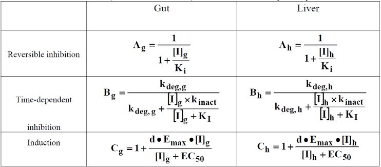
When static mechanistic models or dynamic models (see appendix, section VII.C.1) are used for
predicting DDIs caused by enzyme inhibition, the models should include the inhibition
mechanism only (i.e., the model should not include concurrent induction predictions for an
4Contains Nonbinding Recommendations
investigational drug that is hypothesized to be both an inducer and inhibitor) to assess the
potential of the investigational drug to inhibit metabolizing enzymes.
C. Determining if the Investigational Drug is an Inducer of Metabolizing
Enzymes
1. Conducting In Vitro Studies
The sponsor should evaluate the potential of an investigational drug to induce CYP1A2,
CYP2B6, CYP2C8, CYP2C9, CYP2C19, and CYP3A4. Initially, sponsors can conduct
experiments to evaluate CYP1A2, CYP2B6, and CYP3A4 only. If no induction of CYP3A4
enzymes is observed, evaluating the induction potential of CYP2C enzymes is not necessary
because both CYP3A4 and CYP2C enzymes are induced via activation of the pregnane X
receptor (PXR). If the investigational drug induces CYP3A4 and the results suggest that a
clinical study is warranted, the sponsor should evaluate the potential of the investigational drug
to induce CYP2C. However, a negative in vivo study with a CYP3A sensitive substrate can be
used to rule out induction potential of an investigational drug on CYP2C enzymes, as long as the
potential of CYP3A inhibition by the drug and its metabolite(s) can be excluded.
2. Data Analysis and Interpretation
The induction results should be evaluated separately for each donor. If the result from at least
one donor exceeds the pre-defined threshold, the sponsor should consider the drug to have
induction potential and conduct a follow-up evaluation. Several basic methods can assess the
potential of an investigational drug to induce metabolizing enzymes (Fahmi, Kish, et al. 2010;
Fahmi and Ripp 2010; Einolf, Chen, et al. 2014; Kenny, Ramsden, et al. 2018). Three are
described in detail below:
1. Fold-change method: The sponsor can examine the fold-change in CYP enzyme mRNA
levels when incubated with the investigational drug by using a cutoff determined from
known positive and negative controls to calibrate the system. For example, a drug is
interpreted as an inducer if: (1) it increased mRNA expression of a CYP enzyme in a
concentration-dependent manner; and (2) the fold change of CYP mRNA expression relative
to the vehicle control is ≥ 2-fold at the expected hepatic concentrations of the drug. Expected
drug concentrations in the liver can be calculated by assuming a certain fold of I max,u (e.g.,
30-fold of mean unbound maximal steady-state plasma concentration at therapeutic dose).
Considering uncertainties in the protein binding measurements, the unbound fraction in
plasma should be set to 1% (f u,p = 0.01) if experimentally determined to be < 1% when
cacluating I max,u .
However, the induction potential should not be ruled out for an investigational drug that
increases CYP enzyme mRNA less than 2-fold the of vehicle control, if the increase is more
than 20% of the response of the positive control. Further evaluation is recommended when
there is an inconclusive finding.
5Contains Nonbinding Recommendations
To calculate the percent of the response to the positive control, the following equation should
be used:
% of positive control = (mRNA fold increase of test drug treated cells - 1) × 100/ (mRNA
fold increase of positive control – 1)
2. Correlation methods: The sponsor may use correlation methods as described in Figure 3
to predict the magnitude of a clinical induction effect (e.g., AUC ratio of index substrate in
the presence and absence of inducers) of an investigational drug according to a calibration
curve of relative induction scores (RIS) or I max,u /EC 50 for a set of known inducers of the
same enzyme. If the predicted magnitude is more than a predefined cut-off (e.g., AUC ratio
≤ 0.8), a drug is considered have induction potential in vivo.
The calibration can be established once for one batch of hepatocytes and does not need to be
determined for each experiment. Sometimes, E max or EC 50 cannot be estimated due to an
incomplete in vitro induction profile (e.g., limited by solubility or cytotoxicity of tested
drug). An alternative correlation approach may be used if the method is validated.
Figure 3: Two Correlation Methods to Assess the Potential of an Investigational Drug to
Induce Metabolizing Enzymes (Fahmi and Ripp, 2010)
Correlation Method 1: Calculate a relative induction score (RIS) using (E max × I max,u ) / (EC 50 +
I max,u )
OR
Correlation Method 2: Calculate I max,u / EC 50 values
Emax is the maximum induction effect determined in vitro.
EC 50 is the concentration causing half-maximal effect determined in vitro.
Imax,u is the maximal unbound plasma concentration of the interacting drug at steady state.*
*Considering uncertainties in the protein binding measurements, the unbound fraction in plasma should be set to 1%
(fraction unbound in the plasma (f u,p ) = 0.01) if experimentally determined to be < 1%.
3. Basic kinetic model: To use this method, the sponsor should calculate the R value
(R 3 ) as described in Figure 4 and compare to a predefined cut-off determined from a set
of inducers and non-inducers. For example, a R 3 value ≤ 0.8 may indicate that the
investigational drug has induction potential in vivo.
Figure 4: An Equation to Calculate the R value for Basic Models of Induction (Kenny,
Ramsden, et al. 2018)
R 3 = 1 / [1 + d × ((E max × 10 × I max,u ) / (EC 50 + 10 × I max,u ))]
R 3 is the predicted ratio of intrinsic clearance values of a probe substrate for an enzymatic pathway in the absence
and presence of an inducer.
d is the scaling factor and is assumed to be 1. A different value can be used if supported by prior experience with
the system used (Vermet, Raoust, et al. 2016).
Emax is the maximum induction effect determined in vitro.
Imax,u is the maximal unbound plasma concentration of the interacting drug at steady state.*
EC 50 is the concentration causing half-maximal effect determined in vitro.
*Considering uncertainties in the protein binding measurements, the unbound fraction should be set to 1% if
experimentally determined to beContains Nonbinding Recommendations
If these methods indicate that the investigational drug has the potential to induce metabolizing
enzymes (using specific cutoff values mentioned above or developed by individual laboratories
for these methods), the sponsor should further investigate the enzyme induction potential of the
investigational drug by using mechanistic models (see appendix, section VII.C.1) or by
conducting a clinical DDI study with a sensitive index substrate. If the predicted AUCR of a
sensitive index substrate in the presence and absence of an investigational drug is ≤ 0.8 based on
static mechanistic models or dynamic mechanistic models (e.g., PBPK models; see appendix,
section VII.C.1), the sponsor should further investigate potential DDIs by conducting a clinical
DDI study using a sensitive index substrate.
When static mechanistic models or dynamic mechanistic models (see appendix, section VII.C.1)
are used for predicting DDIs caused by enzyme induction, the models should include the
induction mechanism only (i.e., the model should not include concurrent inhibition predictions
for an investigational drug that is hypothesized to be both an inducer and inhibitor) to assess the
potential of an investigational drug to induce metabolizing enzymes.
3. Additional Considerations
The AUCR cutoffs of > 0.8 (for induction) and < 1.25 (for inhibition) using mechanistic models
are the suggested default values to indicate that the investigational drug has no effect on the
levels of metabolizing enzymes.
When evaluating whether an investigational drug is an inhibitor of multiple CYP enzymes, the
sponsor can prioritize in vivo DDI evaluations for various CYP enzymes with sensitive index
substrates of respective pathways (see the January 2020 FDA guidance for industry Clinical
Drug Interaction Studies — Cytochrome P450 Enzyme- and Transporter-Mediated Drug
Interactions) based on rank-ordered R 1 , R 2 , or the predicted AUCR values, preferably using the
in vitro inhibition parameters obtained in the same study. 4 That is, the sponsor may first carry
out an in vivo study with a sensitive index substrate of the CYP with the largest R or AUCR
value. If this in vivo study shows no interaction, in vivo evaluations of other CYPs with lower
potencies (e.g., smaller R or AUCR) are not needed. However, if this in vivo study shows a
positive interaction between the drug and the sensitive index CYP substrate, the sponsor should
conduct additional in vivo studies for other CYPs, starting with the CYP with the next largest R
or AUCR value. Alternatively, the sponsor can use a mechanistic dynamic model to inform the
need for additional studies. The sponsor should verify and update dynamic models to
demonstrate that the model can adequately describe the observed findings from the first in vivo
study with a sensitive index substrate. In the presence of inhibitory metabolites of an
investigational drug, their contribution and rank order of metabolite R values should also be
considered when determining what in vivo studies should be conducted.
4
An orally administered drug may inhibit intestinal metabolic enzymes (e.g., CYP3A) in addition to hepatic
enzymes. Therefore, in vivo DDI for CYP3A inhibition should be considered if R1,gut is greater than or equal to 11,
even if R1 for CYP3A is not the largest value among the major CYPs evaluated.
7Contains Nonbinding Recommendations
Concurrent prediction of inhibition and induction using mechanistic static models or dynamic
models (see appendix, section VII.C.1) can be considered for predicting the net effect of an
investigational drug that is hypothesized to be both an inhibitor and an inducer of metabolizing
enzymes. However, there is a concern with concurrent predictions, as over-prediction of
inhibition may mask the induction effect leading to a false negative prediction of the overall
effect (Einolf, Chen, et al. 2014). If the induction potential is over-predicted, it will mask the
inhibition effect.
In vitro induction studies may also detect enzyme down-regulation. However, research in this
area is presently very limited, and the mechanisms behind these effects are unclear. If
concentration-dependent down-regulation is observed in vitro and is not attributable to
cytotoxicity, additional in vitro or in vivo studies may be needed to understand the potential
clinical consequences (Hariparsad, Ramsden, et al. 2017).
IV. EVALUATING TRANSPORTER-MEDIATED DRUG INTERACTIONS
Membrane transporters can have clinically relevant effects on the pharmacokinetics and
pharmacodynamics of a drug in various organs and tissues by controlling its absorption,
distribution, and elimination (Giacomini, Huang, et al. 2010; Giacomini and Huang 2013). In
contrast to drug metabolizing enzymes that are largely expressed in the liver and small intestines,
transporters are expressed in tissues throughout the human body and govern the access of
endogenous and exogenous substances to various sites in the body. In concert with metabolizing
enzymes, transporters can govern a drug’s disposition and pharmacological action. Conversely,
a drug can also modulate transporter expression or activity, resulting in altered disposition of
endogenous (e.g., creatinine, glucose) or exogenous substances.
Several transporters interact with drugs in clinical use (Giacomini, Huang, et al. 2010; Giacomini
and Huang 2013), for example:
• P-glycoprotein (P-gp or Multi-drug Resistance 1 (MDR1) protein)
• Breast cancer resistance protein (BCRP)
• Organic anion transporting polypeptide 1B1/1B3 (OATP1B1/OATP1B3)
• Organic anion transporter 1/3 (OAT1/OAT3)
• Multidrug and toxin extrusion (MATE) proteins (MATE1/MATE2-K)
• Organic cation transporter 2 (OCT2)
Understanding whether the drug is a substrate or inhibitor of these key transporters can explain
some clinical consequences, such as increased toxicity or altered efficacy, that result from altered
tissue distribution of a drug that is a substrate of a transporter. This section focuses on
transporters that have clinical evidence suggesting their involvement in drug interactions
(Giacomini, Huang, et al. 2010; Brouwer, Keppler, et al. 2013; Giacomini and Huang 2013;
Tweedie, Polli, et al. 2013; Zamek-Gliszczynski, Lee, et al. 2013). The sponsor should evaluate
the interactions between investigational drugs acting as substrates and/or inhibitors of these
transporters as outlined below. The timing of the in vitro evaluation of each transporter may
vary depending on the therapeutic indications of the investigational drug. For example, if the
8Contains Nonbinding Recommendations
intended population is likely to use statins, the sponsor should examine the potential of the
investigational drug to interact with OATP1B1/1B3 before initiation of clinical studies in
patients. If in vitro experiments indicate a low potential for an interaction between the
transporter and investigational drug, subjects taking statins may be included in clinical studies to
better represent the intended patient population.
A. Determining if the Investigational Drug is a Substrate of the Transporters P-
gp and BCRP
P-gp and BCRP are expressed in various tissues including the gastrointestinal tract, liver, kidney,
and brain. Thus, both transporters have the potential to impact the oral bioavailability, the tissue
distribution, and the hepatic and renal elimination of substrates.
1. Conducting In Vitro Studies
Sponsors should evaluate most investigational drugs in vitro to determine whether they are
substrates of P-gp and BCRP using the experimental systems described in the appendix, section
VII.B. P-gp and BCRP are generally not expected to impact the oral bioavailability of highly
permeable and highly soluble drugs. In vitro assessment of these drugs as P-gp or BCRP
substrates is not suggested unless there are potential safety concerns with the drug distributing
into tissues (e.g., the kidney and brain). See the 2017 FDA guidance for industry entitled Waiver
of In Vivo Bioavailability and Bioequivalence Studies for Immediate-Release Solid Oral Dosage
Forms Based on a Biopharmaceutics Classification System to determine if the investigational
drug can be classified as highly permeable and/or highly soluble (e.g., biopharmaceutics
classification system class 1 drugs).
2. Data Analysis and Interpretation
The following results suggest that an investigational drug is an in vitro P-gp substrate:
• A net flux ratio (or efflux ratio (ER)) of ≥ 2 for an investigational drug in cells that
express P-gp (e.g., Caco-2 cells or transfected cells overexpressing P-gp) 5
• A flux that is inhibited by at least one known P-gp inhibitor at a concentration at least 10
times its K i or IC 50 (e.g., the ER decreases to < 50% of the ER in the absence of inhibitor
or the flux reduced to unity).
When using Caco-2 cells that express multiple efflux transporters, the sponsor should use more
than one P-gp inhibitor to determine the specificity of the efflux. The sponsor may use a net flux
ratio cutoff other than 2 or a specific relative ratio to positive controls if prior experience with
the cell system justifies these alternative methods.
5
The ER can be calculated as the ratio of the basal to apical (B-A) transport rate to the apical to basal (A-B)
transport rate. The net flux ratio can be calculated as the ratio of the ER between cells expressing the transporter of
interest to cells not expressing the transporter.
9Contains Nonbinding Recommendations
If in vitro studies indicate that a drug is a P-gp substrate, the sponsor should consider whether to
conduct an in vivo study based on the drug’s safety margin, therapeutic index, and likely
concomitant medications that are known P-gp inhibitors in the indicated patient population (see
the January 2020 FDA guidance for industry entitled Clinical Drug Interaction Studies —
Cytochrome P450 Enzyme- and Transporter-Mediated Drug Interactions).
The sponsor may also use the above procedures to determine whether the drug is a BCRP
substrate by using known BCRP inhibitors. If in vitro studies indicate that a drug is a BCRP
substrate, the sponsor should consider whether to conduct an in vivo study based on the drug’s
safety margin, therapeutic index, and likely concomitant medications that are known BCRP
inhibitors in the indicated patient population (see the January 2020 FDA guidance for industry
entitled Clinical Drug Interaction Studies — Cytochrome P450 Enzyme- and Transporter-
Mediated Drug Interactions).
B. Determining if the Investigational Drug is a Substrate of the Hepatic
Transporters OATP1B1 and OATP1B3
OATP1B1 and OATP1B3 are key uptake transporters expressed on the sinusoidal membrane of
hepatocytes and play an important role in the hepatic uptake of various drugs.
1. Conducting In Vitro Studies
If in vitro studies or human/animal absorption, distribution, metabolism, and/or excretion
(ADME) data suggest that an investigational drug’s hepatic uptake or elimination is significant
(i.e., the drug’s clearance through hepatic metabolism or biliary secretion is ≥ 25% of the total
drug’s clearance), or the drug’s uptake into the liver is clinically important (e.g., for
biotransformation or to exert a pharmacological effect), the sponsor should evaluate the
investigational drug in vitro to determine whether it is a substrate for the hepatic uptake
transporters OATP1B1 and OATP1B3 (see the appendix, section VII.B). Other factors to be
considered include the drug’s physiological properties, e.g., low passive membrane permeability,
high hepatic concentrations relative to other tissues, organic anion/charged at physiological pH,
which support the importance of active uptake of the drug into liver.
2. Data Analysis and Interpretation
An investigational drug is considered an in vitro substrate for OATP1B1 or OATP1B3 if: (1) the
uptake of the drug in OATP1B1- or OATP1B3-transfected cells is ≥ 2-fold of the drug’s uptake
in empty vector-transfected cells; and (2) a known inhibitor (e.g., rifampin) can decrease the
drug’s uptake to ≤ 50% at a concentration at least 10 times that of the K i or IC 50 . The sponsor
may justify alternative cutoff ratios based on its prior experience with the cell system.
If in vitro studies indicate that a drug is an OATP1B1 or OATP1B3 substrate, the sponsor should
consider whether to conduct an in vivo study based on the drug’s safety margin, therapeutic
index, and likely co-medications that are known OATP1B1 or OATP1B3 inhibitors in the
indicated patient populations (see the 2019 FDA guidance for industry entitled Clinical Drug
Interaction Studies — Cytochrome P450 Enzyme- and Transporter-Mediated Drug Interactions).
10Contains Nonbinding Recommendations
C. Determining if the Investigational Drug is a Substrate of the Renal
Transporters OAT, OCT, and MATE
OAT1, OAT3, and OCT2 are renal transporters expressed on the basolateral membrane of the
renal proximal tubule. MATE1 and MATE2-K are expressed on the brush border membrane.
All the aforementioned renal transporters can play a role in the active renal secretion of
investigational drugs.
1. Conducting In Vitro Studies
If the investigational drug’s ADME data suggest that active renal secretion is significant for a
drug (i.e., active secretion of the parent drug by the kidney is ≥ 25% of the systemic clearance),
the sponsor should evaluate the drug in vitro to determine whether it is a substrate of OAT1/3,
OCT2, MATE1 and MATE2-K (see appendix, section VII.B). See Figure 5 for the equation to
calculate active secretion.
Figure 5: An Equation to Calculate Active Secretion*
Active secretion = CLr – (f u,p × GFR)
Cl r is the renal clearance.
f u,p is the unbound fraction in plasma.
GFR is the glomerular filtration rate.
*This equation is valid assuming that there is no re-absorption (e.g., no active re-absorption and passive re-
absorption is equal to passive secretion). The GFR is set as 125 mL/min for subjects with normal renal function if
the GFR is not measured.
2. Data Analysis and Interpretation
The investigational drug is an in vitro substrate for the above renal transporters if: (1) the ratio
of the investigational drug’s uptake in the cells expressing the transporter versus the drug’s
uptake in control cells (or cells containing an empty vector) is ≥ 2; and (2) a known inhibitor of
the transporter decreases the drug’s uptake to ≤ 50% at a concentration at least 10 times its K i or
IC 50 . The sponsor may justify alternative cutoff ratios based on its prior experience with the cell
system.
If in vitro studies indicate that a drug is a substrate of one or more of these renal transporters, the
sponsor should consider whether to conduct an in vivo study based on the drug’s safety margin,
therapeutic index, and likely concomitant medications that are known inhibitors of these renal
transporters in the indicated patient populations (see the January 2020 FDA guidance for
industry entitled Clinical Drug Interaction Studies — Cytochrome P450 Enzyme- and
Transporter-Mediated Drug Interactions).
D. Determining if the Investigational Drug is an Inhibitor of a Transporter
11Contains Nonbinding Recommendations
1. Conducting In Vitro Studies
The sponsor should conduct in vitro studies to evaluate whether an investigational drug is an
inhibitor of P-gp, BCRP, OATP1B1, OATP1B3, OCT2, MATEs (MATE1, MATE2-K), OAT1,
and OAT3 (see appendix, section VII.B for considerations regarding in vitro systems).
2. Data Analysis and Interpretation
P-gp and BCRP: The sponsor should conduct studies to determine if an investigational drug
inhibits the efflux ratio or net flux of a known P-gp or BCRP substrate in Caco-2, P-gp- or
BCRP-overexpressed cells or inhibits uptake of substrate when membrane vesicles are used, and
determine the drug’s inhibition potency (i.e., IC 50 or K i ). The investigational drug has the
potential to inhibit P-gp or BCRP in vivo if the investigational drug is administered orally, and
the I gut /IC 50 or K i ≥10 where I gut = dose of inhibitor/250 mL. If a metabolite of the drug is an
inhibitor or the investigational drug is administered by parenteral route, in vivo inhibition of P-
gp or BCRP may occur if the I 1 /IC 50 or K i ≥ 0.1, where I 1 is the C max of the metabolite or the
inhibitor drug. These cutoff values are based on a limited dataset (Zhang, Zhang, et al. 2008;
Tachibana, Kato, et al. 2009; Agarwal, Arya, et al. 2013; Ellens, Deng, et al. 2013). The sponsor
may calibrate its internal in vitro systems with known inhibitors and non-inhibitors and propose a
different cutoff value with proper justification.
If in vitro studies indicate that a drug is a P-gp or BCRP inhibitor, the sponsor should consider
whether to conduct an in vivo study based on likely concomitant medications that are known P-
gp or BCRP substrates in the indicated patient populations (see the January 2020 FDA guidance
for industry entitled Clinical Drug Interaction Studies — Cytochrome P450 Enzyme- and
Transporter-Mediated Drug Interactions).
OATP1B1 and OATP1B3: The sponsor should conduct studies to determine the inhibition
potency (i.e., IC 50 or K i ) of the investigational drug on the uptake of a known OATP1B1 or
OATP1B3 substrate in cells overexpressing the relevant transporter. Time-dependent inhibition
has been demonstrated for a few OATP1B1/3 inhibitors (Amundsen, Christensen, et al. 2010;
Gertz, Cartwright, et al. 2013; Izumi, Nozaki, et al. 2015; Pahwa, Alam, et al. 2017). Sponsors
may consider adding a pre-incubation step as part of assay validation when determining IC 50
values for an investigational drug. The investigational drug has the potential to inhibit
OATP1B1/3 in vivo if the R value (as described in Figure 6 below) is > 1.1.
12Contains Nonbinding Recommendations
Figure 6: Equation to Calculate the R Value of the Investigational Drug to Determine the
Potential to Inhibit OATP1B1/3*
R=1+ ((f u,p × I in,max )/IC 50 ) ≥1.1
f u,p is the unbound fraction in plasma.
IC 50 is the half-maximal inhibitory concentration.
Iin,max is the estimated maximum plasma inhibitor concentration at the inlet to the liver. It is calculated as:
I in,max = I max +(F a ×F g ×k a ×Dose)/Qh /RB
Fa is the fraction absorbed.
Fg is the intestinal availability.
k a is the absorption rate constant.
Q h is the hepatic blood flow rate.
R B is the blood-to-plasma concentration ratio.
*If unknown, Fa= 1, Fg = 1 and k a = 0.1/min can be used as a worst-case estimate.
Considering uncertainties in the protein binding measurements, the unbound fraction (f u,p ) should be set to 1% if
experimentally determined to be less than 1%.
The cutoff value described in Figure 6 is based on limited published data (Yoshida, Maeda, et al.
2012; Tweedie, Polli, et al. 2013; Vaidyanathan, Yoshida, et al. 2016). Sponsors may calibrate
their internal in vitro systems with known inhibitors and non-inhibitors of these transporter
systems and propose a specific cutoff value with proper justification.
If in vitro studies indicate that a drug is an OATP1B1 or OATP1B3 inhibitor, the sponsor should
consider whether to conduct an in vivo study based on whether the likely concomitant
medications used in the indicated patient populations are known OATP1B1or OATP1B3
substrates (see the January 2020 FDA guidance for industry entitled Clinical Drug Interaction
Studies — Cytochrome P450 Enzyme- and Transporter--Mediated Drug Interactions).
OAT, OCT, and MATE: Sponsors should conduct studies to determine the inhibition potency
(i.e., IC 50 or K i ) of the investigational drug on the uptake of a known substrate for renal
transporters (i.e., OAT1, OAT3, OCT2, MATE1, and MATE2-K) in cells overexpressing these
transporters. The investigational drug has the potential to inhibit these transporters in vivo if the
I max,u /IC 50 value is ≥ 0.1. 6 These cutoff values are based on limited data (Dong, Yang, et al.
2016a; Dong, Yang, et al. 2016b). Sponsors may calibrate their unique in vitro systems with
known inhibitors and non-inhibitors of these transporter systems and propose a different cutoff
value with proper justification. Creatinine is also a substrate for OCT2, MATEs, and OAT2
(Lepist, Zhang, et al. 2014). Elevated serum creatinine levels observed in clinical studies could
be due to inhibition of these transporters by the investigational drug (Chu, Bleasby, et al. 2016;
Mathialagan, Rodrigues, et al. 2017; Arya, Yang, et al. 2014). Confirmation of the mechanism
of an increase in serum creatinine with the investigational drug requires additional evidence such
as clinical mechanistic studies.
6
Considering uncertainties in the protein binding measurements, the unbound fraction should be set to 1% if
experimentally determined to be less than 1%.
13Contains Nonbinding Recommendations
If in vitro studies indicate that a drug is an inhibitor of these renal transporters, the sponsor
should consider whether to conduct an in vivo study based on whether the likely concomitant
medications used in the indicated patient populations are known substrates of these renal
transporters (see the January 2020 FDA guidance for industry entitled Clinical Drug Interaction
Studies — Cytochrome P450 Enzyme- and Transporter-Mediated Drug Interactions).
E. Determining if the Investigational Drug is an Inducer of a Transporter
Certain transporters such as P-gp are induced through mechanisms similar to those for CYP
enzymes (e.g., by activation of specific nuclear receptors). Because of these similarities,
information from CYP3A induction studies can inform P-gp induction studies (see the January
2020 FDA guidance for industry entitled Clinical Drug Interaction Studies — Cytochrome P450
Enzymes and Transporters-Mediated Drug Interactions). At this time, the in vitro methods to
evaluate the induction of P-gp and other transporters are not well established, therefore
recommendations for in vitro evaluation of investigational drugs as transporter inducers are not
provided.
V. EVALUATING THE DDI POTENTIAL OF METABOLITES
Sponsors should evaluate the DDI potential of an investigational drug’s metabolites for their
impact on the drug’s safety and efficacy using a risk-based assessment that considers safety
margins, likely concomitant medications, and therapeutic indications.
A metabolite with significant plasma exposure or pharmacological activities may need to be
evaluated for its DDI potential as a substrate or as a perpetrator of metabolizing enzymes (see
sections below). In vitro studies normally use a synthesized or purified metabolite standard.
Alternative methods are acceptable if the sponsor can justify that the DDI potential of the
metabolites can be adequately assessed (Callegari, Kalgutkar, et al. 2013; Yu and Tweedie 2013;
Yu, Balani, et al. 2015). If basic models suggest that the metabolite(s) may have in vivo DDI
liability and a static or dynamic mechanistic modeling approach (e.g., PBPK) is used for DDI
assessment of a drug, metabolite(s) should be incorporated in these models.
Published data have shown that some Phase II metabolites can be better substrates (more polar
than the parent) or inhibitors of various transporters leading to a higher chance of DDIs than the
parent drug (Zamek-Gliszczynski et al, 2014). Therefore, the DDI potential of a metabolite as a
substrate or a perpetrator of major drug transporters should be assessed on a case-by-case basis.
The same principles and strategies mentioned above for the parent drug should be applied where
applicable.
A. Metabolite as a Substrate
1. Conducting In Vitro Studies
14Contains Nonbinding Recommendations
The risk of a clinically relevant DDI through altered formation or elimination of metabolites
should be investigated if changes in metabolite exposure levels may result in clinically
meaningful alteration of efficacy or safety in vivo. The risk of a DDI when the metabolite acts
as a substrate should be evaluated for a pharmacologically active metabolite that contributes to ≥
50% of the overall activity. Both the in vitro receptor potency and the in vivo unbound systemic
exposure (expressed in molar unit) of a metabolite relative to the parent drug need to be taken
into consideration when evaluating the contribution of the metabolite to efficacy. If plasma
protein binding of the parent drug and the metabolite is high, it is preferred to determine their
protein binding in the same study to reduce inter-study variability. If available, data related to
target tissue distribution of parent drug and the metabolite may need to be considered when
evaluating the contribution of metabolite to in vivo efficacy.
2. Data Analysis and Interpretation
The sponsor should consider in vivo DDI studies of the metabolite based on in vitro assessments
using the same strategies as those for the parent drugs (see section III.A).
B. Metabolite as an Inhibitor
1. Conducting In Vitro Studies
If in vitro assessments suggest that the parent drug inhibits major CYP enzymes and transporters
and in vivo DDI studies are warranted, in vitro assessments of metabolites as enzyme or
transporter inhibitors may not be needed because the in vivo inhibition potential of metabolites
would be evaluated in vivo along with the parent drug, unless clinically relevant exposures of the
metabolite cannot be adequately represented in the in vivo DDI study (i.e., the study duration
does not allow the metabolite to accumulate). However, if in vitro assessments suggest that the
parent drug alone will not inhibit major CYP enzymes or transporters, in vivo DDIs caused by
metabolites may still be possible. In this situation, the sponsor should evaluate the in vitro
inhibition potential of a metabolite on CYP enzymes taking into account the systemic exposure
(in molar unit) and polarity (e.g., measured or predicted LogP, the elution order on the
chromatogram of reverse phase-high performance liquid chromatography) of the metabolite
relative to the parent drug. The sponsor should conduct an in vitro CYP enzyme inhibition study
if: (1) the metabolite is less polar than the parent drug and the AUC metabolite ≥ 25% of AUC parent ;
or (2) the metabolite is more polar than the parent dug and the AUC metabolite ≥ AUC parent . A
lower cut-off value for the metabolite-to-parent AUC ratio may also be considered for
metabolites with structural alerts for potential mechanism-based inhibition (Orr, Ripp, et al.
2012; Yu and Tweedie 2013; Yu, Balani, et al. 2015).
2. Data Analysis and Interpretation
Based on the results of in vitro DDI assessments of the metabolite, the sponsor should consider
an in vivo DDI study of the metabolite using the same strategies as those for the parent drug
except that R 1,gut may not be applicable (see section III.B).
15Contains Nonbinding Recommendations
VI. LABELING RECOMMENDATIONS
The Prescribing Information must include a summary of drug interaction information that is
essential for the safe and effective use of the drug product by the health care provider and must
be based on data derived from human experience whenever possible. 7 In the absence of clinical
information, the sponsor should include in vitro information regarding the characterization of
metabolic and transporter pathways as well as PK interactions between the drug and other
prescription drugs, over-the-counter drugs, classes of drugs, dietary supplements, and foods or
juices (including inhibition, induction, and genetic characteristics) in the Prescribing
Information, if clinically significant. In addition, the results of pertinent in vitro studies that
establish the absence of an effect must be included. 8 In vitro information that has been
superseded by clinical information should not be included in the Prescribing Information unless
it is essential to understanding the clinical results.
In vitro information should generally be placed under the 12.3 Pharmacokinetics subsection of
the CLINICAL PHARMACOLOGY section. In rare cases, the clinical significance of the in
vitro information may require placement in other sections of the Prescribing Information (e.g.,
BOXED WARNING, CONTRAINDICATIONS, WARNINGS AND PRECAUTIONS, and/or
DRUG INTERACTIONS sections).
See the following FDA guidances for industry for labeling recommendations relevant to drug
metabolism and transporter pathways as well as clinical DDIs:
• Clinical Pharmacology Labeling for Human Prescription Drug and Biological Products
— Considerations, Content, and Format (December 2016)
• Clinical Drug Interaction Studies — Cytochrome P450 Enzyme- and Transporter-
Mediated Drug Interactions (January 2020)
7
21 CFR 201.56(a)(3).
8
21 CFR 201.57(c)(13)(c)(i)(C).
16Contains Nonbinding Recommendations
VII. APPENDICES
A. Evaluating Metabolism-Based Drug Interactions In Vitro
Various hepatic in vitro systems can be used to evaluate the drug interaction potential of an
investigational drug, including:
(1) Subcellular human liver tissue fractions such as reconstituted microsomal systems,
supernatants after 9000 g centrifugation of liver homogenate (S9), and cytosol (adding
appropriate co-factors as necessary)
(2) Recombinant human CYP enzymes in various expression systems that can identify the
production of individual drug metabolites and the involvement of certain classes of enzymes
(3) Human liver tissues, including freshly prepared hepatocytes and cryopreserved
hepatocytes that preserve enzyme architecture and contain the full complement of Phase I
and Phase II drug metabolizing enzymes
Although the main focus of this guidance is on CYP and hepatic metabolism, sponsors should
consider non-CYP, enzyme-based metabolism (e.g., Phase II enzymes) and metabolism
occurring in extra-hepatic tissues when relevant for their investigational drugs.
1. Determining if the Investigational Drug is an Enzyme Substrate
Drug metabolizing enzyme identification studies, often referred to as reaction phenotyping
studies, are a set of in vitro experiments that identify the specific enzymes responsible for the
metabolism of a drug. Along with other information (e.g., in vivo pharmacokinetics, enzyme
polymorphism or DDI data), in vitro phenotyping data are often used to quantify elimination
pathways of an investigational drug.
a. Conducting metabolic pathway identification experiments
Metabolic pathway identification experiments identify the number and structures of metabolites
produced by a drug and whether the metabolic pathways are parallel or sequential. These
experiments use intact human liver systems (e.g., human hepatocytes), human liver
microsomes, or recombinant enzyme systems. Data obtained from metabolic pathway
identification experiments help to determine whether and how to conduct a reaction
phenotyping study.
b. Identifying the enzymes that metabolize an investigational drug
The sponsor should conduct in vitro experiments to identify specific metabolizing enzymes that
are involved in the metabolism of an investigational drug, preferably before first-in-human
studies. There are two widely used methods for identifying the individual CYP enzymes
responsible for a drug's metabolism: (1) the first method uses chemicals, drugs, or antibodies as
17
01/21/20Contains Nonbinding Recommendations
specific enzyme inhibitors in human liver microsomes or hepatocytes (e.g., a pool of more than
10 donors); and (2) the second method uses individual human recombinant CYP enzymes. The
sponsor should consider the following recommendations when performing reaction phenotyping
experiments:
• The sponsor should use both methods to identify the specific enzymes responsible for a
drug's metabolism.
• When using individual human recombinant CYP enzymes, the sponsor should consider
the difference in the amount and enzyme activity of CYPs between the recombinant CYP
enzyme systems and the human liver (Venkatakrishnan, von Moltke, et al. 2000; Chen,
Liu, et al. 2011).
• The in vitro system for these studies should: (1) be robust and reproducible; and (2) be
characterized with in vitro probe substrate to prove the activity of each enzyme. A list of
probe substrates can be found on the FDA’s Web site on Drug Development and Drug
Interactions. 9
• Whenever possible, the sponsor should conduct all experiments with drug concentrations
deemed appropriate by kinetic experiments, relevant to clinical setting, and under initial
rate conditions (linearity of metabolite production rates with respect to time and enzyme
concentrations). The sponsor should conduct an adequate number of replicates (e.g.,
three or more replicates per drug concentration) in a single study.
• When conducting an in vitro study to examine the contribution of individual CYP
enzymes to the overall metabolism of an investigational drug, there are two widely used
methods: measurement of parent drug depletion; and measurement of metabolite
formation. For the latter method, it is desirable that all of the major metabolites have
been identified and quantified in metabolite formation experiments.
• When conducting in vitro studies to examine the contribution of individual CYP enzymes
to the formation of a specific metabolite, the sponsor should measure the formation rate
of the metabolite.
• The sponsor should develop validated and reproducible analytical methods to measure
levels of the parent drug and each metabolite.
• The use of a radiolabeled drug substrate is advantageous because samples can be
analyzed using liquid chromatography coupled with a radioactivity detector and a mass
spectrometer to identify and quantify drug-related species.
9
A list of probe substrates:
https://www.fda.gov/Drugs/DevelopmentApprovalProcess/DevelopmentResources/DrugInteractionsLabeling/ucm0
93664.htm#table1.
18
01/21/20Contains Nonbinding Recommendations
• The sponsor should separately evaluate individual isomers of racemic drugs when it is
important to understand the different disposition characteristics of each isomer (e.g.,
when two isomers have different pharmacological activities).
• Most chemical inhibitors are not specific for an individual CYP enzyme. The sponsor
should verify the selectivity and potency of inhibitors in the same experimental
conditions using probe substrates for each CYP enzyme. Commonly used in vitro CYP
enzyme inhibitors can be found on the FDA’s Web site on Drug Development and Drug
Interactions. 10
• The sponsor should test the inhibitory effect of an antibody to a CYP enzyme at
sufficiently low and high concentrations to establish a titration curve and ensure the
maximal inhibition of a particular pathway (ideally resulting in greater than 80 percent
inhibition). The sponsor should verify the effect of an antibody using probe substrates of
each CYP isoform and with the same experimental conditions.
2. Determining if the Investigational Drug is an Enzyme Inhibitor or Inducer
a. Conducting in vitro enzyme inhibition studies
The potential of an investigational drug to inhibit CYP enzymes is usually investigated in human
liver tissue systems using probe substrates to determine the inhibition mechanisms (e.g.,
reversible or time-dependent inhibition (TDI)) and inhibition potencies (e.g., K i for reversible
inhibition, and K I and k inact for TDI). The in vitro systems used for these studies include human
liver microsomes, microsomes obtained from recombinant CYP-expression systems, or
hepatocytes (Bjornsson, Callaghan, et al. 2003).
Kinetic data from in vitro inhibition studies of an investigational drug can be used in quantitative
models to predict the investigational drug’s effects on the pharmacokinetics of other drugs in
humans. These analyses inform the decision on whether to conduct an in vivo DDI study using
sensitive enzyme index substrates (see section III.B.2).
The sponsor should consider the following recommendations when designing an in vitro CYP
inhibition study:
• A probe substrate should be selective (e.g., predominantly metabolized by a single
enzyme in pooled human liver microsomes or recombinant CYPs) and have simple
metabolic schemes (ideally, the drug does not undergo sequential metabolism).
10
Examples of in vitro selective inhibitors for P450-mediated metabolism:
https://www.fda.gov/Drugs/DevelopmentApprovalProcess/DevelopmentResources/DrugInteractionsLabeling/ucm0
93664.htm#table1-2
19
01/21/20You can also read