MCP Papers in Press. Published on August 2, 2018 as Manuscript RA118.000792
←
→
Page content transcription
If your browser does not render page correctly, please read the page content below
MCP Papers in Press. Published on August 2, 2018 as Manuscript RA118.000792

Title: Identification of tumor antigens among the HLA peptidomes of Glioblastoma tumors
and plasma

Running Title: The HLA peptidome of Glioblastoma

Authors: Bracha Shraibman1, Eilon Barnea1, Dganit Melamed Kadosh1, Yael Haimovich1, Gleb
Slobodin2, Itzhak Rosner2, Carlos López-Larrea3, Norbert Hilf4, Sabrina Kuttruff4, Colette Song4,
Cedrik Britten5,12, John Castle5, Sebastian Kreiter5, Katrin Frenzel5, Marcos Tatagiba6, Ghazaleh
Tabatabai6, Pierre-Yves Dietrich7, Valérie Dutoit7, Wolfgang Wick8, Michael Platten8, Frank
Winkler8, Andreas von Deimling8, Judith Kroep9, Juan Sahuquillo10, Francisco Martinez-Ricarte10,

                                                                                                    Downloaded from http://www.mcponline.org/ by guest on December 12, 2018
Jordi Rodon10, Ulrik Lassen13, Christian Ottensmeier11, Sjoerd H. van der Burg9,12, Per Thor
Straten13, Hans Skovgaard Poulsen14, Berta Ponsati15, Hideho Okada16, Hans-Georg Rammensee6,
Ugur Sahin5, Harpreet Singh4 and Arie Admon1

Addresses
1. Biology, Technion – Israel Institute of Technology, Haifa 32000, Israel
2. Rheumatology Unit Bnai Zion Medical Center, Haifa 31048, Israel
3. Hospital Universitario Central de Asturias, 33011 Oviedo, Asturias, Spain
4. Immatics Biotechnologies GmbH, Paul-Ehrlich-Str. 15,72076 Tuebingen, Germany
5. BioNTech AG, Holderlinstr. 8,55131 Mainz, Germany
6. Eberhard Karls Universität Tübingen, Department of Immunology, Auf der Morgenstelle
   15,72076 Tubingen, Germany
7. Université de Genève, Rue Gabrielle Perret Gentil 4; 1211 Geneve 14, Switzerland
8. Heidelberg University Medical Center, Im Neuenheimer Feld 672, D-69120 Heidelberg,
   Germany
9. Leiden University Medical Center, Department of Medical Oncology, Albinusdreef 2, 2333 ZA
   Leiden, The Netherlands
10. Vall d’Hebron University Hospital, Institut Catala de la Salut, Pg. Vall d’Hebron 119-129,
   08035 Barcelona, Spain
11. Cancer Sciences Division, Faculty of Medicine, University of Southampton, Southampton, UK
12. Association for Cancer Immunotherapy (CIMT), Langenbeckstr. 1,55131 Mainz, Germany

                                                1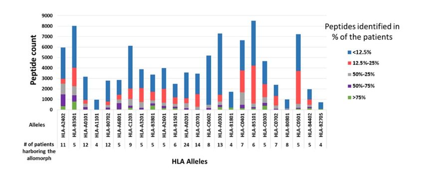
13. Region Hovedstaden (Center for Cancer Immune Therapy (CCIT), Herlev Hospital, Herlev
   Ringvej 75, DK-2730, Copenhagen, Denmark
14. Rigshospitalet, Departments of Radiation Biology and Oncology, Rigshospitalet 9,
   Blegdamsvej, DK-2100, Copenhagen, Denmark
15. BCN Peptides, Pol. Ind. Els Vinyets-Els Fogars II. 08777 Sant Quinti de Mediona (Barcelona),
   Spain
16. University of California, San Francisco, CA 94131USA

Corresponding author: Arie Admon, Department of Biology, Technion – Israel Institute of
Technology, Haifa 32000, Israel. Email: admon@technion.ac.il

                                                                                                   Downloaded from http://www.mcponline.org/ by guest on December 12, 2018
Abbreviations: MHC – major histocompatibility complex; HLA – human leukocytes antigen;
CTA – cancer/testis antigen; GBM – glioblastoma; TSA – tumor specific antigens; TAA – tumor
associated antigens; sHLA – soluble human leukocytes antigen; mHLA – membranal human
leukocytes antigen; FDR – false discovery rate; BBB – blood brain barrier.

                                               2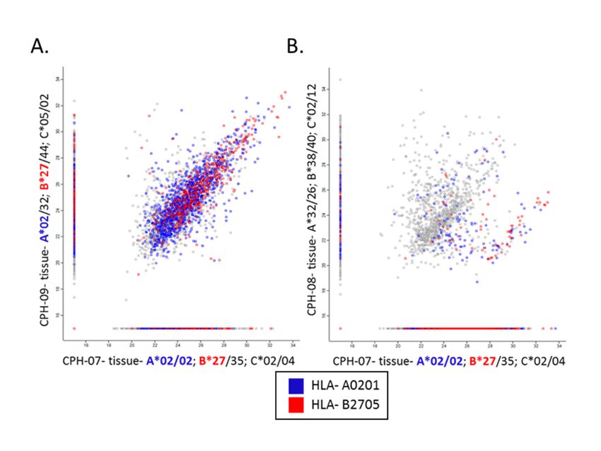
Summary

Glioblastoma multiforme (GBM) is the most aggressive brain tumor with poor prognosis to most
patients. Immunotherapy of GBM is a potentially beneficial treatment option, whose optimal
implementation may depend on familiarity with tumor specific antigens, presented as HLA
peptides by the GBM cells. Furthermore, early detection of GBM, such as by a routine blood test,
may improve survival, even with the current treatment modalities. This study includes large-scale
analyses of the HLA peptidome (immunopeptidome) of the plasma-soluble HLA molecules
(sHLA) of 142 plasma samples, and the membranal HLA of GBM tumors of 10 of these patients’
tumor samples. Tumor samples were fresh-frozen immediately after surgery and the plasma

                                                                                                      Downloaded from http://www.mcponline.org/ by guest on December 12, 2018
samples were collected before, and at multiple visits after surgery. In total, this HLA peptidome
analysis involved 52 different HLA allotypes and resulted in the identification of more than 35,000
different HLA peptides. Strong correlations were observed in the signal intensities and in the
repertoires of identified peptides between the tumors and plasma-soluble HLA peptidomes of the
individual patients, while low correlations were observed between these HLA peptidomes and the
tumors’ proteomes. HLA peptides derived from Cancer/Testis Antigens (CTAs) were selected
based on their presence among the HLA peptidomes of the patients and absence of expression of
their source genes from any healthy and essential human tissues, except from immune-privileged
sites. Additionally, peptides were selected as potential biomarkers if their levels in the plasma-
sHLA peptidome were significantly reduced after the removal of tumor mass. The CTAs identified
among the analyzed HLA peptidomes provide new opportunities for personalized immunotherapy
and for early diagnosis of GBM.

                                                3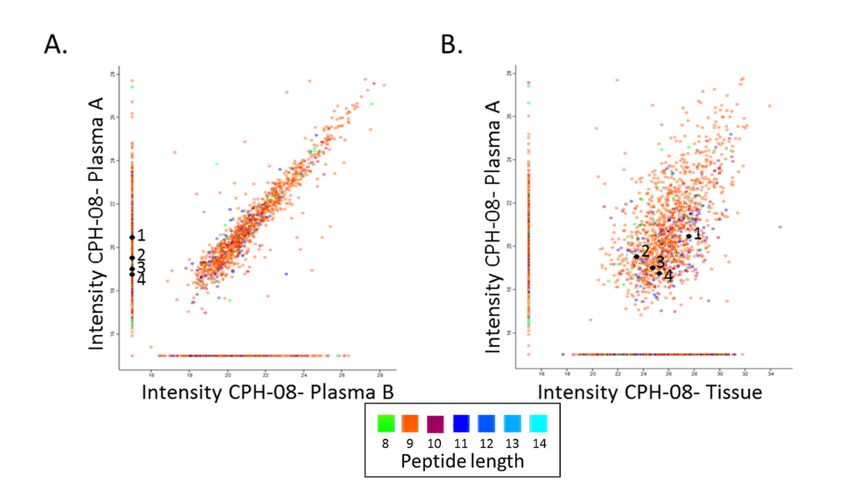
Introduction

Glioblastoma multiforme (GBM) is the most common and aggressive primary brain tumor, with a
median survival of about 15 months (1, 2). The grim prognosis facing most GBM patients calls
for improvements in targeted therapeutic treatments, as well as in discovery of proper biomarkers
for early detection of the disease. Recently, immunotherapy of different cancers, including GBM,
has drawn significant attention (3, 4), reviewed in (5–13). Much of the recent excitement about
cancer immunotherapy stems from the success in treating patients with immune checkpoint
modulators, which induce anti-cancer T cell immune reactions that can break tolerance and bring
about complete responses in increasingly larger percentages of patients (14, 15). Identification of

                                                                                                      Downloaded from http://www.mcponline.org/ by guest on December 12, 2018
tumor antigens is needed for development of effective cancer immunotherapy, including GBM,
and a good source for such antigens are the pools of HLA-bound peptides presented preferentially,
or even exclusively, by the tumor cells (16), reviewed in (17–20). Tumor-specific antigens (TSA)
and tumor-associated antigens (TAA) that can serve as candidates for cancer immunotherapeutics
have been searched for extensively. Indeed, many such antigens were already identified, yet none
has induced sufficiently strong anti-cancer immune reaction to eradicate the tumors in large
cohorts of patients (18, 20, 21). One subset of preferred TAAs are Cancer/Testis Antigens (CTA),
which are abnormally expressed in the malignant cells, and are normally expressed only in fetal
tissues and in immune privileged sites, such as the male germ cells, placenta and ovary, but are
absent from the normal somatic cells of any healthy tissue (22), reviewed in (23). Another special
groups of tumor antigens are neoantigens, which are attracting significant attention as potential
targets for cancer immunotherapy (24). These are more likely common in tumors with higher
mutational load and therefore less frequently found in GBM.

       HLA class I molecules are predominantly expressed as membrane anchored, cell-surface
proteins (mHLA). Yet, the HLA molecules are also released from cells (25, 26), reviewed in (27),
and can be recovered from the soluble HLA molecules in human plasma (sHLA) for analysis of
their bound peptidome (28–30). Confounding factors that limit the use of the sHLA peptidome as
a source for biomarkers are the presence of small amounts of sHLA molecules in the plasma of all
healthy individuals (27, 31), and in addition, some of the sHLA alleles are released to the
circulation in larger amounts than others (32, 33).

                                                 4
It has been suggested that shedding of the sHLA molecules represents a mechanism for
evasion of immune recognition of tumor cells, which release larger amounts of sHLA molecules
relative to the healthy cells (33). Since the sHLA molecules carry with them their original load of
bound peptides, the immunopeptidomes of the sHLA molecules of cancer patients include
disproportionally large fractions of peptides originating from the tumor cells, even though the
tumors constitute just small fraction of the body mass. It is therefore intuitive to assume that
biomarkers of disease can be found among those sHLA peptides. Such plasma-sHLA molecules
could then be a useful source of cancer biomarkers for early detection of recurrence during follow-
up (28).
Downloaded from http://www.mcponline.org/ by guest on December 12, 2018
Analyses of the HLA peptidomes, based on immunoaffinity purification of the mHLA
molecules from cell lines, tumor tissues or patients’ plasma, followed by analysis of the bound
peptides by chromatography and tandem mass spectrometry (34) currently results in identification
of thousands of HLA bound peptides (35–38). HLA peptides were previously isolated from GBM
cell lines (39) and fresh tumors, including GBM (4, 16). Similarly, the HLA peptidomes of tumors,
such as melanoma (40, 41), renal cell carcinoma (42, 43), leukemia (44) and multiple myeloma
(45) have been characterized. Furthermore, several thousands of peptides have successfully been
isolated from cancer patients’ plasma-sHLA molecules (28, 30).
In this research, more than thirty-five thousand plasma-sHLA and tumor-mHLA peptides
from GBM patients were identified. These large HLA peptidomes included several hundred
peptides, derived from both known TAAs and newly defined potential CTAs. We propose that
these may serve as useful biomarkers and candidates for immunotherapeutics for GBM, as well as
for other cancers.
Experimental Procedures
Experimental Design and Statistical Rationale: 106 plasma samples were obtained from 52
different GBM patients, collected prior to tumor removal and at different time points after the
surgery (Supplemental Table S1). Additionally, control plasma of healthy donors (N=6) and
patients diagnosed with ankylosing spondylitis (N=30), as an exemplar inflammatory process,
5were used for this study. In addition, tumor samples were obtained from 10 of the 52 GBM patients
for mHLA peptidome analysis, as well as for tumor proteome analysis.
Patient Characterization: Peripheral blood (PB) and tumor tissues samples from GBM patients
(University Hospital Heidelberg, Leiden University Medical Centre, Vall d’Hebron University
Hospital, Université de Genève, Southampton Universities Hospital, Heidelberg University
Medical Center, Herlev Hospital and Rigshospitalet) and non-cancerous controls (Universitario
Central de Asturias and Bnei-Zion Hospital). The HLA tissue typing of the GBM patients and non-
cancerous controls, healthy, and Ankylosing Spondylitis patients are listed only with gender, age,
and stage of disease in Supplemental Table S1. All human bio-specimens were obtained with
Downloaded from http://www.mcponline.org/ by guest on December 12, 2018
informed consent, and after approval of the relevant ethics committees.
Plasma and Tumor Cells Collection: Peripheral blood samples were collected into EDTA tubes
and cleared of the cells by centrifugation for 10 min at 1,200g, at room temperature. The cleared
plasma was stored frozen at -80°C until use for sHLA purification. Tumor samples were frozen in
liquid nitrogen immediately after dissection, and stored at -80°C until use, for both mHLA
peptidome and proteome analyses of the same tissue extracts.
Affinity Purification of HLA Molecules: Membranal HLA class I molecules were purified from
GBM tumor tissues by immunoaffinity from the cells after lysis with 0.25% sodium deoxycholate,
0.2 mM iodoacetamide, 1 mM EDTA, 1:200 Protease Inhibitors Mixture (Sigma), 1 mM PMSF,
and 1% octyl-D-glucopyranoside (Sigma) in PBS at 4 °C for 1 hour. The lysate was cleared by
centrifugation for 45 min at 18,000 rpm, at 4 °C. HLA class I molecules from the cleared lysate or
from the fresh human plasma were immunoaffinity purified using the W6/32 mAb bound to
Amino-Link beads (Thermo-Fisher Scientific, Waltham, MA) as in (28, 46). The HLA molecules
with their bound peptides were eluted from the affinity column with five column volumes of 1%
TFA. The eluted HLA class I proteins, and the released peptides were loaded on disposable C18
micro-columns (Harvard Apparatus, Holliston, MA), and the peptides fraction was recovered with
30% acetonitrile in 0.1% TFA, while the protein fraction was recovered with 80% acetonitrile in
0.1% TFA, as in (46). The peptide fractions were dried using vacuum centrifugation, reconstituted
in 100 μl of 0.1% TFA, reloaded on C18 Stage-Tips (47), eluted with 80% acetonitrile, dried, and
reconstituted with 0.1% formic acid for LC-MS/MS analysis.
6Proteomics analysis was performed after trypsin digestion of the proteins in gel slices. Briefly,
40 micrograms protein sample from each tissue was ran in 10% acrylamide gel, stained with
Coomassie, and each lane of the gel was sliced into five slices. The proteins in the gel slices were
reduced with 2.8 mM DTT at 60 °C for 30 min and carbamidomethylated with 8.8 mM
iodoacetamide in 100 mM ammonium bicarbonate at room temperature for 30 min and digested
overnight at 37 °C in 10% acetonitrile and 10 mM ammonium bicarbonate with modified trypsin
(Promega) at a 1:10 (wt/wt) enzyme-to-substrate ratio. This was repeated for another 4 hours with
a similar amount of trypsin, followed by 150 min gradients of LC-MS/MS of the tryptic peptides
from each gel slice.
Downloaded from http://www.mcponline.org/ by guest on December 12, 2018
Identification of the HLA and tryptic peptides: Both the HLA and tryptic peptides were
resolved by capillary chromatography and electrospray tandem mass spectrometry using a Q-
Exactive-Plus mass spectrometer fitted with a capillary Ultimate 3000 RSLC nano-capillary
UHPLC (Thermo-Fisher Scientific). The reversed phase chromatographs were with about 30 cm
long, 75 micron inner diameter capillary columns, home-packed with 3.5 m silica ReproSil-Pur
C18-AQ resin (Dr. Maisch GmbH, Ammerbuch-Entringen, Germany) as in (48). HLA and tryptic
peptides were eluted with a linear gradient of 5–28% of acetonitrile in 0.1% formic acid. For both
the HLA peptides and for the in-gel digested fractions, the capillary HPLC gradients were run at
flow rates of 0.15 µl/minutes during 120 min. Data was acquired using a data-dependent “top 10”
method, fragmenting the peptides by higher-energy collisional dissociation (HCD). Full scan MS
spectra were acquired at a resolution of 70,000 at 200 m/z with a target value of 3*106 ions. MS/MS
ions were accumulated to an AGC target value of 105 with a maximum injection time of 100 msec.
For the HLA peptides with unassigned precursor ion charge states, or charge states of four and
above, no fragmentation was performed. For tryptic peptides, the fragmentation was performed on
charge states between 2 to 7. The peptide match option was set to Preferred. Normalized collision
energy was set to 25% and MS/MS resolution was 17,500 at 200 m/z. Fragmented m/z values were
dynamically excluded from further selection for 20 seconds.
Data Analysis: The MS data was analyzed by the MaxQuant computational proteomics platform
(49) version 1.5.3.8 and searched with the Andromeda search engine (50). Peptide identifications
were based on the human section of the Uniprot database (http://www.uniprot.org/) of July 2015
7containing 69,693 entries. Proteins were declared positive identification with at least two identified
tryptic peptides per protein. Mass tolerance of 4.5 ppm for the precursor masses and 20 ppm for
the fragments were allowed. Methionine oxidation was accepted as variable modification for both
tryptic and HLA peptides. Carbamidomethyl cysteine was accepted as a fixed modification for the
proteomics data and as a variable modification for the HLA peptidome data. Methionine sulfoxide
and n-acetylation were set as variable modifications for both the proteomics and HLA peptidomics
analyses. Minimal peptide length was set to seven amino acids and a maximum of two
miscleavages was allowed for tryptic peptides. The false discovery rate (FDR) was set to 0.01 for
protein identifications, and 0.05 for the HLA peptides, since it resulted in identification of about
twice as many true HLA ligands (see Discussion). The resulting identified protein tables were
Downloaded from http://www.mcponline.org/ by guest on December 12, 2018
filtered to eliminate the identifications derived from the reverse database, as well as common
contaminants.
Normalization of the HLA peptidome and proteome Data: The HLA peptidome of each of the
LC-MS/MS runs was normalized according to its median. The medians were zeroed, and down-
regulated HLA peptides were defined as those changing by at least two folds in the plasma samples
collected immediately before surgery relative to samples collected at later clinic visits. The
proteomics LC-MS/MS data was normalized using the LFQ and iBAQ tools of the MaxQuant
software (49).
HLA Typing: DNA was extracted from blood of GBM patients using the DNeasy Blood & Tissue
kit (Qiagen, Hilden, Germany) as per the kit instructions. The HLA typing was conducted with
this DNA at the DKMS Life Science Lab (Germany). The samples were processed within high-
throughput workflow using next generation sequencing. HLA typing was based on the regions
covering the peptide-binding domains. Regions sequenced were Exons 2 and 3 for HLA-A, -B, -
C, -DQB1, -DRB1 and -DPB1 (51).
Definition of the Tumor Associated Antigen (TAA) Group: Selection of TAAs was based on
the cancer tumor database (CT gene database) (http:// www.cta.lncc.br/) (52) comprised of 277
different TAAs (data accumulated between the years 2005–2009) and the Tumor T cell Antigen
database (TANTIGEN) (http://cvc.dfci.harvard.edu/tadb), comprised of 259 different TAAs (53).
8Data Availability: The mass spectrometry proteomics data of the tissue proteomes, the mHLA
and the sHLA peptidomes have been deposited to the ProteomeXchange Consortium (54)
(http://proteomecentral.proteomexchange.org) via the PRIDE partner repository with the data set
identifier PXD008127.
Results
Large HLA peptidomes are identified from tumors and plasma of GBM patients
Tumor samples were obtained from 10 different patients undergoing surgery. These freshly frozen
tumor tissues were used for analysis of their membranal HLA peptidomes and their tumor
Downloaded from http://www.mcponline.org/ by guest on December 12, 2018
proteomes. In addition, 106 plasma samples from these 10 patients, and other 42 GBM patients
(52 patients in total) were used for the analysis of their plasma-sHLA bound peptidomes.
Additionally, control plasma of healthy donors (N=6) and patients diagnosed with ankylosing
spondylitis (N=30), as an exemplar inflammatory process, were used for this study (Fig. 1,
Supplemental Table S1). The study was performed as part of the GAPVAC project (The European
Glioma Actively Personalized Vaccine Consortium), which aimed to develop a personal
immunotherapy treatment based on administration of synthetic copies of selected HLA peptides
derived from tumor antigens to GBM patients. Here we describe the results of the plasma-soluble
HLA (sHLA) peptidome analyses, the tumor membranal HLA (mHLA) peptidome and proteome
analyses, performed in parallel with the GBM tumors. Overall, the tumor-mHLA peptidome
analyses resulted in the identification of 24,650 different HLA peptides, derived from 7,217
different source proteins, and the plasma-sHLA peptidome analyses resulted in the identification
of 32,089 different HLA peptides, derived from 8,281 different source proteins (Supplemental
Table S2). The proteomics analysis of these 10 GBM tumor samples resulted in identification of
7,351 different proteins (Supplemental Table S3).
The GBM HLA peptidomes include many peptides from multiple Tumor Antigens
Among the patients’ plasma-sHLA and the tumor-mHLA peptidomes, 904 different HLA peptides
derived from 143 known TAAs were identified. The reference set of TAAs was based on the cancer
tumor database (CT gene database) (http://www.cta.lncc.br/) comprising 277 different TAAs (data
9accumulated between 2005-2009) (52) and the Tumor T cell Antigen database (TANTIGEN)
(http://cvc.dfci.harvard.edu/tadb) (53) with 259 different TAAs, for a total set of 496 TAAs.
Derived from these 496 TAAs, a total of 763 plasma-sHLA and 611 tumor-membranal HLA
peptides were identified. Importantly, up to 78.8% of the plasma-sHLA peptides, derived from this
TAA group, were also detected among the tumor-mHLA peptidomes of the different patients.
These results indicate that the plasma-sHLA peptidome may indeed provide a useful source of
tumor antigens for diagnosis and immunotherapeutics (selected examples in Table 1, and the entire
list in Supplemental Table S4). 405 out of the 904 identified HLA peptides derived from TAAs
were detected only in GBM plasma and tissue samples but not in any non-cancerous donors’
Downloaded from http://www.mcponline.org/ by guest on December 12, 2018
plasma samples, supporting their potential significance as authentic tumor antigens.
Among the TAAs expressed in the tumor cells, a subset of genes, normally expressed only
in germline, embryonic and placenta cells, can be defined as CTAs, which are expressed at levels
below a threshold of nine gcrma units (expression units using background adjustment: GC content
adjusted with Robust Multiarray Average, described in the BioGPS website) in all normal and
essential tissues. Such CTAs were further defined as those derived from genes whose transcripts
are expressed at significantly higher levels in the tumors tissues (according to BioGPS (55, 56),
see Discussion). Using this filtration, a list of 145 CTAs was established (Supplemental Table S5).
In the HLA peptidome analysis described here, 19 different HLA peptides, belonging to 11 CTAs,
were identified. Of these, 15 HLA peptides were identified only in the GBM plasma and/or tumor
samples (for example ODF1, Supplemental Fig. S1A and the entire list in Supplemental Table S2).
In addition, HLA peptides derived from proteins expressed only in fetal tissues were observed in
the GBM samples, and not at all, or in less than 5% of the control plasma samples. An example
for such antigen is the transcription factor SOX11 gene (SOX11) that is expressed only in fetal
brain, according to BioGPS (Supplemental Fig. S1B). In this analysis, two of its derived HLA
peptides, AHSASEQQL and NFSDLVFTY, were observed in the plasma-sHLA and tumor-
mHLA of the GBM patients, and were detected in less than 5% of the non-cancerous blood donors’
plasma-sHLA peptidomes (Table 1). Moreover, HLA peptides derived from male tissues such as
testis or prostate, and identified in women’s plasma or GBM tissue, or HLA peptides derived from
female tissues, such as placenta and ovaries, and identified in the men’s plasma or GBM tissue,
can serve as potential immunotherapeutics candidates (Table 2). For example, eight different HLA
10peptides originating from the ETV5 gene were identified in this analysis. This gene is expressed
only in placenta but not at any other tissue in the body. Moreover, the eight ETV5 HLA peptides
were identified mostly among the male GBM sHLA and mHLA samples but not at any of the non-
cancerous donors, suggesting their relevance to serve as proper biomarkers for the disease.
The levels of potential biomarker plasma-sHLA peptides are reduced following surgical
removal of the tumors
The presence of the plasma-sHLA peptides derived from tumor antigens may serve as surrogate
biomarkers for different cancers, including GBM. Peptides detected among the plasma-sHLA
peptidome, derived from tumor antigens that are not normally expressed at elevated levels in
Downloaded from http://www.mcponline.org/ by guest on December 12, 2018
normal tissues, should be reduced in their levels after removal or reduction of the tumor load by
treatments (57) such as the surgery performed for the GBM patients studied here. Most of the
plasma-sHLA peptides of the individual patients remain relatively stable before and after surgery,
and even a few months later, since these are self-peptides of the healthy tissues (example in Fig.
2A). This facilitated focusing on the minority of HLA peptides that were reduced in their levels
relative to the rest of the sHLA peptidome, in order to search among them for potential biomarkers.
In this study, 94 different plasma samples were collected prior to, and following surgery of the
same 34 GBM patients as part of the GAPVAC project. The sHLA molecules of the different
plasma samples were affinity purified and their bound peptidomes were analyzed separately by
LC-MS/MS. As many as 250 sHLA peptides, derived from 236 proteins (out of the 8,532 sHLA
peptides identified only in the GBM plasma samples), were down regulated by at least two folds
in the plasma samples collected before and a few weeks after surgery, and remained low in the
plasma samples collected at subsequent visits to the clinic. These HLA peptides were derived from
genes/proteins from which no sHLA peptides were detected in any of the plasma samples of the
non-cancerous donors (Supplemental Table S23, column B). Examples for such down-regulated
peptides are displayed in Fig. 2A. Some of the down-regulated peptides were derived from known
cancer related genes, such as the HLA-A*32:01 peptide RVNPLVKSF of FRMD3, which is not
expressed in any of the normal tissues of the body, according to BioGPS. The disappearance of
sHLA peptides from the plasma samples after surgery can be due to real reduction in their amounts,
or due to chance misidentifications caused by the shotgun LC-MS/MS approach used here
(examples in Fig. 2A).
11The plasma-sHLA and tumor-mHLA peptidomes of the same patients are highly correlated
The large HLA peptidomes of the patients’ tumors-mHLA and plasma-sHLA enabled comparisons
between these patients’ peptidomes. The peptidome analyses of the ten patients, from which both
types of samples were available, indicated overlaps of up to 50% between the identified peptides
of the individual patients (Fig. 2B). Multiple plasma samples of each patient were also considered
as biological replica, and their LC-MS intensities were averaged. Many of the mHLA and the
sHLA peptides of the individual patients were shared between both types of samples
(Supplemental Fig. S2A). In addition, the LC-MS signal intensities of these shared sHLA and
mHLA peptides, of the individual patients, were significantly correlated with Pearson correlations
Downloaded from http://www.mcponline.org/ by guest on December 12, 2018
between 0 and 0.53 (Supplemental Fig. S2B). Most of these shared peptides were true HLA ligands
and received NetMHC scores below rank of 2%, according to the patient’s HLA alleles. For
example, in patient “11-002”, as many as 1083, out of the 1204 (about 90%) shared sHLA peptides
of plasma A (taken before surgery) and tissue mHLA peptidome, fitting this definition as HLA
ligands (Supplemental Table S2). Importantly, many of the shared HLA peptides were derived
from the TAA group (Supplemental Table S4).
Similarity between HLA peptidomes of individuals sharing HLA allotypes
As expected, both the tumor-mHLA and plasma-sHLA peptidomes of different blood donors were
more similar when they shared HLA allotypes (examples in Fig. 3) and these patients shared more
TAAa and CTAs peptides (Supplemental Table S6) even though only small fractions of the
peptidomes were shared among most patients (Fig. 4, Supplemental Table S7). The shared peptides
identified in the plasma-sHLA peptidomes of people harboring very different HLA allotypes that
belong to dissimilar HLA supertypes, are likely contaminating peptides, co-purifying with the
HLA molecules during the affinity purification. Furthermore, peptides that fit the sequence motifs
of the HLA allotypes of the patients are more likely authentic HLA ligands of these allotypes (Fig.
3).
The HLA peptidomes do not correlate with the proteomes of the tumors
The protein repertoires and their levels (measured in iBAQ values) of the different tumors, were
much better correlated than the HLA peptidomes of the different tumors (Fig. 5, Supplemental Fig.
12S3). As many as 4,855 different proteins were identified in all ten tumors analyzed, out of the total
number of 7,351 proteins identified (Supplemental Table S3). Importantly, 1,302 different HLA
peptides, derived from 706 of the proteins identified in all of the tumor samples, were also detected
in the tumor tissues mHLA and plasma sHLA peptidomes. None of these peptides was derived
from the source genes of the sHLA peptides that were detected in the non-cancerous donors’
plasma ( Supplemental Table S2, column E). Among these 1,302 different peptides, a subset of 32
HLA peptides belong to the TAA group (Table 3). An example for such peptide is AHIKGVETI
of Outer dense fiber protein 1 (ODF1). The mRNA of ODF1 is not expressed in any of the essential
healthy tissues, except the testis, in healthy individuals according to the BioGPS (Supplemental
Fig. S1A) and its protein was observed by the proteome analyses in all the ten analyzed tumors.
Downloaded from http://www.mcponline.org/ by guest on December 12, 2018
Furthermore, no peptides derived from this gene were observed in any of the plasma-sHLA
peptidome of the non-cancerous donors. HLA peptides derived from this gene were detected in the
plasma-sHLA of two female GBM patients (Table 2). Therefore, this gene and similar genes like
it, whose proteins products are expressed in the tumor tissues and their derived sHLA peptides
appear in the plasma-sHLA peptidomes, may serve as both, potential biomarkers and candidates
for immunotherapeutics.
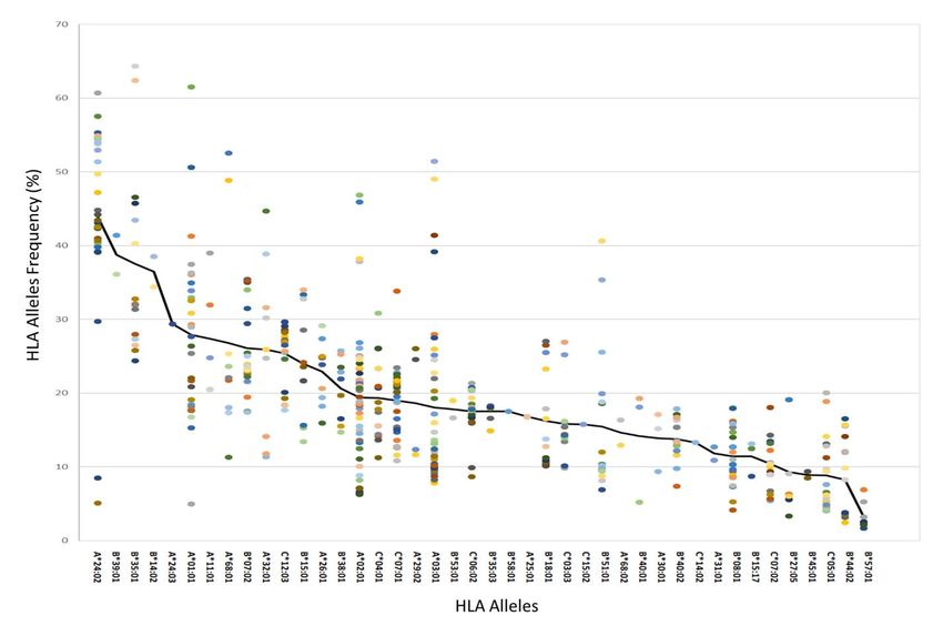
Some HLA allomorphs present larger diversity of peptides in both tumors and plasma
The 52 different GBM samples and 36 different non-cancerous samples have undergone a
complete HLA typing analysis based on DNA sequencing (51). This analysis facilitated the use of
the NetMHC platform to fit the HLA peptides sequences to their likely presenting HLA allomorphs
by selecting peptides with NetMHC rank equal or better than 2
(http://www.cbs.dtu.dk/services/NetMHC/). HLA allomorphs with a consensus sequence motif
such as HLA-B*35:01 and HLA-A*24:02 presented larger numbers of peptides relative to
allomorphs such as HLA-B*27:05 or HLA-B*40:01 (Fig. 4 and Supplemental Table S8). This
phenomenon was observed in both GBM and in the non-cancerous donors’ plasma samples, as
well as the GBM tissues (Fig. 6, Supplemental Fig. S4). The HLA-A*24:02 allele is common
among glioma patients (58) and therefore HLA peptides presented by these alleles may become
useful for treatment of larger groups of patients.
Discussion
13This study is an extensive immunopeptidome analysis of 106 GBM plasma samples, 10 GBM
tumor tissues, and 36 control plasma samples. The analysis led to high confidence identification
of 35,545 unique HLA peptides. It was suggested before that the plasma-sHLA peptidome
represents the tumor-mHLA peptidome, since the tumors’ cells shed larger amounts of sHLA
molecules relative to the uninvolved tissues (28). Similarly, in this study, a significant overlap was
observed between mHLA peptidomes of the tumor tissues and matched plasma-sHLA peptidomes
of each of the patients, demonstrating that the plasma-sHLA peptidome contains many peptides
derived from the tumors. These correlations, between the repertoires and LC-MS intensities of the
mHLA and sHLA peptidomes of the individual patients, were much larger than the correlations
between the HLA peptidomes and the proteomes of the patients’ tumors (Supplemental Fig. S3).
Downloaded from http://www.mcponline.org/ by guest on December 12, 2018
Low correlations in both repertoires and expression levels, between HLA peptidomes, proteomes,
and transcriptomes, of the same cells, were suggested in previous publications (59, 60) but were
found to be higher in others (61–64). Additionally, we demonstrate that the plasma-sHLA
peptidomes are relatively stable, can be analyzed reproducibly, and represent the tumor-mHLA
peptidomes. Comparative analysis of both mHLA and sHLA peptidomes may provide candidate
peptides, potentially useful for immunotherapy and as biomarkers. Precision medicine based on
large-scale body-fluid biomarkers may help to identify patients that are most likely to benefit from
specific treatments, including, but not limited to immunotherapy (65–67). It is expected that
different HLA peptides derived from the TAA and CTA genes will be presented among the
patients’ sHLA allomorphs’ peptidomes. Thus, the search among the sHLA peptidomes of
different people for disease biomarkers is not limited to specific peptides, but to the presence of
different peptides derived from the selected TAA/CTA genes. A few of the 250 down-regulated
sHLA peptides observed in this study may serve as biomarkers for early detection of the disease
or relapse. Indeed, potentially useful TAA can be selected based on different criteria. In our
opinion, first and outmost are peptides that are derived from genes that not expressed in any healthy
adult tissue, other than the immune privileged sites, such as the germline cells. Such HLA peptides
are potentially useful for both immunotherapy and diagnosis. Plasma sHLA peptides that disappear
after treatment or remission are potentially also useful for diagnosis, for early detection and for
relapse. sHLA and mHLA peptides that appear in tumor tissues and not in the sHLA peptidome of
the healthy donors are clearly more significant targets for further research since such peptides are
more likely (but not necessarily) tumor specific. However, only a few of the down-regulated
14peptides observed here are TAAs. Such selected biomarker peptides do not need to be derived
from bona fide CTAs, but need to be affected similarly in a large cohort of patients in order to
become useful for clinical exploitation. One should consider that many peptides were not detected
in this study in the plasma-sHLA peptidomes after surgery, also due to the nature of shotgun
peptidomics methodology used here. Performing targeted LC-MS/MS analysis with the same or
with different sets of samples is still required to exclude this possibility. The alternative use of
data-independent analysis of HLA peptidome was already used for analyses of HLA peptidomes
at higher reproducibility (68–71), and SRM were used to obtain more accurate presentation levels
for the selected peptides in multiple analyses (72, 73). Smaller numbers of HLA peptides were
identified with the LC-MS/MS data collected in this study when the FDR was set to 0.01 instead
Downloaded from http://www.mcponline.org/ by guest on December 12, 2018
of 0.05 in the Andromeda search (50), performed within the MaxQuant analysis tool (49). This is
expected, since the MS/MS fragmentations of many peptides are suboptimal in the data-dependent
(shotgun) analysis performed in this study. The use of data-independent analysis for the HLA
peptidome analysis may help to solve some of the data loss incurred (69, 71, 74). Alternatively,
one can increase the FDR to 0.05 and facilitate this way the discovery of candidates for vaccine or
biomarkers HLA peptides that would have been lost with 0.01 FDR (75). Here as well, a significant
fraction of the peptides that were lost by use of 0.01 FDR, instead of 0.05, are true ligands of the
HLA allomorphs of the patient (Supplemental Table S9), as defined by the similarity of sequence
motifs to the peptides included in the 0.01 FDR (Supplemental Fig. S5).
The blood-brain barrier (BBB) prevents entrance and exit of some cells and molecules (5,
6) to the brain and it is unknown if it allows passage of the circulating sHLA-peptide complexes.
However, the BBB in GBM patients is partially broken by the local inflammatory conditions, and
differs in its tumor vessels morphology and in the hyper-permeability of its endothelial cells (5).
The loss of functional integrity of the BBB allows passage of low molecular weight compounds,
including different chemotherapeutics into the brain (76). Such changes in the BBB may also
facilitate the release of sHLA molecules with their bound peptides into the circulation, thus
allowing their detection in the plasma for further evaluations as tumor markers.
Ideally, HLA peptides useful to serve as immunotherapeutics or biomarkers should be
derived from genes expressed at sufficient levels in the malignant tissues, but not at all in any of
the other essential healthy tissues (18, 23). Fresh-frozen tumors can be used for exome,
15transcriptome, proteome and HLA peptidome analyses, allowing identification of neoepitopes and
tumor antigens (4, 16, 40–45, 63, 77). Selection of CTA candidates for immunotherapy can be
based on high expression levels of their HLA peptides, mRNA and proteins in the tumors, and no
expression in healthy tissues. Even though tumors and plasma are good sources of HLA peptides,
healthy tissues are not normally available for analysis of their HLA peptidomes, while data about
the gene expression levels is available in public databases. Very large and relatively accurate
databases are publicly available, including data on gene and protein expression in many tissues, of
numerous people. Examples include BioGPS (55, 56), TANTIGEN (53) and HPR
(https://www.proteinatlas.org/) (78, 79). Although these databases are based on numerous studies,
some discrepancies were observed between them in regards to different selected CTAs studied
Downloaded from http://www.mcponline.org/ by guest on December 12, 2018
here. Using additional gene expression databases may alleviate some of these concerns and provide
candidate immunotherapeutics with lower risk of inducing adverse effects.
The preferred CTAs, providing potentially useful HLA peptides discovered in this study,
were defined as those derived from genes whose transcripts are expressed at levels below nine
gcrma units in all normal, essential tissues (according to BioGPS) and are expressed at
significantly higher levels in the tumors tissues. Nine gcrma units of mRNA levels of expression
were selected here as sufficiently low, since this is the highest measured mRNA expression level
of many well-characterized CTAs in healthy (non-testis) adult tissues. Some of these CTAs,
including CTAG1A (NY-ESO-1) and MAGE-A1 are well known TAAs, whose expression levels
in healthy tissues are below 9 gcrma units. These CTAs were already used in multiple clinical
studies without observable autoimmune reactions (19, 80–82) suggesting that genes expressed
below these levels are possibly safe for clinical use.
The tumor-mHLA and plasma-sHLA peptidomes of different people can be compared
while looking for HLA peptides shared between larger cohorts of individuals. HLAs with
completely distinct binding motifs are not expected to bind and present shared peptide ligands.
Importantly, significant similarities were observed in this study between the HLA peptidomes of
different patients that share some of their HLA alleles (Fig. 3). In contrast, shared peptides,
detected in the HLA peptidomes of different people who do not have any common HLA alleles or
have HLA alleles belonging to different HLA supertypes (83) are more likely to be defined as
contaminants, rather than true HLA ligands. For example, it is very unlikely that HLAs such as
16HLA-A*2, B*7 and B*27 will share any peptide ligands (83). On the other hand, shared tumor
antigens that are detected in multiple patients that have similar HLA allotypes, are extremely
important, due to their potential to become useful for treatment of multiple patients.
The HLA molecules of some HLA allomorphs, such as HLA A*24:02, are more abundant
in the plasma of carriers of these alleles (32). In addition, some HLA allomorphs present more
numerous peptides than others do (84). Indeed, here we observe similar phenomena at the HLA
peptidome levels, some HLA allomorphs, such as HLA-A24, HLA-B35 and HLA-B51 present
more diverse repertoires of peptides than others do, in both the tumors and the plasma. This implies
that the use of plasma-sHLA peptidome analysis is probably more efficient for carriers of these
Downloaded from http://www.mcponline.org/ by guest on December 12, 2018
alleles, since larger sHLA peptidomes can be recovered and identified from their plasma samples
(Fig. 4 and Fig. 6). It may also mean that there is larger immune tolerance in the HLA-A*24
patients and, therefore, it may be more difficult to break the immune tolerance induced by
circulating sHLA molecules, when attempting immunotherapy. Importantly, HLA-A*24 was
claimed to be associated genetically with glioma (58). Therefore, the use of the sHLA peptidome
analysis to search for disease biomarkers will need to be adjusted accordingly, in order to take into
account the particular HLA allotypes of the individual patients.
In conclusion, the data described here suggests a useful method for selection of biomarkers
and cancer immune-therapeutics for GBM, and provides large lists of such candidates. Such
methodologies are likely useful for discovery of biomarkers and immune-therapeutics candidates
for other cancers. Most importantly, the identification of numerous sHLA peptides derived from
CTAs can represent a promising noninvasive strategy for the monitoring of patients’ response and
progression during standard treatment modalities.
Acknowledgements
GAPVAC project was funded by the European Union Framework 7 Program. We thank Ilana
Navon from the Smoler Proteomics Center at the Technion for performing the LC-MS/MS
experiments. The authors acknowledge the advice and discussion with the members of the
GAPVAC consortium, Stefan Stevanovic and Cécile Gouttefangeas.
17References
1. Alifieris, C., and Trafalis, D. T. (2015) Glioblastoma multiforme: Pathogenesis and
treatment. Pharmacol. Ther. 152, 63–82
2. Thakkar, J. P., Dolecek, T. A., Horbinski, C., Ostrom, Q. T., Lightner, D. D., Barnholtz-
Sloan, J. S., and Villano, J. L. (2014) Epidemiologic and molecular prognostic review of
glioblastoma. Cancer Epidemiol. Biomarkers Prev. 23, 1985–96
3. Terasaki, M., Shibui, S., Narita, Y., Fujimaki, T., Aoki, T., Kajiwara, K., Sawamura, Y.,
Kurisu, K., Mineta, T., Yamada, A., and Itoh, K. (2011) Phase I trial of a personalized
Downloaded from http://www.mcponline.org/ by guest on December 12, 2018
peptide vaccine for patients positive for human leukocyte antigen-A24 with recurrent or
progressive glioblastoma multiforme. J. Clin. Oncol. 29, 337–344
4. Neidert, M. C., Schoor, O., Trautwein, C., Trautwein, N., Christ, L., Melms, A., Honegger,
J., Rammensee, H. G., Herold-Mende, C., Dietrich, P. Y., and Stevanović, S. (2013) Natural
HLA class i ligands from glioblastoma: Extending the options for immunotherapy. J.
Neurooncol. 111, 285–294
5. Patel, M. A., and Pardoll, D. M. (2015) Concepts of immunotherapy for glioma. J.
Neurooncol. 123, 323–30
6. Cohen-Inbar, O., and Zaaroor, M. (2016) Immunological Aspects of Malignant Gliomas.
Can. J. Neurol. Sci. / J. Can. des Sci. Neurol. 43, 494–502
7. Polivka, J., Holubec, L., Kubikova, T., Priban, V., Hes, O., Pivovarcikova, K., and
Treskova, I. (2017) Advances in Experimental Targeted Therapy and Immunotherapy for
Patients with Glioblastoma Multiforme. Anticancer Res. 37, 21–33
8. Swartz, A. M., Batich, K. A., Fecci, P. E., and Sampson, J. H. (2015) Peptide vaccines for
the treatment of glioblastoma. J. Neurooncol. 123, 433–40
9. Ampie, L., Woolf, E. C., and Dardis, C. (2015) Immunotherapeutic advancements for
glioblastoma. Front. Oncol. 5, 12
1810. Oh, T., Sayegh, E. T., Fakurnejad, S., Oyon, D., Lamano, J. B., DiDomenico, J. D., Bloch,
O., and Parsa, A. T. (2015) Vaccine therapies in malignant glioma. Curr. Neurol. Neurosci.
Rep. 15, 508
11. Srinivasan, V. M., Ferguson, S. D., Lee, S., Weathers, S.-P., Kerrigan, B. C. P., and
Heimberger, A. B. (2017) Tumor Vaccines for Malignant Gliomas. Neurotherapeutics 14,
345–357
12. Kamran, N., Calinescu, A., Candolfi, M., Chandran, M., Mineharu, Y., Asad, A. S.,
Koschmann, C., Nunez, F. J., Lowenstein, P. R., and Castro, M. G. (2016) Recent advances
and future of immunotherapy for glioblastoma. Expert Opin. Biol. Ther. 16, 1245–64
Downloaded from http://www.mcponline.org/ by guest on December 12, 2018
13. Ott, P. A., Hu, Z., Keskin, D. B., Shukla, S. A., Sun, J., Bozym, D. J., Zhang, W., Luoma,
A., Giobbie-Hurder, A., Peter, L., Chen, C., Olive, O., Carter, T. A., Li, S., Lieb, D. J.,
Eisenhaure, T., Gjini, E., Stevens, J., Lane, W. J., Javeri, I., Nellaiappan, K., Salazar, A. M.,
Daley, H., Seaman, M., Buchbinder, E. I., Yoon, C. H., Harden, M., Lennon, N., Gabriel,
S., Rodig, S. J., Barouch, D. H., Aster, J. C., Getz, G., Wucherpfennig, K., Neuberg, D.,
Ritz, J., Lander, E. S., Fritsch, E. F., Hacohen, N., and Wu, C. J. (2017) An immunogenic
personal neoantigen vaccine for patients with melanoma. Nature, 1–25
14. Sharma, P., and Allison, J. P. P. (2015) Immune checkpoint targeting in cancer therapy:
toward combination strategies with curative potential. Cell 161, 205–214
15. Palucka, A. K., and Coussens, L. M. (2016) The Basis of Oncoimmunology. Cell 164,
1233–1247
16. Dutoit, V., Herold-Mende, C., Hilf, N., Schoor, O., Beckhove, P., Bucher, J., Dorsch, K.,
Flohr, S., Fritsche, J., Lewandrowski, P., Lohr, J., Rammensee, H.-G., Stevanovic, S.,
Trautwein, C., Vass, V., Walter, S., Walker, P. R., Weinschenk, T., Singh-Jasuja, H., and
Dietrich, P.-Y. (2012) Exploiting the glioblastoma peptidome to discover novel tumour-
associated antigens for immunotherapy. Brain 135, 1042–54
17. Bassani-Sternberg, M., and Coukos, G. (2016) Mass spectrometry-based antigen discovery
for cancer immunotherapy. Curr. Opin. Immunol. 41, 9–17
1918. Rammensee, H.-G., and Singh-Jasuja, H. (2013) HLA ligandome tumor antigen discovery
for personalized vaccine approach. Expert Rev. Vaccines 12, 1211–7
19. Pol, J., Bloy, N., Buqué, A., Eggermont, A., Sautès-fridman, C., Galon, J., Tartour, E.,
Kroemer, G., Galluzzi, L., Pol, J., Bloy, N., Buqué, A., Eggermont, A., Cremer, I., Sautès-
fridman, C., Galon, J., Tartour, E., Zitvogel, L., Kroemer, G., Galluzzi, L., Watch, T., Pol,
J., Bloy, N., Buqué, A., Eggermont, A., Cremer, I., Zitvogel, L., Pol, J., Bloy, N., Buqu, A.,
Buque, A., Eggermont, A., Cremer, I., Sautes-Fridman, C., Galon, J., Tartour, E., Zitvogel,
L., Kroemer, G., and Galluzzi, L. (2015) Trial Watch : Peptide-based anticancer vaccines
Trial Watch : Peptide-based anticancer vaccines. Oncoimmunology 4, e974411
Downloaded from http://www.mcponline.org/ by guest on December 12, 2018
20. Comber, J. D., and Philip, R. (2014) MHC class I antigen presentation and implications for
developing a new generation of therapeutic vaccines. Ther Adv Vaccines 2, 77–89
21. Heemskerk, B., Kvistborg, P., and Schumacher, T. N. M. (2012) The cancer antigenome.
EMBO J. 32, 194–203
22. Chen, Y. T., Scanlan, M. J., Sahin, U., Türeci, O., Gure, A. O., Tsang, S., Williamson, B.,
Stockert, E., Pfreundschuh, M., and Old, L. J. (1997) A testicular antigen aberrantly
expressed in human cancers detected by autologous antibody screening. Proc. Natl. Acad.
Sci. U. S. A. 94, 1914–8
23. Whitehurst, A. W. (2014) Cause and consequence of cancer/testis antigen activation in
cancer. Annu. Rev. Pharmacol. Toxicol. 54, 251–72
24. Schumacher, T. N., and Schreiber, R. D. (2015) Neoantigens in cancer immunotherapy.
Science 348, 69–74
25. Charlton, R. K., and Zmijewski, C. M. (1970) Soluble HL-A7 antigen: localization in the
beta-lipoprotein fraction of human serum. Science 170, 636–7
26. van Rood, J. J., van Leeuwen, A., and van Santen, M. C. (1970) Anti HL-A2 inhibitor in
normal human serum. Nature 226, 366–7
27. Tabayoyong, W. B., and Zavazava, N. (2007) Soluble HLA revisited. Leuk Res 31, 121–5
2028. Bassani-Sternberg, M., Barnea, E., Beer, I., Avivi, I., Katz, T., and Admon, A. (2010)
Soluble plasma HLA peptidome as a potential source for cancer biomarkers. Proc Natl Acad
Sci U S A 107, 18769–18776
29. Ritz, D., Gloger, A., Weide, B., Garbe, C., Neri, D., and Fugmann, T. (2016) High-
sensitivity HLA class I peptidome analysis enables a precise definition of peptide motifs
and the identification of peptides from cell lines and patients ’ sera. Proteomics 16, 1570–
1580
30. Ritz, D., Gloger, A., Neri, D., and Fugmann, T. (2017) Purification of soluble HLA class I
complexes from human serum or plasma deliver high quality immuno peptidomes required
Downloaded from http://www.mcponline.org/ by guest on December 12, 2018
for biomarker discovery. Proteomics 17, 1–6
31. Puppo, F., Scudeletti, M., Indiveri, F., and Ferrone, S. (1995) Serum HLA class I antigens:
markers and modulators of an immune response? Immunol Today 16, 124–127
32. Adamashvili, I. M., Fraser, P. A., and McDonald, J. C. (1996) Association of serum
concentration of soluble class I HLA with HLA allotypes. Transplantation 61, 984–7
33. Campoli, M., and Ferrone, S. (2008) Tumor escape mechanisms: potential role of soluble
HLA antigens and NK cells activating ligands. Tissue Antigens 72, 321–334
34. Hunt, D. F., Henderson, R. A., Shabanowitz, J., Sakaguchi, K., Michel, H., Sevilir, N., Cox,
a L., Appella, E., and Engelhard, V. H. (1992) Characterization of peptides bound to the
class I MHC molecule HLA-A2.1 by mass spectrometry. Science (80-. ). 255, 1261–3
35. Granados, D. P., Laumont, C. M., Thibault, P., and Perreault, C. (2015) The nature of self
for T cells-a systems-level perspective. Curr. Opin. Immunol. 34, 1–8
36. Schumacher, F.-R., Delamarre, L., Jhunjhunwala, S., Modrusan, Z., Phung, Q. T., Elias, J.
E., and Lill, J. R. (2017) Building proteomic tool boxes to monitor MHC class I and class
II peptides. Proteomics 17, 1600061
37. de Verteuil, D., Granados, D. P., Thibault, P., and Perreault, C. (2012) Origin and plasticity
of MHC I-associated self peptides. Autoimmun. Rev. 11, 627–635
2138. Fritsche, J., Rakitsch, B., Hoffgaard, F., Römer, M., Schuster, H., Kowalewski, D. J.,
Priemer, M., Stos-Zweifel, V., Hoerzer, H., Satelli, A., Sonntag, A., Goldfinger, V., Song,
C., Mahr, A., Ott, M., Schoor, O., Weinschenk, T., Hörzer, H., Satelli, A., Sonntag, A.,
Goldfinger, V., Song, C., Mahr, A., Ott, M., Schoor, O., and Weinschenk, T. (2018)
Translating Immunopeptidomics to Immunotherapy-Decision-Making for Patient and
Personalized Target Selection. Proteomics, 1700284
39. Shraibman, B., Kadosh, D. M., Barnea, E., and Admon, A. (2016) Human Leukocyte
Antigen (HLA) Peptides Derived from Tumor Antigens Induced by Inhibition of DNA
Methylation for Development of Drug-facilitated Immunotherapy. Mol. Cell. Proteomics
Downloaded from http://www.mcponline.org/ by guest on December 12, 2018
15, 3058–70
40. Weinschenk, T., Gouttefangeas, C., Schirle, M., Obermayr, F., Walter, S., Schoor, O.,
Kurek, R., Loeser, W., Bichler, K., Wernet, D., Stevanović, S., and Rammensee, H.-G.
(2002) Integrated functional genomics approach for the design of patient-individual
antitumor vaccines. Cancer Res. 62, 5818–27
41. Bassani-Sternberg, M., Bräunlein, E., Klar, R., Engleitner, T., Sinitcyn, P., Audehm, S.,
Straub, M., Weber, J., Slotta-Huspenina, J., Specht, K., Martignoni, M. E., Werner, A.,
Hein, R., H. Busch, D., Peschel, C., Rad, R., Cox, J., Mann, M., Krackhardt, A. M., H
Busch, D., Peschel, C., Rad, R., Cox, J., Mann, M., and Krackhardt, A. M. (2016) Direct
identification of clinically relevant neoepitopes presented on native human melanoma tissue
by mass spectrometry. Nat. Commun. 7, 13404
42. Seliger, B., Dressler, S. P., Massa, C., Recktenwald, C. V., Altenberend, F., Bukur, J.,
Marincola, F. M., Wang, E., Stevanovic, S., and Lichtenfels, R. (2011) Identification and
characterization of human leukocyte antigen class I ligands in renal cell carcinoma cells.
Proteomics 11, 2528–41
43. Klatt, M. G., Kowalewski, D. J., Schuster, H., Di Marco, M., Hennenlotter, J., Stenzl, A.,
Rammensee, H.-G., and Stevanović, S. (2016) Carcinogenesis of renal cell carcinoma
reflected in HLA ligands: A novel approach for synergistic peptide vaccination design.
Oncoimmunology 5, e1204504
2244. Kowalewski, D. J., Schuster, H., Backert, L., Berlin, C., Kahn, S., Kanz, L., Salih, H. R.,
Rammensee, H.-G., Stevanovic, S., and Stickel, J. S. (2015) HLA ligandome analysis
identifies the underlying specificities of spontaneous antileukemia immune responses in
chronic lymphocytic leukemia (CLL). Proc. Natl. Acad. Sci. U. S. A. 112, E166-75
45. Walz, S., Stickel, J. S., Kowalewski, D. J., Schuster, H., Weisel, K., Backert, L., Kahn, S.,
Nelde, A., Stroh, T., Handel, M., Kohlbacher, O., Kanz, L., Salih, H. R., Rammensee, H.,
and Stevanović, S. (2015) The antigenic landscape of multiple myeloma: mass spectrometry
(re)defines targets for T-cell-based immunotherapy. Blood 126, 1203–13
46. Milner, E., Gutter-Kapon, L., Bassani-Strenberg, M., Barnea, E., Beer, I., and Admon, A.
Downloaded from http://www.mcponline.org/ by guest on December 12, 2018
(2013) The effect of proteasome inhibition on the generation of the human leukocyte antigen
(HLA) peptidome. Mol. Cell. Proteomics 12, 1853–64
47. Ishihama, Y., Rappsilber, J., and Mann, M. (2006) Modular stop and go extraction tips with
stacked disks for parallel and multidimensional Peptide fractionation in proteomics. J.
Proteome Res. 5, 988–94
48. Ishihama, Y., Rappsilber, J., Andersen, J. S., and Mann, M. (2002) Microcolumns with self-
assembled particle frits for proteomics. J. Chromatogr. A 979, 233–239
49. Cox, J., and Mann, M. (2008) MaxQuant enables high peptide identification rates,
individualized p.p.b.-range mass accuracies and proteome-wide protein quantification. Nat.
Biotechnol. 26, 1367–72
50. Cox, J., Neuhauser, N., Michalski, A., Scheltema, R. A., Olsen, J. V., and Mann, M. (2011)
Andromeda: a peptide search engine integrated into the MaxQuant environment. J.
Proteome Res. 10, 1794–805
51. Lange, V., Böhme, I., Hofmann, J., Lang, K., Sauter, J., Schöne, B., Paul, P., Albrecht, V.,
Andreas, J. M., Baier, D. M., Nething, J., Ehninger, U., Schwarzelt, C., Pingel, J., Ehninger,
G., and Schmidt, A. H. (2014) Cost-efficient high-throughput HLA typing by MiSeq
amplicon sequencing. BMC Genomics 15, 63
52. Almeida, L. G., Sakabe, N. J., deOliveira, A. R., Silva, M. C., Mundstein, A. S., Cohen, T.,
23Chen, Y. T., Chua, R., Gurung, S., Gnjatic, S., Jungbluth, A. A., Caballero, O. L., Bairoch,
A., Kiesler, E., White, S. L., Simpson, A. J., Old, L. J., Camargo, A. A., and Vasconcelos,
A. T. (2009) CTdatabase: a knowledge-base of high-throughput and curated data on cancer-
testis antigens. Nucleic Acids Res 37, D816-9
53. Zhang, G. L., Sun, J., Chitkushev, L., and Brusic, V. (2014) Big data analytics in
immunology: a knowledge-based approach. Biomed Res. Int. 2014, 437987
54. Serrano, A., Tanzarella, S., Lionello, I., Mendez, R., Traversari, C., Ruiz-Cabello, F., and
Garrido, F. (2001) Rexpression of HLA class I antigens and restoration of antigen-specific
CTL response in melanoma cells following 5-aza-2’-deoxycytidine treatment. Int. J. cancer
Downloaded from http://www.mcponline.org/ by guest on December 12, 2018
94, 243–51
55. Wu, C., Jin, X., Tsueng, G., Afrasiabi, C., and Su, A. I. (2016) BioGPS: building your own
mash-up of gene annotations and expression profiles. Nucleic Acids Res. 44, D313-6
56. Wu, C., Orozco, C., Boyer, J., Leglise, M., Goodale, J., Batalov, S., Hodge, C. L., Haase,
J., Janes, J., Huss, J. W., Su, A. I., Huss 3rd, J. W., and Su, A. I. (2009) BioGPS: an
extensible and customizable portal for querying and organizing gene annotation resources.
Genome Biol 10, R130
57. Admon, A., and Bassani-Sternberg, M. (2011) The Human Immunopeptidome Project, a
Suggestion for yet another Postgenome Next Big Thing. Mol. Cell. Proteomics 10,
O111.011833
58. Nitta, T., Ebato, M., and Sato, K. (1994) Association of malignant glioma with the human
leukocyte antigen, HLA-A24(9). Neurosurg. Rev. 17, 211–5
59. Bourdetsky, D., Schmelzer, C. E. H., and Admon, A. (2014) The nature and extent of
contributions by defective ribosome products to the HLA peptidome. Proc. Natl. Acad. Sci.
U. S. A. 111, E1591-9
60. Shraibman, B., Kadosh, D. M., Barnea, E., and Admon, A. (2016) Human Leukocyte
Antigen (HLA) Peptides Derived from Tumor Antigens Induced by Inhibition of DNA
Methylation for Development of Drug-facilitated Immunotherapy. Mol. Cell. proteomics
24MCP 15, 3058–3070
61. Bassani-Sternberg, M., Pletscher-Frankild, S., Jensen, L. J., and Mann, M. (2015) Mass
spectrometry of human leukocyte antigen class I peptidomes reveals strong effects of
protein abundance and turnover on antigen presentation. Mol. Cell. Proteomics 14, 658–73
62. Pearson, H., Daouda, T., Granados, D. P., Durette, C., Bonneil, E., Courcelles, M.,
Rodenbrock, A., Laverdure, J.-P., Côté, C., Mader, S., Lemieux, S., Thibault, P., and
Perreault, C. (2016) MHC class I-associated peptides derive from selective regions of the
human genome. J. Clin. Invest. 126, 4690–4701
Downloaded from http://www.mcponline.org/ by guest on December 12, 2018
63. Khodadoust, M. S., Olsson, N., Wagar, L. E., Haabeth, O. A. W., Chen, B., Swaminathan,
K., Rawson, K., Liu, C. L., Steiner, D., Lund, P., Rao, S., Zhang, L., Marceau, C., Stehr,
H., Newman, A. M., Czerwinski, D. K., Carlton, V. E. H., Moorhead, M., Faham, M., Kohrt,
H. E., Carette, J., Green, M. R., Davis, M. M., Levy, R., Elias, J. E., and Alizadeh, A. A.
(2017) Antigen presentation profiling reveals recognition of lymphoma immunoglobulin
neoantigens. Nature 543, 723–727
64. Schuster, H., Peper, J. K., Bösmüller, H.-C., Röhle, K., Backert, L., Bilich, T., Ney, B.,
Löffler, M. W., Kowalewski, D. J., Trautwein, N., Rabsteyn, A., Engler, T., Braun, S., Haen,
S. P., Walz, J. S., Schmid-Horch, B., Brucker, S. Y., Wallwiener, D., Kohlbacher, O., Fend,
F., Rammensee, H.-G., Stevanović, S., Staebler, A., and Wagner, P. (2017) The
immunopeptidomic landscape of ovarian carcinomas. Proc. Natl. Acad. Sci. U. S. A. 114,
E9942–E9951
65. Sawyers, C. L. (2008) The cancer biomarker problem. Nature 452, 548–52
66. Perez-Gracia, J. L., Sanmamed, M. F., Bosch, A., Patiño-Garcia, A., Schalper, K. A.,
Segura, V., Bellmunt, J., Tabernero, J., Sweeney, C. J., Choueiri, T. K., Martín, M., Fusco,
J. P., Rodriguez-Ruiz, M. E., Calvo, A., Prior, C., Paz-Ares, L., Pio, R., Gonzalez-
Billalabeitia, E., Gonzalez Hernandez, A., Páez, D., Piulats, J. M., Gurpide, A., Andueza,
M., de Velasco, G., Pazo, R., Grande, E., Nicolas, P., Abad-Santos, F., Garcia-Donas, J.,
Castellano, D., Pajares, M. J., Suarez, C., Colomer, R., Montuenga, L. M., and Melero, I.
(2016) Strategies to design clinical studies to identify predictive biomarkers in cancer
25You can also read