The Cuban Human Brain Mapping Project, a young and middle age population-based EEG, MRI, and cognition dataset - Nature
←
→
Page content transcription
If your browser does not render page correctly, please read the page content below
www.nature.com/scientificdata

               OPEN           The Cuban Human Brain Mapping
Data Descriptor               Project, a young and middle age
                              population-based EEG, MRI, and
                              cognition dataset
                              Pedro A. Valdes-Sosa 1,2,4 ✉, Lidice Galan-Garcia2,4, Jorge Bosch-Bayard1,2,3,4,
                              Maria L. Bringas-Vega 1,2,4, Eduardo Aubert-Vazquez2,4, Iris Rodriguez-Gil2, Samir Das3,
                              Cecile Madjar3, Trinidad Virues-Alba2, Zia Mohades3, Leigh C. MacIntyre3, Christine Rogers3,
                              Shawn Brown3, Lourdes Valdes-Urrutia2, Alan C. Evans 3,5 & Mitchell J. Valdes-Sosa1,2,5

                              The Cuban Human Brain Mapping Project (CHBMP) repository is an open multimodal neuroimaging
                              and cognitive dataset from 282 young and middle age healthy participants (31.9 ± 9.3 years, age range
                              18–68 years). This dataset was acquired from 2004 to 2008 as a subset of a larger stratified random
                              sample of 2,019 participants from La Lisa municipality in La Habana, Cuba. The exclusion criteria
                              included the presence of disease or brain dysfunctions. Participant data that is being shared comprises
                              i) high-density (64–120 channels) resting-state electroencephalograms (EEG), ii) magnetic resonance
                              images (MRI), iii) psychological tests (MMSE, WAIS-III, computerized go-no go reaction time), as well
                              as iv,) demographic information (age, gender, education, ethnicity, handedness, and weight). The EEG
                              data contains recordings with at least 30 minutes in duration including the following conditions: eyes
                              closed, eyes open, hyperventilation, and subsequent recovery. The MRI consists of anatomical T1 as
                              well as diffusion-weighted (DWI) images acquired on a 1.5 Tesla system. The dataset presented here is
                              hosted by Synapse.org and available at https://chbmp-open.loris.ca.

                              Background & Summary
                              In the past decade several neuroimaging databases (ADNI, HCP, UK Biobank, CAMCAN, ABCD, PPMI), as well
                              as consortia (ENIGMA), have been launched. They aim to accelerate insights into neurodevelopment and physi-
                              opathology and to allow the identification of new biomarkers. An essential ingredient, lacking in many projects,
                              is the inclusion of the electroencephalograms (EEG), an informative and direct measurement of brain activity.
                              The EEG is cost-effective, accessible, and applicable to underserved populations throughout the world. It is a
                              technique of choice for extensive population screening in any economic setting. It is not surprising, therefore, that
                              interest in EEG has increased, and this modality has been included in new multimodal neuroimaging datasets,
                              such as the mind-brain-body dataset CMI (Babayan et al.1) and the open resource for transdiagnostic research in
                              pediatric mental health LEMON (Alexander et al.)1,2.
                                  Here we present a new multimodal neuroimaging dataset3 that includes, this time information from a
                              Latin American middle-income country. It was led by the Cuban Human Brain Mapping Project (CHBMP)4,
                              a population-based, multi-decade longitudinal brain health data-gathering effort in Havana Cuba. This ongo-
                              ing project is organized by the Cuban Ministry of Public Health (MINSAP) and coordinated by the Cuban
                              Neuroscience Center (CNEURO). The CHBMP focuses on the development of tools and health applications
                              based on multimodal neuroimaging. This data corresponds to a young to middle-age adult population. Due to an

                              1
                               The Clinical Hospital of Chengdu Brain Sciences, University of Electronic Sciences and Technology of China,
                              Chengdu, China. 2Cuban Neuroscience Center, La Habana, Cuba. 3McGill Centre for Integrative Neurosciences MCIN.
                              Ludmer Centre for Mental Health. Montreal Neurological Institute, McGill University, Montreal, Canada. 4These
                              authors contributed equally: Pedro A. Valdes-Sosa, Lidice Galan-Garcia, Jorge Bosch-Bayard, Maria L. Bringas-
                              Vega, Eduardo Aubert-Vazquez. 5These authors jointly supervised this work: Alan C. Evans, Mitchell J. Valdes-Sosa.
                              ✉e-mail: pedro.valdes@neuroinformatics-collaboratory.org

  Scientific Data |   (2021) 8:45 | https://doi.org/10.1038/s41597-021-00829-7                                                                   1
www.nature.com/scientificdata/ www.nature.com/scientificdata
Age Interval Number of participants %
10 < xwww.nature.com/scientificdata/ www.nature.com/scientificdata
Categories N (%)
Female 87 (30.85%)
Gender
Male 195 (69.14%)
Right 238 (84.39%)
Handedness by Left 26 (9.21%)
preference Ambidextrous 8 (2.38%)
Missing 10 (3.54%)
Primary school 16 (5.67%)
Secondary School 72 (25.53%)
Education level High school 145 (51.41%)
University 39 (13.82%)
Missing 10 (3.54%)
Table 2. Demographic description of the sample.
Activity Sample size
Selection of one municipality with the closest match in gender, age, ethnic characteristics of the general Cuban
population. This was also a municipality with a large proportion of persons originally from other provinces according
Stage 1 30,000
to the MINSAP and Statistics National Office. La Lisa municipality was selected. A random sample of 30,000
participants from the total population was selected based on the ID card serial numbers.
From the 30,000 participants selected in the previous stage, a random subsample of 2019 was selected stratified by
Stage 2 2019
age, sex, ethnic origin, educational level, and socioeconomic status.
The Doctor and nurses of the Family Program evaluated the willingness to participate and any health conditions of
the candidates from the subsample (N = 2019). In detail:
1) The Family nurses visited each household to provide both printed and verbal information about the project
objectives and enquired about the preliminary willingness to participate in the study.
2) Those willing to participate attended an interview at their local Polyclinic center for further explanation and
Stage 3 signed the Informed Consent. 1439
3) The Health Questionnaire was then applied by the nurse to carry out a further screening of pathology, cognitive
complaints, use of pharmaceutical agents, heavy smoking, etc.
4) According to the exclusion criteria, Table 3, a further selection of the subsample was carried out for the next stage.
Participants with health problems remained in the overall study, and continue to be followed up to this day with
further clinical evaluation and treatment. However, they were excluded from the normative part of the project.
At the local Polyclinic center psychiatrist/neurologist applied:
• A standardized and structured psychiatry tool, the MINI International Interview (MINI)
• MINIMENTAL State Evaluation (MMSE)
Stage 4 530
• The Edinburg Handedness Questionnaire to classify dexterity
A psychometrist applied the:
• The Weschler Adult Intelligence Scale (WAIS) III
At the local Polyclinic center, blood samples for genetic studies were extracted and additional measures: the blood
pressure, body temperature, frequency and heart rate, body weight and height were obtained (data not included in
Stage 5 530
this version of the database). The weight in pounds was included in demographic file.
Digital EEG recording was the final step at the designated Policlinics
MRI collection was carried out at the CIMEQ Hospital followed by an MRI evaluation by a neuroradiologist. Due to
Stage 6 394
a request of the Ministry of Health the number of males selected at this stage was more than double of females.
Table 3. General procedure for recruitment.
Stage 1: In agreement with the Ministry of Public Health (MINSAP), and due to logistical constraints, a single
municipality of the province of Havana City was selected for the study, together with the whole structure of the
Family Doctor and Polyclinic Centers. Towards this end, a study was carried out with a committee from MINSAP
and the National Office for Population Studies to assess the distribution of the following variables of all inhabit-
ants in every municipality in the Province of Habana City: ethnicity, sex, province of origin, and socioeconomic
status. Based on these distributions, the Municipality of La Lisa, was selected for the study since it had the closest
match to the general Cuban population. A sample of 30,000 inhabitants in this region was randomly selected from
the National Identity Card registry.
Stage 2: From the original roster of 30,000, a random subsample of N = 2019 was then selected for further
processing, is stratified by age, gender, socio-economic status.
Family Doctors (Stage 3) then examined the participant’s records to exclude persons whom they already
had ascertained to have health issues. All the remaining participants were visited by the Family nurses who left
a printed description of the project and gave a detailed verbal explanation of its aims. As usual for population
studies in Cuba, it was explained that there would be no payment for the study, but if a participant needed to be
absent from the workplace, the local government guaranteed this as a fully paid day. They were also informed
about all data acquisition protocols as well as safety measures with a special focus on MRI acquisition and safety.
To a great degree, the success of this project was due to the close contact of the Family Doctor and Nurse with the
local population, as well as the abundant information provided from the media to the general public about the
Cuban Neuroscience Center and its project. This explains a 93% initial willingness to participate in the project.
For those participants that gave written consent, a health questionnaire was applied for further screening, and
Scientific Data | (2021) 8:45 | https://doi.org/10.1038/s41597-021-00829-7 3www.nature.com/scientificdata/ www.nature.com/scientificdata
Criteria Description
Malignant systemic disease requiring chemotherapy, diabetes, thyroid dysfunction (hyper or hypothyroidism),
rheumatic disease, muscular dystrophies, liver cirrhosis, sickle cell anemia, Wilson’s disease, lupus erythematosus
(SLE), malnutrition, cardiovascular diseases, arterial hypertension, infectious diseases, AIDS, respiratory diseases,
Medical conditions
history of reactions to medications with hypersensitivity type I, current pregnancy or lactation, permanent metal
appliances, cardiac pacemakers or other types of metal in any part of the body (fixed prosthesis, fragments of a
bullet, etc.)
Chronic systemic diseases of the Central Nervous System (CNS), seizures or other attacks, malignant expansive
processes of the SNC-radiotherapy, more than one loss of consciousness, cerebrovascular accidents, transient
Neurological disease
cerebral ischemia, migraine or frequent headaches, severe neuropathies, history of cranial trauma with or without
loss of consciousness.
History of previous psychiatric treatments, tics, stuttering, intellectual disability, suicide attempt, depression,
Psychiatric Disorder sociopathic behavior, anxiety disorders, panic attacks, schizophrenia, obsessive-compulsive disorder, drug use and
abuse, alcoholism, hyperactivity and attention deficit, dementia.
Prenatal and perinatal Difficult pregnancy as indicated by the obstetrician, preterm delivery, gestational hypertension, diabetes, obesity,
antecedents early membrane rupture in pregnancy, placenta previa, retroplacental hematoma, other infections.
Sleep disorders Nocturnal terrors, somnambulism, and others.
Familiar pathological
Epilepsy, neurodegenerative diseases, multiple sclerosis, Wilson’s disease, schizophrenia, manic depressive disorder.
background
Alcohol abuse according to DSM-IV definition, non-legal drugs, smoker of more than 12 cigarettes per day, drugs
Drug addiction affecting the CNS, direct and chronic exposure to toxic substances such as pesticides, heavy metals, phosphorus, and
organic solvents.
Neurological physical Any abnormality in the neurological physical examination (hypertonia, hypotonia, asymmetry of reflexes, decrease
examination in visual acuity, nystagmus, etc.).
Table 4. Exclusion Criteria.
WAIS below 70
Cognitive MMSE below 24
Abnormalities in the background EEG activity characterized by any of the following: a) out of range average amplitude or
Visual EEG dominant frequency, b) absence of normal modulation patterns, c) inadequate anterior/posterior organization according to
age, d) paroxysms, e) lack of reactivity to eye-opening and/or hyperventilation procedures.
MRI Neuroradiological reports of lesions or atypical findings.
Table 5. The exclusion criteria according to cognitive, EEG, and MRI examination.
consequently, 580 persons were excluded at this stage from the normative study. In this, as well as in subsequent
stages, all participants that did not continue in the normative study followed a separate workflow to ensure spe-
cialized diagnostic and intervention by units of the health system, with the same protocol as those continuing in
the study. The exclusion criteria used for this stage are listed in Table 4. The most prevalent health conditions to
exclude participants were diagnosed with metabolic syndrome, psychiatric conditions, personal history of severe
illnesses, and sensory and motor disabilities.
In Stage 4, participants continuing in the study were examined at the polyclinic by specialists in Neurology
and Psychiatry, to rule out chronic diseases (e.g. addictions, including heavy smoking) or any disorders of the
nervous system that would invalidate their participation. Neurological examination was performed following
the procedure described in the guidelines published by the U.S. Department of Health and Human Services in
199717 and the Mini-Mental State Examination18 (MMSE) for global cognitive screening. The Mini-International
Psychiatric Interview was used for psychiatric evaluation19 (Spanish version) and the Weschler Adult Intelligence
Scale (WAIS) III for intelligence.
In Stage 5, EEG recordings were carried out at the polyclinic. Other measures were obtained the same day
such as anthropometric (height) and blood pressure. Additional blood samples were extracted, following a
join protocol from CNEURO with the National Center for Medical Genetics https://www.ecured.cu/Centro_
Nacional_de_Gen%C3%A9tica_M%C3%A9dica for further genetic studies of the Cuban population (to be pub-
lished separately).
During stages 4 and 5 specific exclusion criteria were employed (see Table 5).
In Stage 6, the MRI studies were carried out at the Center for Medical and Surgical Research la
Habana, Cuba (in Spanish: Centro Investigaciones Medico-Quirurgicas, CIMEQ https://www.ecured.cu/
Centro_de_Investigaciones_M%C3%A9dico_Quir%C3%BArgicas_
(CIMEQ). Additionally, a psychiatrist/psychometrist applied the computerized Reaction Time test at the end
of the study for a subsample (N = 56).
The participants who presented hypertension during this research study were included in a separate study
and underwent more specific evaluations such as carotid flow, white matter hyperintensities, eye fund, optic and
blood vessel impairments, and a set of extra measurements. The analysis of the results of this hypertension study
was partially published in4.
Procedure. Workflow.Examinations for final participants (Stages 4–6) were carried out in a five-day schedule.
See Fig. 1.
Scientific Data | (2021) 8:45 | https://doi.org/10.1038/s41597-021-00829-7 4www.nature.com/scientificdata/ www.nature.com/scientificdata
Assessment Day Assessment Day Assessment Day Assessment Day Assessment Day
1 2 3 4 5
Physical exam MMSE Blood drawing MRI preparation Reaction time
Paradigm Go NO-
Neurological WAIS III Blood pressure 1.5 T MRI GO
screening acquisition
Anthropometry
Psychiatric
screening MINI EEG preparation
EEG recordings
Fig. 1 Flow-chart of 5 days assessment.
Measurement Number
250.
EEG 170 participants with 64 and 80 with
120 channels.
MRI 203
WAIS 167
MMSE 156
RT 56
EEG + MRI 171
EEG + MRI + WAIS 142
EEG + MRI + MMSE 132
Table 6. Number of participants with each type of measurement.
The final sample count for this study (N = 282) included all the participants who completed all the requisites,
after all the steps. The total participants included in each measurement and the conjunction between modalities
can be found in Table 6.
Psychological tests. MMSE. The Mini-Mental State Examination MMSE18 is a quick and easy measure
of cognitive functioning that has been widely used in clinical evaluation and research involving patients with
dementia. In our study, the MMSE was employed as a screening test to exclude participants with cognitive impair-
ment. The total score of the participants is available in the file MMSE.csv with also the individual items for 52
subjects.
Intelligence test. Intelligence was assessed using the fully validated and translated to the Spanish language
version of the David Wechsler Adult Intelligence Scale WAIS-III printed and distributed in Mexico by El
Manual Moderno (https://www.worldcat.org/title/wais-iii-escalaweschler-de-inteligencia-para-adultos-iii/
oclc/54053545). This scale provided scores for a Full-Scale IQ (FSIQ), Verbal IQ (VIQ), and Performance IQ
(PIQ) along with four secondary indices: PO, PS, VC, and WM. The subtests included in each index were as fol-
lows: PO: picture completion, block design, matrix reasoning; PS: digit-symbol coding and symbol search; VC:
vocabulary, similarities, information, comprehension; and WM: arithmetic, digit span, letter-number sequencing.
The intelligence raw measures were scored according to the official normative data included in the printed
version of WAIS-III. However, to avoid cultural bias, they were subsequently standardized with information from
the Cuban sample to produce scores of the specific performance, adjusted for age for our population. The results
included subtests, the verbal and performance subscales and full scale IQ for all the subjects, available in the
WAIS_III.csv file. This dataset was employed in one study about how the white matter (FA-tracts based) predicts
fluid and crystallized intelligence20.
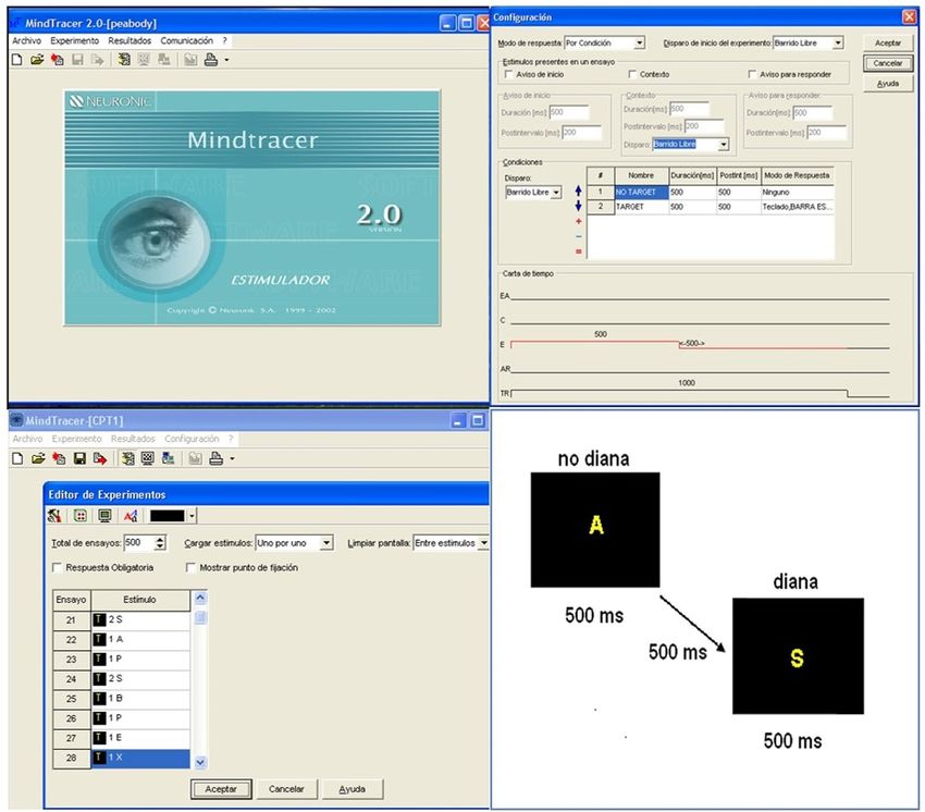
Go No-Go test. For a subset of 56 participants, reaction times were recorded using a Go No-Go paradigm which
consisted in a visual attention task, implemented using the psychophysiology software for cognitive stimulation
Mindtracer21 (N_P-SW 1.3 v.2.1.0.0 Neuronic S.A.).
The task consisted of 500 trials, 25% in GO condition and 75% in NO-GO condition, where the stimuli was a
set of letters: P B X E A S. The instructions for the participants were: Simple Reaction Time (RT1) consign: “Press
the spacebar when “S” appears at the screen”. Complex Reaction time (RT2) consign: “Press the spacebar only
when the letter “S” appears preceded by the letter “A”. The two tasks were presented to all the participants in the
same consecutive order. For a description of the facilities of the software see Fig. 2. The results included in the file
Reaction_Time.csv are: mean, standard deviation and skewness of the simple and complex reaction time tasks
in milliseconds.
Scientific Data | (2021) 8:45 | https://doi.org/10.1038/s41597-021-00829-7 5www.nature.com/scientificdata/ www.nature.com/scientificdata
Fig. 2 Top left: The main display of the software MINDTRACER for the preparation and presentation of the
stimuli of the reaction time task. Top right.- Software options to design the experimental task. Bottom left.-
Description of the different trials. Bottom right: Design of the presentation of the complex stimuli.
Equipment MEDICID 5
Number of channels 64/120
Gain 20,000/1000
Impedanceswww.nature.com/scientificdata/ www.nature.com/scientificdata
Fpz
Fp1 Fp2
AF7 AF8
AF3 AFz AF4
AFF5h AFF6h
F7 AFF1h AFF2h F8
F5 F6
F3 F4
F1 Fz F2
FFT7h FFT8h
FFC5h FFC6h
FFC3h FFC4h
FFC1h FFC2h FT8
FT7
FC5 FC3 FC6
FC1 FCz FC2 FC4
FTT7 FCC5 FCC5h FCC6 FTT8
FCC3h FCC1h FCC2h FCC4h FCC6h
T9 T7 C5 C3 Cz
C1 C2 C4 C6 T8 T10
CCP1h CCP2h
TTP7h CCP5h CCP3h CP3h CCPz CP4h CCP4h CCP6h TTP8h
CP5h CP1h CP2h CP6h
CP5 CP3 CP1 CPz CP2 CP4 CP6
TP7 CPP1h CPP2h TP8
P3h P4h
TPP9h TPP7 Pz
TPP8
TPP10h
P1 P2
P3 P4
P5 P6
P7 PO3h PO4h P8
PPO3h PPO4h
PO5 PO3 POz PO4 PO6 PO8
PO7
POO3 POO1 POO2 POO4
POO5 POO6
O1 O2
Oz
Fig. 3 Schematic representation of the position of the electrode. In black the subset of electrodes employed
in the 64 channels (maximum 62 cephalic electrodes). In white, the other electrodes were used for the 120
channels montage (maximum 120 cephalic electrodes). Both configurations use three additional channels
which are employed to record the Electro-oculogram and Electrocardiogram.
Electrode placement. Two different montages were employed, one with 64 channels and other with 120
channels as illustrated in Fig. 3 with different colors black (64) and white (120) to identify each montage. The
nomenclature of the electrodes employed in the MEDICID system and their standardization is included in
online-only Table 1 “Nomenclature of electrodes position.xlxs”
Description of the EEG protocol comprising the following participant condition:
1. Baseline: resting-state EEG with closed Eyes (state A), 10 minutes
2. Reactivity: this test consisted of the consecutive opening and closing eyes with an interval of 12 seconds.
Open eyes (state B), 5 minutes, where the participant was instructed to look at a point, keeping the pupils
fixed.
3. Hyperventilation (HPV): Dividing it in the first minute HPV1 (state C), the subject was instructed to start
taking air through the nose and to breathe deeply. The second minute HPV2 (state D), and HPV3 (state E),
this last one minute with more frequent and shallow inspirations. Total 3 minutes.
4. Recovery (state F): The last step is the recovery of the patient after the HPV, which lasted around one and a
half minutes, but was recorded for 2 full minutes.
Note that the subject’s recordings were monitored continuously by the technician, to avoid contamination of
the EEG with the electromyogram interference, other changes in the direct current level due to sweating, and also
to prevent drowsiness. Any of these artifacts were annotated online by the technician.
Therefore, recordings of at least half an hour were ensured. A design requirement was to have enough valid
EEG to carry subsequent frequency domain analysis. For this, quasi-stationary EEG epochs were selected, each
consisting of 512-time samples, or 2.56 seconds being marked online continuous EEG recordings. Due to the high
density of electrodes, the number of epochs for further analysis was guaranteed to be at least 50 windows for 64
channels and 80 windows for 120 channels (For details on analysis see12)
Scientific Data | (2021) 8:45 | https://doi.org/10.1038/s41597-021-00829-7 7www.nature.com/scientificdata/ www.nature.com/scientificdata
Protocol Description
• Patient position: First Supine Head
• Sequence: 3D MPRAGE
• FOV: 256 × 256 × 160 mm
• Voxel dimensions: 1 × 1 × 1 mm
• Inversion Time (TI): 1100 ms
T1w
• Repetition Time (TR): 3000 ms
• Echo Time (TE): 3.93 ms
• Flip Angle: 15 degrees
• Series Name: t13d_anatVOL
• Name of the sequence: tfl3d1_ns brusque and remarkable.
• Sequence: 2D Double-echo gradient-echo field map
• FOV: 224 × 224 mm
• Imaging matrix: 64 × 64
• In-plane resolution: 3.5 × 3.5 mm
• Number of slices: 40
• Slice orientation: Axial
• Slice thickness: 3.5 mm
• Slice gap: 0 mm
Field Maps
• Repetition Time (TR): 672 ms
• First Echo Time (TE1): 7.71 ms
• Second Echo Time (TE2): 12.47 ms
• Acquisitions: 1
• Flip Angle: 60 degrees
• Image type: Magnitude and Phase
• Name of the Series: fieldmap_pha or fieldmap_mag depending on the type
• Name of the Sequence: fm2d2r.
• Sequence: 2D Single-Shot Gradient Echo Echo-Planar-Imaging (GE-EPI)
• Parallel acquisition: none
• Multiband: none
• FOV: 256 × 256 mm
• Imaging matrix: 128 × 128
• Phase-Encoding direction: AP
• In-plane resolution: 2 × 2 mm
• Number of slices: 25
• Slice orientation: Axial
• Slice thickness: 3 mm
• Slice gap: 3 mm
• Repetition Time (TR): 7000 ms
Diffusion-weighted images • Echo Time (TE): 160 ms
(DWI) • Flip Angle: 90 degrees
Variant 1 • Acquisitions: 6
• Diffusion gradient scheme: Siemens MDDW
• Number of diffusion gradient directions: 12
• Echo Spacing: 1.149 ms
• Readout Time: 145.92 ms
• Image type: Magnitude
• Series Name: ep2d_diff12dir
• Name of the Sequences: ep_b0, ep_b1200 # 0… 11
This DWI sequence was repeated in a second run with the same parameters. The only difference was
the position of the slices. They were translated parallel to the axis normal to the slice (axial plane) so
that the single from the gaps of the first run were acquired in the second run. Consequently, the gaps
of the second run occupied the regions from which the signal in the first run was acquired. In this way,
the entire brain was covered with a total of 50 slices.
• Sequence: 2D Single-Shot Gradient Echo Echo-Planar-Imaging (GE-EPI)
• Parallel acquisition: none
• Multiband: none
• FOV: 256 × 256 mm
• Imaging matrix: 128 × 128
• Phase-Encoding direction: AP
• In-plane resolution: 2 × 2 mm
• Number of slices: 25
• Slice orientation: Axial
Diffusion-weighted images
• Slice thickness: 3 mm
(DWI)
• Slice gap: 0 mm
Variant 2
• Repetition Time (TR): 7000 ms
• Echo Time (TE): 160 ms
• Flip Angle: 90 degrees
• Acquisitions: 6
• Diffusion gradient scheme: Siemens MDDW
• Number of diffusion gradient directions: 12
• Echo Spacing: 1.41 ms
• Image type: Magnitude
This sequence was not repeated.
Table 8. MRI Protocol.
MRI procedure. MRI: Magnetic resonance imaging (MRI) was performed on a 1.5 Tesla scanner
(MAGNETOM Symphony Siemens Erlangen Germany). Over the course of MRI data acquisition, the scanner
remained stable and did not undergo any major maintenance or updates which would systematically affect the
quality of data provided here. The total measurement time was 45 minutes. See the MRI protocol used in Table 8.
Table 7 summarizes the technical specifications for the acquisition of the MRI. Note that only 203 participants
have useful MRI images. Concerning diffusion images, we collected 201 participants with two variants: Variant
Scientific Data | (2021) 8:45 | https://doi.org/10.1038/s41597-021-00829-7 8www.nature.com/scientificdata/ www.nature.com/scientificdata
1 was performed on 148 subjects and Variant 2 on 53 subjects. The DWI specific parameters can be found in the
metadata provided by BIDS for each participant. All DWIs were visually inspected and those which presented
either technical or pathological defects were discarded.
Several EEG and MRI studies using this dataset has been published. One such study demonstrated how the
cortical surface area could explain the morphological connectivity of brain networks23. Other studies explained
the substantial inter-individual variability on the neuroanatomical determinants of EEG spectral properties using
the DWI-fractional anisotropy24. Two papers studied the human brain anatomical network via diffusion-weighted
MRI and Graph Theory characterizing brain anatomical connections25,26. A general framework for the tensor
analysis of single-modality model inversion and multimodal data fusion was presented using our neuroimaging
data as an example27.
Data Records
BIDS (Brain Imaging Data Structure) is the new standard for the organization and description of the datasets
containing neuroimaging (MRI, MEG, EEG, iEEG, NIRS, PET) and behavioral information28.
Based on this BIDS structure, we developed a methodology with the following steps:
1. Anonymization of EEG recordings and MRI scans.
2. Defacing of the MRI scans
3. Conversion of EEG recordings and MRI to BIDS-EEG
4. Validation of the BIDS structure
Anonymization. We developed an application (anomplg.exe) to erase all the personal information stored in
the EEG recordings in plg format, which could facilitate the identification of the participants. This application
generated a secured copy of the personal information before its elimination.
The anonymization of the MRI neuroimages was performed using the script Dicat.py V 1.2 https://github.
com/aces/DICAT developed by MCIN (McGill Center for Integrative Neuroscience) Montreal, Canada. All the
data was anonymized with DICAT in 4 folders (ID, Patient_Name, Sex, Birth_Date).
Defacing. The defacing process consisted in the elimination of the section with the face of the subject inside
the anatomical MRI. This prevents the identification of the subject if a posterior 3D rendering is employed with
the MRI scan. The software employed was the Mri_deface V 1.2, del FreeSurfer https://surfer.nmr.mgh.harvard.
edu/fswiki/mri_deface.
Conversion to BIDS. EEG. We developed an ad-hoc application (plg2bids.exe) to read the original EEG
recordings in NEURONIC format and convert them into BIDS structure. This application is designed to read
either individual EEG recordings or folders with multiple recordings and can update a current BIDS structure
with new recordings.
MRI. The conversion of MRI neuroimages to BIDS structure was using the Dcm2Bids https://github.com/cbe-
detti/Dcm2Bids which generates the MRI BIDS structure with the original data in format DICOM.
The BIDS-EEG and MRI-BIDS structures were combined in only one structure using a software to combine
EEG-MRI-BIDS (joinbids.exe).
Validation. The final step was the validation of the BIDS structure using the web Bids-validator. https://
bids-standard.github.io/bids-validator/
The dataset is hosted at Synapse.org, see1 https://doi.org/10.7303/syn22324937 and complete access is possible
after login in the system. The dataset is also imported in the Longitudinal Online Research and Imaging System
(LORIS) v20.2. https://mcin.ca/technology/loris/
Technical Validation
For the quality control of the EEG, the MRI and psychological tests data of this study were implemented in a workflow:
EEG.
1. The ocular movements and other incidences were annotated by the technician during the recordings.
2. A Board of Certified clinical neurophysiologist reviewed the recorded raw EEG by visual inspection to provide:
a. Overall assessment of the EEG recordings quality to determine whether the recording should be repeated.
b. Scoring of semi-quantitative scales for abnormalities which could motivate the exclusion of the par-
ticipant from the normative sample.
c. Selection of artifact-free EEG segments useful for further analysis in the continuous recordings.
MRI.
1. Automatic inspection was performed to check the protocol parameters of the MRI images using in-house
software which generate a file with the value of the parameters.
2. Visual inspection by several clinical radiologists to detect abnormalities to decide if the participant should
be excluded.
Scientific Data | (2021) 8:45 | https://doi.org/10.1038/s41597-021-00829-7 9www.nature.com/scientificdata/ www.nature.com/scientificdata
3. As part of the MRI quality control process, several MRI T1 images studies were fixed when a wrap-around
artifact (without overlapping with the head) was detected by using the in-house Quality Control MRI app.
Psychological tests and behavioral information.
1) Supervision of the assessment sessions by one board-certified Clinical Neuropsychologist.
2) After input to the system, curation of the clinical, psychological, and demographical data was carried by a
CNEURO team, assisted by statistical summary tools, to ensure quality control.
Usage Notes
Each participant was assigned one ID with the structure CBMxxxx 3 characters identifying “Cuban Brain
Mapping” and four digits indicating the number or order of the participant in the dataset. Note that the code is
the same as the different data modalities of the subject. The dataset included BIDS files, the in-house programs,
the psychological (WAIS-III, MMSE and reaction time), and the demographic and handedness data (∗.csv) avail-
able at Synapse.org. See reference3. You can visualize the data at https://doi.org/10.7303/syn22324937. To down-
load them you need to be registered at the synapse.org website.
All the datasets have also been stored in the McGill Centre for Integrative Neuroscience (MCIN) network. The
dataset will be available by request at https://chbmp-open.loris.ca
As mentioned before, due to an official request, there is a gender imbalance in the sample. This should be
taken into consideration when doing statistical analysis.
While this data will provide greater ethnic and geographical diversity by providing a large multimodal dataset
from a Latin-American setting, it is important to consider differences in EEG and MRI recording equipment. The
interindividual variability of EEG characteristics seems to be much higher than country or equipment specific
sources of variance5,29–32. However as mentioned in the section on EEG recording the characteristics of the EEG
amplifiers used are provided to facilitate more detailed analyses of these factors. The issue of site and MRI scanner
harmonization must be taken into consideration33,34.
We do note that this is a normative sample, selected to be functionally healthy with exclusion of any possible
factor impacting brain health. The participants also had the benefit of an adequate life-long diet, free national
educational and health system, most subjects with early stimulation programs during their infancy as provided by
daycare centers. When comparing this data to other populations this is something to be taken into consideration.
In an early multinational study29 the most important factor predicting deviations from EEG normality was socio
economic status. The availability of this dataset can be useful in further studies of this type, now incorporating
MRI information.
Code availability
The software developed by EAV is at https://github.com/eduardo-aubert/CHBMP-Code and https://doi.
org/10.7303/syn22324937. The codes are:
1) Anomplg.exe: to anonimize the EEG records, deleting all the personal information stored in the EEG
recordings, which could facilitate the identification of the participants.
2) Plg2bids.exe: to read the original EEG recordings in NEURONIC format and convert them to BIDS
structure.
3) Joinbids.exe: to combine BIDS-EEG and MRI-BIDS into only one structure EEG-MRI-BIDS.
Received: 2 September 2020; Accepted: 12 January 2021;
Published: xx xx xxxx
References
1. Babayan, A. et al. Data descriptor: A mind-brain-body dataset of MRI, EEG, cognition, emotion, and peripheral physiology in
young and old adults. Sci. Data 6, 1–21 (2019).
2. Alexander, L. M. et al. Data Descriptor: An open resource for transdiagnostic research in pediatric mental health and learning
disorders. Sci. Data 4, 1–26 (2017).
3. Valdes-Sosa, P. A. et al. CHBMP_database. Synapse https://doi.org/10.7303/syn22324937 (2020).
4. Hernandez-Gonzalez, G. et al. Multimodal quantitative neuroimaging databases and methods: the Cuban Human Brain Mapping
Project. Clin. EEG Neurosci. 42, 149–159 (2011).
5. John, E. R. et al. Neurometrics. Science (80-.). 196, 1393–1410 (1977).
6. Valdés, P. et al. QEEG in a Public Health system. Brain Topogr. 4, 259–266 (1992).
7. Bosch-Bayard, J., Galan, L., Aubert Vazquez, E., Virues Alba, T. & Valdes-Sosa, P. A. Resting State Healthy EEG: The First Wave of
the Cuban Normative Database. Front. Neurosci. 14 (2020).
8. Valdés, P. et al. Frequency domain models of the EEG. Brain Topogr. 4, 309–19 (1992).
9. Szava, S. et al. High resolution quantitative EEG analysis. Brain Topogr. 6, 211–219 (1994).
10. Evans, A. et al. 3D statistical neuroanatomical models from 305 MRI volumes. in Proceedings of IEEE- Nuclear Science Symposium
and Medical Imaging Conference 1813–1817 (1993).
11. Bosch-Bayard, J. et al. 3D statistical parametric mapping of EEG source spectra by means of variable resolution electromagnetic
tomography (VARETA). Clin. Electroencephalogr. 32, 47–61 (2001).
12. Bosch-Bayard, J. et al. A Quantitative EEG Toolbox for the MNI Neuroinformatics Ecosystem: Normative SPM of EEG Source
Spectra. Front. Neuroinform. 14 (2020).
13. Park, H. J. et al. Statistical parametric mapping of LORETA using high density EEG and individual MRI: Application to mismatch
negativities in schizophrenia. Hum. Brain Mapp. 17, 168–178 (2002).
14. Das, S., Zijdenbos, A. P., Harlap, J., Vins, D. & Evans, A. C. LORIS: a web-based data management system for multi-center studies.
Front. Neuroinform. 5, 1–11 (2012).
15. Sherif, T. et al. CBRAIN: a web-based, distributed computing platform for collaborative neuroimaging research. Front. Neuroinform.
8, 54 (2014).
Scientific Data | (2021) 8:45 | https://doi.org/10.1038/s41597-021-00829-7 10www.nature.com/scientificdata/ www.nature.com/scientificdata
16. World Medical Association. World Medical Association Declaration of Helsinki. Ethical Principles for Medical Research Involving
Human Subjects. Bull. World Health Organ. 79, 373–374 (2001).
17. Department, U. H. and H. Neurological Single Examination. (1997).
18. Folstein, M., Robins, N. & Helzer, J. The mini-mental state examination. Arch. Gen. Psychiatry 40, 812 (1983).
19. Lecrubier, Y. et al. La Entrevista Neuropsiquiátrica Internacional Reducida (MINI). Una entrevista diagnóstica estructurada breve:
fiabilidad y validez según la CIDI. Eur. Psychiatry Spanish Ed. 5, 13–21 (1998).
20. Góngora, D., Jahanshahi, M., Vega-hernández, M., Valdés-sosa, P. A. & Bringas-vega, M. L. Crystallized and fluid intelligence are
predicted by microstructure of specific white-matter tracts. Hum. Brain Mapp. 1–11, https://doi.org/10.1002/hbm.24848 (2019).
21. Borrego Hernández, M., Díaz-Comas Martínez, L. & Bobes León, M. A. Mindtracer 2.0, Sistema De Estimulación Para Estudios
Cognitivos. in BioInformatica2007 (CD)BIO030).ISBN: 978-959-286-002-5. 1–10 (2007).
22. Pernet, C. R. et al. EEG-BIDS, an extension to the brain imaging data structure for electroencephalography. Sci. data 6, 103 (2019).
23. Sanabria-Diaz, G. et al. Surface area and cortical thickness descriptors reveal different attributes of the structural human brain
networks. Neuroimage 50, 1497–510 (2010).
24. Valdés-Hernández, P. A. et al. White matter architecture rather than cortical surface area correlates with the EEG alpha rhythm.
Neuroimage 49, 2328–39 (2010).
25. Iturria-Medina, Y. et al. Characterizing brain anatomical connections using diffusion weighted MRI and graph theory. Neuroimage
36, 645–660 (2007).
26. Iturria-Medina, Y., Sotero, R. C., Canales-Rodríguez, E. J., Alemán-Gómez, Y. & Melie-García, L. Studying the human brain
anatomical network via diffusion-weighted MRI and Graph Theory. Neuroimage 40, 1064–1076 (2008).
27. Karahan, E., Rojas, P. A., Bringas-vega, M. L., Valdes-Hernandez, P. A. & Valdes-Sosa, P. A. Tensor Analysis and Fusion of
Multimodal Brain Images. Proceeding IEEE 103 (2015).
28. Gorgolewski, K. J. et al. The brain imaging data structure, a format for organizing and describing outputs of neuroimaging
experiments. Sci. Data 3, 160044 (2016).
29. Harmony, T. et al. EEG maturation on children with different economic and psychosocial characteristics. Int J Neurosci 41, 103–113
(1988).
30. John, E. R., Harmony, T., Prichep, L. & Valdes, P. Analysis of electroencephalographic maturation. in Machinery of the mind,
Birkhauser, Boston, 360–375 (Birkhauser, Boston, 1990).
31. Koenig, T. et al. Millisecond by Millisecond, Year by Year: Normative EEG Microstates and Developmental Stages. Neuroimage 16,
41–48 (2002).
32. Hu, S., Ngulugulu, A., Bosch-Bayard, J., Bringas-Vega, M. L. & Valdes-Sosa, P. A. Multinational qEEG developmental surfaces.
bioRxiv (2019).
33. Pomponio, R. et al. Harmonization of large MRI datasets for the analysis of brain imaging patterns throughout the lifespan.
Neuroimage 208, 116450 (2020).
34. Yamashita, A. et al. Harmonization of resting-state functional MRI data across multiple imaging sites via the separation of site
differences into sampling bias and measurement bias. PLoS Biol. 17, e3000042 (2019).
Acknowledgements
The second wave of the CHBMP was carried out during the National Program of Disabilities initiated by
the Ministry of Public Health in 2004 and directed by Vice-Minister Marcia Cobas Ruiz. Our gratitude to all
the Cuban participants who volunteered to participate. Very special thanks to the nurses and the staff of the
Policlinics “Elpidio Berovides” and “Aleida Fernandez” of the municipality of La Lisa, La Habana Cuba who
were indispensable to carry out this project. We would like to thank the contributions of principal investigators,
researchers, and staff personnel of the Cuban Neuroscience Center and former members of the CHBM project
who are not authoring this paper: Pedro A. Valdes Hernandez, Yasser Iturria Medina, Lester Melie-Garcia and
Gertrudis Hernandez Gonzalez. This project was carried out in collaboration with the National Center for
Clinic Genetic directed by Beatriz Marcheco. The authors would like to thank the support from the National
Natural Science Foundation of China (NSFC) projects (81861128001, 61871105, 61673090, and 81330032) and
the CNS Program of UESTC (No. Y0301902610100201). This work is part of the Cuba China Canada (CCC)
axis platform of neuroinformatics. JBB, SD, CM, ZM, LCM, CR, SB and ACE were supported by Brain Canada
(243030), the Fonds de recherche du Québec (FRQ) HBHL FRQ/CCC Axis (246117) and Helmholtz (252428)
during the realization of this work. JBB, SD, CM, ZM, LCM, CR, and ACE were also supported by the CFREF/
HBHL HIBALL. Special thanks to Ayelen Bosch-Castro who helped with the preparation of the figures.
Author contributions
P.V.S. was the principal investigator of this project. P.V.S. and M.V.S. with J.B.B. and L.G.G. conceived this
project, directed the data collection, and wrote the first version of this article. M.L.B. designed and directed
the neuropsychological assessment and the analysis of cognitive data. I.R.G. and E.A.V. were in charge of the
cooperation to upload the dataset in the MCIN inside LORIS. E.A.V. did the processing and visualization of
neuroimaging data; T.V.A. was in charge of the protocols, recordings, and visual inspection of the EEG; L.V.U.
was in charge of logistics and organization of the study. A.C.E. gave the strategic and institutional support of
MCIN, and S.D., C.M., Z.M., L.C.M., C.R. and S.B. were the Canadian team in charge of the CBRAIN platform
and LORIS software integration of the CHBMP data. P.V.S., L.G.G., J.B.B. and M.L.B. wrote the final version of
the paper.
Competing interests
The authors declare that the research was conducted in the absence of any commercial or financial relationship
that could be construed as a potential conflict of interest.
Additional information
Correspondence and requests for materials should be addressed to P.A.V.-S.
Reprints and permissions information is available at www.nature.com/reprints.
Scientific Data | (2021) 8:45 | https://doi.org/10.1038/s41597-021-00829-7 11www.nature.com/scientificdata/ www.nature.com/scientificdata
Publisher’s note Springer Nature remains neutral with regard to jurisdictional claims in published maps and
institutional affiliations.
Open Access This article is licensed under a Creative Commons Attribution 4.0 International
License, which permits use, sharing, adaptation, distribution and reproduction in any medium or
format, as long as you give appropriate credit to the original author(s) and the source, provide a link to the Cre-
ative Commons license, and indicate if changes were made. The images or other third party material in this
article are included in the article’s Creative Commons license, unless indicated otherwise in a credit line to the
material. If material is not included in the article’s Creative Commons license and your intended use is not per-
mitted by statutory regulation or exceeds the permitted use, you will need to obtain permission directly from the
copyright holder. To view a copy of this license, visit http://creativecommons.org/licenses/by/4.0/.
The Creative Commons Public Domain Dedication waiver http://creativecommons.org/publicdomain/zero/1.0/
applies to the metadata files associated with this article.
© The Author(s) 2021
Scientific Data | (2021) 8:45 | https://doi.org/10.1038/s41597-021-00829-7 12You can also read