Potential zoonotic pathogens hosted by endangered bonobos - Nature
←
→
Page content transcription
If your browser does not render page correctly, please read the page content below
www.nature.com/scientificreports
OPEN Potential zoonotic pathogens
hosted by endangered bonobos
Hacène Medkour1,2, Sergei Castaneda1,2, Inestin Amona2,3, Florence Fenollar2,3,
Claudine André4, Raphaël Belais4, Paulin Mungongo4, Jean‑Jacques Muyembé‑Tamfum5,
Anthony Levasseur2,3, Didier Raoult1,2, Bernard Davoust1,2 & Oleg Mediannikov1,2*
Few publications, often limited to one specific pathogen, have studied bonobos (Pan paniscus),
our closest living relatives, as possible reservoirs of certain human infectious agents. Here, 91
stool samples from semicaptive bonobos and bonobos reintroduced in the wild, in the Democratic
Republic of the Congo, were screened for different infectious agents: viruses, bacteria and parasites.
We showed the presence of potentially zoonotic viral, bacterial or parasitic agents in stool samples,
sometimes coinfecting the same individuals. A high prevalence of Human mastadenoviruses (HAdV-C,
HAdV-B, HAdV-E) was observed. Encephalomyocarditis viruses were identified in semicaptive
bonobos, although identified genotypes were different from those identified in the previous fatal
myocarditis epidemic at the same site in 2009. Non-pallidum Treponema spp. including symbiotic
T. succinifaciens, T. berlinense and several potential new species with unknown pathogenicity were
identified. We detected DNA of non-tuberculosis Mycobacterium spp., Acinetobacter spp., Salmonella
spp. as well as pathogenic Leptospira interrogans. Zoonotic parasites such as Taenia solium and
Strongyloides stercoralis were predominantly present in wild bonobos, while Giardia lamblia was
found only in bonobos in contact with humans, suggesting a possible exchange. One third of bonobos
carried Oesophagostomum spp., particularly zoonotic O. stephanostomum and O. bifurcum-like
species, as well as other uncharacterized Nematoda. Trypanosoma theileri has been identified in
semicaptive bonobos. Pathogens typically known to be transmitted sexually were not identified. We
present here the results of a reasonably-sized screening study detecting DNA/RNA sequence evidence
of potentially pathogenic viruses and microorganisms in bonobo based on a noninvasive sampling
method (feces) and focused PCR diagnostics.
Human-animal-environmental interactions play a major role in understanding the spread of infectious agents
that are pathogenic to humans1, and these interactions were the origin of the emergence of the "One Health"
concept. To understand these interactions, samples from apes containing genetic material are required in order
to conduct relevant studies and to assess the health status of a given population2,3. This was the case when gorillas
and common chimpanzees4 were discovered to be the origin of devastating human pathologies such as HIV5,
malaria6,7 and E
bola8. Today, the habitats of great apes are affected by extensive agriculture, mining, deforestation
and the emission of toxic substances such as mercury and c yanide9. This leads to a decrease in the habitat area
of primate populations and their population d ensity10. In addition, there is continued poaching by humans. As
they are genetically extremely close to humans and share the same environment, nonhuman primates (NHPs)
promote the exchange and dispersal of pathogens with humans11–13. It is therefore essential to identify the full
spectrum of microorganisms hosted by these primates to assess common interests. Discovering their natural
cycles, virulence and viability are required to better understand infectious diseases in primates, implement control
strategies and, probably, to predict possible spill-over e pisodes14,15. However, invasive methods to collect tissue
samples from primates are not very feasible. Regulations and restrictions are in place to protect these animals
from any potential damage.
The bonobo (Pan paniscus), discovered in 1929, is, along with the common chimpanzee (Pan troglodytes),
the closest genetic relative of humans16. It is among the most endangered species according to the International
Union for Conservation of N ature17. Bonobo populations are endemic only in the central lowland basin of
equatorial Africa, south of the Congo River, in the Democratic Republic of the Congo (DRC). Dispersed in a
1
Aix Marseille Univ, IRD, AP-HM, MEPHI, IHU-Méditerranée Infection, Marseille, France. 2IHU-Méditerranée
Infection, Marseille, France. 3Aix Marseille Univ, IRD, AP-HM, SSA, VITROME, IHU-Méditerranée Infection,
Marseille, France. 4Les Amis des Bonobos du Congo, Kinshasa, Democratic Republic of the Congo. 5National
Institute of Biomedical Research (INRB), Kinshasa, Democratic Republic of the Congo. *email: olegusss1@
gmail.com
Scientific Reports | (2021) 11:6331 | https://doi.org/10.1038/s41598-021-85849-4 1
Vol.:(0123456789)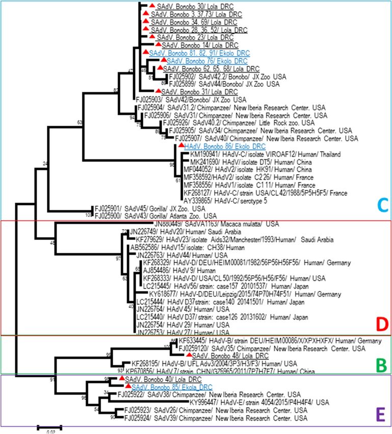
www.nature.com/scientificreports/
Infectious agents Tests Size and nature of samples
Viruses
Human respiratory syncytial virus18 PCR and sequence fragments 8 carcasses of wild bonobos
Simian T-cell lymphotropic virus (STLV)19 PCR and sequence fragments 633 fecal samples from wild-living bonobos
Herpesviridae20 PCR and sequence fragments 21 blood samples from captive bonobos
21 Histopathology, immunohistochemistry, PCR and full genome
Papillomavirus Tissue samples from a lesion in the oral cavity of a captive bonobo
sequence
Hepatitis E22 Serological cross reactivity (ELISA) 25 sera from captive bonobos
Adenovirus23 Virus isolation and full genome sequences Number of fecal samples not specified of captive bonobos
Adenovirus24 PCR and sequence fragments 84 fecal samples from semicaptive bonobos
Histopathology, immunohistochemistry and PCR and sequence
Encephalomyocarditis virus25 2 carcasses of semicaptive bonobos
fragments
26
Cytomegalovirus PCR and sequence fragments 33 fecal samples from semicaptive bonobos
Merkel cell polyomavirus27 PCR and full genome sequences 26 fecal samples from semicaptive bonobos
Bacteria
Streptococcus pneumoniae18 PCR and sequence fragments 8 carcasses of wild bonobos
Shigella spp.28 Macroscopy 34 bonobos, articular surface
Protozoa
Plasmodium falciparum, P. malariae29 PCR and sequence fragments 42 bonobo blood samples from Lola Ya sanctuary
Plasmodium (Laverania) lomamiensis30 PCR and sequences fragments 1556 fecal samples from wild bonobos
Sheather’s flotation and merthiolate-iodine-formaldehyde sedi-
Balantidium coli31 23 fecal samples from wild bonobos
mentation
Table 1. Summary of previous studies conducted on bonobo (Pan paniscus) according to the size and nature
of the samples used.
fragmented manner along the river and its tributaries, gene flow is limited by these major environmental barriers.
Poaching is the main threat these populations face, particularly due to the civil war that has taken place in the
country. Population migration, habitat alteration through agricultural and commercial practices, and disrup-
tion of the education and health systems that replaced the traditional vision of indigenous bonobo protection
are the main factors that are likely to reduce their populations in the coming decades17. A few publications have
studied bonobos as possible reservoirs of certain pathogens. Each study was dedicated to the search for a specific
pathogen (Table 1).
However, the number of samples explored varies; studied animals lived in captivity (in most cases) far from
their original distribution area in DRC. In addition, studies involving a larger number of samples concentrated on
the search for one pathogen and did not provide information in terms of the potential of bonobos to be a reservoir
for various microorganisms. Here, we intend to contribute to a better understanding of the role of bonobos as
reservoirs/hosts of human pathogens by exploring the spectrum of associated pathogenic microorganisms using
a noninvasive method. We looked for a wide range of zoonotic viral, bacterial, and parasitic agents, based on the
available literature. We also compared two bonobo populations: bonobos from an orphan bonobo sanctuary,
Lola Ya Bonobo (living in semicaptivity), and bonobos reintroduced in a natural reserve living in the wild (wild
bonobos), Ekolo Ya Bonobo.
Results
For screening purposes, we used whole DNA/RNA extracted from stool samples diluted in DNAse/RNAse-free
water at 1:10. Indeed, pure DNA/RNA extracts from stool samples may contain a considerable amount of poly-
merase inhibitors32. The choice to use the dilution at 1:10 is therefore explained by the fact that we have sought
to limit the polymerase inhibitors while having a sufficient DNA/RNA q uantity33. Dilution at 1:100 would have
allowed us to further limit the amount of inhibitor but with less DNA/RNA available. Nevertheless, confirma-
tion of positive results was made using two other samples: pure DNA/RNA extracts and dilution at 1:100. The
prevalence was calculated based on the results of the screening by qPCR.
Viruses. Among 15 viruses screened, three groups have been identified, namely, Astroviruses, Encephalo-
myocarditis virus (ECMV) and Adenoviruses (AdVs), in bonobo stool samples collected from the two sites in
DRC (Fig. 1a).
Astrovirus was only found in 2.2% (2/91) of samples from the semicaptive bonobos of Lola Ya Bonobo (Lola).
The prevalence of ECMVs was 16.5% (15/91) including 12.5% (2/16) from wild bonobos of Ekolo Ya Bonobo
(Ekolo) and 17.3% (13/75) from Lola. A high prevalence of AdVs was detected, 83.5% (76/91), and all samples
from Ekolo were positive 100% (16/16) versus 80% (60/75) positive in Lola (Z-test; P < 0.000001). All samples
were negative for Sarbecoviruses including SARS-CoV-2, Enteroviruses, Hepatitis A and E viruses, Noroviruses,
Parechoviruses, Poxviruses, Rotaviruses, SIV and HPV.
The primers F1 and P1 were designed to amplify a part of the viral polymerase gene (3D gene) of EMCVs.
Phylogenic analysis of the obtained sequences showed an identical strain in wild and semicaptive bonobos, and
Scientific Reports | (2021) 11:6331 | https://doi.org/10.1038/s41598-021-85849-4 2
Vol:.(1234567890)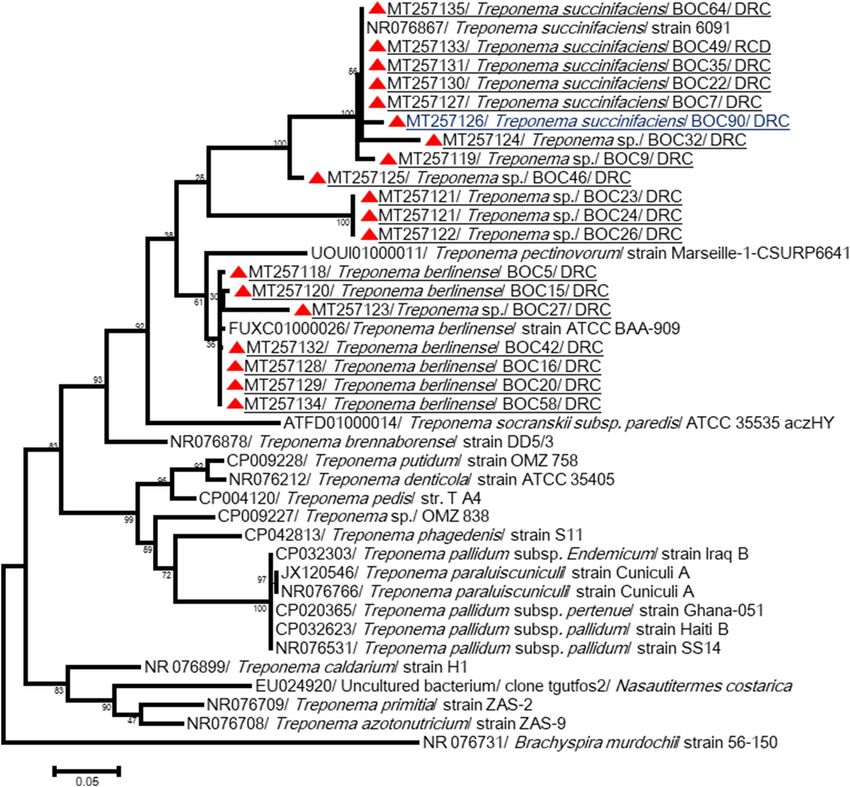
www.nature.com/scientificreports/
Figure 1. Large screening results; (a) Prevalence of viruses; (b) Prevalence of Bacteria; (c) Prevalence of
parasites. ***Significative difference between Lola and Ekolo with p. value < 0.0001. **p. value < 0.03. *p.
value < 0.05. Bars represent the error bars for percentages, they are showed by the lower half part of bars.
this was very similar to the ECMV Strain ATCC VR-129B (KM269482) isolated from a captive chimpanzee
from Florida. Additionally, this strain showed > 99% similarity with other strains detected in pigs, tiger and a
dog from China (Fig. 2).
Based on the DNA polymerase gene of AdVs, and by optimization of a nested-PCR protocol using the outer/
escribed23, the obtained sequences in the present study were different from each other
inner primers previously d
and from the available AdV sequences deposited in GenBank. Ten sequences (eight from Lola and two from
Ekolo) were almost identical to each other and showed high similarity (92 to 98%) with SAdV-42.1 (FJ025903)
and SAdV-42.2 (FJ025902) isolated from bonobos’ guts at Jacksonville Zoo, USA, while one AdV sequence
detected in Ekolo (Bonobo86) shared only 93% identity with its closest relative Human adenovirus sp. isolate
DT5 (MK241690) from a Chinese man. One sequence obtained from Lola Ya Bonobo was similar to SAdV-35
(FJ0259120) isolated from a chimpanzee from New Iberia Research Center, USA, and a strain (KF633445)
detected in a human from Germany, within the HAdV-B species. In addition, two sequences, one from a semi-
captive bonobo and another from a wild one, were similar to each other and close to HAdV-E detected in chim-
panzees (SAdV-26 and SAdV-39) from New Iberia Research Center and a man from the USA (Fig. 3, Table 2).
Bacteria. Screening detected 27 bacterial agents listed in Table 3, including principal zoonotic agents. Patho-
genic Leptospira spp. have been detected in 89% (81/91) of bonobos, and the prevalence was higher in Lola than
in Ekolo (100% vs 37%, P < 0.001). Mycobacterium spp. (non-tuberculosis) have been detected in 73.6% (67/91),
and again, the prevalence was higher in Lola (84%) vs (25%) Ekolo (Z-test; P < 0.001). In addition, the highest
prevalence (88%) was noted for Treponema spp. (non-pallidum), and animals from Lola were more frequently
infected (92%) than bonobos from Ekolo (87%) (P = 0.049). Acinetobacter spp. (non-baumanii) DNA was found
in 69% of bonobos with a prevalence almost equal at the two sites. Finally, Salmonella spp. (non-typhi/paraty-
phi) and Mycoplasma spp. were detected in 3.3% and 6.6% of bonobos, respectively; all of them were from Lola
(Fig. 1b). DNA of Bartonella, Borrelia, Chlamydia, Anaplasma, Wolbachia and Rickettsia spp., Rickettsia felis,
Coxiella burnetii, Helicobacter pylori, Campylobacter spp., Mycobacterium tuberculosis, Acinetobacter baumannii,
Salmonella paratyphi/typhi, Treponema pallidum, Tropheryma whipplei, Clostridium difficile, Neisseria gonor-
rhoeae, Atopobium vaginae, Gardnerella vaginalis, Listeria monocytogenes, Vibrio cholerae and Yersinia pestis was
not detected.
Leptospira spp.-positive samples were amplified by PCRs targeting six genes: LipL 32, LipL 41, Adk, Icda, rrs
2 and sec-Y34. LipL 41 partial gene-positive 460–520-bp samples were sequenced. Five good quality sequences
obtained from Lola were almost similar to each other, and they showed > 99% identity to highly pathogenic
Leptospira kirschneri (previously called L. interrogans serovar Grippotyphosa) (JQ690557) (Fig. 4).
The 16S rRNA of Treponema spp. was amplified, but sequencing has not been successful, probably due to
the nonspecificity of primers (multiple peaks superposition). The primer sets for the 23S rRNA gene were more
specific and allowed a total of 19 sequences of 787–1039 bp, exhibiting 83–100% identity to each other, to be
obtained. Five sequences obtained from samples from Lola showed > 99.5% identity with T. succinifaciens strain
Scientific Reports | (2021) 11:6331 | https://doi.org/10.1038/s41598-021-85849-4 3
Vol.:(0123456789)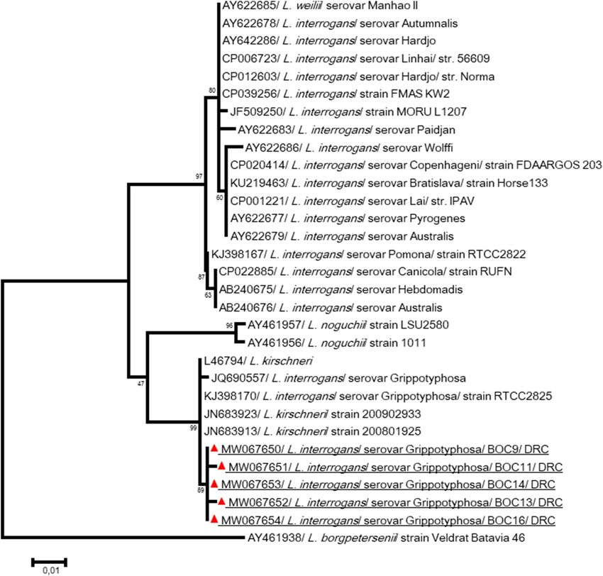
www.nature.com/scientificreports/
Figure 2. Phylogenetic tree for the partial sequence 3D of the Encephalomyocarditis viruses. The evolutionary
history was inferred by using the Maximum Likelihood method based on the Tamura 3-parameter model.
Sequences are identified as follows: accession number/virus/strain/host/country. Obtained sequences on
samples from wild or semi-captive bonobos were closely identical to each other and to the available sequences in
GenBank detected on other mammals (chimpanzee, pigs, tiger, rodents and dog). By contrast, they were distinct
ola25. The tree is drawn to scale, with
from the strain SPU64/03 (in bold), responsible of the fatal epizooty in L
branch lengths measured in the number of substitutions per site. The analysis involved 27 nucleotide sequences.
All positions containing gaps and missing data were eliminated. There were a total of 228 positions in the final
dataset. Evolutionary analyses were conducted in M EGA761.
609 (NR076867). The sequences also exhibited > 97.5% similarity with Treponema spp. detected in other African
great apes and monkeys (Algerian macaque, hamadryas from Djibouti, Guinea baboon and a green monkey
from Senegal, gorilla and human individuals from the Congo) (Medkour et al. submitted). Three others, includ-
ing one sequence obtained from an Ekolo bonobo (MT257126), were close and showed 95–98% identity to T.
succinifaciens (NR076867) as well as the sequences of NHPs described above. One other sequence (MT257125)
forms a single branch and was 93% similar to T. succinifaciens DSM 2489 (CP002631). Three similar sequences
constitute another branch, and they were 83% identical to T. succinifaciens DSM 2489 and T. brennaborense
DSM 12,168 (CP002696). They were almost identical (98%) to Treponema spp. detected in the feces of a gorilla
in the Congo and a macaque in Algeria. Finally, seven sequences were 94.5–100% identical to each other and
clustered with other sequences from Treponema spp. detected in African NHPs, such as clone G06B (MT257098)
from a gorilla in the Congo, clone RS18 (MT257249) from an Algerian macaque, and clone Bab3 (MT257103)
and clone CH32 (MT257113) detected in a Senegalese baboon and chimpanzee, respectively. In addition, the
sequences showed 78–88% similarity with the official strain T. brennaborense and 81–99% with T. berlinense
(FUXC01000026) (Fig. 5, Table 2).
Parasites. Samples were screened for the main zoonotic parasites (Table 3). Bonobos from Ekolo carried
more parasites than in Lola, except for Giardia lamblia (9.9% of bonobos), which has been detected only in
individuals living in semicaptivity (12% in Lola versus 0% in Ekolo; Z test; P = 0.03). The prevalences (in Ekolo
versus Lola; Z test P-value) were 4.4% for Strongyloides stercoralis (25% versus 0%; P < 0.001) including two
samples detected as positive by qPCR-Nematoda, 1.1% for Taenia solium (6.3% versus 0%; P < 0.001), 15.4% for
Nematoda spp. (31.3% versus 12%; P > 0.05) and 5.5% for Kinetoplastida spp. (6.3% versus 5.3%; P > 0.05) includ-
ing 4.4% positive for Trypanosoma spp. (Fig. 1c).
Samples were also screened using specific qPCRs for Filarioidea, Mansonella spp., Loa loa, Physaloptera spp.,
Ancylostoma duodenale, Ascaris lumbricoides, Cryptosporidium parvum/C. hominis, Cyclospora cayetanensis,
Entamoeba histolytica, Enterobius vermicularis, Leishmania spp., Plasmodium spp., Piroplasmida spp., Necator
americanus, Schistosoma mansoni, Toxoplasma gondii, and Trichuris trichiura, and all of them were found to be
negative.
To identify nematodes detected by pan-Nematoda qPCR, the partial Cox1 and 18S rRNA genes were success-
fully amplified. Possibly due to multiple coinfections by more than one Nematode species, electropherograms
containing double peaks were difficult to analyze. However, for one sample from Ekolo, an 18S rRNA sequence
Scientific Reports | (2021) 11:6331 | https://doi.org/10.1038/s41598-021-85849-4 4
Vol:.(1234567890)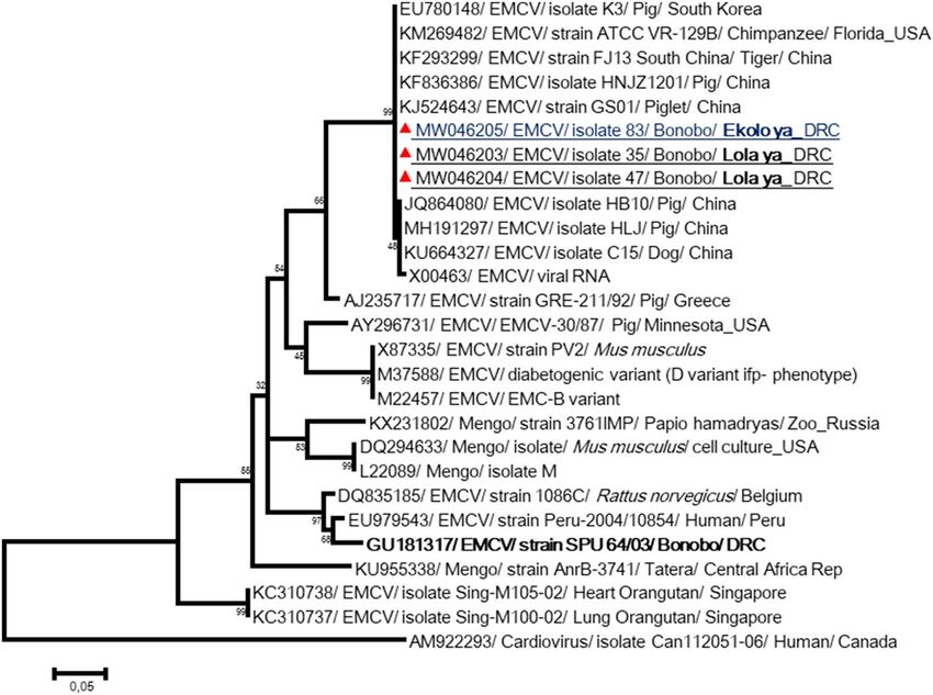
www.nature.com/scientificreports/
Figure 3. Phylogenetic analysis of Adenoviruses. The evolutionary history, based on 250 bp of DNA PoL gene,
was inferred using the Neighbor-Joining method. Adenoviruses obtained sequences here were different in each
other and with those available in GenBank database. Sequences are identified as follows: accession number/
virus/strain/host/country.A similarity rate of 92 to 98% was obtained when comparing the obtained sequences
from bonobos in Lola to reference strain of Simian adenovirus 42.1 (FJ025903) within HAdV-C species isolated
from the gut of bonobo from Jacksonville zoo, USA23. Adenovirus strain (Bonobo 86) detected in Ekolo was
93% identity with a Human adenovirus sp. isolate DT5 (MK241690) isolated in a human from China. This
suggests a possible “jump” of human strain to bonobos. Other species (HAdV-B and HAdV-E) were detected
Lola and/or Ekola bonobos. The differences in the composition bias among sequences were considered in
evolutionary comparisons. The analysis involved 39 nucleotide sequences. All positions containing gaps and
missing data were eliminated. There were a total of 213 positions in the final dataset. Evolutionary analyses were
conducted in MEGA761.
Scientific Reports | (2021) 11:6331 | https://doi.org/10.1038/s41598-021-85849-4 5
Vol.:(0123456789)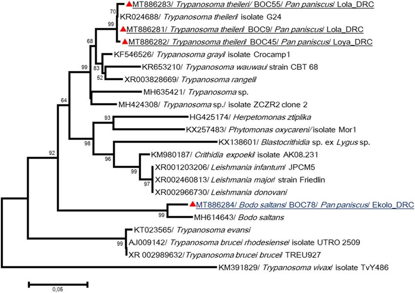
www.nature.com/scientificreports/
Best blast hits
(genebank acc
Agents Used primers Length and Tm Obtained sequences Amplified fragment New sequence ID number)
P1-CCCTACCTCACG
GAATGGGGCAAAG
F1-TTATWATTAGGG
Nested PCR: P1/F1 > 99% with ECMV
CIGGYTTG MW046203–
EMCVs (400 bp) at 55 °C; then 3 250 bp of 3D Strain ATCC VR-129B
F2-CTAGCAAAGACA MW046205
F2/R2 (250 bp) at 55 °C (KM269482)
GGRTAYAA
R2-ACGRAAIGGG
GCAAAGAG
92 to 98% with SAdV-
Fw Outer-TGATGC 42.1 (FJ025903) and
GYTTCTTACCTYTGG 10
SAdV-42.2 (FJ025902)
TYTCCATGAG belonged to AdV-C
Rw Outer-AGTT YT
ACATGCTGGGCTCTT Nested PCR: outer prim- 93% with HAdV sp.
ACCG ers (≈ 1400 bp) at 58 °C, 1 isolate DT5 (MK241690)
Adenoviruses 250 bp of DNA pol – belonged to AdV-C
Fw inner-GTGACA then Inner primers (≈
AAGAGGCTGTC-CGT 250 bp) at 55 °C SAdV-35 (FJ0259120)
GTCCCCGTA 1
belonged to AdV-B
Rw inner- TCACGT
GGCCTACACTTACA- SAdV-26 (FJ025923) and
AGCCAATCAC 2 SAdV-39 (FJ025924)
belonged to AdV-E
F- TAGGAAATTGCG
> 99% with L. interrogans
CAGCTACA MW067650–
Leptospira spp. F/R (520 bp), 53 °C 5 460–520 bp of LipL41 serovar Grippotyphosa
R- GCATCGAGAGGA MW067654
(JQ690557)
ATTAACATCA
> 99.5% with T. suc-
cinifaciens strain 609
(NR076867) and > 97.5%
5
with Treponema spp.
from other African
NHPs
95–98% with T. succinifa-
3
ciens (NR076867)
F1-GGGAGTGAGACT
93% with T. succini-
GCGIGCG
Treponema spp. F1/R2 (900 bp), 57 °C 1 787–1039 bp of 23S MT257118–MT257135 faciens DSM 2489
R2- GGTGTCASCM-
(CP002631)
CCTATACGTCYCAT
83% with T. succinifa-
ciens DSM 2489 and
3
T. brennaborense DSM
12,168 (CP002696)
78–88% with T. bren-
naborense and 81–99%
7
with T. berlinense
(FUXC01000026)
Fwd.18S.631-TCGTCA 99.5% and 99.4%
TTGCTGCGGTTAAA with O. aculeatum
Rwd.18S.1825r- GGT 1127–1155 bp, 54 °C 1 1100 bp of 18S MT89 0583 (AB677956) and O. mun-
TCAAGCCACTGC tiacum NSMT:As4470
GATTAA (LC415112)
Oesophagostumum spp. NC1-TTAGTTTCTTTT > 99% with O. stephanos-
27
CCTCCGCT Hemi-nested tomum (KR149651)
NC2-ACGTCTGGT PCR: NC1/ NC2
268–307 bp of ITS2 MW040123– 97% with O. bifurcum
TCAGGGTTGTT (280–400 bp), 50 °C;
region MW040151 (MT184890) and
OesophITS2-21TGT NC2/ OesophITS2-21 2
RACACTGTTTGT (260 bp), 55 °C with O. cf. aculeatum
CGAAC (AB586134)
> 99% identity with
F720-GTTAAAGGG 3 MT886281–MT886283 Trypanosoma theleiri
TTCGTAGTTGAA F720/R1425 (≈ 750 bp), (KR024688)
Kinetoplastida sp. 475–522 bp of 18S
R1425- GACTACAAT 50 °C
GGTCTCTAATCA > 99% Bodo saltans
1 MT886284
(MH614643)
Table 2. Generated sequences in this study and their identification.
of 1100 bp was obtained and showed 99.5% and 99.4% identity with Oesophagostomum aculeatum (AB677956)
and O. muntiacum NSMT:As4470 (LC415112), respectively (Fig. S1). This sequence showed > 99.7% identity
with Oesophagostomum isolates CC09 and CC37 (MT260066 and MT260068) detected in Barbary macaques
from Algeria35. After that, all samples were screened using a nested PCR targeting the ITS2 region of oesophago-
stumum spp. and positive were sequenced. We found 31.9% of positive including 75% in Ekolo versus 22.7% in
Lola (P < 0.0001). All obtained sequences, except two, were almost similar and showed > 99% with O. stephanos-
tomum (KR149651). The two other sequences showed 97% identity with O. bifurcum (MT184890) and with O.
cf. aculeatum (AB586134) (Fig. 6).
The 18S and 28S rRNA partial genes for Kinetoplastida spp. were also amplified. Three 18S rRNA sequences
obtained from bonobos from Lola were almost identical to each other and showed > 99% identity with
Scientific Reports | (2021) 11:6331 | https://doi.org/10.1038/s41598-021-85849-4 6
Vol:.(1234567890)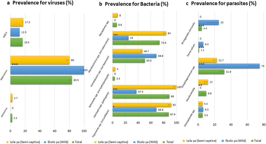
www.nature.com/scientificreports/
Bacteria Parasites Viruses
Acinetobacter spp. Ancylostoma duodenale Adenovirus
Acinetobacter baumannii Ascaris lumbricoides Astrovirus
Anaplasmataceae Cryptosporidium parvum, C. hominis Encephalomyocarditis virus
Bartonella spp. Cyclospora cayetanensis Enterovirus
Borrelia spp. Entamoeba histolytica Hepatitis A virus
Chlamydia spp. Enterobius vermicularis Hepatitis E virus
Coxiella burnetii Filarioidea Norovirus
Helicobacter pylori Giardia lamblia Parechovirus
Mycobacterium spp. Kinetoplastida Poxvirus
Mycoplasma spp. Leishmania spp. Rotavirus
M. genitalium Loa loa Sapovirus
Rickettsia spp. Mansonella spp. Simian immunodeficiency virus (SIV)
Rickettsia felis Necator americanus Papillomavirus (HPV)
Salmonella spp. Nematoda Sarbecovirus
Salmonella paratyphi/typhi Plasmodium spp. Sars-Cov 2
Staphylococcus aureus Schistosoma mansoni Herpes simplex virus
Treponema spp. Strongyloides stercoralis
Treponema pallidum Taenia saginata
Tropheryma whipplei Taenia solium
Vibrio cholerae Toxoplasma gondii
Wolbachia spp. Trichuris trichiura
Yersinia pestis Physaloptera spp.
Neisseria gonorrhoeae Oesophagostomum spp.
Atopobium vaginae
Gardnerella vaginalis
Listeria monocytogenes
Leptospira spp.
Clostridium difficile
Table 3. Pathogens for which tests have been carried out.
Trypanosoma theleiri (KR024688). In addition, in Ekolo, one other sequence was closely identical to Bodo sal-
tans (MH614643) (Fig. 7, Table 2).
Discussion
The objective of this study was to identify the spectrum of zoonotic bacteria, parasites and viruses in two popula-
tions of bonobos, one semicaptive (Lola) living every day in contact with humans and the other living in the wild
(Ekolo), and to compare the pathogen distribution between the two sites. Ekolo Ya Bonobo is the only bonobo
reintroduction site in the world. The first group led by the "female Alfa" Etumbe was reintroduced in 2008. The
expansion of the protected area (Ekolo) will ensure sufficient space to continue to reintroduce bonobo groups
rescued from the bushmeat trade and rehabilitated in Lola Ya Bonobo.
The use of stool samples, a noninvasive sampling technique, to study pathogens is very important. In addition
to enteric pathogens that are logically easy to find in stool samples, this technique allows the identification of
microorganisms usually residing in blood ( malaria6, Leishmania36, filaria35, etc.) and in the urogenital system,
such as Leptospira (this study). This sampling technique is absolutely noninvasive and easily authorized for
NHPs. In this study, 91 bonobo stool samples were treated. This is the first large-scale systematic screening by
PCR/RT-PCR of zoonotic pathogens in bonobos using fecal samples. Previous publications reported infectious
agents found in various kinds of samples from bonobos including blood, stool, serum, secretion swab, urine,
cadaver, etc. (Table 1).
Here, adenovirus carriage was very high in both wild and semicaptive bonobos, and great genetic diversity
was highlighted. Human mastadenovirus C (HAdV-C) was present in bonobos cohabiting with humans and those
released in the forest. These results reinforce the exchange between human and bonobo viruses, since we detected
a HAdV-C strain in a bonobo very close to strains detected in humans (Fig. 3). In addition, the presence of AdVs
in wild bonobos is evidence of the continuous circulation of adenoviruses in the bonobo population; we have
identified the same viruses in individuals living in semicaptivity (in Lola) as well as those that were released in
the wild 4 years ago. It is highly likely that bonobos released in Ekolo were already infected by these adenoviruses
at that time, so it seems that we observed the persistence of HAdV-C in bonobos. This is not surprising because
in humans, HAdV species C has the capacity to establish persistent infection in intestinal T lymphocytes of the
digestive tract37. Furthermore, the presence of persistent and/or latent AdV infections in the gut of great apes,
including bonobos, has been observed23 and should be considered in the design and interpretation of human
and NHP studies (including vaccine development) with adenovirus vectors.
Scientific Reports | (2021) 11:6331 | https://doi.org/10.1038/s41598-021-85849-4 7
Vol.:(0123456789)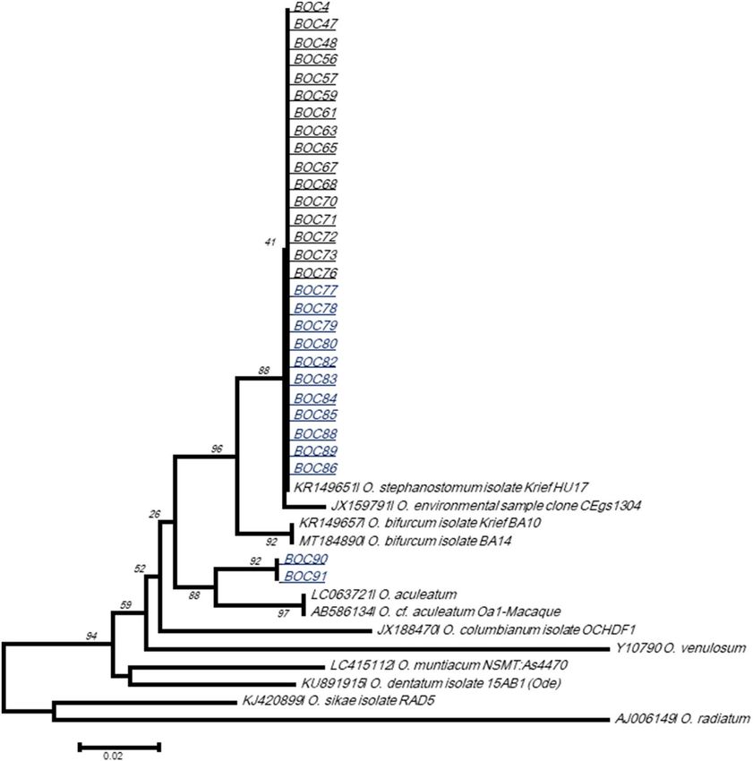
www.nature.com/scientificreports/
Figure 4. Phylogenetic analysis of Leptospira spp. from bonobo fecal samples. The evolutionary history,
based on LipL 41 partial gene, was inferred by using the Maximum Likelihood method based on the Tamura
3-parameter model. Initial treefor the heuristic search were obtained automatically by applying Neighbor-Join
and BioNJ algorithms to a matrix of pairwise distances estimated using the Maximum Composite Likelihood
(MCL) approach, and then selecting the topology with superior log likelihood value. Sequences are identified
as follows: accession number/species/strain/host/country. Obtained sequences in the present study showed
almost similarity in each other and with Leptospira interrogans serovar Grippotyphosa RTCC2825 (KJ398170).
The analysis involved 31 nucleotide sequences. All positions containing gaps and missing data were eliminated.
EGA761.
There were a total of 436 positions in the final dataset. Evolutionary analyses were conducted in M
In our recent study on AdV circulation in African humans and NHPs, HAdV-B, HAdV-C, HAdV-D and
HAdV-E were found in both humans and/or NHPs from the Congo. We demonstrated a possible jump of a
human strain of HAdV-C to gorillas, and, vice versa, a gorilla strain of HAdV-C to humans sharing the same
living area in the C ongo33. HAdV-B members were detected in Lola and HAdV-E in Ekolo, and they were
previously reported in captive bonobos from the Jacksonville Zoo, U SA23. In a study of 800 fecal samples from
wild African great apes and humans to investigate the evolutionary history and zoonotic potential of hominine
HAdVs, HAdV-B and -E were frequently detected in wild gorillas (55%) and chimpanzees (25%), respectively. It
was shown that HAdV-B circulating in humans are of zoonotic origin and have probably affected global human
health for most of our species lifetime24. The finding in HAdVs in bonobo which have been reintroduced into
the wild is very alarming and shows the risk of such reintroduction programs. In our study, the Lola Ya sanctu-
ary followed the international recommendations for reintroduction38. In addition, because of the methodology,
Scientific Reports | (2021) 11:6331 | https://doi.org/10.1038/s41598-021-85849-4 8
Vol:.(1234567890)www.nature.com/scientificreports/
Figure 5. Molecular Phylogenetic analysis for Treponema spp. detected on bonobos from DRC. The
evolutionary history, based on partial 23S rRNA gene, was inferred by using the Maximum Likelihood method
based on the Tamura 3-parameter model. Initial tree for the heuristic search were obtained automatically by
applying Neighbor-Join and BioNJ algorithms to a matrix of pairwise distances estimated using the Maximum
Composite Likelihood (MCL) approach, and then selecting the topology with superior log likelihood value.
Sequences are identified as follows: accession number/species/strain/host/country. Sequences in this study are
highlighted by black circle and underlined. Sequence wrote in bleu was obtained on a wild bonobo sample while
all the others obtained on semi-captive bonobo samples. In addition, in bolt are the Treponema spp. sequences
from African NHPs. The tree is drawn to scale, with branch lengths measured in the number of substitutions
per site. The analysis involved 48 nucleotide sequences. All positions containing gaps and missing data were
eliminated. There were a total of 721 positions in the final dataset. Evolutionary analyses were conducted in
MEGA761.
it could be possible that very divergent segments were not picked up by the primers, especially those of simian
AdVs. Therefore, the nonidentified AdVs here could be SAdVs or AdVs from other species, which were not
amplified by the applied primers. Furthermore, a DNA polymerase fragment was selected in order to be highly
conserved. It is therefore no surprise that the fragment was found to be considerably conserved.
In 2009, a devastating epizooty due to ECMV in Lola led to fatal myocarditis in bonobos25. Here, EMCV
genotypes detected in Lola and Ekolo bonobos were identical and close to other strains detected in mammals,
including humans and chimpanzees. By contrast, they were very divergent to EMCV strain SPU 64/03 responsible
for Lola bonobo mortality, which confirms the circulation of more than one EMCV strain in this area (Fig. 2).
Highly divergent EMCVs were isolated from orangutans after fatal myocarditis in S ingapore39. Several species
Scientific Reports | (2021) 11:6331 | https://doi.org/10.1038/s41598-021-85849-4 9
Vol.:(0123456789)www.nature.com/scientificreports/
Figure 6. Phylogenetic analysis of Oesophagostomum based on ITS2 rDNA (260 bp) sequences. The
evolutionary history was inferred using the Neighbor-Joining method. Sequences in this study are named
“BOC”, those in black are from Lola and those in bleu are from Ekolo. The tree is drawn to scale, with branch
lengths in the same units as those of the evolutionary distances used to infer the phylogenetic tree. The
evolutionary distances were computed using the Tamura-Nei method and are in the units of the number of base
substitutions per site. The differences in the composition bias among sequences were considered in evolutionary
comparisons. The analysis involved 41 nucleotide sequences. All positions containing gaps and missing data
were eliminated. Evolutionary analyses were conducted in MEGA761.
of captive NHPs are susceptible to highly fatal EMCV myocarditis including the chimpanzee (Pan troglodytes),
African green monkeys (Chlorocebus), squirrel monkeys (Saimiri sciureus), baboons (Papio spp.), macaques
(Macaca fascicularis and Macaca sylvanus) and orangutan (Pongo pygmaeus)25. EMCV cases require particular
attention. Taking into consideration the extremely high mortality due to viral encephalomyocarditis in apes and
monkeys, especially those kept in captivity and, in general, the severe clinical picture, it seems to be urgent to
accumulate epidemiological data concerning the circulation of these viruses. Virtually nothing is known about
the epidemiology of these viruses in simians, for example, their transmission, origins, reservoirs or possibility
of infecting humans. Bonobo dormitories were exposed to rates that can be the origin of this infection. Rodents
were suspected as reservoirs and diagnosed epidemics in African wildlife. A study showed a striking temporal
correlation between the occurrence of a population explosion (as evidenced by markedly increased catch rates
per trap-night) and a surge in prevalence of antibody to EMCVs in rodents, and the occurrence of the outbreak
of disease in elephants in South Africa40.
Bonobos host a variety of Treponema species. We identified at least seven genomospecies of Treponema in
their feces including T. succinifaciens, which was identified in all bonobos, as well as previously reported for
African gorilla, chimpanzee, green monkey, Guinea baboon, hamadryas, macaque and the human gut (Medkour
et al. submitted). It seems that this species is a part of the gut microbiota, but its role is poorly understood. We
also showed the existence of potential new species with unknown pathogenicity (Fig. 5). Spirochaetes has been
reported in the gut microbiota of NHPs41. Treponema species have been detected in a ncient42 and traditional
rural human p opulations43,44. All traditional rural populations were enriched for T. succinifaciens in a recent
study45, and other species clustered with Treponema reported from termites44. The roles of different Treponema
species in the gut still need to be explored.
Scientific Reports | (2021) 11:6331 | https://doi.org/10.1038/s41598-021-85849-4 10
Vol:.(1234567890)www.nature.com/scientificreports/
Figure 7. Phylogenetic analysis of Kinetoplastida spp. detected in this study. The evolutionary history, based on
18S rRNA gene, was inferred using the Neighbor-Joining method. The obtained sequences here were compared
to sequences of Kinetoplastida spp. available in GenBank. Sequences are identified as follows: accession
number/species/strain/host/country. Sequences in Lola were almost identical and presented > 99% identity with
Trypanosoma theleiri (KR024688), known as pathogen for ruminants. Whereas, in Ekolo, one other sequence
was closely identical to Bodo saltans (MH614643), a free living nonpathogenic kinetoplastid. The analysis
involved 22 nucleotide sequences. All positions containing gaps and missing data were eliminated. There were a
EGA761.
total of 483 positions in the final dataset. Evolutionary analyses were conducted in M
The presence of pathogenic Leptospira in great apes’ stool has never been reported. The high Leptospira
spp. (pathogenic) prevalence observed and the identification of L. interrogans serovar Grippotyphosa in semi-
captive bonobo feces was surprising (Fig. 4). It is important to note that Lola bonobo dormitories may be easily
approachable for different species of wild and peridomestic rodents that can be a source of bonobo infection. In
addition, bonobo feces could be contaminated by their own urine. NHPs might be sensitive to Leptospira infec-
tion, as an outbreak of severe leptospirosis was reported in capuchin (Cebus) monkeys46. Leptospira in the feces
of wild bonobos could also be due to environmental contamination as samples were collected from the ground.
Furthermore, the extent of Leptospira transmission between humans and NHPs is unknown.
Strongyloides stercoralis was found only in wild bonobos. Recent studies revealed the presence of S. stercoralis
in human communities in contact with gorillas in the C ongo35 and with long-tailed macaques in Th ailand47.
African NHPs were reported to be reservoirs/hosts of Oesophagostomum roundworms35,48. Eight species
of Oesophagostomum have been recognized so far to occur in NHPs49. Among them, O. bifurcum, O. stepha-
nostomum and O. aculeatum are also reported in h umans50. Central African gorillas and chimpanzees were
reported to be infected (with sometimes fatal outcomes) by O. stephanostomum and, probably, by human-borne
O. bifurcum51. The same two species seem to be responsible for endemic human esophagostomiasis in Ghana
and Togo52. Subsequently, isolated cases have been described in Malaysia, Indonesia, Brunei, Brazil and several
African countries (Ghana, Togo but also Zimbabwe, Ethiopia, Cote d’Ivoire, Uganda and Nigeria)53. We identify
in our case O. stephanostomum in semicaptive and wild bonobos, and O. bifurcum in wild bonobos. The ques-
tion concerning the role of great apes in the epidemiology of human nodular esophagostomosis remains open.
Only the analysis of parasite population genetics can resolve the extent to which zoonotic transmission occurs.
Uncommonly, T. solium was identified in both bonobo populations. Cysticerci, presumably caused by T.
solium, have been described in apes (gibbons and chimpanzees), New World monkeys (squirrel monkeys and
marmosets), Old World monkeys (rhesus monkeys, baboons, mangabeys, patas monkeys, langurs, and vervets)
and prosimians (lemurs)54.
Trypanosoma theileri was identified in Lola sanctuary bonobos. Usually, T. theileri infects Bovinae (cattle, buf-
orld55. A previous study suggested that
falo, yaks, and some antelopes) and is prevalent in cattle throughout the w
trypanosomiasis has been recorded among humans within the area of occurrence of bonobos and appears to be
the most important disease shaping the area of occupancy of bonobos within their overall extent of o ccupancy56.
Here, however, we also cannot exclude the possibility of contamination of bonobo feces by Trypanosoma-infected
arthropods. Uncharacterized Nematoda and Kinetoplastida spp. are found in the two sites and need further
exploration. The surveillance of parasitic infection in bonobos is of great importance for conservation and
public health. Using the primate–parasite network, the role of different NHPs was evaluated for the probability
Scientific Reports | (2021) 11:6331 | https://doi.org/10.1038/s41598-021-85849-4 11
Vol.:(0123456789)www.nature.com/scientificreports/
of sharing parasitic infectious diseases with humans. Apes, as well as monkeys, such as baboons and macaques,
were shown to be infected with many parasites identified as emerging infectious diseases in h umans57.
One of the strengths of this study is the analysis of 91 stool samples from two different collection sites, Lola
and Ekolo. By comparing the two collection sites, it was possible to establish a significant difference between Lola
(animals in semicaptivity) and Ekolo (reintroduced into the wild), markedly in the case of Giardia lamblia, which
was detected only in captive populations, as well as Mycoplasma and Salmonella spp. However, for the latter two
pathogens, no significant difference between the two sites could be demonstrated possibly due to the number
of samples from Ekolo being too low. It is, however, important to mention that there is possible degradation of
the samples due to their collection in nature, and the prevalence of these microorganisms remains underesti-
mated, even more so for extraintestinal microorganisms. Treponema spp. (non-pallidum) and Mycobacterium spp.
(non-tuberculosis) were more significantly identified in Lola bonobos, suggesting possible transmission between
humans and bonobos. Indeed, Lola is a sanctuary for the conservation and protection of orphaned bonobos. The
animals cared for in the sanctuary are animals with an immature immune system, which makes them sensitive
to possible exchange of bacterial flora with personnel. This characteristic should be considered when identify-
ing bonobos as a potential reservoir of emerging infectious diseases. In addition, it has been shown that direct
contact is not necessary to contaminate b onobos58. In Lola, bonobos are cared for by people who have to clean
and prepare the housing areas. This maintenance work involves constant direct contact between them, which
increases the risk of sharing pathogens and interspecies transmission.
Additionally, we looked for sexually transmitted pathogens in the current study (Chlamydia spp., T. pallidum,
N. gonorrhea, Simian HIV and papillomaviruses) because of sex-based conciliation practices in bonobos. All
results were negative. The diversity of the nature of the samples would also broaden the range of pathogens not
found in stool and provide a clearer diagnostic vision in bonobos. It would then be interesting in the future to
use other types of excreta and biological fluids from these animals, at least for the populations in the sanctuary.
Two samples were of particular interest because they were positive for Astrovirus and other agents, including
Treponema spp., Mycobacterium spp. and O. stephanostomum. One was also found to be positive for Acinetobacter
spp. and another for Giardia lamblia. Both animals were from Lola. The fact that these pathogens were found
in feces may suggest a latency period that allows pathogens to better adapt and/or induce pathogenicity in the
host, the host tolerates infection and has an unrelated r esponse59,60, or the pathogens simply are commensal and
part of the animal’s intestinal microbiota.
For the first time, bacteria such as Mycobacterium spp., Salmonella spp., Acinetobacter spp., and Mycoplasma
spp. have been found in bonobo feces, as well as protozoa such as T. theileri, B. saltans and G. lamblia or Astrovi-
rus. Finally, the results presented above can also be explained by the sample collection method, their transport
and storage. Standardized collection conditions were maintained, and samples were then transported in alcohol
from the DRC and maintained at − 80 °C. In addition, a number of missed organisms because of PCR failures
due to primer mismatches is possible. Some microorganisms are highly diverse, especially in this part of the
world (example: picornaviruses) and we can imagine primers design against a narrow set of these microorgan-
isms would miss a lot of diversity. Consequently, it would be interesting to combine these results with a set of
fresh stool samples collected from the same sites from which microorganisms were isolated.
Methods
Ethic statement, animals and study area. This study was based on 91 samples of bonobo feces col-
lected in August 2017 from two collection sites in the Democratic Republic of Congo (DRC): 75 samples were
collected in Lola Ya Bonobo (Lola), Kinshasa suburbs, a sanctuary for the protection, rehabilitation and reintro-
duction of orphaned bonobos; and 16 in Ekolo Ya Bonobo (Ekolo), Equateur region, a 20,000-hectare section
of tropical forest dedicated to the reintroduction of bonobos (Fig. 8). The samples were aliquoted in alcohol and
stored at − 80 °C upon arrival at the IHUMéditérannée Infection lab, Marseille.
The Public Health Ministry of the DRC has given its agreement for sample export (N° 482 INRB/DG of
01/09/17). Bouches-du-Rhône prefecture, in Marseille (France), has authorized the import of samples (N° 16/17
of 06/27/17). Finally, the feces of this species are not subject to an import–export permit for international
circulation.
DNA and RNA extraction. Nucleic acids (DNA/RNA) were extracted using the Qiagen Virus Mini Kit
v2.0 (Qiagen, Courtaboeuf, France) for viral nucleic acid and the QIAamp DNA Mini Kit (Qiagen) for bacteria
and parasites using an EZ1 biorobot (Qiagen). The DNA and RNA from each sample were extracted twice. This
protocol included sample preparation with proteinase K, followed by mechanical stool lysis with tungsten beads
(Qiagen, Courtaboeuf, France) using a FastPrep-24 5G Grinder. The supernatant was then recovered and incu-
bated overnight at 56 °C. According to the manufacturer’s instructions, 130 µL of viral DNA/RNA and 200 µL
of extracted DNA were collected in elution tubes, aliquoted in individual PCR tubes to an amount of 50 µL of
pure extracted DNA/RNA, with another aliquot of 50 µL of DNA/RNA diluted to one-tenth, and finally a third
aliquot of 50 µL of DNA/RNA diluted to one-hundredth. The original elution tubes containing the pure extrac-
tions were stored at − 20 °C for DNA and − 80 °C for RNA.
Bacterial DNA extraction was controlled by amplifying the 16S rRNA gene for all bacteria using a real-time
PCR system (qPCR). Viral nucleic acid extraction was performed after adding 10 µL of internal controls in
the extraction tubes, namely, Enterobacteria phage T4 (T4) and Enterobacteria phage MS2 for DNA and RNA
controls, respectively. The extraction and dilutions were controlled by qPCR targeting the phages T4 and MS2
(Table S3).
Scientific Reports | (2021) 11:6331 | https://doi.org/10.1038/s41598-021-85849-4 12
Vol:.(1234567890)www.nature.com/scientificreports/
Figure 8. Study area. Samples from Lola Ya Bonobo (n = 75) where animals live in a sanctuary dedicated to the
rehabilitation of orphan bonobos near Kinshasa, they are in permanent contact with humans. Samples from
site Ekolo Ya bonobo (n = 16) were collected in nature where bonobos were introduced into the wild in the
equatorial region. Figure modified from: https://docplayer.fr/162905495-Republique-democratique-du-congo-
ministere-de-l-environnement-et-developpement-durable.html.
Reverse transcription (cDNA synthesis). First-strand cDNA was synthesized using the MMLV-RT kit
(Invitrogen, Carlsbad, CA, USA) according to the manufacturer’s protocol. The reaction mixture was prepared
in a volume of 50 µL including 11 µL of M
gCl2, 5 µL of Buffer 10×, 10 µL of dNTP (10 mM), 2.5 µL of hexameres
at 1:10 dilution, 1.25 µL of RT-Multiscribe, 1 µL of RNAse, 9.25 µL of ultra-purified DNAse-RNAse-free water
and finally 10 µL of DNA/RNA template extracted by EZ1. The RT-reaction was performed in a thermocycler
(Applied Biosystem) with three thermal steps: 25 °C for 10 min, 48 °C for 30 min and 95 °C for 5 min followed
by a pause step at 4 °C.
Quantitative real‑time PCR assays (qPCR). The qPCR amplifications were performed in a CFX96
Real-Time system (Bio-Rad Laboratories, Foster City, CA, USA) after activating the readers of the dyes (FAM
and/or VIC) used in each qPCR system. This method was used for the detection of parasites, viruses and bacteria
of interest, using PCR systems for the detection of the pathogens studied here (Tables S1–3). The qPCR reactions
were carried out in a final volume of 20 µL, containing 5 µL of DNA/cDNA template and 10 µL of Master Mix
Roche (Eurogentec). The volume of each primer per reaction was 0.5 µL, with 0.5 µL of both UDG and each
probe, and finally, the volume was brought to 20 µL using ultra-purified DNAse-RNAse-free water. The TaqMan
cycling conditions included two hold steps at 50 °C for 2 min, followed by 95 °C for 15 min and 40 cycles of two
steps each (95 °C for 30 s and 60 °C for 30 s). The PCR systems used for the study are detailed in (Table S4). Each
PCR plate contains 96 wells; however, it was decided to run 50 samples per plate to avoid contamination. To con-
firm the results, samples that tested positive were retested using pure solutions and diluted to one-hundredth.
Genetic amplification by standard PCR and sequencing. For gene amplifications, PCRs were per-
formed in a total volume of 50 µL, consisting of 25 µL of AmpliTaq Gold master mix, 18 µL of ultra-purified
water DNAse-RNAse free, 1 µL of each primer and 5 µL of DNA/cDNA template. The thermal cycling condi-
tions were as follows: incubation step at 95 °C for 15 min, 40 cycles of 1 min at 95 °C, 30 s for the annealing at a
different melting temperature for each PCR assay, 30 s to 1.5 min of elongation time at 72 °C (according to the
fragment length), followed by a final extension for 5 min at 72 °C (Table S4). PCR amplification was performed
in a Peltier PTC-200 model thermal cycler (MJ Research Inc., Watertown, MA, USA). The results of amplifica-
tion were visualized by electrophoresis on a 2% agarose gel. The purification of PCR products was performed
using NucleoFast 96-well PCR plates (Macherey Nagel EURL, Hoerdt, France) according to the manufactur-
er’s instructions. The amplicons were sequenced using the Big Dye Terminator Cycle Sequencing Kit (Perkin
Elmer Applied Biosystems, Foster City, CA, USA) with an ABI automated sequencer (Applied Biosystems). The
obtained electropherograms were assembled and edited using ChromasPro software (ChromasPro 1.7, Tech-
nelysium Pty Ltd., Tewantin, Australia) and compared with those available in the GenBank database by NCBI
BLAST (https://blast.ncbi.nlm.nih.gov/Blast.cgi). Obtained sequences for each gene, for each pathogen, from
Scientific Reports | (2021) 11:6331 | https://doi.org/10.1038/s41598-021-85849-4 13
Vol.:(0123456789)www.nature.com/scientificreports/
positive samples were aligned with those available in the GenBank database for the same gene. Maximum-
likelihood or the neighbor joining method was used to infer the phylogenetic analyses, and tree reconstruction
was performed using MEGA software version 7 (https://www.megasoftware.net/)61. Bootstrap analyses were
conducted using 1000 replicates.
For Treponema spp., since the 16S rRNA-based P CR62 did not allow identification, we developed a set of
primers (F1, F2, R1, and R2) (Table S4) targeting the 23S rRNA of Treponema spp. First, sequences (Supple-
mentary material S1) were aligned using BioEdit v 7.0.5.3 s oftware63 to reveal conserved areas suitable as target
regions for specific primers. This region was submitted to Primer3 software v. 0.4.0 (http://primer3.ut.ee/) to
determine valuable candidate primers and probes, and selection was based on the criteria for primer and probe
design. Degenerated nucleotides were used to achieve the maximum sensitivity within the genus Treponema. In
the same manner, we developed sets of primers targeting the 16S, LipL 32, LipL 41, LipL 71, and Sec Y genes of
Leptospira spp. (Table S4).
Settings for the PCR primers were in accordance with the guidelines as described by Apte and D aniel64 and as
recommended by Invitrogen and Applied Biosystems. Melting temperatures, secondary structures and the pos-
sibility for primer-dimers were tested using the free online software Oligo Analyzer 3.165. All primer sequences
were also checked for their specificity in an NCBI BLAST nucleotide sequence similarity s earch66. Furthermore,
they were checked within the DNA databases of metazoans (taxid:33208), vertebrates (taxid:7742), bacteria
(taxid:2), arthropods (taxid:6656), primates (taxid:9443), Canidae (taxid:9608), Felidae (taxid:9682) and humans
(taxid:9605) as previously d escribed67. Primers were synthesized by Eurogentec (Liège, Belgium).
Microorganisms screened. To optimize time and resources, zoonotic pathogens and, in particular, those
routinely researched at the IHU Marseille lab, were studied (Table 3).
Statistical analyses. To determine if there is a difference in the frequency of microorganisms between the
bonobos living in semicaptivity (Lola) and the wild (Ekolo), a Z test was performed. Significant differences were
considered at p < 0.05.
Data availability
All data are included in the manuscript. The newly generated sequences were deposited in the GenBank database
under the accession numbers: MW046203-MW046205 (3D pol) of EMCV; MW067650-MW067654 (LipL41) of
Leptospira spp.; MT257118-MT257134 (23S) of Treponema spp.; MW040123-MW040123 (ITS2) of Oesophago-
stumum spp.; MT890583 (18S) of Oesophagostumum spp.; MT886281-MT886283 (18S) of Trypanosma theileri;
MT886284 (18S) of Bodo saltans.
Received: 16 October 2020; Accepted: 5 March 2021
References
1. Morse, S. S. et al. Prediction and prevention of the next pandemic zoonosis. Lancet 380, 1956–1965 (2012).
2. Levinson, J. et al. Targeting surveillance for zoonotic virus discovery. Emerg. Infect. Dis. 19, 743–747 (2013).
3. Wolfe, N. D. et al. Bushmeat hunting, deforestation, and prediction of zoonoses emergence. Emerg. Infect. Dis. 11(12), 1822–1827
(2005).
4. Mossoun, A. et al. Contact to non-human primates and risk factors for zoonotic disease emergence in the Taï Region, Côte d’Ivoire.
EcoHealth 12, 580–591 (2015).
5. Karesh, W. B. et al. Ecology of zoonoses: Natural and unnatural histories. Lancet 380, 1936–1945 (2012).
6. Liu, W. et al. Origin of the human parasite Plasmodium falciparum in gorillas (Author Manuscript). Nature 467, 420–425 (2010).
7. Prugnolle, F. et al. African great apes are natural hosts of multiple related malaria species, including Plasmodium falciparum. Proc.
Natl. Acad. Sci. USA. 107, 1458–1463 (2010).
8. Anthony, S. J. et al. Non-random patterns in viral diversity. Nat. Commun. 6, 1–7 (2015).
9. Devaux, C. A. et al. Infectious disease risk across the growing human-non human primate interface: A review of the evidence.
Front. Public Health 7, 1–22 (2019).
10. Odeniran, P. O. et al. A review of wildlife tourism and meta-analysis of parasitism in Africa’s national parks and game reserves.
Parasitol. Res. 117, 2359–2378 (2018).
11. Narat, V. et al. Using physical contact heterogeneity and frequency to characterize dynamics of human exposure to nonhuman
primate bodily fluids in central Africa. PLoS Negl. Trop. Dis. 12, 1–25 (2018).
12. Calvignac-Spencer, S. et al. Wild great apes as sentinels and sources of infectious disease. Clin Microbiol Infect. 18(6), 521–527
(2012).
13. Kowalewski, M. M. & Gillespie, T. R. Primatology, Biocultural Diversity and Sustainable Development in Tropical Forests. ISBN:
978-607-7579-82-3 (2018).
14. Woolhouse, M. & Gaunt, E. Ecological origins of novel human pathogens. Crit. Rev. Microbiol. 33, 231–242 (2007).
15. Narat, V. et al. Rethinking human-nonhuman primate contact and pathogenic disease spillover. EcoHealth 14(4), 840–850 (2017).
16. Prüfer, K. et al. The bonobo genome compared with the chimpanzee and human genomes. Nature 486, 527–531 (2012).
17. Fruth, A. et al. The IUCN Red List of Threatened Species 2016: e.T15932A102331567. Pan paniscus 8235, (2017).
18. Grützmacher, K. S. et al. Human respiratory syncytial virus and Streptococcus pneumoniae infection in wild bonobos. EcoHealth
15, 462–466 (2018).
19. Ahuka-Mundeke, S. et al. Genetic diversity of STLV-2 and interspecies transmission of STLV-3 in wild-living bonobos. Virus Evol.
2, vew011 (2016).
20. Lavergne, A. et al. African Great Apes are naturally infected with roseoloviruses closely related to human herpesvirus 7. J. Virol.
88, 13212–13220 (2014).
21. Hoffmann, M. et al. Disease manifestation and viral sequences in a bonobo more than 30 years after papillomavirus infection.
Pathogens 8, 13 (2019).
22. Spahr, C. et al. Detection of HEV-specific antibodies in four non-human primate species, including great apes, from different zoos
in Germany. Epidemiol. Infect. 146, 119–124 (2018).
Scientific Reports | (2021) 11:6331 | https://doi.org/10.1038/s41598-021-85849-4 14
Vol:.(1234567890)You can also read