SO2- D2.5.2.1 - AESI Case Definition Companion Guide for 1st Tier AESI Acute Disseminated Encephalomyelitis (ADEM) - AESI Case Definition ...
←
→
Page content transcription
If your browser does not render page correctly, please read the page content below
Safety Platform for Emergency vACcines

       SO2- D2.5.2.1 - AESI Case Definition Companion
                   Guide for 1st Tier AESI

        Acute Disseminated Encephalomyelitis (ADEM)

Work Package: WP2 Standards and tools
V1.0 – February 11th, 2021
Authors: Barbara Law
Nature: Report | Diss. level: Public
V1.0. 11-Feb-2021 | Diss. level: Public

TABLE OF CONTENTS

DOCUMENT INFORMATION……………………………………………………………………………………………………………………..2
DOCUMENT HISTORY……………………………………………………………………………………………………………………………….3
DEFINITIONS & ACRONYMS………………………………………………………………………………………………………………………4
INTRODUCTION………………………………………………………………………………………………………………………………………. 5
1.      BACKGROUND……………………………………………………………………………………………………………………………………….5
2.      OBJECTIVES OF THIS DELIVERABLE…...…………………………………………………………………..………………………………..6
3.      METHODS…………………………………………………………………………………………………………………………………………….6
4.      RESULTS……………………………………………………………………………………………………………………………………………… 6
5.      RECOMMENDATIONS & DISCUSSION………………………………………………………………………………………………………… 6
6.      REFERENCES………………………………………………………………………………………………………………………………………… 7

APPENDIXES

APPENDIX 1. ADEM RISK FACTORS ..………………………………………………………………………………………………….……….….…9
APPENDIX 2. ADEM BACKGROUND RATES .………………………………………………………………………………………………….….10
APPENDIX 3. ADEM CASE DEFINITION KEY CAVEATS FOR DIAGNOSIS, DATA ANALYSIS AND PRESENTATION ……………..…..13
APPENDIX 4. ADEM DIAGNOSTIC CODES: ICD-9/10-CM AND MEDDRA ……………..………………………………………………16
APPENDIX 5. ADEM DATA ABSTRACTION AND INTERPRETATION FORM FOR MEDICAL CHART REVIEW ………………..………. 17
APPENDIX 6. ADEM TABULAR CHECKLIST FOR KEY CASE DEFINITION CRITERIA AND LEVEL OF CERTAINTY ALGORITHM……. 26
APPENDIX 7. ADEM PICTORIAL LEVEL OF CERTAINTY ALGORITHM ………………………………………………………………………… 28
APPENDIX 8. METHODOLOGY: BRIEF SUMMARY ………………………………………………………………………………………………… 29

                                                          THIS PROJECT HAS BEEN FUNDED IN WHOLE BY CEPI.   1
V1.0. 11-Feb-2021 | Diss. level: Public
DOCUMENT INFORMATION
Master Service Agreement Service order SO2
Project acronym SPEAC Full project title Safety Platform for Emergency Vaccines
CEPI Project Lead Nadia Tornieporth / Jakob Cramer
CEPI Project Manager Brett Barnett
CEPI Contract Manager Nishat Miah
Deliverable number SO2 D2.5.2.1 Title Transform Tier 1 AESI Tools
Work package number WP2 Title Standards and tools
Delivery date Actual date February 11th 2021
Changes on due date ☑
Status Draft ☐ Final ☑ Old due date: Version 1.0
Nature Report ☑ Toolbox ☐ List ☐ Template ☐ Guidance ☐ Handbook ☐ Questionnaire ☐
Dissemination
Public ☑ Confidential ☐
Level
SPEAC Project Lead Robert Chen E-mail: robert.chen@cepi.net
Scientific Coordinator Miriam Sturkenboom E-mail: miriam.sturkenboom@cepi.net
Reviewer 1 Marc Gurwith E-mail: @cepi.net
Reviewer 2 E-mail: @cepi.net
Main Author(s) Barbara Law E-mail: barbara.law@cepi.net
WP Leader Barbara Law E-mail: barbara.law@cepi.net
This deliverable collates into a single document all SPEAC Acute ADEM resources (Risk factors,
background rates, ICD9/10-CM & MedDRA codes), tools (data abstraction & interpretation
Description form, tabular summary of key case definition criteria and algorithm for level of certainty
of the determination, pictorial level of certainty algorithm) and guidance (real time investigation,
deliverable data collection, analysis and presentation). This guide can be used by stakeholders to
assess the occurrence of Acute Disseminated Encephalomyelitis (ADEM) in several settings
including as an adverse event following immunization.
ADEM, Brighton case definition, risk factors, background rates, ICD-9-CM, ICD-10-CM,
Key words
MedDRA, case definition level of certainty.
THIS PROJECT HAS BEEN FUNDED IN WHOLE BY CEPI. 2V1.0. 11-Feb-2021 | Diss. level: Public
DOCUMENT HISTORY
NAME OF DOCUMENT DATE VERSION CONTRIBUTOR(S) DESCRIPTION
SO2-D2.5.2.1 Transform Tier 1 Barbara Law
30 January 2021 V0.1 First draft
AESI Tools Marta Rojo Villaescusa
SO2-D2.5.2.1 Transform Tier 1
7 February 2021 V0.1 Marc Gurwith Review
AESI Tools
SO2-D2.5.2.1 Transform Tier 1
11 February 2021 V1.0 Barbara Law Consolidation
AESI Tools
THIS PROJECT HAS BEEN FUNDED IN WHOLE BY CEPI. 3V1.0. 11-Feb-2021 | Diss. level: Public
DEFINITIONS & ACRONYMS
ADEM Acute Disseminated Encephalomyelitis
AESI Adverse Events of Special Interest
AIDS Acquired Immunodeficiency Syndrome
BC Brighton Collaboration
CD Case Definition
CEPI Coalition for Epidemic Preparedness and Innovation
CM Clinical Modification (Relates to numbered versions of ICD codes)
CMV Cytomegalovirus
CNS Central Nervous System
CT Computed Tomography
CUI Concept Unique Identifier
DTaP Diptheria Tetanus acellular Pertussis (vaccine)
EBV Epstein Barr Virus
EEG Electroencephalogram
EMG Electromyogram
HHV-6 Human Herpes Virus type 6
HIV Human Immunodeficiency Virus
HPV Human Papilloma Virus
HSV Herpes Simplex Virus
ICD International Classification of Diseases
ICU Intensive Care Unit
L Left
MedDRA Medical Dictionary for Regulatory Activities
MMR Measles Mumps Rubella (Vaccine)
MRI Magnetic Resonance Imaging
MS Multiple Sclerosis
NA Not Applicable
R Right
SPEAC Safety Platform for Emergency Vaccines
spp species (relates to bacteria/fungi where species isn’t names)
Tdap Tetanus diptheria acellular pertussis (Vaccine)
UMLS Unified Medical Language System
VZV Varicella Zoster Virus
THIS PROJECT HAS BEEN FUNDED IN WHOLE BY CEPI. 4V1.0. 11-Feb-2021 | Diss. level: Public
INTRODUCTION
1. Background
CEPI has contracted with the Brighton Collaboration, through the Task Force for Global Health, to harmonize the safety
assessment of CEPI-funded vaccines via its Safety Platform for Emergency vACcines (SPEAC) Project.
A key aspect of this harmonization has been creation of lists of priority potential adverse events of special interest (AESI)
that are relevant to vaccines targeting CEPI target diseases.
SPEAC Work Package 2 is creating resources and tools for the AESI including:
1. Tabular summaries of risk factors and background rates for each AESI.
2. Guidance on AESI real time investigation, data collection, analysis and presentation.
3. Spreadsheet summaries of ICD9/10 and MedDRA codes for each AESI.
4. Tools to facilitate capturing the specific clinical data needed to meet AESI case definitions across a variety of
settings applicable to clinical trials, epidemiologic studies and individual case causality assessment. These include:
a. Data abstraction and interpretation forms to facilitate capturing data from medical charts and applying it
to determine a given AESI case definition level of certainty.
b. Tabular checklists that are a stand-alone tool useful for summarizing key clinical data needed to determine
the level of diagnostic certainty for a given case definition.
c. Tabular logic and pictorial decision tree algorithms, also stand-alone tools, to facilitate correct application
of key clinical data to determine the level of diagnostic certainty for each AESI.
d. Glossary of terms relevant to anaphylaxis and the neurologic AESI.
To guide timelines for the activities above, the AESIs have been prioritized into 4 tiers as shown in the Table below (process
described in SO1-D2.0 Addendum to SO1-D2.2 & 2.3 Landscape Analyses Priority Tiers for All CEPI Vaccine Development
AESI). This is available in the Developers Toolbox and on the Brighton Collaboration website.
TABLE 1. AESI PRIORITIZED BY TIER
Tier 1 Tier 2 Tier 3 Tier 4
Vaccine associated Acute/Chronic
Anaphylaxis Sensorineural hearing loss
enhanced disease inflammatory rheumatism
Acute respiratory distress
Thrombocytopenia Anosmia/ageusia Total/partial loss of vision
syndrome
Generalized convulsion Acute cardiovascular injury Chilblain like lesions Optic neuritis
Aseptic meningitis Coagulation disorder Erythema multiforme Alopecia
Encephalitis Acute kidney injury Acute aseptic arthritis Neonatal sepsis
Single organ cutaneous
Myelitis Acute liver injury Neonatal encephalopathy
vasculitis
Acute disseminated Neonatal neuro-
Stillbirth Maternal death
encephalomyelitis developmental delay
Guillain Barré & Miller Spontaneous abortion and
Neonatal death
Fisher Syndromes ectopic pregnancy
Pathways to Preterm birth
Peripheral facial nerve palsy
& Preterm birth
THIS PROJECT HAS BEEN FUNDED IN WHOLE BY CEPI. 5V1.0. 11-Feb-2021 | Diss. level: Public
To simplify access to AESI specific tools and resources, companion guides to the Brighton AESI case definition are now being
prepared for each AESI separately. That is the purpose of this deliverable, which focuses on ADEM.
2. Objective of this deliverable
To collate SPEAC & BC tools, resources and guidance that have been developed for ADEM.
3. Methods
The methods for developing each of the tools included in this guide were detailed in previously completed SPEAC
deliverables as follows:
• ADEM risk factors and background rates and risk factors: SO1-D2.4 Tier 1 AESI: Risk Factors and Background Rates
• ADEM Case definition key caveats for diagnosis, data analysis and presentation: SO1-D2.7 Guidance for CEPI
Developers
• ADEM Diagnostic Codes: SO2-D2.3 Tier 1 AESI: ICD-9/10-CM and MedDRA Codes
• ADEM Data Abstraction, Tabular checklist and Level of Certainty algorithms: SO2-D2.5.1.1-Tools for Tier 1 AESI Data
Collection and Interpretation
The methods are briefly described in Appendix 8 of this Guide along with links to source documents which have more
detailed methodology.
4. Results
The outputs are provided as separate appendices to simplify printing as needed. These are provided as shown below.
1. ADEM Risk Factors
2. ADEM Background Rates
3. ADEM Case Definition key caveats for diagnosis, data analysis and presentation
4. ADEM Diagnostic Codes: ICD-9CM, ICD-10CM, MedDRA
5. ADEM Data Abstraction and Interpretation Form for Medical Chart Review
6. ADEM Tabular checklist for key case definition criteria and level of certainty algorithm
7. ADEM Pictorial level of certainty algorithm
8. Summary of methods. Also provides links, as appropriate, to the original deliverable documents with more
detailed methodology.
5. Recommendations & discussion
This guide brings together many resources and tools related to the AESI of ADEM including risk factors, background
rates, guidance for real time investigation, ICD-9/10-CM and MedDRA codes for data entry or database searching and
provides tools for collecting and interpreting clinical data to apply the Brighton ADEM case definition and determine
the level of diagnostic certainty. The choice of tabular or pictorial algorithm is up to the user in terms of what is best
suited to the situation and the assessor. SPEAC recommends that the tools be used in order to assign level of certainty
for all identified AEFI with features of ADEM. This standard, harmonized approach will facilitate signal detection and
assessment as well as the capacity to combine data across trials for meta-analyses.
One particular point to be noted for ADEM is that it may present with features that indicate central nervous system
involvement including encephalitis or myelitis. These three entities are defined in a single Brighton case definition1,
but each has their own definition with levels of certainty. Similarly, it makes sense to present risk factors and
background rates separately. Thus, separate companion guides are available for encephalitis and myelitis. The three
THIS PROJECT HAS BEEN FUNDED IN WHOLE BY CEPI. 6V1.0. 11-Feb-2021 | Diss. level: Public
guides can be used together for data collection and assessment of level of certainty as appropriate to the clinical
presentation of illness.
6. References
1. Sejvar JJ, Kohl KS, Bilynsky R et al. Encephalitis, myelitis and acute disseminated encephalomyelitis (ADEM): Case
definitions and guidelines for collection, analysis and presentation of immunization safety data. Vaccine 2007;
25:5771-5792. Doi: 10.1016/j.vaccine.2007.04.060.
2. Cole J, Evans E, Mwangi M. Acute disseminated encephalomyelitis in children: an updated review based on current
diagnostic criteria. Pediatric Neurology 2019; https://doi.org/10.1016/j.pediatrneurol.2019.06.017
3. Pohl D, Alper G, Van Haren K et al. Acute disseminated encephalomyelitis: updates on an inflammatory CNS
syndrome. Neurology 2016; 87 (Suppl2): S38-S45.
4. Pellegrino P, Radice S, Clementi E. Geoepidemiology of acute disseminated encephalomyelitis. Epidemiology 2014;
25(6): 928-929.
5. Menge T, Kieseier BC, Nessler S et al. Acute disseminated encephalomyelitis: an acute hit against the brain. Curr Opin
Neurol 2007; 20:247-254.
6. Tenembaum SN. Acute disseminated encephalomyelitis. Handbook of Clinical Neurology 2013; 112:1253-1262.
7. Hemachudha T, Griffin DE, Giffles D et al. Myelin basic protein as an encephalitogen in encephalomyelitis and neuritis
following rabies vaccination. NEJM 1987; 316:369-374.
8. IOM (Institute of Medicine). 2011. Adverse effects of vaccines: Evidence and Causality. Washington, DC: The national
Academies Press.
9. Dudley MZ, Halsey NA, Omer SB et al. The state of vaccine safety science: systematic reviews of the evidence. Lancet
ID 2020; published online April 9. https://doi.org/10.1016/S1473-3099(20)30130-4.
10. Baxter R, Lewis E, Goddard K et al. Acute demyelinating events following vaccines: a case-centered analysis. Clin Infect
Dis 2016; 63(11): 145601462.
11. Rowhani-Rahbar A, Klein NP, Dekker CL et al. Biologically plausible and evidence-based risk intervals in immunization
safety research. Vaccine 2012; 31:271-7.
12. Leake JAD, Albani S, Kao AS et al. Acute disseminated encephalomyelitis in childhood: epidemiologic, clinical and
laboratory features. Pediatr Infect Dis J 2004; 23: 756 – 764 10.1097/01.inf.0000133048.75452.dd
13. Banwell B, Kennedy J, Sadovnick D et al. Incidence of acquired demyelination of the CNS in Canadian children.
Neurology 2009; 72: 232 – 239 https://doi.org/10.1212/01.wnl.0000339482.84392.bd
14. Torisu H, Kira R, Ishizaki Y et al. Clinical study of childhood acute disseminated encephalomyelitis, multiple sclerosis,
and acute transverse myelitis in Fukuoka Prefecture, Japan. Brain Dev 2010; 32: 454 – 462
10.1016/j.braindev.2009.10.006
15. Xiong CH, Yan Y, Liao Z, et al. Epidemiological characteristics of acute disseminated encephalomyelitis in Nanchang,
China: a retrospective study. BMC Public Health 2014;14:111.
16. Pavone P, Pettoello-Mantovano M, Pira AL, Giardino I, Pulvirenti A, Giugno R, et al. Acute Disseminated
Encephalomyelitis: A Long-Term Prospective Study and Meta-Analysis. Neuropediatrics 2010;41(06):246–55.
17. Pohl D, Hennemuth I, von Kries R, Hanefeld F. Paediatric multiple sclerosis and acute disseminated encephalomyelitis
in Germany: results of a nationwide survey. Eur J Pediatr 2007;166:405–12.
18. Willame C, Codd C, van der Aa L et al. Incidence rates of autoimmune diseases in European Healthcare databases: a
contribution of the ADVANCE project. Drug Safety 2021, Jan 19. https://doi.org/10.1007/s40264-020-01031-1.
19. Becker BFH, Avillach P, Romio S, van Mulligen EM, Weibel D, Sturkenboom MCJM, Kors J. CodeMapper: Semi-
automatic coding of case definitions. A contribution from the ADVANCE project. Pharmacoepidemiology and
Drug Safety, 2017 (8) 26: 998-1005. Doi:10.1002/pds.4245
THIS PROJECT HAS BEEN FUNDED IN WHOLE BY CEPI. 7V1.0. 11-Feb-2021 | Diss. level: Public
20. McCray AT, Burgun A, Bodenreider O. Aggregating UMLS semantic types
for reducing conceptual complexity. Studies Health Technology Information, 2001 84(Pt 1): 216-20. PMID: 11604736;
PMCID: PMC4300099.
21. Rogers F. Medical subject headings. Bull Med Libr Assoc, 1963. 51(1): 114-6. PMID: 13982385; PMCID: PMC197951.
22. Brown EG, Wood L, Wood S. The medical dictionary for regulatory activities (MedDRA). Drug Safety, 1999. 0(2):109-
17. Doi: 10.2165/00002018-199920020-00002.
23. Schuemie MJ, Jelier R, Kors JA. Peregrine: Lightweight gene name normalization by dictionary lookup. In: Proc of the
Second BioCreative Challenge Evaluation Workshop., 2007. 131–133.
THIS PROJECT HAS BEEN FUNDED IN WHOLE BY CEPI. 8V1.0. 11-Feb-2021 | Diss. level: Public
APPENDIX 1.
ADEM Risk Factors
1.1. ADEM Risk Factors
TABLE 1. ADEM RISK FACTORS 1-11
Historically, highest incidence has been in children with onset typically in the first decade of life
Age 2,3,5
Gender Some evidence for a slightly higher incidence in boys than girls but not a uniform finding.
Geography Incidence noted to increase as distance from equator increases 4
• 55-86% of pediatric ADEM cases have preceding symptoms of systemic viral illness2,3,5,6
o Known association following vaccine preventable infections: about 1/1000 wild type
measles or varicella; 1/5000 rubella1
o Also noted to follow EBV, CMV, HSV, Hepatitis A, Enterovirus, HHV-6, HIV, Influenza,
Infection
Dengue, West Nile Virus1-3,5,6
• Has also been noted to follow bacterial (Mycoplasma pneumoniae, Campylobacter jejuni,
Chlamydia pneumoniae, Borrelia burgdorferi, Legionella pneumoniae, Leptospira spp, beta-
hemolytic Group-A Streptococcus) and parasitic (Malaria, Toxoplasmosis) infections 1-3,5,6
• Only proven association with vaccine is with the now unavailable Semple rabies vaccine
derived from sheep or mouse brains 1,7
• Institute of Medicine reviewed evidence for a link between MMR, VZV, influenza, Hepatitis
A/B, HPV, D/T/aP, meningococcal vaccines and ADEM and concluded evidence was inadequate
to accept or reject a causal relationship. They noted that immune-mediated mechanisms
included autoantibody, T cells and molecular mimicry.8
• An updated review of evidence published since the 2011 IOM report for the same vaccines had
a similar conclusion to IOM regarding no evidence to accept/reject causality9
Vaccine
• A recent US Vaccine safety data link study found a possible association of ADEM following Tdap
vaccine, but the excess risk was no more than 1.16 cases/million vaccine doses administered10
• Risk window for ADEM as a vaccine product related reaction11
o Inactivated or subunit vaccines: recommended risk window for individuals is 2-42 days
and for epidemiologic studies 5-28 days for primary analysis, and 2-42 days for
secondary analysis
o Live attenuated vaccines – this should be based on the incubation period for the
vaccine strain, adding as above, 5-28 days for primary analysis and 2-42 days for
secondary analysis following the end of the incubation period.
THIS PROJECT HAS BEEN FUNDED IN WHOLE BY CEPI. 9V1.0. 11-Feb-2021 | Diss. level: Public
APPENDIX 2.
ADEM Background Rates
2.1 ADEM Background Rates
TABLE 1. ADEM BACKGROUND RATES12-18
Population Incidence rate per 100,000 person years
Study
Country reference
(age in [95% confidence interval] (total cases)
years
years) All Males Females
AMERICAs
0-4 0.6
1991-
USA (California) 12
5-9 0.8
2000
All (V1.0. 11-Feb-2021 | Diss. level: Public
(Aarhus University 2-4 3.8 [3.1-4.6]
Hospital and Staten 2003- 5-14 2.0 [1.8-2.4]
Serum Institute) 2014 15-24 2.5 [2.3-2.8]
for all 25-44 6.7 [6.3-7.0]
45-64 7.1 [6.7-7.5]
≥65 6.4 [5.9-6.8]
All ages 5.4 [5.2-5.5] (3866)
0-1 5.8 [5.14-9.06]
2-4 5.8 [4.54-7.53]
5-14 4.2 [3.58-4.96]
Italy
15-24 12.1 [10.99-13.31]
(Agenzia regionale di
25-44 20.2 [19.37-21.01]
sanità)
45-64 15.0 [14.26-15.68]
≥65 8.6 [8.02-9.21]
All ages 13.5 [13.11-13.82] (5521)
0-1 1.4 [0.20-10.17]
2-4 4.8 [2.01-11.57]
5-14 2.8 [1.50-5.18]
Italy 15-24 10.1 [7.23-14.17]
(Val Padana) 25-44 22.9 [20.18-25.97]
45-64 14.4 [12.33-16.84]
≥65 8.6 [6.86-10.73]
All ages 13.4 [12.35-14.65] (527)
0-1 1.2 [0.17-8.76]
Italy
2-4 1.0 [0.14-6.86]
(Pedianet)
5-14 1.9 [0.73-5.15]
All 0-14 2.1 [1.93-2.26] (6)
0-1 1.3 [0.67-2.49)
2-4 0.2 [0.06-0.90]
Spain
5-14 0.7 [0.47-1.14]
(Base de Datos para la
15-24 0.8 [0.56-1.06]
Ivestigación
25-44 1.2 [1.01-1.45]
Farmacoepidemiológica
45-64 2.2 [1.87-2.58]
en Atención Primaria)
≥65 6.8 [6.02-7.59]
All ages 2.1 [1.93-2.26] (619)
0-1 0.8 [0.25-2.45]
2-4 1.2 [0.55-2.73]
UK 5-14 1.3 [0.83-1.95]
(Royal College of General 15-24 1.6 [1.06-2.28]
Practitioners Research 25-44 3.1 [2.57-3.66]
and Surveillance Centre) 45-64 2.9 [2.42-3.50]
≥65 2.5 [1.97-3.23]
All ages 2.4 [2.19-2.70] (353)
0-1 0.6 [0.28-1.11]
2-4 1.2 [0.79-1.79]
5-14 0.7 [0.52-093]
UK
15-24 1.0 [0.76-1.25]
THIS PROJECT HAS BEEN FUNDED IN WHOLE BY CEPI. 11V1.0. 11-Feb-2021 | Diss. level: Public
(The Health 25-44 1.1 [0.96-1.29]
Improvement Network) 45-64 1.2 [1.08-1.43]
≥65 0.9 [0.77-1.16]
All ages 1.0 [0.96-1.13] (601)
THIS PROJECT HAS BEEN FUNDED IN WHOLE BY CEPI. 12V1.0. 11-Feb-2021 | Diss. level: Public
APPENDIX 3
ADEM Case Definition Key Caveats for Diagnosis, Data Analysis and Presentation
3.1. ADEM Case Definition1 Key Caveats for Diagnosis, Data Analysis and Presentation
• Key elements of Case Definition (CD)
• There are 3 levels of certainty based on clinical signs, brain imaging with MRI and duration of follow-up for
recurrence or relapse. ADEM is a diagnosis of exclusion and the CD identifies 4 separate exclusionary criteria
based on alternative diagnoses, acute infectious etiologies for the disease (as opposed to an infectious illness
several weeks prior to onset of ADEM which is consistent with the diagnosis), histopathologic or neuroradiologic
imaging findings that are considered incompatible with ADEM and/or a temporal course that has relapse or
recurrence of illness 3 months or longer after the acute presentation. The table below provides a listing of the
differential diagnoses that would exclude ADEM as a consideration.
• ADEM may be accompanied by evidence of myelitis. If so, consult the companion guide for myelitis in order to
assess the corresponding level of certainty.
• There is a great deal of overlap between ADEM and encephalitis and all cases with encephalopathy should be
assessed against both case definitions (see the separate companion guide for encephalitis). A level 3A of
certainty can be used to specify cases where there are insufficient data to allow distinction between Level 3
encephalitis and Level 3 ADEM. However, if one of the two entities achieve a higher level of certainty that should
be the basis for categorization: e.g., level 2 ADEM and level 3 encephalitis should be reported as level 2 ADEM.
TABLE 3.1 Differential Diagnoses of ADEM (all considered exclusionary to ADEM as a case; not an exhaustive list)2,3,5,6
Underlying Process Possible causes
• Acute meningoencephalitis: viral, bacterial, parasitic (see Encephalitis Companion
Guide for etiologic possibilities)
• CMV subacute encephalitis
Infection • HIV associated encephalopathy
o Primary HIV disease
o Opportunistic neurologic infection in AIDS patients
o Progressive multifocal leukoencephalopathy
• Subacute sclerosing panencephalitis
• Multiple sclerosis
• Marburg disease (acute variant of Multiple sclerosis)
Other central
• Neuromyelitis Optica
nervous system
demyelinating
• Acute demyelinating brainstem encephalitis
disorders • Acute demyelinating cerebellitis
• Neurosarcoidosis
• Neurologic Behcet’s disease
Autoimmune • Anti NMDA receptor encephalitis
encephalitides
Central nervous • Systemic vasculitis with neurologic involvement (e.g., Systemic lupus erythematosus)
system vasculitic • Anti-phospholipid antibody syndrome
disorders • Primary isolated central nervous system angiitis
THIS PROJECT HAS BEEN FUNDED IN WHOLE BY CEPI. 13V1.0. 11-Feb-2021 | Diss. level: Public
• Moyamoya disease
• Carotid artery dissection
• Gliomatosis cerebri
Central nervous
system malignancy
• Primary CNS lymphoma
• Histiocytosis
• Carbon monoxide poisoning
• Vitamin B12 deficiency
• Folate deficiency
• Mercury poisoning
Toxic, Nutritional or • Organic acidurias
Metabolic Disorders • Mitochondrial encephalopathy with lactic acidosis and stroke like episodes
• Inherited leukodystrophies
• Radiation induced leukoencephalopathy
• Grave’s disease
• Hashimoto encephalopathy
Other Posterior reversible leukoencephalopathy
• Recommendations for real time assessment
• Neurologic consultation should be obtained when possible, as early as possible in the illness course.
In addition to notes summarizing the neurologic exam findings, neurologic status should be measured using
Glasgow Coma Scale/Pediatric Coma Score, Mini-Mental State Examination, Barthel Index, Modified Rankin
Functional Score. (All can be found in the Brighton published CD1 and are reproduced in Appendix 5 at the end
of the data abstraction and interpretation form.
• Follow-up should be for a minimum of 3 months from the time of clinical nadir, but a longer duration is
recommended since it may enable identification of alternate illnesses, primarily multiple sclerosis, or
neuromyelitis optica, should there be a recurrence or relapse of illness after 3 months.
• Recommended frequency of neurologic assessment is at initial presentation to medical care, at the clinical nadir
(defined as when clinical status is at the worst), at all subsequent points of significant change in neurologic status
until the end of the clinical course (recovery, death or end of follow-up).
• Recent ADEM review publications provide recommendations for investigation and summarize clinical, laboratory
and radiologic features that may point to one of many entities in the differential diagnosis, especially MS.2,3
However, there are no absolute features that rule in ADEM and as noted, it is a diagnosis of exclusion.
• Data Collection Guidelines
• Document all ADEM case definition criteria that are met by each case. As an aid, the data abstraction form in
appendix 5 can be used to record the data including:
o Neurologic symptoms/signs plus all relevant (to the case definition criteria) laboratory results including
neuroimaging and/or histopathologic features (include test dates). Relevant results include brain biopsy,
brain CT and MRIs, EEG, EMG & Nerve Conduction studies, relevant autopsy findings if applicable, and all
tests done for illness etiology or exclusionary criteria for alternate causes.
o Identify the initial neurologic findings that enabled the first fulfilment of case definition criteria including start
and end dates.
o Characterize the temporal nature of the onset of encephalopathy as either acute (evolving over minutes-
hours to hours-days) or subacute (evolving over hours-days to days-weeks).
THIS PROJECT HAS BEEN FUNDED IN WHOLE BY CEPI. 14V1.0. 11-Feb-2021 | Diss. level: Public
o Identify the level of consciousness at the clinical nadir.
• Document any concurrent signs, symptoms and diseases other than the event described.
• Document the neurologic/functional outcome and disposition at last observation.
• Data Analysis Guidelines
• When there is one or a few cases, individual case summaries or case reports represent the ideal method of
assessment for each case of ADEM. Include specification of the following intervals:
o Days from immunization to onset of prodromal symptoms
o Days from immunization to onset of neurologic signs
o Days from onset of neurologic signs to clinical nadir
o Days with a Glasgow Coma Scale scoreV1.0. 11-Feb-2021 | Diss. level: Public
APPENDIX 4
ADEM Diagnostic Codes: ICD-9/10-CM and MedDRA
4.1 ADEM Diagnostic Codes: ICD-9/10-CM and MedDRA
TABLE 1. NARROW SEARCH TERMS ACUTE DISSEMINATED ENCEPHALOMYELITIS (ADEM)
UMLS Concept Diagnostic Coding System Term and Codes
CUI Name Term MedDRA ICD9CM ICD10CM
C0014059 Encephalomyelitis, Acute Acute disseminated encephalomyelitis 10000709
Disseminated
C1719722 Infectious acute disseminated encephalomyelitis (ADEM) 323.61
C2875015 Acute disseminated encephalitis and encephalomyelitis, unspecified G04.00
G04.81
Acute disseminated demyelination, unspecified G04.90
341.9 G36.9
C3263956 Postinfectious acute disseminated encephalitis and G04.01
encephalomyelitis (postinfectious ADEM)
C3263957 Postimmunization acute disseminated encephalitis, myelitis and G04.02
encephalomyelitis
THIS PROJECT HAS BEEN FUNDED IN WHOLE BY CEPI. 16V1.0. 11-Feb-2021 | Diss. level: Public
APPENDIX 5
ADEM Data Abstraction and Interpretation Form
for Medical Chart Review
5.1. ADEM Data Abstraction and Interpretation Form for Medical Chart Review
Instructions are provided with each table. The focus is on the specific data needed to meet and/or exclude ADEM based on the Brighton case definition.1 This form
will be most applicable to situations where a hospital/other institutional chart is available and used retrospectively to gather the information needed to validate that
a case coded as ADEM meets or does not meet the Brighton case definition. It may also serve as a guide for the type of data to be collected and investigations to be
done at the time a possible case is identified or reported during a clinical trial or active surveillance for cases as part of pharmacovigilance. ADEM may be difficult to
distinguish from encephalitis and may be accompanied by myelitis. Separate companion guides are available in both the Developers’ toolbox and Brighton
collaboration website and should be used for assessing such cases. The numbering of the lettered criteria is consistent across the data abstraction and interpretation
forms and the algorithms for encephalitis, myelitis and ADEM in each of their respective companion guides. For example, the histopathologic criterion A includes A1
and A2 which relate to findings of inflammation and demyelination in brain biopsies typical for encephalitis and ADEM respectively and A3 which relates to similar
findings in spinal cord biopsy. Similarly, the exclusion criteria X1 applies to all 3 entities whereas X2, X3 and X4 apply to ADEM only. A glossary of neurologic term is
available as a separate document. Four tables are included in the form:
• Table 1 is a guide to likely sources of information for the key case definition clinical and laboratory criteria.
• Table 2 is the main data abstraction form. Use it to record data from the chart and based on the evidence to assign a value to each case definition criterion.
Space is limited and additional paper can be used as appropriate to capture key clinical and laboratory data.
• Table 3 should be used to summarize the criterion values as determined once table 2 is completed.
• Table 4 is the key to determine the level of certainty based on the summary data in Table 3. It follows the logic of the Brighton case definition.
• Tables 5 A, B & C: Glasgow Coma Scoring for Adults and Children
• Tables 6 A & B: Mini-mental state examination.
• Tables 7 A & B: Disease outcome overall severity (Modified Rankin Scale) and functional outcome (Barthel index)
TABLE 1. MYELITIS KEY CASE DEFINITION CRITERIA, LIKELY AND ACTUAL SOURCES OF INFORMATION
Criterion Criterion category Likely sources of information Actual sources of information
Surgical procedure(s) to obtain tissue samples; laboratory results –
A Brain histopathology
specifically pathology/histopathology reports; post-mortem findings
B Encephalopathy
Admitting history&physical; neurologic consultation(s); other
Focal neurologic consultation(s); discharge summary;
C
symptoms/signs
F MRI MRI findings, report(s)
THIS PROJECT HAS BEEN FUNDED IN WHOLE BY CEPI. 17V1.0. 11-Feb-2021 | Diss. level: Public
Progress reports in chart, repeat neurologic assessments to determine
Temporal pattern of
G date of clinical nadir (worst clinical status); date of admission to ICU, need
illness
for mechanical ventilation, death.
Exclusion criteria Follow-up post discharge: neurologic or other clinic visits; illness
(follow-up, investigation recurrence; hospital readmission, new diagnoses. Investigations/specialty
X for infectious etiologies consultations for acute infectious diseases as well as alternative diagnoses
/ alternative diagnosis (neoplasm, vascular disorder, toxic/metabolic encephalopathy); Discharge
summary; Discharge diagnosis;
TABLE 2. ACUTE ADEM DATA ABSTRACTION FORM: NOTE: GLOSSARY OF TERMS AVAILABLE AS A SEPARATE DOCUMENT
1. Record specific information, to the extent possible, for all column 1 criteria in the results column 2 below.
2. Use recorded results to circle most appropriate BCCD criterion value based on the formulae in column 3.
1.Data Category 2.Results (NOTE: glossary of neurologic term is available as a separate document.) 3.BCCD Criteria Value Determination
a) Date of first symptom(s) onset: (dd/mon/yy): __ / ___ /__
Onset of
b) Hospital admission? ___Yes ___No ___Uncertain NA
neurologic illness
If yes date of admission: (dd/mon/yy): __ / ___ /__
Admitting diagnosis:
Diagnosis NA
Discharge diagnosis:
Clinical Criteria
B. Level of consciousness (LOC)
B1-a. Depressed LOC for >24 hours: Yes No Unknown B1 = YES IF ≥ 1 of B1(a,b,c OR d) = Yes
Criterion B1 B1-b. Altered LOC for > 24 hours: Yes No Unknown B1 = NO IF B1(a + b + c + d) = No
Encephalopathy B1-c. Lethargy for > 24 hours: Yes No Unknown B1 = UNKNOWN IF B1(a+b+c+d) = unknown
B1-d. Personality change for > 24 hours: Yes No Unknown OR there is a combination of No and unknown
for B1(a + b + c + d)
Glasgow Coma Best Eye Response:
Score (if assessed Best Verbal Response:
during acute Not a specific criterion but if known may help
Best Motor Response:
illness – see to complete section B2
Total Glasgow Coma Score:
appendix)
Criterion B2 B2-a. Decreased or absent response to loud noise or painful stimuli: B2 = YES IF ≥ 1 of B2(a-e) = Yes
THIS PROJECT HAS BEEN FUNDED IN WHOLE BY CEPI. 18V1.0. 11-Feb-2021 | Diss. level: Public
Accompanying __Yes __No __Unknown __Not tested
encephalopathy – B2-b. Inconsistent or absent response to other external stimuli: B2 = NO IF B2[a + b + c + d + e] = No
choose best __Yes __No __Unknown __Not tested
answer for each of B2-c. Decreased or absent eye contact: __Yes __No __Unknown __Not tested B2 = UNKNOWN IF B2[a + b + c + d + e] = Not
B2-a B2-d. Decreased arousability: __ Yes __No __Unknown__Not tested tested or unknown OR there is a mixture of
through B2-e B2-e. LOC was associated with a seizure? __Yes __No __Unknown No and Not tested /unknown
C. Focal or Multifocal CNS Abnormalities (Criterion C)
Focal cortical signs: __Yes(specify below) __No __Not tested __Unknown C = YES IF
C1 Focal cortical signs ___Aphasia/Dysphasia ___Alexia ___Agraphia ___Acalculia ___Agnosia ≥ 1 of (C1,C2,C3,C4,C5,C6,C7 OR C8) = Yes
(see glossary for ___Agraphesthesia ___Apraxia ___Aprosodia ___Astereognosia ___Cortical
definitions) blindness ___Disconnection/neglect syndrome C = NO IF
(C1+C2+C3+C4+C5+C6+C7+C8) = No
Cranial nerve dysfunction: __Yes(specify below) __No __Not tested__Unknown
__I.Olfactory __II.Optic __III.Oculomotor __IV.Trochlear
C2 Cranial nerves C = UNKNOWN IF
__V.Trigeminal __VI.Abducens __VII.Facial __VIII.Vestibulocochlear
(C1+C2+C3+C4+C5+C6+C7+C8) = Not tested or
__IX.Glossopharyngeal __X.Vagus __XI.Accessory __XII.Hypogloxssal
Unknown OR is a combination of No or Not
C3 Visual fields Visual field defect: __Yes(specify below) __No __ Not tested __Unknown
tested/Unknown
Specify sidedness __central scotoma ( R L ) __hemianopia ( R L) ___quadrantopia ( R L)
(same as C5 below) Other:
Primitive reflex present: __Yes(specify below) __No __Not tested __unknown
C4 Primitive reflexes
__Babinski __Glabellar __Snout __Sucking __Other:
C5 Motor weakness Strength Normal Weak (specify worst grade1 if known) Not tested Unknown Use this space to provide ‘other details’ as
Specify sidedness as Leg appropriate for C1-C8.
right(R), left (L), or Arm
both(R+L)
C6 Sensory Present (describe) Not Not Unknown
abnormalities present tested
Specify sidedness
Leg
(same as for C5)
Arm
THIS PROJECT HAS BEEN FUNDED IN WHOLE BY CEPI. 19V1.0. 11-Feb-2021 | Diss. level: Public
Site Absent Decreased Normal Increased Not Unknown
C7 Altered deep tested
tendon reflexes
Ankle
Specify sidedness
Knee
(same as for C5)
Biceps
If ‘Other’ describe
Triceps
below table
Other
Cerebellar dysfunction: __Yes (specify below) __No __Not tested __Unknown
___Ataxia (___incoordination___postural instability ___broad stance gait)
C8 Cerebellar ___Dysmetria ___dysdiadochokinesis ___Cerebellar nystagmus ___Intention
dysfunction tremor ___Other (describe)
Laboratory Criteria
Brain Histopathology A2. Brain biopsy results: check all that apply below A2 = NO if biopsy not done, done but results
Criteria A2 1___acute inflammation of brain parenchyma unavailable OR unknown if done
AND exclusion criterion 2___meningeal involvement in the inflammation
X2 3___diffuse areas of demyelination A2 = YES IF 3 OR 4 OR 5 checked
(only complete this
4___multifocal areas of demyelination
section if done and A2 = NO IF 7 checked
results are available) 5___focal area of demyelination
6___histopathology is inconsistent with diagnosis of ADEM X2 = MET IF 6 checked
NOTE: A1 is the criterion 7___normal histopathology Caveat 1: if only 2 and /or 8 checked will need
relevant to encephalitis 8___other: Describe expert help to assign criterion A2
and A3 to myelitis – both
fully defined in the Caveat 2: if 1 and/or 2 checked assess case for
relevant companion acute encephalitis as well.
guides.
F1. Brain MRI results F = YES IF F1 = 2
F. Demyelination
__0. Not done or done but results unavailable/uninterpretable
Criterion F1
__1. Normal F = NO IF F1 = 1
And
__2. Diffuse or multifocal white matter lesions / demyelination on T2-weighted,
Exclusion criterion X2
diffusion-weighted (DWI) or fluid-attenuated inversion recovery (FLAIR) sequences. F = UNKNOWN IF F1 = 0
THIS PROJECT HAS BEEN FUNDED IN WHOLE BY CEPI. 20V1.0. 11-Feb-2021 | Diss. level: Public
(± gadolinium enhancement on T1 sequences)
X2 = Met IF F1= 3
__3. Inconsistent with diagnosis of ADEM
__4. Other (describe)
Temporal and Other Exclusionary Criteria
Criterion G G1. If known, date of symptomatic nadir: (dd/mon/yy) __/___/__
G = YES IF G3 = ≥ 3months
Exclusion criterion X3 If exact date unspecified estimate date based on hospital course (e.g. if admitted to
ICU use date of that admission as the nadir). G = NO IF G2 = No OR unknown
NOTE: Symptomatic OR if G3 =V1.0. 11-Feb-2021 | Diss. level: Public
TABLE 3. Based on information recorded in Table 2 above, circle status for each of the listed criteria below and fill in final disposition column
I. Diagnostic Criteria: Use the information gathered in the data abstraction form and the Additional decisions regarding Final Criterion
instructions in column 3 ‘BCCD’ criteria to choose the status for each lettered criterion below diagnostic criteria disposition
A. Brain / Spinal Cord A2 Yes No A2 =
histopathology
B. Encephalopathy B1 Yes No Unknown B = YES IF B1 AND B2 both = ‘Yes’; B=
B2 Yes No Unknown B = NO IF B1 AND B2 both = ’No’;
Else B = ‘UNKNOWN’
C. Focal/multifocal CNS C Yes No Unknown C=
abnormalities
F. Demyelination on MRI F Yes No Unknown F=
G. ≥ 3 months Follow-up G Yes No Unknown G=
X. Exclusion Criteria X1 Met Not met X1 =
X2 Met Not met X2 =
X3 Met Not met X3 =
X4 Met Not met X4 =
TABLE 4. Apply Criterion values from checklist above to formulae below to determine level of certainty (LOC)
LOC
Level 1 I. A2=YES AND [X1-X4] = NOT MET OR UNKNOWN
Either I. OR ii. meets level 1
(2 ways to meet Level1) II. [B &/OR C]=YES AND F=YES AND G=YES AND [X1-X4] = NOT MET
Level 2 [B &/OR C]=YES AND F=YES AND G=[NO OR UNKNOWN] AND [X1-X4] = NOT MET
Level 3 [B &/OR C]=YES AND F=[NO OR UNKNOWN] AND [X1-X4] = NOT MET
Level 4 Reported as ADEM but insufficient evidence to meet any level of the case definition level AND [X1-4]=NOT MET
Level 5 - Not a case [B & C=No] OR [meets level 1 OR 2 OR 3 but any of X1, X2, X3 OR X4 = MET]
1
ADEM may be difficult to distinguish from encephalitis and the criteria B and C are identical for the two entities plus demyelination on brain MRI doesn’t rule out
encephalitis. ADEM may also have a myelitis component. With encephalopathy or focal/multifocal CNS findings complete the encephalitis data abstraction form to
assess the LOC for encephalitis; and with myelopathy use the abstraction form for myelitis and assess the LOC. If case meets both level 3 ADEM and encephalitis
classify as level 3A. In cases where level 1 of ADEM is met and level 2 or 3 of encephalitis and/or myelitis are met, classify the case as level 1 ADEM.
THIS PROJECT HAS BEEN FUNDED IN WHOLE BY CEPI. 22V1.0. 11-Feb-2021 | Diss. level: Public
5.2 Supplemental material1
5.2.1 Glasgow coma score
TABLE 5A. GLASGOW COMA SCORE – ADULT (From CD1 appendix; source Teasdale G, Jennett B. Assessment of coma and impaired consciousness. Lancet 1974)
Score Best Eye Response (E) Best Verbal Response (V) Best Motor Response (M)
6 __Obeys commands
5 __Oriented __Localising pain
4 __Eyes open spontaneously __Confused __Withdrawal from pain
3 __Eye opening to verbal command __Inappropriate words __Flexion to pain
2 __Eye opening to painful stimulus __Incomprehensible sounds __Extension to pain
1 __No eye opening __No verbal response __No motor response
Score ___E + ____V + ____M = ____total Glasgow Coma Score (GCS)
TABLE 5B. PEDIATRIC COMA SCALE (from CD appendix; source Simpson D, Reilly P. Paediatric Coma Scale, Lancet 1982; 2:450)
Score Eyes Open Best Verbal Response Best Motor Response
5 Orientated Obeys command
4 Spontaneously Words Localizes pain
3 o speech Vocal sounds Flexion to pain
2 To pain Cries Extension to pain
1 None None None
Score ___E + ____V + ____M = ____total Glasgow Coma Score (GCS)
TABLE 5C. Best achievable normal scores for age: (13+ = mild brain injury; 9-12=moderate; 5 yrs Orientated = 5 Obeys command = 5 14
Adult Orientated=5 Obeys command=6 15
THIS PROJECT HAS BEEN FUNDED IN WHOLE BY CEPI. 23V1.0. 11-Feb-2021 | Diss. level: Public
5.2.2 Mental State Examination
TABLE 6A. MINI-MENTAL STATE EXAMINATION (From CD1 appendix; Source: Folstein M, Folstein S. McHugh P. Mini-mental state – a practical method for
grading the cognitive state of patients for the clinician. J Psychiatric Res 1975; 12:189-98.)
Maximum
Ability Task Points assigned
points
Orientation Identify current: Year, Season, Date, Day of week, Month, Town or city, County or district, 1 point for each correct 10
State or Province, Hospital or Clinic, specific floor of hospital or clinic. response
1. Examiner names 3 objects, spoken distinctly and with brief pause (e.g. apple, table, 1 point for each correct 3
penny) response in step 2
Registration
2. Patient repeats all three
(up to 3 points)
3. Examiner repeats process until all 3 objects named correctly; record how many trials
needed to learn the 3 objects
Examiner asks patient to spell WORLD backwards; 1 point for each correct 5
Attention and
letter until first error (e.g.
Calculation
DLORW scores 2)
Recall Examiner asks patient to recite the 3 objects learned in the Registration section 1 point for each 3
1. Examiner shows 2 objects and asks patient to name them (e.g. pencil, watch) 1. 1 point each 1. 2
2. Examiner says a short sentence and asks patient to repeat (e.g. “No ifs ands or buts”) 2. 1 point 2. 1
3. Examiner asks patient to follow a three-stage command: (e.g. “take a paper in your
Language right hand, fold it in half, put in on the floor”) 3. 1 point each 3. 3
4. Examiner gives patient a sheet to read and obey containing: ‘Close your eyes, write a
sentence, copy the design (picture of 2 overlapped pentagons) 4. 1 point each 4. 3
All 30
TABLE 6B. Interpretation of score: Normal = 24 and higher; but can adjust per education/age norms
Education 18-69 years 70-79 years >79 years
4th grade 22-25 21-22 19-20
th
8 grade 26-27 25 23-25
High School 28-29 27 25-26
College 29 28 27
THIS PROJECT HAS BEEN FUNDED IN WHOLE BY CEPI. 24V1.0. 11-Feb-2021 | Diss. level: Public
5.2.3 Disease outcome measures
TABLE 7A. MODIFIED RANKIN SCALE (FROM CD1 APPENDIX; SOURCE: RANKIN J. CEREBRAL VASCULAR ACCIDENTS IN PATIENTS OVER THE AGE OF 60:
PROGNOSIS. SCOTT MED J 1957; 2:200-215)
Score Status
0 No symptoms at all
1 No significant disability despite symptoms; able to carry out all usual duties and activities
2 Slight disability; unable to carry out all previous activities, but able to look after own affairs without assistance
3 Moderate disability; requiring some help but able to walk without assistance
4 Moderately severe disability; unable to walk without assistance and unable to attend to own bodily needs without assistance
5 Severe disability; bedridden, incontinent and requiring constant nursing care and attention
6 Dead
TABLE 7B. BARTHEL INDEX FOR FUNCTIONAL OUTCOME (FROM CD1 APPENDIX; SOURCE: MAHONEY FT, BARTHEL D. FUNCTIONAL EVALUATION: BARTHEL
INDEX. MD STATE MED J 1965; 14:61-5) MAXIMUM SCORE = 100
Skill 0 pts 5pts 10pts 15pts
Feeding Unable Needs help cutting/spreading butter or needs modified diet Independent
Bathing Dependent Independent
Grooming Needs help with personal care Independent face, hair, teeth, shaving
Dressing Dependent Needs help but can do about half unaided Independent (buttons/zips/laces)
Bowels Incontinent or needs enemas Occasional accident Continent
Incontinent, catheterized or
Bladder Occasional accident Continent
unable to manage alone
Independent(on+off/dressing/
Toilet Use Dependent Needs some help but can do something alone
wiping)
Transfers Unable, no sitting balance Major help (1-2 people, physical), can sit Minor help (verbal / physical) Independent
Mobility Independent
Walks with help of 1 person
(on level Immobile or 50yds (may use
(verbal or physical) >50yds
surfaces) aid) >50 yds
Stairs Unable Needs help (verbal, physical, carrying aid) independent
THIS PROJECT HAS BEEN FUNDED IN WHOLE BY CEPI. 25V1.0. 11-Feb-2021 | Diss. level: Public
APPENDIX 6
ADEM Tabular Checklist for Key Case Definition Criteria and Level of Certainty Algorithm
6.1 ADEM Tabular Checklist for Key Case Definition Criteria and Level of Certainty Algorithm
TABLE 1. STEP 1: USE AVAILABLE CLINICAL DATA TO ASSIGN VALUES FOR CRITERIA IN THE TABLE. YES’ OR ‘MET’ MEANS CRITERION AS DESCRIBED IS DOCUMENTED
TO BE PRESENT; ‘NO’ MEANS IT IS DOCUMENTED TO BE ABSENT; ‘UNKNOWN’ MEANS THERE WAS NO DOCUMENTATION OF CLINICAL FINDINGS OR A TEST WAS
NOT DONE OR IT IS UNKNOWN IF THE TEST WAS DONE OR TEST RESULTS ARE UNAVAILABLE. ‘NOT MET’ CAN EQUAL ‘NO’ OR ‘UNKNOWN’ AS DEFINED ABOVE.
I. Diagnostic Criteria: (Note – the alphanumeric criterion codes match those in the data abstraction and interpretation Additional rules to Criterion
form and the pictorial algorithm for level of certainty) assign Criterion value Value
A. Brain histopathology diffuse, multifocal of focal areas of demyelination A2* YES NO UNKNOWN A2 =
B. Encephalopathy (LOC = level of consciousness)
>24hrs of ≥1 of: depressed LOC; altered LOC; lethargy; personality change B1 YES NO UNKNOWN
≥1 of: decreased or absent response to loud noise or painful stimuli; absent or B=YES IF [B1 & B2] = ‘YES’;
B=NO IF [B1 OR B2] = ’NO’; B=
inconsistent response to other external stimuli; decreased or absent eye B2 YES NO UNKNOWN Else B = ‘UNKNOWN’
contact; decreased arousability; decreased LOC was associated with a seizure
C. Focal/multifocal CNS abnormalities ≥1 of: focal cortical sign; cranial nerve
dysfunction; visual field defect; primitive reflex; motor weakness; sensory C YES NO UNKNOWN C=
abnormality; cerebellar dysfunction; altered deep tendon reflexes.
F1. MRI shows diffuse, multifocal or focal white matter lesions/demyelination F1 YES NO UNKNOWN F1 =
G. ≥ 3 months Follow-up past the symptomatic nadir (worst point) G YES NO UNKNOWN G=
X. Exclusion Criteria
1. Alternative diagnosis for illness (cancer, vascular disorder, toxic or
X1 MET NOT MET X1 =
metabolic process, infectious process)
2. Brain histopathology &/OR MRI inconsistent with diagnosis of ADEM X2 MET NOT MET X2 =
3. Illness relapse occurred 3 months or more after the symptomatic nadir X3 MET NOT MET X3 =
4. There is evidence for an acute concurrent infectious process X4 MET NOT MET X4 =
* A2 is the histopathologic finding relevant to ADEM. A1 relates to encephalitis and A3 to myelitis – these are fully characterized in the corresponding case definition
companion guides.
THIS PROJECT HAS BEEN FUNDED IN WHOLE BY CEPI. 26V1.0. 11-Feb-2021 | Diss. level: Public
TABLE 2. STEP 2: APPLY CRITERION VALUES FROM CHECKLIST ABOVE TO FORMULAE BELOW TO DETERMINE LEVEL OF CERTAINTY (LOC)
LOC
Level 1 III. A2=YES AND [X1-X4] = NOT MET OR UNKNOWN
Either I. OR ii. meets level 1
(2 ways to meet Level1) IV. [B &/OR C]=YES AND F=YES AND G=YES AND [X1-X4] = NOT MET
Level 2 [B &/OR C]=YES AND F=YES AND G=[NO OR UNKNOWN] AND [X1-X4] = NOT MET
Level 3 [B &/OR C]=YES AND F=[NO OR UNKNOWN] AND [X1-X4] = NOT MET
Level 4 Reported as ADEM but insufficient evidence to meet any level of the case definition level AND [X1-4]=NOT MET
Level 5 - Not a case [B & C=No] OR [meets level 1 OR 2 OR 3 but any of X1, X2, X3 OR X4 = MET]
1
ADEM may be difficult to distinguish from encephalitis and the criteria B and C are identical for the two entities plus demyelination on brain MRI doesn’t rule out
encephalitis. ADEM may also have a myelitis component. With encephalopathy or focal/multifocal CNS findings assess the LOC for encephalitis; and with myelopathy
assess the LOC for myelitis. If case meets both level 3 ADEM and encephalitis classify as level 3A. In cases where level 1 of ADEM is met and level 2 or 3 of encephalitis
and/or myelitis are met, classify the case as level 1 ADEM. Companion Guides with similar algorithms specific to encephalitis and myelitis are available.
THIS PROJECT HAS BEEN FUNDED IN WHOLE BY CEPI. 27V1.0. 11-Feb-2021 | Diss. level: Public
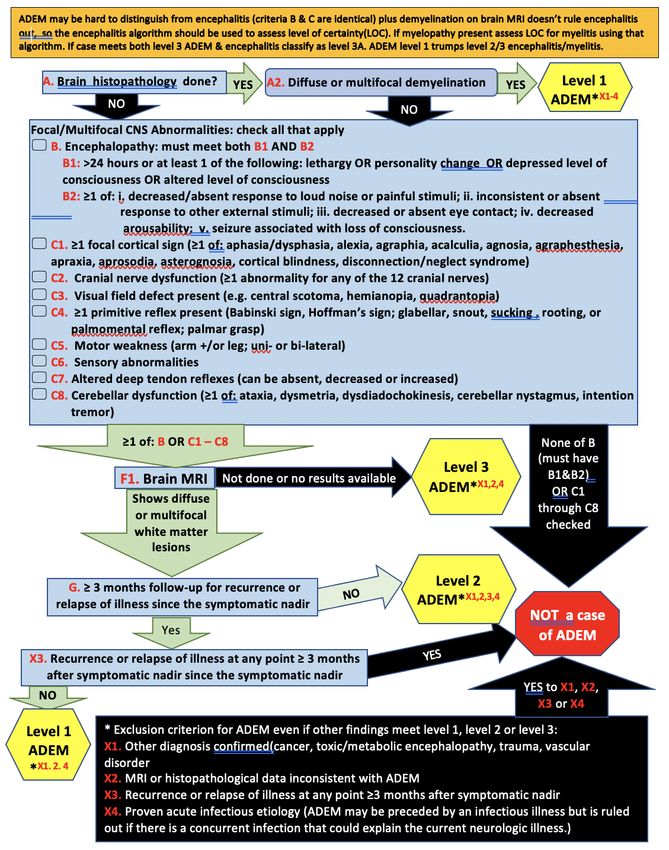
APPENDIX 7
ADEM Pictorial Level of Certainty Algorithm
7.1 ADEM Pictorial level of certainty algorithm
Use available clinical history, examination and laboratory investigation results to determine level of diagnostic certainty for
ADEM. Companion Guides with similar algorithms specific to encephalitis and myelitis are available.
THIS PROJECT HAS BEEN FUNDED IN WHOLE BY CEPI. 28V1.0. 11-Feb-2021 | Diss. level: Public
APPENDIX 8.
Methodology: Brief Summary
8.1. ADEM Risk Factors 1-11
A risk factor is “an exposure, behavior, or attribute that, if present and active, clearly alters the occurrence of a particular
disease compared with an otherwise similar group of people who lack the risk factor”. According to James Last dictionary of
epidemiology version 4, a risk factor is an aspect of personal behavior or lifestyle, an environmental exposure, or an inborn
or inherited characteristic, that, on the basis of epidemiologic evidence, is known to be associated with health-related
condition(s) considered important to prevent. The term risk factor is rather loosely used, with any of the following
meanings:
1. An attribute or exposure that is associated with an increased probability of a specified outcome, such as the occurrence
of a disease. Not necessarily a causal factor. A RISK MARKER.
2. An attribute or exposure that increases the probability of occurrence of disease or another specified outcome. A
DETERMINANT.
3. A determinant that can be modified by intervention, thereby reducing the probability of occurrence of disease or other
specified outcomes. To avoid confusion, it may be referred to as a modifiable risk factor.
Risk factors can include infection, medication, diet, surgical or medical procedure, environmental location, stress, toxins,
trauma and vaccine. Attribute includes genetic makeup, age, gender, ethnicity, social status, occupation. Behavior includes
smoking, drinking, other substance abuse, sexual practices, level of physical activity. A standard tabular format, as shown
in the appendices was used to summarize the key known risk factors for each AESI. Risk factors are only included if there is
evidence for an association with the AESI.
The published Brighton Case definition1 for ADEM was reviewed for evidence related to associated risk factors. In addition,
review articles published after the Brighton case definition were retrieved and reviewed in depth regarding known risk
factors for acute ADEM.2-11
8.2. ADEM Background Incidence12-18
A systematic literature search to estimate the incidence of acute ADEM in the population was conducted using the following
search strategy:
("Encephalomyelitis, Acute Disseminated"[Mesh:noexp] OR "acute disseminated encephalomyelitis"[ti] OR "acute
disseminated encephalomyelitides"[ti] OR "ADEM"[ti]) AND ("Incidence"[Mesh:noexp] OR "incidence"[tiab]) AND
English[lang] AND ("2000/01/01"[PDAT] : "3000/12/31"[PDAT]) AND ("Meta-Analysis"[Publication Type] NOT
("animals"[Mesh:noexp] NOT "humans"[Mesh:noexp]) NOT ("Coronavirus"[Mesh:noexp] OR "coronavirus"[ti] OR
"nCoV"[ti] OR "COVID"[ti] OR "SARS-CoV-2"[ti]) NOT ("therapy"[ti] OR "therapies"[ti] OR "therapeutic"[ti] OR "treatment"[ti]
OR "treatments"[ti] OR "drug"[ti] OR "drugs"[ti] OR trial[ti] OR "trials"[ti] OR "prevention"[ti] OR "prevent"[ti] OR
"prevents"[ti] OR "surgery"[ti] OR "procedure"[ti] OR "procedures"[ti]).
Articles had to meet the following criteria:
1. Original research/meta-analysis
2. Population-based study (selecting the entire population or using probability-based sampling methods)
3. Reported an incidence estimate (or raw numbers that allowed the calculation of an estimate).
If multiple articles reported data from the same study population, the most comprehensive data were used. When studies
reported on different data collection years or subgroups (sex, age), efforts to include all nonoverlapping data were
THIS PROJECT HAS BEEN FUNDED IN WHOLE BY CEPI. 29You can also read