FAPI-74 PET/CT USING EITHER 18F-ALF OR COLD-KIT 68GA LABELING: BIODISTRIBUTION, RADIATION DOSIMETRY, AND TUMOR DELINEATION IN LUNG CANCER PATIENTS
←
→
Page content transcription
If your browser does not render page correctly, please read the page content below
FAPI-74 PET/CT Using Either 18F-AlF or Cold-Kit 68Ga
Labeling: Biodistribution, Radiation Dosimetry, and Tumor
Delineation in Lung Cancer Patients
Frederik L. Giesel*1, Sebastian Adeberg*2–5, Mustafa Syed2,4, Thomas Lindner1, Luis David Jiménez-Franco6,
Eleni Mavriopoulou1, Fabian Staudinger1, Eric Tonndorf-Martini2,4, Sebastian Regnery2,4, Stefan Rieken2,5,7,
Rami El Shafie2,5, Manuel Röhrich1, Paul Flechsig1, Andreas Kluge6, Annette Altmann1, Jürgen Debus2–5,
Uwe Haberkorn1,8, and Clemens Kratochwil1
1Department  of Nuclear Medicine, University Hospital Heidelberg, Heidelberg, Germany; 2Heidelberg Institute of Radiation
Oncology, Heidelberg, Germany; 3Heidelberg Ion-Beam Therapy Center, Heidelberg, Germany; 4Department of Radiation Oncology,
University Hospital Heidelberg, Heidelberg, Germany; 5Clinical Cooperation Unit Radiation Oncology, German Cancer Research
Center, Heidelberg, Germany; 6ABX-CRO Advanced Pharmaceutical Services Forschungsgesellschaft mbH, Dresden, Germany;
7Department of Radiation Oncology, University Hospital Göttingen, Göttingen, Germany; and 8Clinical Cooperation Unit Nuclear

Medicine, German Cancer Research Center, Heidelberg, Germany

                                                                                  Key Words: FAPI PET/CT; cancer-associated fibroblasts; lung
68Ga-fibroblast  activation protein inhibitors (FAPIs) 2, 4, and 46 have          cancer; radiation dosimetry; cold kit
already been proposed as promising PET tracers. However, the                      J Nucl Med 2021; 62:201–207
short half-life of 68Ga (68 min) creates problems with manufacture                DOI: 10.2967/jnumed.120.245084
and delivery. 18F (half-life, 110 min) labeling would result in a more
practical large-scale production, and a cold-kit formulation would
improve the spontaneous availability. The NOTA chelator ligand
FAPI-74 can be labeled with both 18F-AlF and 68Ga. Here, we de-
scribe the in vivo evaluation of 18F-FAPI-74 and a proof of mecha-
nism for 68Ga-FAPI-74 labeled at ambient temperature. Methods: In
10 patients with lung cancer, PET scans were acquired at 10 min, 1
                                                                                  F     ibroblast activation protein (FAP) is highly expressed in the
                                                                                  stroma of a variety of human cancers and is therefore considered
h, and 3 h after administration of 259 ± 26 MBq of 18F-FAPI-74.                   promising for guiding targeted therapy (1). Quinoline-based FAP
Physiologic biodistribution and tumor uptake were semiquantita-                   inhibitors (FAPIs) specifically bind to the enzymatic domain of
tively evaluated on the basis of SUV at each time point. Absorbed                 FAP and are then internalized (2). Methods for conjugation of
doses were evaluated using OLINDA/EXM, version 1.1, and QDOSE
                                                                                  quinoline-based FAP ligands with chelators suitable for radiolab-
dosimetry software with the dose calculator IDAC-Dose, version
2.1. Identical methods were used to evaluate one examination after                eling with various radiometals were developed (3,4). Labeled with
injection of 263 MBq of 68Ga-FAPI-74. Results: The highest con-                   the positron emitter 68Ga, these novel FAP-targeted tracers dem-
trast was achieved in primary tumors, lymph nodes, and distant                    onstrated tumor-to-nontumor contrast ratios that were equal to or
metastases at 1 h after injection, with an SUVmax of more than 10.                even higher than those attained with 18F-FDG PET/CT (5).
The effective dose per a 100-MBq administered activity of 18F-FAPI-                  Although 68Ga is available via approved 68Ge/68Ga generators,
74 was 1.4 ± 0.2 mSv, and for 68Ga-FAPI-74 it was 1.6 mSv. Thus,                  which allow batch production of approximately 2–3 patient doses
the radiation burden of a diagnostic 18F-FAPI-74 PET scan is even                 per elution, the relatively short half-life of 68Ga (68 min, 1.90-MeV
lower than that of PET scans with 18F-FDG and other 18F tracers;
68Ga-FAPI-74 is comparable to other 68Ga ligands. FAPI PET/CT                     positron energy) poses some disadvantages with respect to produc-
supported target volume definition for guiding radiotherapy. Con-                 tion capacity and nuclear decay properties. The short half-life man-
clusion: The high contrast and low radiation burden of FAPI-74                    dates in-house production, making delivery to remote centers
PET/CT favor multiple clinical applications. Centralized large-scale              challenging. In large centers with high patient throughput, several
production of 18F-FAPI-74 or decentralized cold-kit labeling of 68Ga-             productions per day are required to meet potential demands, occu-
FAPI-74 allows flexible routine use.                                              pying a skilled workforce of radiochemists and radiopharmacists
                                                                                  over a protracted period of the work day. If 68Ga-FAPI PET were
                                                                                  to replace 18F-FDG PET in clinical routine, multiple generators
  Received Mar. 13, 2020; revision accepted May 27, 2020.
  For correspondence or reprints contact: Uwe Haberkorn, Department of            would be needed, thus multiplying costs. Labeling with 18F (half-
Nuclear Medicine, University Hospital Heidelberg, Im Neuenheimer Feld 400,        life, 110 min, 0.65-MeV positron energy) would solve these issues.
69120 Heidelberg, Germany.
Email: uwe.haberkorn@med.uni-heidelberg.de                                        Centers with an on-site cyclotron can produce 18F at moderate cost,
  *Contributed equally to this work.                                              and commercial sites can distribute 18F-labeled compounds over a
  Published online Jun. 26, 2020.                                                 wide metropolitan area, eliminating the need for on-site radiochem-
  Immediate Open Access: Creative Commons Attribution 4.0 International
License (CC BY) allows users to share and adapt with attribution, excluding       istry (6). The lower positron energy of 18F could theoretically im-
materials credited to previous publications. License: https://creativecommons.    prove spatial resolution (7).
org/licenses/by/4.0/. Details: http://jnm.snmjournals.org/site/misc/permission.
xhtml.                                                                               As described in a dedicated chemistry/preclinical article (sub-
  COPYRIGHT © 2021 by the Society of Nuclear Medicine and Molecular Imaging.      mitted for publication simultaneously), attempts to label FAPIs

                                                                           FAPI-74 BIODISTRIBUTION        IN   PATIENTS • Giesel et al.           201
with covalently attached 18F were initially unsuccessful by demon-        reconstruction kernel (B30) with CareDose (Siemens). All patients
strating poor tumor uptake. In contrast, chelation of AlF, an ap-         were imaged at 10 min, 1 h, and 3 h after injection of either 259 6
proach that was proposed several years ago and has now been               26 MBq (range, 198–290 MBq) of 18F-FAPI-74 (in 10 patients) or 263
optimized with regard to labeling yield and specific activity (8),        MBq of 68Ga-FAPI-74 (in 1 patient).
presented favorable results in combination with the NOTA-containing
                                                                          FAPI-Based Target Volume of Primary Tumors
FAPI-74. The NOTA chelator also allows chelation with 68Ga at room
                                                                              The acquired 18F-FAPI-74 PET/CT examinations were used to as-
temperature, which would also simplify local on-demand production         sist tumor volume delineation for guiding radiotherapy in patients
in centers that already own a 68Ge/68Ga generator.                        with lung cancer, similar to previous use of 18F-FDG PET/CT
   The aim of this work is to analyze the time-dependent tumor            (9,10). Target volume was defined using Siemens Syngo.via software (Sie-
uptake and tracer biodistribution and to estimate absorbed dose for       mens). CT-based gross tumor volumes (GTVs) were contoured on soft-
18F-FAPI-74 PET/CT scans using examinations that were done
                                                                          tissue and lung windows using contrast-enhanced examinations. PET-based
under a medical indication to assist tumor volume delineation             GTVs (FAPI GTVs) were assessed by comparing tumor SUVs with
for guiding radiotherapy in lung cancer patients. In addition, we         healthy surrounding tissue using Syngo’s auto-contour algorithm at various
demonstrate proof of mechanism for 68Ga-FAPI-74 PET/CT after              SUV thresholds. Two segmentation approaches were considered: either the
tracer labeling at ambient temperature.                                   background level multiplied by a certain number or the percentage of
                                                                          SUVmax. Contours were manually adjusted, checked for plausibility, and
MATERIALS AND METHODS                                                     corrected for false-positive or -negative uptake by 2 experienced radiation
                                                                          oncologists and 2 nuclear medicine physicians, board-certified in their re-
Patients
                                                                          spective specialties. In clinical practice, because defining the radiation field
   This analysis includes 10 patients (4 male, 6 female) with lung        is inherently a subjective task, a consensus of expert readers is usually
cancer (8 with adenocarcinoma, 2 with squamous cell carcinoma) and        considered the best applicable standard of reference.
a median age of 65 y (range, 45–77 y). All patients gave written
informed consent to undergo 68Ga-FAPI-74 PET/CT following national        Biodistribution
regulations and the Declaration of Helsinki. The radiopharmaceutical         For calculation of the SUV, circular regions of interest were drawn
was produced in accordance with the German Pharmaceuticals Act,           around the tumor lesions with focally increased uptake in transaxial
x13(2b). All patients were referred by a radiation oncologist, in order   slices and automatically adapted to a 3-dimensional volume of interest
to improve tumor delineation for radiotherapy planning of central pul-    (VOI) with e.soft software (Siemens) at a 40% isocontour. The tracer
monary lesions that would presumably have been challenging to dis-        biodistribution in patients was quantified by SUVmean and SUVmax at
criminate from the myocardium with 18F-FDG PET. The retrospective         10 min, 1 h, and 3 h after injection of 18F-FAPI-74. The normal organs
evaluation of data acquired under clinical indication was approved by     (brain, oral mucosa, parotid, thyroid, lung, heart muscle, aortic lumen
the ethical committee of Heidelberg University (permit S016/2018).        content, liver, pancreas, spleen, kidney, colon, muscle, fat, and spinal cord)
                                                                          were evaluated with a 2-cm sphere placed inside the organ parenchyma.
Radiopharmaceuticals                                                      Statistical analysis and graphic output were performed with SigmaPlot.
    Chelation with 18F-AlF was performed using the method of McBride et
al. (8) as follows: 2–10 GBq of 18F fluoride (ZAG Cyclotron AG) in 4 mL   Radiation Dosimetry Estimate
of water were trapped on an anion exchange cartridge (Waters Accel           The dosimetry analysis was performed using the QDOSE dosimetry
Plus QMA Light cartridge, preconditioned with 5 mL of 0.5 M               software suite, version 1.1.4 (ABX-CRO).
NaOAc, pH 3.9, and 10 mL of water) and eluted with 0.30 mL of                After all PET and corresponding CT data were imported into the
0.5 M NaOAc, pH 3.9. The solution was incubated with 6 mL of              QDOSE software, CT images were coregistered using an automatic
AlCl3 in water (10 mM) and 300 mL of dimethyl sulfoxide (Simga-           rigid coregistration algorithm. PET images were coupled to the CT
Aldrich) for 5 min at room temperature before 20 mL of a solution of      image of the corresponding imaging time point and manually coregis-
FAPI-74 (4 mM) were added. The reaction was performed at 95C             tered to this CT image when necessary. The frame acquisition time was
for 15 min, cooled to room temperature, diluted with 5 mL of water,       adjusted from the start of the scan (standard for DICOM header) to the
and worked up by solid-phase extraction (Waters Oasis HLB Plus            middle of the acquisition frame (difference of 9.6 6 1.2 min), which
Light cartridge). The final product was eluted with 0.5 mL of ethanol     appears more appropriate for pharmacokinetic evaluation.
and 5 mL of 0.9% saline and spiked with phosphate buffer before              Kidneys, liver, spleen, urinary bladder content, red marrow, heart
sterile filtration (Filtropur S 0.2; Sarstedt).                           content, and remainder of body were considered source organs. According
    Chelation with 68Ga was achieved by adding 1.00 mL of 68Ge/68Ga       to an established model, the red marrow activity was approximated by
generator eluate (0.6 M hydrochloric adic; ;1.2 GBq) to a mixture of      extrapolating activity retrieved from VOIs in lumbar vertebrae 1–5
15 mL of FAPI-74 solution (4 mM in water), 310 mL of sodium acetate       (;12.3% of the red marrow space) to the total red marrow (11).
(2.5 M in water), and 0.50 mL of ethanol. After incubation for 15 min        Because the limbs were cropped by the limited field of view of the
                                                                          PET scan, the total-body cumulated activity (A      ~ Total_Body), which is
at room temperature, the reaction was worked up by solid-phase ex-
traction as described for 18F-FAPI-74.                                    important to determine the cumulated activity in the remainder of
                                                                          the body for dose calculations, was estimated using the injected ac-
PET/CT Imaging                                                            tivity (A) and the effective half-life (Teff) of a VOI covering most the
   All imaging was performed on a Biograph mCT Flow scanner               body. Thus, the total-body cumulated activity was calculated as:
(Siemens). PET was performed in 3-dimensional mode (matrix, 200 ·
200) using FlowMotion (Siemens). The emission data were corrected                             ~ Total_Body 5
                                                                                              A                 A  Teff =ðLnð2ÞÞ:
for randoms, scatter, and decay. Reconstruction was performed with
ordered-subset expectation maximization using 2 iterations and 21            For segmentation of the source organs, VOIs were defined for the
subsets, as well as Gauss filtering to a transaxial resolution of 5 mm    kidneys (left and right), liver, spleen, urinary bladder, heart, lumbar
in full width at half maximum; attenuation was corrected using the        vertebrae (L1–L5) and total body. Tumor areas were not considered in
unenhanced low-dose CT images. The CT scans were reconstructed to         the segmented VOIs. Each source organ was manually segmented on
a slice thickness of 5 mm and an increment of 3 mm using a soft-tissue    the PET images at each time point, and activity values were retrieved

202        THE JOURNAL     OF   NUCLEAR MEDICINE • Vol. 62 • No. 2 • February 2021
to determine the time–activity curves for the organs. The volumes of drug-related pharmacologic effects or physiologic responses oc-
the liver, kidneys, and spleen were determined from segmentation in curred. All observed parameters remained normal and unchanged
the CT images. The calculation of the masses (assuming a density of during and after the examination. No patient reported subjective
1.06 g/cm3) was automatically performed in QDOSE on the basis of symptoms during the 3.5-h observation period after tracer injection.
the segmented VOIs in the CT images.
The time–activity curve for the kidneys was automatically calcu- Normal-Organ Biodistribution and Tumor Uptake
lated as the sum of the activities in the left and right kidneys. Mono- The biodistribution of 18F-FAPI-74 in normal organs and tumor
exponential curve fitting was then applied to all organ time–activity is presented in Figure 1 and illustrated as time-dependent maxi-
curves. The fitted time–activity curves were then integrated from time mum-intensity projections in Figure 2. In contrast to the previous
~ values. The
0 min to infinity to obtain the cumulated time–activity (A) 68Ga-FAPI-2 and 68Ga-FAPI-4 (5), the oral mucosa uptake did not
~ values of the total body and red marrow were added as organs into
A exceed the background in muscle and connective tissue. Another
QDOSE as external calculations for these organs were performed. The difference was a moderately higher blood-pool uptake, both on the
~ of the remainder of the body was automatically calculated by sub-
A initial imaging and on the delayed imaging. Blood-pool and mus-
tracting the A~ of all source organs from the total-body A. ~ Residence
cle uptake did not differ from that with 68Ga-FAPI-2/4, but with
~
times were calculated by dividing the A of each source organ by the 18F-FAPI-74, vessels were definable at all time points. According
injected activity and further exported to OLINDA/EXM, version 1.1 to our previous FAPI tracers, there was no uptake of 18F-FAPI-74 in
(12), for dose calculation with this software.
the liver or spleen exceeding the perfusion-dependent background.
Absorbed and effective dose were calculated using OLINDA/EXM
Within this small sample size, the tumor uptake of adenocarcinoma
(12), with the residence times exported from QDOSE. In addition, the
versus squamous cell carcinoma showed no difference, nor was it
IDAC-Dose, version 2.1, dose calculator (13) integrated in QDOSE was
different from that found previously with 68Ga-FAPI-4 (16). In pri-
also used to perform dose estimations. IDAC-Dose is based on the adult
reference computational phantoms of the International Commission on mary lung tumors, the average SUVmax was 11.8 at 10 min, 12.7 at 1
Radiological Protection (ICRP) (14) and on the ICRP specific absorbed h, and 11.3 at 3 h after injection. Lymph node metastases had an
fractions (15). Organ masses for the kidneys, liver, and spleen, obtained SUVmax of 9.9 at 10 min, 10.7 at 1 h, and 9.4 at 3 h. Distant
from the segmentation in the CT images, were individually adapted for metastases demonstrated an average SUVmax of 11.8 at 10 min,
each patient both in QDOSE (using IDAC-Dose) and in OLINDA/EXM 11.8 at 1 h, and 11.4 at 3 h. Therefore, the uptake generally peaked
to obtain more accurate dose estimations. later than 10 min after injection, but there was already some washout
from tumor tissue between 1 and 3 h after injection; therefore, the
RESULTS best contrast between tumor and background was achieved at 1 h after
injection, and this time point was consecutively used to evaluate GTV
Adverse Events delineation for guiding radiotherapy. The patient receiving 68Ga-
The mean administered activity of 18F-FAPI-74 was 259 6 26 FAPI-74 is presented in Figure 3 and presents similar kinetics, with
MBq (range, 198–290 MBq); for the 68Ga-FAPI-74 examination, tumor SUVmax being 10.4 at 10 min, 11.4 at 1 h, and 8.7 at 3 h.
it was 263 MBq. After quality control, the specific activities of
18F-FAPI-74 were 20–50 nmol/GBq (14.7–36.8 mg/GBq); the spe- Automated Target Volume Delineation of FAPI GTVs
cific activity of 68Ga-FAPI-74 was about 100 nmol/GBq (73.6 mg/ Contouring primary lung tumors on CT resulted in a median
GBq) and was only moderately worsened by physical decay dur- GTV of 67.4 cm3 (range, 25.9–343.4 cm3). For a cutoff at 3-fold
ing the short delay between on-site labeling and injection. Thus, the background, 18F-FAPI-74 PET traced a median GTV of 69.8 cm3
administered masses of FAPI-74 (735.8 g/mol) were about 5–40 mg (P 5 0.21; range, 5.0–527.0 cm3; Fig. 4). Considering a mean back-
per patient dose. All patients tolerated the examination well. No ground SUV of 2 and a mean tumor SUV of 12, the GTVs segmented
FIGURE 1. Time-dependent biodistribution of 18F-FAPI-74 in normal organs and tumor.
FAPI-74 BIODISTRIBUTION IN PATIENTS • Giesel et al. 203Thus, the examinations, which were con-
ducted with 198–290 MBq of 18F-FAPI-
74, translated into effective doses of about
3–4 mSv per examination based on the
OLINDA/EXM mean effective dose. For
68Ga-FAPI-74, the effective dose was 1.6
mSv/100 MBq with OLINDA/EXM and
1.4 mSv/100 MBq with IDAC-Dose. Be-
cause of a rapid renal tracer clearance and
low nonspecific uptake in normal organs,
the radiation dosimetry estimate of 18F-
FAPI-74 compares favorably with most
other 18F-labeled PET tracers in clinical
use, whereas 68Ga-FAPI-74 is in the same
range as other 68Ga-labeled tracers, includ-
ing FAPI-2/4/46 (Table 2).
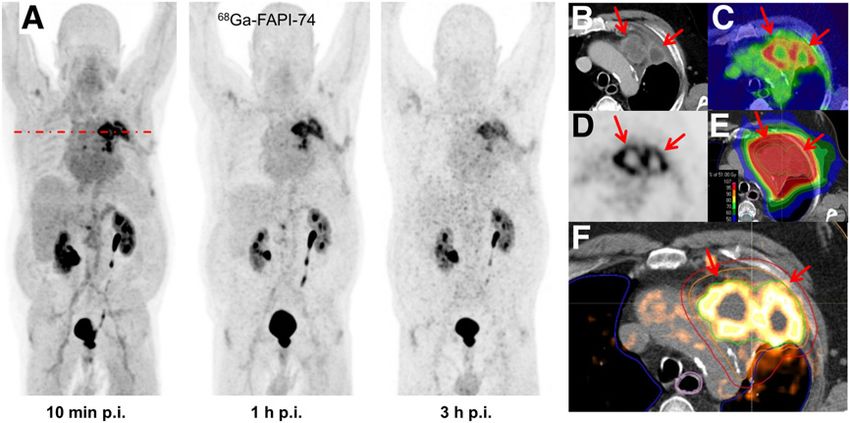
FIGURE 2. (A) Maximum-intensity projections of 18F-FAPI-74 PET at 10 min, 1 h, and 3 h after
DISCUSSION
injection. (B) FAPI PET/CT presents favorable discrimination between tumor and myocardium. (C–
E) Some FAPI-positive lesions were confirmed by CT correlate (C), whereas additional bone In this work, we evaluated the biodis-
lesions were only detected per FAPI PET (D and E). All highlighted arrows represent FAPI uptake tribution and radiation dosimetry of 18F-
with morphological correlation. p.i. 5 after injection.
FAPI-74 PET and demonstrated its possible
value for guiding radiotherapy. In addition,
using a 3-fold background threshold are equal to GTVs segmented at labeling of 68Ga-FAPI-74 at ambient temperature was established,
40%–50% tumor SUVmax. In consensus with the radiation oncologist, and its in vivo performance was evaluated using identical methods.
these PET-segmented volumes were considered more likely to reflect On the basis of the time-dependent biodistribution of 68Ga- and
actual tumor volumes than the corresponding CT image. One patient 18F-FAPI-74 in tumor and normal organs, optimal tumor-to-back-
who was initially considered oligometastatic per the CT image was ground ratios at limited noise were achieved by image acquisition
upstaged and transferred to chemotherapy after additional tumor le- 1 h after injection. This is in contrast to previous experience with
sions were found on 18F-FAPI-74 PET imaging (Fig. 2). 68Ga-FAPI-2/4, for which no improvement in tumor uptake be-
Radiation Dosimetry tween 10 min and 1 h after injection was observed. In normal
The OLINDA/EXM-based dosimetry estimates are presented in organs, the time-dependent biodistribution was nearly identical
Table 1. Calculations according to the IDAC-Dose calculator are to that of other quinoline-based FAPIs (5).
presented in Supplemental Table 1 (supplemental materials are With a mean normalized effective dose of 1.4 mSv/100 MBq
available at http://jnm.snmjournals.org). For 18F-FAPI-74, the nor- (3.5 mSv for a typical 250-MBq examination), the radiation
malized effective dose was 1.4 6 0.2 mSv/100 MBq (range, burden of an 18F-FAPI-74 PET scan is lower than that of PET
1.1–1.7 mSv/100 MBq) with OLINDA/EXM and 1.2 6 0.1 scans with 18F-FDG, the current standard in oncologic imaging
mSv/100 MBq (range, 1.0–1.4 mSv/100 MBq) with IDAC-Dose. (Table 2). The faint physiologic cerebral and hepatic uptake of
18F-FAPI-74 likely accounts for a lower
radiation exposure. The effective dose of 1.6
mSv/100 MBq (3.0 mSv for a typical 185-
MBq examination) for a 68Ga-FAPI-74 PET
scan is within the range for PET imaging with
68Ga-FAPI-2, 68Ga-FAPI-4, and 68Ga-FAPI-
46—a finding that was expected, as all share
a similar biodistribution and tracer kinetics.
We used 18F-FAPI-74 PET/CT to plan
radiotherapy in patients with lung cancer.
Currently, 18F-FDG PET/CT is the stan-
dard for staging and target volume delin-
eation in lung cancer. With 18F-FDG PET/
CT, it is possible to identify additional distant
metastases in about 5%–30% of patients
(17), and its high sensitivity for mediastinal
lymph nodes, 90%–100%, is considered suf-
ficient to limit the target volume to involved
FIGURE 3. (A) Maximum-intensity projections of 68Ga-FAPI-74 PET at 10 min, 1 h, and 3 h after regions. The specificity of 18F-FDG PET/CT
injection. (B–E) Direct comparison of contrast-enhanced CT (B), fusion imaging (C), and FAPI PET
is about 80% because of false-positive find-
(D). (E and F) Superior tumor delineation consecutively improved dose application to tumor
volume using volumetrically modulated arc therapy. Positive FAP uptake is marked by arrows ings (18–24). Our preliminary experience in
(B–F). Green outline 5 GTV; orange outline 5 clinical target volume; red outline 5 planning target 11 patients is not yet sufficient to calculate
volume. p.i. 5 after injection. the sensitivity, specificity, and accuracy of
204 THE JOURNAL OF NUCLEAR MEDICINE • Vol. 62 • No. 2 • February 2021TABLE 1
Dose Estimates for 18F-
and 68Ga-FAPI-74 According to
OLINDA/EXM
Mean 18F-FAPI-
74 ± SD 68Ga-FAPI-74
Target organ (n 5 10) (n 5 1)
Adrenals 1.15 ± 0.09 1.29
Brain 0.78 ± 0.09 1.05
Breasts 0.78 ± 0.07 1.04
Gallbladder wall 1.17 ± 0.10 1.33
Lower large intestine wall 1.23 ± 0.16 1.31
Small intestine 1.16 ± 0.12 1.29
Stomach wall 1.06 ± 0.10 1.24
Upper large intestine wall 1.13 ± 0.11 1.27
Heart wall 2.29 ± 0.28 3.40
Kidneys 2.94 ± 0.79 3.51
Liver 1.50 ± 0.36 1.33
Lungs 0.96 ± 0.07 1.16
FIGURE 4. GTV automatically segmented per FAPI PET at different Muscle 0.94 ± 0.10 1.14
SUV thresholds (x 5 blood-pool–fold) in comparison to CT-based stan- Ovaries 1.25 ± 0.16 1.33
dard of reference.
Pancreas 1.18 ± 0.10 1.32
Red marrow 1.12 ± 0.11 1.11
Osteogenic cells 1.53 ± 0.14 1.70
18F-FAPI-74 PET/CT. However, similar to 18F-FDG PET, with 18F-
Skin 0.73 ± 0.08 1.00
FAPI-74 PET it was possible to identify additional distant metastases
compared with a diagnostic CT scan (Fig. 2). In a recent case report, Spleen 1.67 ± 0.44 1.19
because of the low physiologic cerebral background uptake, 68Ga- Testes 0.99 ± 0.13 1.16
FAPI-4 PET/CT made it possible to identify brain metastases from Thymus 1.02 ± 0.09 1.21
lung cancer (25). Thus, the oncologic application of 18F-FAPI-74 Thyroid 0.91 ± 0.09 1.13
PET/CT appears promising. By applying various cutoffs, the best Urinary bladder wall 7.58 ± 2.84 9.86
correlation between CT and 18F-FAPI-74 PET–guided GTV segmen-
Uterus 1.49 ± 0.25 1.46
tation was found at uptakes that were 3-fold the background level,
which equals 40%–50% of SUVmax (Fig. 4). This finding perfectly Total body 0.97 ± 0.09 1.22
corresponds to several publications about 18F-FDG PET, which rec- Effective dose 1.41 ± 0.22 1.61
ommend delineating the 3-dimensional metabolic target volume at
41%–50% of SUVmax (26–29). Data are absorbed dose (mSv/100 MBq).
On the basis of the first DOTA-modified tracer, FAPI-2, the
derivatives FAPI-4 and FAPI-46 were developed with a focus on the
therapeutic option. The NOTA derivative FAPI-74 was developed as would allow large-scale batch production and distribution via satel-
an exclusive diagnostic ligand, accepting slightly shorter tumor lite concepts. Another characteristic of the NOTA chelator in FAPI-74
retention than the previous theranostic agents. Nevertheless, at early is the possibility for 68Ga labeling at ambient temperature. Standard-
imaging time points, the diagnostic performance should be very ized cold kits would allow chargewise constancy tests as required by
similar. Well in line with our expectations, the tumor SUVs of 68Ga- regulatory bodies and would increase flexibility for local on-demand
and 18F-FAPI-74 are almost equal to that of FAPI-4 when compar- production using approved 68Ge/68Ga generators. Thus, in our center,
ing lung cancer patients (16). In a recent investigation, the accuracy we consider FAPI-74 to be the final evolutionary stage of diagnostic
of FAPI-4 PET/CT was directly compared with 18F-FDG PET/CT, FAP-targeted ligands.
and better tumor-to-background contrast and a higher detection rate Appropriate approximation of the radiation dosimetry of a
for primary tumors, lymph nodes, and visceral metastases was novel radiopharmaceutical is mandatory before prospective clin-
found for FAPI PET than for 18F-FDG PET. In this study, histo- ical trials can take place, and this investigation focused on high
pathologic examination of biopsy or surgical specimens served as methodical standards for the dosimetry part, such as by consid-
the gold standard for the final patient classification (30). In addition ering individually segmented organ masses for all patients. Yet,
to its oncologic application, FAPI PET was also found promising for only a few investigations directly comparing 68Ga-FAPI-2/4 ver-
the evaluation of immune-related and heart diseases (31,32). As a sus 18F-FDG with histopathologic correlation have been reported
practical (i.e., independent from blood sugar and physical activity), (5,30). For the still-limited patient numbers that have been avail-
multipurpose tracer, production capacities could soon become a able so far, the accuracy of FAPI PET/CT appears promising.
relevant issue. One additional advantage of FAPI-74 over previous However, additional research evaluating the clinical impact of
ligands is its greater suitability for labeling with 18F-AlF, which FAPI PET/CT for particular clinical indications, compared with
FAPI-74 BIODISTRIBUTION IN PATIENTS • Giesel et al. 205TABLE 2 the technical assistants of the PET/CT group at Heidelberg
Effective Dose of 18F-FAPI-74 and 68Ga-FAPI-74 in University Hospital for performing the PET scans.
Comparison to Other PET Tracers
Effective KEY POINTS
dose
PET tracer (mSv/MBq) Reference QUESTION: What are the biodistribution and dosimetry charac-
teristics of a FAPI variant that can be used for both 18F and 68Ga
18F-FAPI-74 0.014 This work labeling?
68Ga-FAPI-74 0.016 This work
PERTINENT FINDINGS: The NOTA chelator within the novel li-
68Ga-FAPI-2/4/46 0.008–0.015 (5,33)
gand FAPI-74 allows labeling with 18F-AlF, as well as the design of
68Ga-PSMA-11 0.023 (34) a cold kit for labeling with 68Ga. In patients with lung cancer, the
68Ga-DOTATOC/-TATE 0.021 (35) new ligands presented performance and radiation dosimetry
18F-FDG 0.020 (36) similar to previous FAPIs.
18F-3′-deoxy-3′-18F-fluorothymidine 0.028 (37) IMPLICATIONS FOR PATIENT CARE: FAPI-74 is our final-stage
O-(2-18F-fluoroethyl)-L-tyrosine 0.016 (38) PET tracer for imaging of fibroblast-activating protein in vivo.
(S)-4-(3-18F-fluoropropyl)-L-glutamic acid 0.032 (39)
18F-PSMA-1007 0.022 (40)
18F-flurbetaben 0.015 (41) REFERENCES
18F-flurpiridaz 0.019 (42) 1. Brennen WN, Isaacs JT, Denmeade SR. Rationale behind targeting fibroblast
18F-fluorocholine 0.031 (43) activation protein-expressing carcinoma-associated fibroblasts as a novel chemo-
18F-fluoromisonidazole
therapeutic strategy. Mol Cancer Ther. 2012;11:257–266.
0.013 (44)
2. Jansen K, Heirbaut L, Cheng JD, et al. Selective inhibitors of fibroblast activation
protein (FAP) with a (4-quinolinoyl)-glycyl-2-cyanopyrrolidine scaffold. ACS
Med Chem Lett. 2013;4:491–496.
PSMA 5 prostate-specific membrane antigen.
3. Lindner T, Loktev A, Altmann A, et al. Development of quinoline-based thera-
nostic ligands for the targeting of fibroblast activation protein. J Nucl Med.
2018;59:1415–1422.
a reliable standard of truth, and including sufficient patient num- 4. Loktev A, Lindner T, Mier W, et al. A tumor-imaging method targeting cancer-
bers, is still pivotal. associated fibroblasts. J Nucl Med. 2018;59:1423–1429.
5. Giesel FL, Kratochwil C, Lindner T, et al. 68Ga-FAPI PET/CT: biodistribution
CONCLUSION and preliminary dosimetry estimate of 2 DOTA-containing FAP-targeting agents
in patients with various cancers. J Nucl Med. 2019;60:386–392.
The high contrast and low radiation burden of 68Ga- and 18F- 6. Kesch C, Kratochwil C, Mier W, et al. 68Ga or 18F for prostate cancer imaging? J
FAPI-74 PET/CT favor multiple clinical applications. Centralized Nucl Med. 2017;58:687–688.
7. Sanchez-Crespo A. Comparison of gallium-68 and fluorine-18 imaging charac-
large-scale 18F-AlF–based production of 18F-FAPI-74 or decentral- teristics in positron emission tomography. Appl Radiat Isot. 2013;76:55–62.
ized cold-kit labeling of 68Ga-FAPI-74 allows flexible routine use. 8. McBride WJ, Sharkey RM, Karacay H, et al. A novel method of 18F radiolabel-
ing for PET. J Nucl Med. 2009;50:991–998.
DISCLOSURE 9. Paulino AC, Johnstone PA. FDG-PET in radiotherapy treatment planning: Pan-
dora’s box? Int J Radiat Oncol Biol Phys. 2004;59:4–5.
Uwe Haberkorn, Thomas Lindner, Clemens Kratochwil, and 10. Nestle U, Schaefer-Schuler A, Kremp S, et al. Target volume definition for 18F-
Frederik Giesel have a patent application for quinolone-based FDG PET-positive lymph nodes in radiotherapy of patients with non-small cell
lung cancer. Eur J Nucl Med Mol Imaging. 2007;34:453–462.
FAP-targeting agents for imaging and therapy in nuclear medicine. 11. Hindorf C, Glatting G, Chiesa C, et al. EANM dosimetry committee guidelines
Uwe Haberkorn, Thomas Lindner, Clemens Kratochwil, and for bone marrow and whole-body dosimetry. Eur J Nucl Med Mol Imaging.
Frederik Giesel also have shares of a consultancy group for 2010;37:1238–1250.
iTheranostics. Frederik Giesel is a medical advisor for ABX 12. Stabin MG, Sparks RB, Crowe E. OLINDA/EXM: the second-generation per-
Advanced Biochemical Compound and Telix Pharmaceuticals. sonal computer software for internal dose assessment in nuclear medicine. J Nucl
Med. 2005;46:1023–1027.
Sebastian Adeberg and Jürgen Debus received grants from Accu- 13. Andersson M, Johansson L, Eckerman K, et al. IDAC-Dose 2.1, an internal
ray International Sàrl, Merck Serono GmbH, and Astra Zeneca dosimetry program for diagnostic nuclear medicine based on the ICRP adult
GmbH outside the submitted work. Sebastian Adeberg received reference voxel phantoms. EJNMMI Res. 2017;7:88.
grants from Novocure outside the submitted work. Jürgen Debus 14. Menzel HG, Clement C, DeLuca P. ICRP publication 110: realistic reference
phantoms—an ICRP/ICRU joint effort. A report of adult reference computa-
received grants from CRI–The Clinical Research Institute GmbH, tional phantoms. Ann ICRP. 2009;39:1–164.
View Ray Inc., Accuray Incorporated, RaySearch Laboratories AB, 15. Bolch WE, Jokisch D, Zankl M, et al. ICRP publication 133: the ICRP compu-
Vision RT Limited, Astellas Pharma GmbH, Solution Akademie tational framework for internal dose assessment for reference adults—specific
GmbH, Ergomed PLC Surrey Research Park, Siemens Healthcare absorbed fractions. Ann ICRP. 2016;45:5–73.
16. Kratochwil C, Flechsig P, Lindner T, et al. 68Ga-FAPI PET/CT: tracer uptake in
GmbH, Quintiles GmbH, Pharmaceutical Research Associates GmbH,
28 different kinds of cancer. J Nucl Med. 2019;60:801–805.
Boehringer Ingelheim Pharma GmbH Co., PTW-Freiburg Dr. Pychlau 17. Sahiner I, Vural GU. Positron emission tomography/computerized tomography
GmbH, and Nanobiotix A.A. outside the submitted work. No other in lung cancer. Quant Imaging Med Surg. 2014;4:195–206.
potential conflict of interest relevant to this article was reported. 18. Gould MK, Maclean CC, Kuschner WG, et al. Accuracy of positron emission
tomography for diagnosis of pulmonary nodules and mass lesions: a meta-analysis.
JAMA. 2001;285:914–924.
ACKNOWLEDGMENTS 19. Behzadi A, Ung Y, Lowe V, et al. The role of positron emission tomography in
the management of non-small cell lung cancer. Can J Surg. 2009;52:235–242.
We thank Peter L. Choyke from the NIH/NCI molecular 20. Fernandes AT, Shen J, Finlay J, et al. Elective nodal irradiation (ENI) vs. in-
imaging program for proofreading and scientific advice. We thank volved field radiotherapy (IFRT) for locally advanced non-small cell lung cancer
206 THE JOURNAL OF NUCLEAR MEDICINE • Vol. 62 • No. 2 • February 2021(NSCLC): a comparative analysis of toxicities and clinical outcomes. Radiother 33. Meyer C, Dahlbom M, Lindner T, et al. Radiation dosimetry and biodistribu-
Oncol. 2010;95:178–184. tion of 68Ga-FAPI-46 PET imaging in cancer patients. J Nucl Med. 2020;61:
21. Goldstraw P, Ball D, Jett JR, et al. Non-small-cell lung cancer. Lancet. 1171–1177.
2011;378:1727–1740. 34. Afshar-Oromieh A, Hetzheim H, Kübler W, et al. Radiation dosimetry of 68Ga-
22. De Ruysscher D, Nestle U, Jeraj R, et al. PET scans in radiotherapy planning of PSMA-11 (HBED-CC) and preliminary evaluation of optimal imaging timing.
lung cancer. Lung Cancer. 2012;75:141–145. Eur J Nucl Med Mol Imaging. 2016;43:1611–1620.
23. Broderick SR, Patterson GA. Performance of integrated positron emission to- 35. Sandström M, Velikyan I, Garske-Román U, et al. Comparative biodistribution
mography/computed tomography for mediastinal nodal staging in non-small cell and radiation dosimetry of 68Ga-DOTATOC and 68Ga-DOTATATE in patients
lung carcinoma. Thorac Surg Clin. 2013;23:193–198. with neuroendocrine tumors. J Nucl Med. 2013;54:1755–1759.
24. De Ruysscher D. PET-CT in radiotherapy for lung cancer. Methods Mol Biol. 36. Quinn B, Dauer Z, Pandit-Taskar N, Schoder H, Dauer LT. Radiation dosimetry
2011;727:53–58. of 18F-FDG PET/CT: incorporating exam-specific parameters in dose estimates.
25. Giesel FL, Heussel CP, Lindner T, et al. FAPI-PET/CT improves staging in a BMC Med Imaging. 2016;16:41.
lung cancer patient with cerebral metastasis. Eur J Nucl Med Mol Imaging. 37. Vesselle H, Grierson J, Peterson LM, Muzi M, Mankoff DA, Krohn KA. 18F-
2019;46:1754–1755. fluorothymidine radiation dosimetry in human PET imaging studies. J Nucl Med.
26. Nestle U, Kremp S, Schaefer-Schuler A, et al. Comparison of different methods for 2003;44:1482–1488.
delineation of 18F-FDG PET-positive tissue for target volume definition in radio- 38. Pauleit D, Floeth F, Herzog H, et al. Whole-body distribution and dosimetry of
therapy of patients with non-small cell lung cancer. J Nucl Med. 2005;46: O-(2-[18F]fluoroethyl)-L-tyrosine. Eur J Nucl Med Mol Imaging. 2003;30:519–524.
1342–1348. 39. Smolarz K, Krause BJ, Graner FP, et al. (S)-4-(3-18F-fluoropropyl)-L-glutamic
27. Erdi YE, Mawlawi O, Larson SM, et al. Segmentation of lung lesion volume acid: an 18F-labeled tumor-specific probe for PET/CT imaging–dosimetry. J Nucl
by adaptive positron emission tomography image thresholding. Cancer. 1997;80: Med. 2013;54:861–866.
2505–2509. 40. Giesel FL, Hadaschik B, Cardinale J, et al. F-18 labelled PSMA-1007: bio-
28. Boellaard R, Krak NC, Hoekstra OS, et al. Effects of noise, image resolution, and distribution, radiation dosimetry and histopathological validation of tumor
ROI definition on the accuracy of standard uptake values: a simulation study. lesions in prostate cancer patients. Eur J Nucl Med Mol Imaging. 2017;44:
J Nucl Med. 2004;45:1519–1527. 678–688.
29. Krak NC, Boellaard R, Hoekstra OS, et al. Effects of ROI definition and re- 41. O’Keefe GJ, Saunder TH, Ng S, et al. Radiation dosimetry of beta-amyloid
construction method on quantitative outcome and applicability in a response tracers 11C-PiB and 18F-BAY94-9172. J Nucl Med. 2009;50:309–315.
monitoring trial. Eur J Nucl Med Mol Imaging. 2005;32:294–301. 42. Maddahi J, Bengel F, Czernin J, et al. Dosimetry, biodistribution, and safety of
30. Chen H, Pang Y, Wu J, et al. Comparison of [68Ga]Ga-DOTA-FAPI-04 and [18F] flurpiridaz F18 in healthy subjects undergoing rest and exercise or pharmaco-
FDG PET/CT for the diagnosis of primary and metastatic lesions in patients with logical stress PET myocardial perfusion imaging. J Nucl Cardiol. 2019;26:
various types of cancer. Eur J Nucl Med Mol Imaging. 2020;47:1820–1832. 2018–2030.
31. Luo Y, Pan Q, Zhang W. IgG4-related disease revealed by 68Ga-FAPI and 18F- 43. DeGrado TR, Reiman RE, Price DT, Wang S, Coleman RE. Pharmacoki-
FDG PET/CT. Eur J Nucl Med Mol Imaging. 2019;46:2625–2626. netics and radiation dosimetry of 18F-fluorocholine. J Nucl Med. 2002;43:
32. Varasteh Z, Mohanta S, Robu S, et al. Molecular imaging of fibroblast activity 92–96.
after myocardial infarction using a 68Ga-labeled fibroblast activation protein 44. Graham MM, Peterson LM, Link JM, et al. Fluorine-18-fluoromisonidazole
inhibitor, FAPI-04. J Nucl Med. 2019;60:1743–1749. radiation dosimetry in imaging studies. J Nucl Med. 1997;38:1631–1636.
FAPI-74 BIODISTRIBUTION IN PATIENTS • Giesel et al. 207You can also read