FDG uptake heterogeneity in FIGO IIb cervical carcinoma does not predict pelvic lymph node involvement
←
→
Page content transcription
If your browser does not render page correctly, please read the page content below
Brooks and Grigsby Radiation Oncology 2013, 8:294
http://www.ro-journal.com/content/8/1/294

 RESEARCH                                                                                                                                       Open Access

FDG uptake heterogeneity in FIGO IIb cervical
carcinoma does not predict pelvic lymph node
involvement
Frank J Brooks1* and Perry W Grigsby1,2,3,4

  Abstract
  Translational relevance: Many types of cancer are located and assessed via positron emission tomography (PET)
  using the 18F-fluorodeoxyglucose (FDG) radiotracer of glucose uptake. There is rapidly increasing interest in
  exploiting the intra-tumor heterogeneity observed in these FDG-PET images as an indicator of disease outcome. If
  this image heterogeneity is of genuine prognostic value, then it either correlates to known prognostic factors, such
  as tumor stage, or it indicates some as yet unknown tumor quality. Therefore, the first step in demonstrating the
  clinical usefulness of image heterogeneity is to explore the dependence of image heterogeneity metrics upon
  established prognostic indicators and other clinically interesting factors. If it is shown that image heterogeneity is
  merely a surrogate for other important tumor properties or variations in patient populations, then the theoretical
  value of quantified biological heterogeneity may not yet translate into the clinic given current imaging technology.
  Purpose: We explore the relation between pelvic lymph node status at diagnosis and the visually evident uptake
  heterogeneity often observed in 18F-fluorodeoxyglucose positron emission tomography (FDG-PET) images of
  cervical carcinomas.
  Experimental design: We retrospectively studied the FDG-PET images of 47 node negative and 38 node positive
  patients, each having FIGO stage IIb tumors with squamous cell histology. Imaged tumors were segmented
  using 40% of the maximum tumor uptake as the tumor-defining threshold and then converted into sets of
  three-dimensional coordinates. We employed the sphericity, extent, Shannon entropy (S) and the accrued deviation
  from smoothest gradients (ζ) as image heterogeneity metrics. We analyze these metrics within tumor volume
  strata via: the Kolmogorov-Smirnov test, principal component analysis and contingency tables.
  Results: We found no statistically significant difference between the positive and negative lymph node groups for
  any one metric or plausible combinations thereof. Additionally, we observed that S is strongly dependent upon
  tumor volume and that ζ moderately correlates with mean FDG uptake.
  Conclusions: FDG uptake heterogeneity did not indicate patients with differing prognoses. Apparent heterogeneity
  differences between clinical groups may be an artifact arising from either the dependence of some image metrics
  upon other factors such as tumor volume or upon the underlying variations in the patient populations compared.
  Keywords: Cervical cancer, FDG-PET, Intra-tumor heterogeneity, Image texture analysis

* Correspondence: fjbrooks@wustl.edu
1
 Department of Radiation Oncology, Washington University School of
Medicine, 4921 Parkview Place, Saint Louis, MO 63110, USA
Full list of author information is available at the end of the article

                                         © 2013 Brooks and Grigsby; licensee BioMed Central Ltd. This is an Open Access article distributed under the terms of the
                                         Creative Commons Attribution License (http://creativecommons.org/licenses/by/2.0), which permits unrestricted use,
                                         distribution, and reproduction in any medium, provided the original work is properly cited. The Creative Commons Public
                                         Domain Dedication waiver (http://creativecommons.org/publicdomain/zero/1.0/) applies to the data made available in this
                                         article, unless otherwise stated.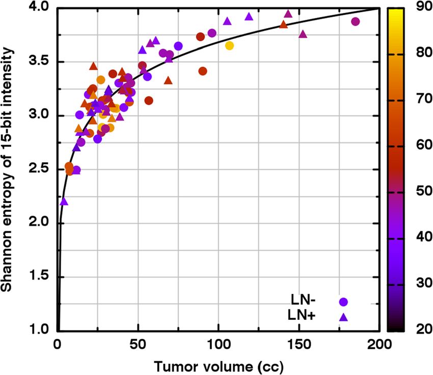
Brooks and Grigsby Radiation Oncology 2013, 8:294                                                                        Page 2 of 8
http://www.ro-journal.com/content/8/1/294

Introduction                                                           where variations in grayscale intensity are clearly visible.
There is evidence that pelvic lymph node status is an                  Of course, each image is a low-resolution view of the
important indicator of both disease free and overall                   underlying biology and is heavily attenuated by the
survival in patients diagnosed with cervical carcinoma                 partial volume effect [12]. Therefore, the image hetero-
[1,2]. There is also a growing belief that the uptake                  geneity observed is unlikely to directly correspond to the
heterogeneity observed within tumors assayed via FDG-                  tumor biology from whence the detected signal came
PET may be of prognostic value [3,4]. A correlation                    [4,13]. For this reason, all FDG uptake heterogeneity
between increased FDG uptake heterogeneity and worse                   analyses should be taken cum grano salis when attempt-
prognosis has been reported for human sarcomas [5,6],                  ing to establish or infer biological meaning from the
non-small cell lung cancer [7], esophageal cancer [8] and              analyses.
cervical cancer [9,10]. Although the relationship is not                  The image heterogeneity observed may be thought of as
well-understood, it seems that tumor state (e.g., quies-               comprising variations in intensity: distribution, arrange-
cence or rapid proliferation) likely is strongly linked to             ment and shape. The grayscale intensity distribution
tumor metabolism [11]. Because FDG uptake is some                      results from some combination of underlying biology,
measure of metabolic activity within the tumor, this                   scanner noise and the partial volume effect. These are the
connection to tumor state offers one plausible means for               shades available to create image detail; less shades means
uptake heterogeneity to indicate disease outcome. We                   that less variation can be conveyed. The spatial arrange-
therefore hypothesize that increased intra-tumor FDG                   ment of the intensities also conveys variation. For ex-
uptake heterogeneity may correlate with lymph node                     ample, intuitively, the smooth gradation of the brightest
involvement. If such a link were established, it will                  image intensities to the dimmest is less varied than
provide a new avenue of studying why only some of                      those same intensities randomly juxtaposed. Overall
cervical carcinoma patients exhibit pelvic lymph node                  shape is distinct from arrangement because shape
involvement and perhaps even provide insight as to why                 defines a priori the bounds of where the intensities
that status so profoundly affects prognosis.                           may be arranged. For example, a tumor growing near
  Heterogeneity in FDG uptake is observed in FDG-PET                   a physical barrier may be unable to achieve the same
images as variations in grayscale intensity with more                  shape a tumor protruding into a void might achieve.
intense regions ostensibly corresponding to increased                  These considerations are crucial when determining if
metabolic activity. Examples may be seen in Figure 1                   two distinct intensity samples (i.e., tumor regions) differ
                                                                       significantly.
                                                                          There have been several previous attempts at quantify-
                                                                       ing uptake heterogeneity seen in FDG-PET images
                                                                       [5-10,14-16]. While we do not exhaustively review them
                                                                       here, we recapitulate some apposite concerns. One study
                                                                       analyzed almost twenty higher-order texture metrics and
                                                                       found only a few to be statistically reproducible on
                                                                       FDG-PET images [17]. This is consistent with another
                                                                       study which showed that dozens of texture metrics
                                                                       applied to FDG-PET images of cervical carcinomas were
                                                                       not useful indicators of prognosis [10]. In general,
                                                                       texture metrics often are inter-related and thus including
                                                                       additional metrics might not include additional infor-
                                                                       mation. For example, the image entropy is (qualitatively)
                                                                       inversely proportional to the image energy [18]. In prac-
                                                                       tice, similar dependences between other metrics may be
                                                                       may not be at all intuitive. For example, it was argued
                                                                       that the slope of the SUV-volume histogram was an
                                                                       intra-tumor heterogeneity metric with prognostic value
                                                                       [9], however, later that same metric was shown to be a
 Figure 1 Shown is an example of a cervical carcinoma imaged
 via FDG-PET. The black and white contour approximately indicates      surrogate for tumor volume [19]. An analogous depend-
 the defined tumor boundary. Within, there are clear variations in     ence upon tumor volume also was illustrated with
 the grayscale intensity. It is these variations researchers seek to   virtual tumor images for the local image entropy [20].
 objectively quantify such that inter-patient comparisons of           Still another metric, the area under the cumulative
 intra-tumor uptake heterogeneity can be made. The image edges
                                                                       SUV-volume histogram, also was presented as a prog-
 correspond to 20 cm within the patient.
                                                                       nostic heterogeneity measure [7,14]. It was argued
Brooks and Grigsby Radiation Oncology 2013, 8:294 Page 3 of 8
http://www.ro-journal.com/content/8/1/294
later that that metric is equivocal in the sense that it Identification of primary tumors
is possible for unequal heterogeneity scenarios to For patients (n = 58) examined between July 2003 and
achieve identical metric values while unequal metric June 2008 diagnostic imaging scanning was performed
values can result from identical heterogeneity scenar- via Siemens Biograph 2 (Munich, Germany) while for
ios [21,22]. Quantification of image heterogeneity is a those examined (n = 27) between October 2008 and
difficult problem in general and has only recently March 2012 were scanned via Siemens Biograph 40
been addressed in the specific context of predicting (Munich, Germany). In both cases, images were recon-
disease outcome from FDG-PET images. Therefore, chal- structed via ordered subset expectation maximization
lenges and refinements to previously proposed metrics with 8 sets; 2 iterations for the former and 4 iterations
(including those we employ) are to be expected as the for the latter. In both cases, a Gaussian smoothing filter
mathematical sophistication of the problem is revealed via was applied post-reconstruction with 5.3 mm and 4 mm
further study. full width at half maximum, respectively. We note that
We analyze our image data via a spatial metric which the 9 most recent Biograph 40 image sets underwent an
has been demonstrated to be both independent of tumor additional point-spread function/time-of-flight correc-
volume and consistent with visual ranking of FDG-PET tion. The primary tumor evident in each FDG-PET
images by human experts [16]. While this metric does image set was identified and segmented specifically for
provide some measure of the variation in intensity the present study by a veteran oncologist using MIM
arrangement within pre-established tumor regions, it version 5.6.3 (MIM Software Inc., Cleveland, OH).
also is sensitive the overall region shape [16]. We Within the manually approximated tumor region, any
employ the sphericity and extent as distinct, volume- image voxel with SUV greater than 40% of the maximum
independent shape metrics which may provide com- region SUV was considered to be part of the tumor [25].
plementary information about the contribution of The oncologist then made slight manual adjustments to
shape to perceived heterogeneity. The sphericity and the ROI to remove any obvious non-tumor pixels such
extent are metrics which can distinguish rounder, as those comprising bladder or bowel regions. For each
more compact regions from highly asymmetrical, por- patient, these data were exported as a set of (x,y,z)
ous regions [23]. We employ the well-established coordinates, each with a single 15-bit grayscale image in-
Shannon entropy [18,24] as the measure of informa- tensity corresponding to radioactivity density in Bq/mL.
tional content within the individual distributions of
grayscale intensities. We note however, that the Shannon Assessment of FDG uptake heterogeneity
entropy is predicted to depend non-linearly upon We assessed observed FDG uptake heterogeneity in sev-
tumor volume and that the exact functional form of eral ways. First, we compute the Shannon informational
that dependence is influenced by both the intensity entropy (S) upon the histogram of grayscale intensities
histogram bin size and image segmentation threshold for each FDG-PET image set [18,24]. The histogram bin
employed [20]. width was defined for each patient individually as the
Freedman-Diaconis bin width. We used the formula
Materials and methods
Patients X
B
This is a retrospective study of 85 patients with newly − pb ln pb ð1Þ
b¼1
diagnosed cervical cancer who underwent FDG-PET
or FDG-PET/CT at Washington University in St. where pb is the probability that a given grayscale inten-
Louis between July 2003 and March 2012. This study sity resides in the bth of a total of B histogram bins.
was approved by the Washington University Human From the (x,y,z) coordinates, we computed the sphericity
Research Protection Office. All patients were evalu- (ψ) and the extent (ξ) for each virtual tumor object. The
ated by history and physical examination, examination sphericity is defined as [23,26]
under anesthesia, and FDG-PET or integrated PET/CT
before initiating treatment. Patients were staged clin- p ffiffiffiffi 2
3
π ð6⋅volumeÞ3
ically, according to FIGO staging (AJCC 2002, 6th ð2Þ
surface area
edition). The selection criteria for inclusion into this
study were FIGO clinical stage IIb tumors and squa- The extent is defined as the ratio of the net object area
mous cell histology. The patients ranged in age from (or volume) to a bounding area (or volume) [23]. We
27 y to 85 y with a median of 49 y. The maximum note one difference from the usual definition of the
SUV of the tumors analyzed ranged from 4 to 56 and extent. Instead of a bounding box, we employed a bound-
approximately followed a log-normal distribution with ing sphere with diameter matching the maximal diameter
median of 13 and variance of 74. of the tumor object and centered at the geometric centerBrooks and Grigsby Radiation Oncology 2013, 8:294 Page 4 of 8
http://www.ro-journal.com/content/8/1/294
of the tumor object. For example, a largely hollow but
basically round tumor will have a low extent (i.e., few
pixels in the bounding sphere) but a high sphericity. Thus,
ψ and ξ together form a set of complementary metrics
which together feasibly discern heterogeneous from
homogeneous tumor geometry. Lastly, we compute the
accrued deviation from smoothest gradients (ζ) [16].
Automated ranking of tumor image via increasing ζ has
been shown to be consistent with rankings done visually
by human experts. As described in Ref. [16], we computed
ζ for our three-dimensional intensity data by using tri-
linear interpolation to approximate the 15-bit intensity
value whenever a fractional (x,y,z) coordinate was required
(recall that only integer coordinates are stored within
single FDG-PET images).
Statistical analysis
Potential differences between the distribution of a het-
erogeneity statistic for distinct patient subgroups defined
Figure 2 The common-axis histograms overlap largely and thus
by pelvic lymph node status were assessed via the two- show that volume alone cannot predict to which lymph node
sample Kolmogorov-Smirnov test. This test of empirical status groups a patient will belong. In other words, we find no
cumulative distribution functions (CDFs) was chosen appreciable difference in distribution of volumes between the
because it: does not require modeling of the distributions LN- and LN+ patient groups. Histogram bin size = 14 cc.
compared, can compare continuous data sets of unequal
size and/or variance and is sensitive to more than just
within each volume stratum. Table 1 shows the results
differences in distribution location [27]. The subgroups
where it is seen that S does not differ significantly within
compared are those with no indication of pelvic lymph
any volume stratum. We repeated this analysis for ψ and
node involvement (LN-) to those with (LN+). For our
ξ separately and again found no significant difference for
population comprising 47 LN- and 38 LN+ patients, a
either variable in any volume stratum (Table 1).
maximum difference (D) between the CDFs greater than
0.286 is significant at the 95% level [28]. In cases where
cross-group ties amongst ranks occur, the p-value given
for D is only an upper-bound of the true p-value [27]. Spe-
cific cases of additional data stratification with differing
significance criteria are detailed in the Results section, as
necessary. Where appropriate, we employ Kendall’s τ as a
measure of variable association and test other potential
variable dependencies via χ2-test applied to contingency
tables. In cases where histograms were constructed,
the Freedman-Diaconis bin width was employed. All
statistical analyses were performed using R version 2.15.2
(R Foundation for Statistical Computing, Vienna, Austria).
Results
In Figure 2, the histograms for the LN- (shaded) and the
LN+ (hatched) groups are shown on the same axes. The
Kolmogorov-Smirnov test (D = 0.121; p ≤ 0.918) implies
no significant difference between these tumor volume
Figure 3 The Shannon informational entropy (S) of each 15-bit
distributions. In Figure 3, S is seen to be strongly grayscale intensity histogram is seen to depend monotonically
dependent upon tumor volume (V). Thus, direct com- upon tumor volume (V) across a wide range of volumes. It is
parison of S for large volumes to S at small volumes is thus seen that by using increasing S values to rank tumors in
not appropriate. We therefore stratified V into quartile increasing heterogeneity, one actually is ranking those tumors
coarsely in terms of volume. The age of the patients in years is
groups of similarly sized tumors and compared the S
indicated by color.
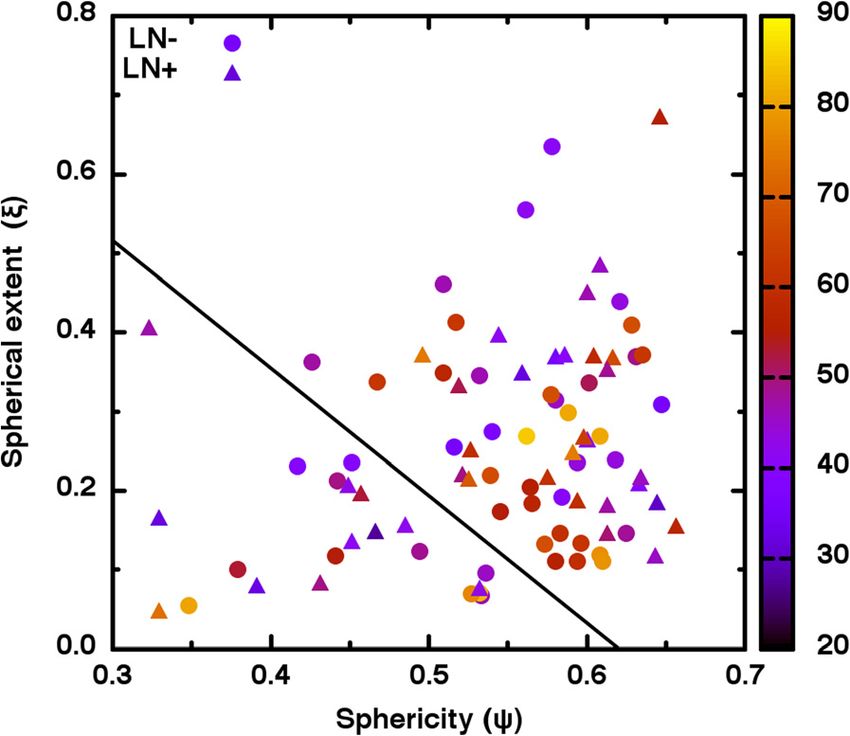
distributions of the lymph node status subgroups strictlyBrooks and Grigsby Radiation Oncology 2013, 8:294 Page 5 of 8 http://www.ro-journal.com/content/8/1/294 Table 1 Within each volume stratum, the Kolmogorov-Smirnov test was used to discern potential differences between lymph node status groups for each of the heterogeneity metrics Volume (in cc) Shannon entropy (S) Sphericity (ψ) Spherical extent (ξ) 4.0 ≤ V < 22.4 D = 0.227; p = 0.886 D = 0.355; p = 0.390 D = 0.200; p ≤ 0.985 22.4 ≤ V < 32.8 D = 0.548; p ≤ 0.102 D = 0.471; p ≤ 0.222 D = 0.269; p ≤ 0.865 32.8 ≤ V < 52.8 D = 0.173; p = 0.987 D = 0.500; p ≤ 0.168 D = 0.539; p = 0.078 52.8 ≤ V < 185.1 D = 0.364; p = 0.479 D = 0.182; p ≤ 0.993 D = 0.273; p ≤ 0.808 The relatively high p-values implies that none of the metrics differ significantly across lymph node status groups. To analyze the potential discerning power of ψ and ξ significant difference between the different lymph node together, we first plot (ψ, ξ) coordinate pairs. As seen in groups (D=0.147; p ≤ 0.752). We did, however, observe Figure 4, there are no obvious clusters of LN- (circles) that ζ is moderately correlated (τ = 0.610) to the mean and LN+ (triangles) and the variables ψ, ξ are only grayscale intensity (g). We investigated this relation in weakly correlated (τ = 0.164). With some subjectivity, two ways. First, we define a new variable ρ as the first one might see a gap in coordinate location indicated by principal component of the matrix of ζ-g correlations. the solid line in Figure 4. However, a contingency table We found that ρ represented 89% of the total ζ-g analysis of lymph node status versus position relative to variance. Proceeding as before, we found (D = 0.215, that line yields χ2 = 0.149, p = 0.699. This indicates that p = 0.239) which indicates no significant difference in we may not reject the null hypothesis that lymph node ρ between lymph node groups. Second, we modified group membership is independent of (ψ, ξ) coordinate the ζ described in Ref. [16] by dividing the average location. In other words, no clearly defined region of deviation from smoothest gradients by the maximal (ψ, ξ) coordinate space reliably corresponds to a particular deviation found for each patient before computing the area lymph node group. the under the curve. This yields a new variable 0 ≤ ζn ≤1 We note that ζ is only weakly correlated with previous which still measures deviations from homogeneity but is shape (τ = −0.271 for ζ-ψ; τ = −0.275 for ζ-ξ) and only weakly dependent upon g (τ = −0.142). Although the intensity heterogeneity metrics (τ = 0.063 for ζ-S) and Kolmogorov-Smirnov test again indicated no difference therefore plausibly might distinguish heterogeneity between lymph node subgroups (D = 0.148; p ≤ 0.744), a groups in ways those metrics could not. We applied natural break in ζn was observed. In Figure 5, distinct the Kolmogorov-Smirnov test to ζ and again found no modes appear across the gap in ζn values between 0.63 Figure 4 Although the combination of shape metric with percent shape-filling metric feasibly might identify heteroge- Figure 5 The distribution of ζn values appears to be bimodal, neous tumors, a contingency table analysis indicated otherwise. thus implying a possible threshold between “small” and “large” As is seen, no clear clustering of LN- or LN+ patients occurs despite ζn. However, a contingency table analysis does not support a the appearance of a clear gap (indicated by the solid line) in shape correspondence between lymph node status and this heterogeneity coordinates. The age of the patients in years is indicated by color. metric. Histogram bin size = 0.09.
Brooks and Grigsby Radiation Oncology 2013, 8:294                                                                Page 6 of 8
http://www.ro-journal.com/content/8/1/294

and 0.72. We therefore segment the global patient group        data set differently. Thus, while reducing image variation
into small and large heterogeneity groups at ζn = 0.675. A     for individuals, population variation actually could be
χ2-test conducted on that 2×2 contingency table (χ2 =          enhanced by bit depth reduction. For example, consider
0.172, p = 0.678) indicates that lymph node status likely      a case where two differently shaped and shaded tumors
does not depend upon ζn.                                       happen to yield similar heterogeneity metric values.
                                                               Because bit depth reduction affects shade more than
Discussion                                                     shape, one tumor can be smoothed significantly more
We now note some important aspects of our uptake               than the other, thus yielding tumors with different
heterogeneity analysis. First, our patient population was      metric values in the new bit depth. This is another way
chosen in order to reduce variation in characteristics         that the statistical distribution of uptake heterogeneity
which affect FDG-PET image heterogeneity. For example,         metric values can depend upon patient population more
one might expect image differences between squamous            strongly than upon intra-image variation (which is what
and non-squamous histology precisely because of the            ostensibly is being measured).
differences in tissue type and/or density. Additionally,          Our main result is that none of the previously estab-
because FIGO stage is known to have prognostic value,          lished uptake heterogeneity metrics we employed yielded
but the relation between stage and FDG-PET uptake is           statistically significant differences between patients with
not well understood, stage could affect the image data in      pelvic lymph node involvement and those without. In
unknown ways. Our patients are all of the same FIGO            other words, we found FDG uptake heterogeneity does
stage and histology. This increases the believability that     not predict membership to groups with differing prog-
the statistical distribution of a given uptake heterogeneity   noses. This is counter to previous claims that mea-
metric comes from intra-tumor variability and not from         surements of intra-tumor uptake heterogeneity generally
comparison of unlike tumors.                                   are of prognostic value [5-10,14]. We posit two main
  Second, we specifically consider the relation between        reasons those analyses yielded contradictory results.
uptake heterogeneity metrics and tumor volume. The             First, the precise uptake heterogeneity found in studies
plot of informational entropy versus volume given in           of other tumor types (such as, e.g., non-squamous small
Figure 3 is but one ready example of how heterogeneity         cell lung carcinomas) simply may not be applicable to,
metrics should be expected to depend upon tumor vol-           or even possible in, the cervical carcinomas we study.
ume [13,20]. Consider that the tumor volume is nothing         Second, and more generally, is the lack of variation in
more than the size of the sample chosen from the dis-          our patient population. In brief, when patient popula-
tribution of FDG-PET intensities possible for a given          tions vary in ways which plausibly affect FDG-PET
tumor type. Decreasing the sample size (volume) must           image heterogeneity metrics, differing heterogeneity was
decrease the believability that the underlying distribution    found; when this population variation was controlled,
has been represented adequately and therefore must             differing heterogeneity was not found. Indeed, it is
also decrease the reliability of comparisons to much           possible that all apparent prognostic FDG uptake hetero-
larger samples. In our analysis, we employed metrics           geneity is a statistical effect arising from the partial vol-
proven to be volume independent or, in the case of the         ume attenuation of vastly differing tumor types, shapes
informational entropy, specifically stratified our patient     and sizes.
data as to diminish the dependence upon volume. We                To objectively isolate tumor from background, we
may therefore be more certain that any observed uptake         employed the 40% of maximum threshold established for
differences are not the result of the comparison of dis-       cervical carcinomas [25]. This threshold is conservative in
parate volumes.                                                that it almost certainly misses some tumor at the periph-
  Third, we analyze the original FDG-PET image data            ery. The point of using a conservative threshold is so that
unaltered via changes in image bit depth. This is crucial      one may be reasonably assured that the data analyzed
because any heterogeneity metric, by definition, must be       actually correspond to intra-tumor regions. As the tumor-
sensitive to the image bit depth. Consider that visually       defining threshold is lowered, so too is the believability
evident differences in tumor heterogeneity might be            that the desired intra-tumor measurements aren’t skewed
described verbally as differences in image: detail, vari-      by inclusion of background pixels. If, instead, the thresh-
ation, texture, tone, contrast and/or grain. By reducing       old were raised significantly, it is unlikely that heterogen-
the number of grayscale shades available to render those       eity even could be measured when so few pixels of so few
qualities, the entire image is smoothed. That is, reducing     shades are given as input. In essence, a super-conservative
bit depth always decreases heterogeneity. However, vast        threshold virtually guarantees homogeneity.
cross-patient differences in tumor shape, tumor size,             Another reasonable concern about our analysis is in
orientation (relative to the scanner) and maximum FDG          our use of image data from two scanners. The sphericity
uptake imply that a bit depth reduction can affect each        and extent were computed using Euclidean distances in
Brooks and Grigsby Radiation Oncology 2013, 8:294 Page 7 of 8
http://www.ro-journal.com/content/8/1/294
centimeters. While is true that the older scanner cannot observed in FDG-PET assays of some cervical carcin-
give as precise an estimate of size as does the current omas. We found that established metrics of image
one, it should not give an inaccurate estimate. Both the heterogeneity do not indicate involvement of pelvic
conversion of radioactivity to grayscale intensity and the lymph nodes. Because pelvic lymph node involvement is
histogram binning of those intensities were uniquely a demonstrated prognostic indicator, our results seem at
determined for each patient. The deviation from smooth- odds with studies which suggest intra-tumor FDG
est gradients is derived on a percent of size, percent of uptake heterogeneity has prognostic value. We argue
intensity scale a priori. For these reasons we expect that that the potential conflict might be explained by the
use of multiple scanners is not a significant source of data heterogenous nature of the patient populations in other
heterogeneity in our analysis. studies influencing the statistical distribution of some
One weakness of our analysis in the creation of the ζn image heterogeneity metrics.
statistic. The original, unbounded heterogeneity metric ζ
Competing interests
[16] only weakly correlates with the normalized metric Frank Brooks and Perry Grigsby have no conflicts of interest with the publication
ζn (τ = 0.040). Therefore while ζ is a metric proven to of this manuscript.
correlate with heterogeneity rankings done visually by
Authors’ contribution
human experts, ζn is unproven. Although it is reasonable FJB drafted the manuscript and performed all mathematical analyses. PWG
that ζn can distinguish image sets, the exact interpret- designed the protocol for the interpretation the FDG-PET images, acquired
ation of ζn or its correspondence with particular visually the image and lymph node status data presented, and provided crucial
medical insight into the data analyzed. Both FJB and PWG read and
perceived image properties is not established. We may approved the final manuscript.
therefore only say that a feasible measure of image vari-
ation (ζn) did not distinguish patient groups of differing Acknowledgements
We wish to formally thank Richard Laforest for his advice on the FDG-PET
lymph node status and that this lack of distinction is image acquisition process.
consistent with the results using proven metrics (S and ζ).
Another potential weakness is in the use of the sphericity Financial support
(ψ) and extent (ξ) to assess heterogeneity in tumor shape. This work was supported by the National Institutes of Health under Grant
1R01-CA136931-01A2.
In general, ψ measures the closeness to spherical shape
and ξ measures the percent filling of that shape. Because Author details
1
neither situation is likely to occur for real tumors, use of ψ Department of Radiation Oncology, Washington University School of
Medicine, 4921 Parkview Place, Saint Louis, MO 63110, USA. 2Division of
and ξ imposes an unrealistic shape constraint. This means Nuclear Medicine, Mallinckrodt Institute of Radiology, Medical Center, Saint
that less variation is possible a priori and genuine differ- Louis, MO, USA. 3Department of Obstetrics and Gynecology, Washington
ences in tumor shape are compressed into a smaller range University Medical Center, Saint Louis, MO, USA. 4Alvin J Siteman Cancer
Center, Washington University Medical Center, Saint Louis, MO, USA.
of variable value. Also, these and similar metrics can
exhibit non-linear jumps in value. When segmenting an Received: 25 September 2013 Accepted: 13 December 2013
image, some thresholding process is employed to discern Published: 23 December 2013
tumor from background. This means, for example, that a References
ring-like cross-sectional shape could be opened to a 1. Stehman FB, Bundy BN, DiSaia PJ, Keys HM, Larson JE, Fowler WC:
horseshoe-like shape simply due to slight signal variations Carcinoma of the cervix treated with radiation therapy: I: a multi-variate
analysis of prognostic variables in the gynecologic oncology group.
which do not surmount the intensity threshold. The result Cancer 1991, 67(11):2776–2785.
is that biologically similar tumors could suddenly become 2. Kidd EA, Siegel BA, Dehdashti F, Rader JS, Mutch DG, Powell MA, et al:
mathematically different when assessed by metrics Lymph node staging by positron emission tomography in cervical
cancer: relationship to prognosis. J Clin Oncol 2010, 28(12):2108–2113.
strongly dependent upon the ratio of interior pixels to 3. Asselin M-C, O’Connor JPB, Boellaard R, Thacker NA, Jackson A: Quantifying
surface pixels. We visually inspected every image of our heterogeneity in human tumours using MRI and PET. Eur J Cancer 2012,
entire data set and found holes and/or disconnected 48(4):447–455.
4. Chicklore S, Goh V, Siddique M, Roy A, Marsden PK, Cook GJR: Quantifying
tumor regions to occur in 218 of the 1,328 images tumour heterogeneity in 18 F-FDG PET/CT imaging by texture analysis.
analyzed. Given the strength of our other results, we do Eur J Nucl Med Mol Imaging 2013, 40:133–140.
not feel that this particular source of error will change our 5. O’Sullivan F, Roy S, Eary J: A statistical measure of tissue heterogeneity
with application to 3D PET sarcoma data. Biostatistics 2003, 4(3):433–448.
overall conclusions, however, this is one more example 6. Eary JF, O’Sullivan F, O’Sullivan J, Conrad EU: Spatial heterogeneity in
of how the analysis of FDG-PET images is inherently sarcoma 18 F- FDG uptake as a predictor of patient outcome. J Nucl Med
more challenging than the commonality of the metrics 2008, 49(12):1973–1979.
7. Van Velden FHP, Cheebsumon P, Yaqub M, Smit EF, Hoekstra OS,
employed belie. Lammertsma AA, et al: Evaluation of a cumulative SUV-volume histogram
method for parameterizing heterogeneous intratumoural FDG uptake in
Conclusion non-small cell lung cancer PET studies. Eur J Nucl Med Mol Imaging 2011,
38(9):1636–1647.
We analyzed a patient population homogeneous in 8. Tixier F, Le Rest CC, Hatt M, Albarghach N, Pradier O, Metges J-P, et al:
qualities which plausibly affect the image heterogeneity Intratumor heterogeneity characterized by textural features on baselineBrooks and Grigsby Radiation Oncology 2013, 8:294 Page 8 of 8
http://www.ro-journal.com/content/8/1/294
18 F-FDG PET images predicts response to concomitant radiochemother-
apy in esophageal cancer. J Nucl Med 2011, 52(3):369–378.
9. Kidd EA, Grigsby PW: Intratumoral metabolic heterogeneity of cervical
cancer. Clin Cancer Res 2008, 14(16):5236–5241.
10. Yang F, Thomas MA, Dehdashti F, Grigsby PW: Temporal analysis of
intratumoral metabolic heterogeneity characterized by textural features
in cervical cancer. Eur J Nucl Med Mol Imaging. Springer-Verlag 2013,
40(5):716–727. doi:10.1007/s00259-012-2332-4. Epub 2013 Jan 23.
11. Vander Heiden MG, Cantley LC, Thompson CB: Understanding the
Warburg effect: the metabolic requirements of cell proliferation. Science
2009, 324(5930):1029–1033.
12. Erlandsson K, Buvat I, Pretorius PH, Thomas BA, Hutton BF: A review of
partial volume correction techniques for emission tomography and their
applications in neurology, cardiology and oncology. Phys Med Biol 2012,
57(21):R119–R159.
13. Brooks FJ: On some misconceptions about tumor heterogeneity
quantification. Eur J Nucl Med Mol Imaging 2013, 40(8):1292–12924.
doi:10.1007/s00259-013-2430-y. Epub 2013 Apr 30.
14. El Naqa I, Grigsby P, Apte A, Kidd E, Donnelly E, Khullar D, et al: Exploring
feature- based approaches in PET images for predicting cancer
treatment outcomes. Pattern Recognit 2009, 42(6):1162–1171.
15. O’Sullivan F, Wolsztynski E, O’Sullivan J, Richards T, Conrad EU, Eary JF: A
statistical modeling approach to the analysis of spatial patterns of
FDG-PET uptake in human sarcoma. IEEE Trans Med Imaging 2011,
30(12):2059–2071.
16. Brooks FJ, Grigsby PW: Quantification of heterogeneity observed in
medical images. BMC Med Imaging 2013, 13:7.
17. Tixier F, Hatt M, Le Rest CC, Le Pogam A, Corcos L, Visvikis D:
Reproducibility of tumor uptake heterogeneity characterization through
textural feature analysis in 18 F-FDG PET. J Nucl Med 2012, 53(5):693–700.
18. Haidekker MA: Advanced biomedical image analysis. 1st edition. Hoboken NJ:
John Wiley and Sons; 2011.
19. Brooks FJ, Grigsby PW: Current measures of metabolic heterogeneity
within cervical cancer do not predict disease outcome. Radiat Oncol
2011, 6:69.
20. Brooks FJ, Grigsby PW: The effect of small tumor volumes upon metabolic
heterogeneity studies. J Nucl Med 2013 [Epub ahead of print].
21. Brooks FJ: Area under the cumulative SUV-volume histogram is not a
viable metric of intratumoral metabolic heterogeneity. Eur J Nucl Med
Mol Imaging 2013, 40(6):967–968.
22. Brooks FJ: Area under the cumulative SUV-volume histogram is not a
viable metric of intratumoral metabolic heterogeneity: further
comments. Eur J Nucl Med Mol Imaging 2013, 40(12):1926–1927.
23. Russ JC: The image processing handbook. 6th edition. Boca Raton: CRC Press;
2011.
24. Cover TM, Thomas JA: Elements of information theory. 2nd edition. Hoboken,
NJ: Wiley-Interscience; 2006.
25. Miller TR, Grigsby PW: Measurement of tumor volume by PET to evaluate
prognosis in patients with advanced cervical cancer treated by radiation
therapy. Int J Radiat Oncol Biol Phys 2002, 53(2):353–359.
26. Wadell H: Volume, shape and roundness of rock particles. J Geol 1932,
40(5):443–451.
27. Lehmann EL, D’Abrera HJM: Nonparametrics: statistical methods based on
ranks. San Francisco: Holden-Day; 1975.
28. Kim P, Jennrich RI: Tables of the Exact Sampling Distribution of the
Two-sample Kolmogorov-Smirnov Criterion Dmn. In Selected Tables in
Mathematical Statistics. 1st edition. Edited by Harter HL, Owen DB, Institute
of Mathematical Sciences. American Mathemematical Society; 1973.
ISBN 0-8218-1901-1. Submit your next manuscript to BioMed Central
and take full advantage of:
doi:10.1186/1748-717X-8-294
Cite this article as: Brooks and Grigsby: FDG uptake heterogeneity in • Convenient online submission
FIGO IIb cervical carcinoma does not predict pelvic lymph node
• Thorough peer review
involvement. Radiation Oncology 2013 8:294.
• No space constraints or color figure charges
• Immediate publication on acceptance
• Inclusion in PubMed, CAS, Scopus and Google Scholar
• Research which is freely available for redistribution
Submit your manuscript at
www.biomedcentral.com/submitYou can also read