In vitro skin culture media influence the viability and inflammatory response of primary macrophages - DORA 4RI
←
→
Page content transcription
If your browser does not render page correctly, please read the page content below
www.nature.com/scientificreports
OPEN In vitro skin culture media influence
the viability and inflammatory
response of primary macrophages
Chiara Griffoni1,2, Berna Neidhart1, Ke Yang1, Florian Groeber‑Becker2,3,
Katharina Maniura‑Weber1, Thomas Dandekar4, Heike Walles3,5 & Markus Rottmar1*
The replacement of animal models for investigation of inflammation and wound healing has been
advancing by means of in vitro skin equivalents with increasing levels of complexity. However, the
current in vitro skin models still have a limited pre-clinical relevance due to their lack of immune cells.
So far, few steps have been made towards the incorporation of immune cells into in vitro skin and the
requirements for immunocompetent co-cultures remain unexplored. To establish suitable conditions
for incorporating macrophages into skin models, we evaluated the effects of different media on
primary keratinocytes, fibroblasts and macrophages. Skin maturation was affected by culture in
macrophage medium, while macrophages showed reduced viability, altered cell morphology and
decreased response to pro- and anti-inflammatory stimuli in skin differentiation media, both in 2D and
3D. The results indicate that immunocompetent skin models have specific, complex requirements for
supporting an accurate detection of immune responses, which point at the identification of a suitable
culture medium as a crucial pre-requisite for the development of physiologically relevant models.
Application of skin models for risk and efficacy testing has gained interest and relevance since the first skin equiv-
alent was described in 1 9811. Since then, in vitro skin culture has greatly evolved and is now a well-established
technology. Although many models have been designed to assess tissue responses to the application of irritants
or upon wounding, the absence of immune cells limits their physiological relevance, underlining the need for
more advanced models better mimicking human physiological responses that would lead to the replacement
of animal models. In vivo, skin response to inflammation not only involves tissue-resident cells, but a range of
immune cells that are recruited after pro-inflammatory signals are released at the site of i njury2. Depending on
the application, different cell types have been included in skin cultures up to the present, however, the inclusion
strategies, scaffolds, cell sources, culture media and culture times are highly heterogeneous3,4.
Macrophages are crucial players during wound healing, being capable of both eliciting and inhibiting the
progression of inflammation due to their intrinsic p lasticity5. Because of their crucial role in tuning inflamma-
tory responses, implementation of primary macrophages into skin equivalents is expected to greatly enhance
the relevance of in vitro models by better recapitulating the physiological behavior of inflammatory and healing
processes. By sensing the environment, macrophages can acquire a spectrum of distinct phenotypes through a
process called polarization, whose representative extremes are pro-inflammatory or M1-like, and anti-inflam-
matory or M2-like p henotypes6,7. M1-like macrophages derive from stimulation with lipopolysaccharide (LPS)
and interferon-γ (IFN-γ) and secrete pro-inflammatory cytokines and chemokines such as IL-1β, IL-6, IL-12
and tumor necrosis factor α, while M2-like macrophages are a result of IL-4 and/or IL-13 stimulation and secrete
anti-inflammatory cytokines such as IL-10 and transforming growth factor β6,7.
To date, only very few studies have described the incorporation of macrophages into in vitro skin c ultures8,9,
reporting the detection of immune cells through immunohistological evaluation and by quantification of the
secreted cytokines. However, the lack of e pidermis9 or the absence of a healthy e pidermis8 limits their relevance.
Skin response to injury is the result of synergistic signaling between epidermis and dermis10–12, thus the lack of a
healthy epidermis affects the signals triggered by skin tissue and the inflammatory process. Additionally, prior to
the incorporation of immune cells, no evaluation of the culture conditions had been performed. The effects of cul-
ture medium on skin equivalents’ maturation have been previously reported, demonstrating that different nutrient
1
Laboratory for Biointerfaces, Empa - Swiss Federal Laboratories for Materials Science and Technology, St. Gallen,
Switzerland. 2Department Tissue Engineering & Regenerative Medicine, University Hospital Würzburg, Würzburg,
Germany. 3Translational Center for Regenerative Therapies, Fraunhofer-Institute for Silicate Research ISC,
Würzburg, Germany. 4Department of Bioinformatics, University of Würzburg, Würzburg, Germany. 5Core Facility
Tissue Engineering, Otto-Von-Guericke-University, Magdeburg, Germany. *email: markus.rottmar@empa.ch
Scientific Reports | (2021) 11:7070 | https://doi.org/10.1038/s41598-021-86486-7 1
Vol.:(0123456789)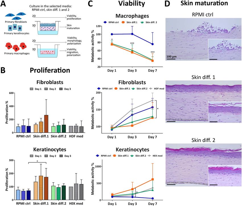
www.nature.com/scientificreports/
combinations affect tissue thickness, stability of dermo-epidermal junctions and number of epidermal l ayers13.
While the ideal conditions to obtain a mature epithelium differ depending on the system used for c ulture14,
diverse supplements have been shown to enhance differentiation. For instance, calcium concentrations above
1 mM promote the formation of tight junctions between keratinocytes and trigger d ifferentiation15,16. Serum
supplementation, conversely, contains factors that both promote and inhibit keratinocyte differentiation17,18,
which led to the use of low serum-containing11,19–21 or even serum-free m edia22,23 for in vitro skin culture. The
generation of in vitro skin models requires a complex formulation of nutrients and growth factors, but thus far
described immunocompetent skin equivalents used media suitable for skin m aturation8,9 and neglected the
potential effect of the medium on the employed immune cells. While the implementation of immune cells into
skin equivalents represents a step towards the generation of more relevant in vitro models, the pre-requisites for
the triple co-culture described by immunocompetent skin models have yet to be established.
We here evaluate the influence of culture medium on in vitro skin maturation as well as macrophages phe-
notype and functionality, to better understand the role of culture conditions on the future generation of a func-
tional immunocompetent in vitro skin model. For this, two skin differentiation media were selected, a serum-
free commercially available medium specifically designed for 3D air-lift culture of skin equivalents and a fully
defined medium described in literature19, which demonstrated excellent skin tissue maturation and the ability to
support a co-culture of skin equivalents and Langerhans cells. The effects of the selected media were compared
to a commonly used medium for standard in vitro macrophage c ulture24–29. Fibroblasts and keratinocytes were
evaluated for their ability to generate mature skin in the selected media, and their viability and proliferation in
single 2D cultures were analyzed. Macrophages were examined for cell viability, morphology and their response
to pro- and anti-inflammatory stimuli in 2D cultures. In addition, macrophages were embedded in collagen
hydrogels to study the effects of the media in an in vivo-like three-dimensional environment. Cell viability, abil-
ity to respond to pro-inflammatory stimuli and ability to migrate through the collagen gels were assessed. The
presented results show that skin differentiation media have inhibitory effects on the inflammatory response, and
at the same time reveal that in vitro skin models have a protective effect against inflammation.
Results and discussion
Skin differentiation media promote skin maturation but decrease macrophage viability. To
understand the influence of culture medium on the viability of single cell types, fibroblasts, keratinocytes and
macrophages were cultured in RPMI control, skin differentiation media 1 and 2 for up to 7 days and were
assessed for their metabolic activity as a measure of the number of viable cells. Additionally, proliferation of
fibroblasts and keratinocytes was assessed after 1, 2 and 3 days of culture (Fig. 1A).
Fibroblast proliferation rates were increased in presence of skin differentiation medium 1, but without any
statistical significance when compared to cells grown in fibroblast medium (Fig. 1B). Similarly, metabolic activity
showed increased values over time for all conditions, with a significant difference (p < 0.05) for skin differentia-
tion medium 1 at 7 days compared to cells grown in fibroblast medium (Fig. 1C). Proliferation of keratinocytes
showed increased values for skin differentiation medium 1, while cells in RPMI control medium had the lowest
values detected (Fig. 1B). Similarly, assessing metabolic activity showed increasing values over time in skin dif-
ferentiation media, with a significant (p < 0.05) difference for skin differentiation medium 1 at 3 days compared to
RPMI control medium (Fig. 1C). Notably, keratinocytes cultured in RPMI control medium displayed decreasing
values over time, indicating that the medium does not support keratinocyte survival. Macrophage metabolic
activity values showed a significant (p < 0.05 at day 1, p < 0.001 at days 3 and 7) decrease already after one day of
culture in either skin differentiation media, with similar values for both (Fig. 1C). In contrast, culture in RPMI
control medium resulted in decreased metabolic activity values only after one week of culture. The decreased
macrophage viability in skin differentiation media, as indicated by the low metabolic activity values, is likely
due to the absence or lower concentration of serum in skin differentiation media, as macrophages require serum
for survival30. No other supplement present in skin differentiation medium 2 has been associated with altered
macrophage viability. To enhance epithelial stratification, for instance, medium is supplemented with vitamin
C22,31–34 or amino acids such as L-carnitine and L-serine11,19,32,34. The mentioned supplements are involved in the
differentiation process of keratinocytes, but their effects on primary immune cells remain largely unexplored.
Similarly, the only known component of skin differentiation 1, hydrocortisone, has never been associated with
decreased macrophage viability. While the influence of serum or other components on macrophage survival is
unknown, the significant decrease in metabolic activity values suggests that skin differentiation media do not
support macrophage culture for extended periods of time.
To investigate the influence of medium on tissue maturation, skin models were cultured in RPMI control, skin
differentiation medium 1 or 2 and evaluated for epidermal stratification. Culture in RPMI control medium did
not promote the formation of a mature epidermis, as no stratified keratinocyte layers were detected (Fig. 1D).
Only few keratinocytes were visible on top of the dermal compartments, likely due to lack of cell attachment,
spreading and proliferation because of insufficient survival cues provided by the medium. Despite the high serum
content of RPMI control medium, no other supplement such as KGF or C aCl2 is present to promote the adhesion
of cells to the collagen gel and to sustain keratinocyte proliferation and differentiation (Tab. S1). Those results
reflect the effects observed for 2D culture in RPMI control medium, which showed a decrease in cell viability of
50% after 7 days and low proliferation rates, confirming that also in 3D this medium does not support keratino-
cyte culture. In contrast, the use of skin differentiation media resulted in epidermal stratification, as indicated
by the presence of several layers of keratinocytes and of a stratum corneum on the apical part of the epidermis
(Fig. 1D). Skin differentiation medium 1 is a serum-free medium specifically designed for the co-culture of
fibroblasts and keratinocytes, and skin differentiation medium 2 contains KGF, C aCl2 and other supplements.
Conversely, RPMI control medium does not contain any specific factor promoting the survival and proliferation
Scientific Reports | (2021) 11:7070 | https://doi.org/10.1038/s41598-021-86486-7 2
Vol:.(1234567890)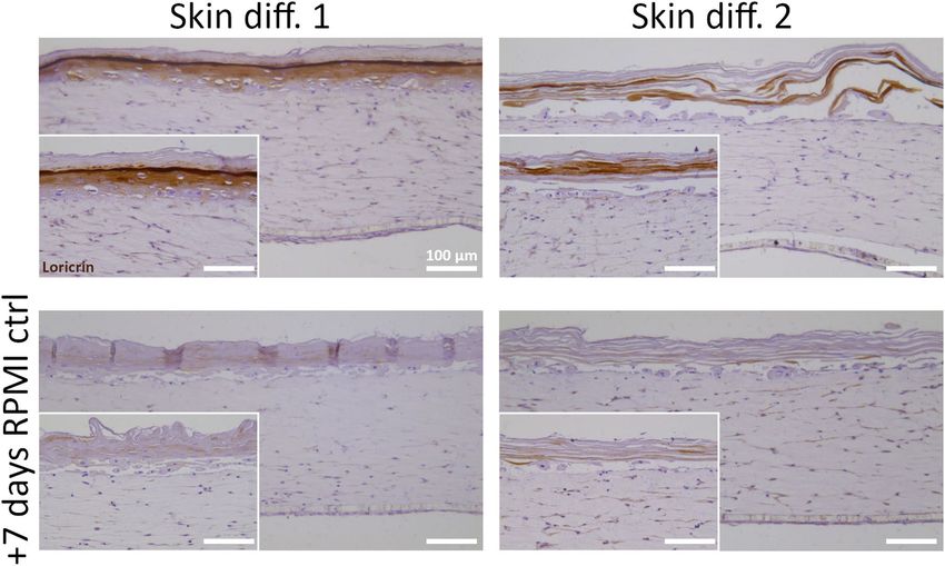
www.nature.com/scientificreports/
Figure 1. Influence of culture medium composition on skin cell viability and proliferation and in vitro skin
maturation. (A) Schematic of the experiments performed. (B) Proliferation and (C) viability of primary
fibroblasts, keratinocytes and macrophages cultured in RPMI control (“RPMI ctrl”), skin differentiation 1
(“Skin diff. 1”) or skin differentiation 2 (“Skin diff. 2”) media. Values are normalized to fibroblasts, keratinocytes
or macrophages cultured for 1 day in fibroblast (“HDF med”), keratinocyte (“HEK med”) or RPMI control
medium, respectively. N = 3, triplicate measurements for fibroblasts and keratinocytes; N = 5, triplicate
measurements for macrophages. Error bars represent standard deviation (two-way ANOVA, Tukey’s multiple
comparisons test). Statistical significance indicated with *p < 0.05, ***p < 0.001. (D) Representative histological
images of skin models cultured in the three media after 7 days of air-lift culture. N = 3, triplicate measurements.
Scale bars: 100 µm.
of keratinocytes, at the same time including a high serum percentage that interferes with differentiation. These
findings are in agreement with previous results from Black et al.33, who demonstrated that the presence of serum
during air-lift culture prevents the maturation of in vitro skin models.
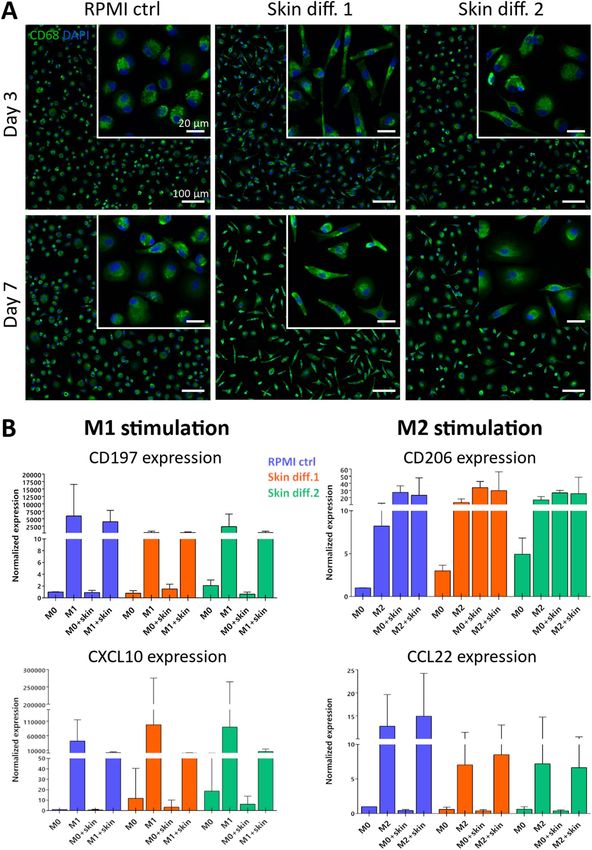
Skin differentiation media affect macrophage morphology and response to stimuli. To under-
stand whether skin media are suitable for macrophage culture, morphology was examined for 7 days and the
cells‘ ability to polarize towards M1-like and M2-like phenotypes after cytokine stimulation was investigated.
Already after 3 days, cell morphology was affected by culture in skin differentiation media. In RPMI control
medium, cells showed a round-shaped morphology, while in skin media they appeared elongated and spindle-
shaped, which was more evident in presence of skin differentiation medium 1 (Fig. 2A). After 7 days of culture,
the same tendency as well as qualitatively a brighter staining intensity of CD68 was observed in presence of skin
medium 1. An elongated cell shape and a brighter CD68 staining indicate opposite effects of the skin media on
cell polarization. In fact, an upregulated CD68 expression was reported during inflammation35,36, indicating
macrophage polarization to a pro-inflammatory or M1 phenotype. At the same time, macrophages with an
elongated morphology were correlated with an anti-inflammatory or M2 p henotype37.
Scientific Reports | (2021) 11:7070 | https://doi.org/10.1038/s41598-021-86486-7 3
Vol.:(0123456789)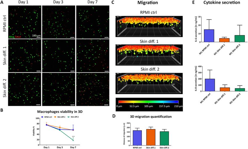
www.nature.com/scientificreports/

          Scientific Reports |   (2021) 11:7070 |   https://doi.org/10.1038/s41598-021-86486-7   4

Vol:.(1234567890)
www.nature.com/scientificreports/
◂Figure 2. Influence of culture medium on primary macrophage morphology and gene expression. (A)
Representative images of macrophages cultured for 3 and 7 days in RPMI control (“RPMI ctrl”), skin
differentiation 1 (“Skin diff. 1”) or skin differentiation 2 (“Skin diff. 2”) media upon staining with CD68 and
DAPI. N = 3, duplicate measurements. Scale bars: 100 µm for the lower magnification, 20 µm for the higher
magnification (insets). (B) Quantification of gene expression of primary macrophages either unstimulated
(“M0”) or polarized into M1- or M2-like phenotypes in the three media, in presence ("M1 + skin"; "M2 + skin")
or absence ("M1"; "M2") of skin as a co-culture. M1-like stimulated cells were analyzed for M1 markers CD197
and CXCL10, and M2-like stimulated cells for M2 markers CD206 and CCL22. N = 10, duplicate measurements
for macrophages cultured alone; N = 5, single measurement for macrophage-skin co-cultures. Error bars
represent standard deviation.
The results observed in macrophage morphology evaluation were further investigated by means of gene
expression analysis. Specific M1 and M2 markers were quantified after culture in the different media, both in
unstimulated cells (Fig. S1) and in response to M1 and M2 stimulation (Fig. S2). Macrophage gene expression
was measured both for monocultures and for macrophages in presence of 3D skin equivalents as a co-culture
(Fig. 2B). Both skin differentiation media showed to affect the expression of pro-inflammatory markers in mac-
rophages monocultures, both in unstimulated cells and upon stimulation with LPS and IFN-γ. Values of the
M1 marker CD197 were decreased in skin differentiation media 1 and 2, whereas CXCL10 showed an increased
expression. Gene expression of M2 markers CD206 and CCL22 showed the same trend in both skin differentia-
tion media, both for unstimulated macrophage monocultures and upon anti-inflammatory stimulation with
IL-4, with an increased CD206 expression and a decreased expression of CCL22 (Fig. 2B).
To exclude that the observed differences in gene expression resulted from the M-CSF supplementation during
the monocyte-to-macrophage differentiation, gene expression of cells generated with granulocyte–macrophage
colony-stimulating factor (GM-CSF) supplementation was also evaluated. Indeed, M-CSF has been shown to
steer cell fate towards M2-like cells, while GM-CSF was indicated to steer towards a M1-like p henotype27. The
results did not indicate that M1 and M2 expression was dependent on the monocyte-to-macrophage differentia-
tion conditions (Fig. S3), pointing at the composition of skin differentiation media to be the cause of such effects.
Isoproterenol and hydrocortisone, both contained in skin differentiation medium 2, are known to downregulate
the inflammatory response to LPS in m acrophages38,39. Skin differentiation medium 1 to our knowledge contains
hydrocortisone, which could explain the decreased inflammatory response. Other supplements present in skin
differentiation medium 2 have not been linked to altered immune cellular responses.
In vivo, monocytes and macrophages are under a continuous stimulation from cytokines, chemokines and
growth factors constitutively secreted by skin tissue to maintain homeostasis2,40. Mimicking this situation, mac-
rophages were co-cultured with 3D skin equivalents to investigate the influence of skin paracrine signaling on
the response of macrophages to stimuli. Macrophage morphology did indicate an altered response to stimuli,
with less rounded and spread cells in presence of 3D skin equivalents (Fig. S4). Gene expression quantification
confirmed that the presence of skin as a co-culture with macrophages influenced the expression of the analyzed
genes (Fig. 2B). M1 markers CD197 and CXCL10 were down-regulated, indicating a further inhibition of mac-
rophages ability to respond to pro-inflammatory stimuli in presence of skin equivalents. At the same time, the M2
marker CD206 showed an upregulation when skin was present as a co-culture, suggesting an enhanced response
to anti-inflammatory stimuli (Fig. 2B). The changes in gene expression levels upon polarization with pro- and
anti-inflammatory stimuli were statistically not significant for macrophages cultured alone and in co-culture with
3D skin equivalents (Fig. S2), however showed statistically significant differences in unstimulated cells (Fig. S1).
Due to the different gene expression upregulation or downregulation patterns within the M1 markers CD197
and CXCL10 and within the M2 markers CD206 and CCL22, we could not confirm the correlation between
cell shape and polarization state. Compared to other studies where the connection between cell morphology
and function was investigated37,41, the present study employed human primary blood monocytes-derived mac-
rophages. The use of primary immune cells implies a relevant donor-to-donor variation, dependent on factors
as the health state at the moment of isolation, which is reflected on the detected heterogeneous cellular response
to inflammation28. The results obtained from macrophage-skin co-cultures are in agreement with recent find-
ings by Limandjaja et al.42, who demonstrated that the co-culture of monocytes below in vitro skin equivalents
results in the differentiation of the immune cells towards an M2 phenotype. While the methods to investigate
macrophage phenotype differ from the present study, the medium composition used in Limandjaja et al.42 for
the co-culture is comparable to skin differentiation medium 2, as it was a 3:1 ratio combination of DMEM/Ham’s
F12 initially supplemented with serum, KGF, insulin, hydrocortisone and isoproterenol, and after 3 days further
supplemented with L-carnitine, L-serine, ascorbic acid, and other components. The observed anti-inflammatory
effect exhibited from skin equivalents further strengthens the correlation between in vitro skin and human skin,
where resident skin cells participate in immunosurveillance to protect the body from i nfection43.
Skin differentiation media influence viability, response to stimuli and migration of 3D‑embed‑
ded macrophages. In an immunocompetent skin model, macrophages would be cultured within a
3D environment. Therefore, cells were embedded in collagen gels to study the influence of a 3D environment on
viability, response to pro-inflammatory stimuli and ability to migrate in different media compositions.
Macrophage viability showed a small decrease over 7 days, with a similar trend for all media until day 3
(Fig. 3A,B). However, by day 7, a large number of dead cells could be observed in skin differentiation medium
2, with viability decreasing below 20%. Surprisingly, skin differentiation medium 2 showed to negatively influ-
ence macrophages survival to a greater extent in 3D compared to 2D cultures. The effects are plausible due to the
Scientific Reports | (2021) 11:7070 | https://doi.org/10.1038/s41598-021-86486-7 5
Vol.:(0123456789)www.nature.com/scientificreports/
Figure 3. Influence of culture medium composition on viability, response to pro-inflammatory stimuli and
migration ability of macrophages in a 3D environment. (A) Representative maximum intensity projections
of macrophages after culture in RPMI control (“RPMI ctrl”), skin differentiation 1 (“Skin diff. 1”) or skin
differentiation 2 (“Skin diff. 2”) media, upon live/dead staining. Scale bars: 100 µm. (B) Quantification of
macrophage viability. N = 3, n = 2. Error bars represent standard deviation. Statistical significance indicated
with ***p < 0.001. (C) Representative 3D reconstructed images of live/dead staining after vertical invasion assay,
color coded for invasion depth. (D) Quantification of macrophage maximal migration distance. N = 3, triplicate
measurements. Error bars represent standard deviation. (E) Quantification of pro-inflammatory cytokines IL-6
and IL-8 secretion in supernatants. N = 3, duplicate measurements. Error bars represent standard deviation.
presence of a 3D matrix around the cells that prevented dead cells to be removed from the area through media
change, increasing the dead cell fraction compared to the 2D setting.
The ability of macrophages to migrate through the tissue is a crucial requirement during the inflammatory
response, therefore we performed a vertical invasion assay. Macrophages showed the ability to migrate through
the dense collagen matrix in all conditions (Fig. 3C), with a maximal migration depth of 182 ± 28 µm, 157 ± 22 µm
and 168 ± 21 µm for skin differentiation medium 1, medium 2 and control, respectively (Fig. 3D). While no
statistically significant differences in migration depth could be observed, a different number of cells attached to
the surface of the gel prior migration. In fact, observation of cell distribution on the collagen gel surface showed
that a lower number of cells adhered in presence of skin differentiation medium 2, compared to gels cultured
in RPMI control and skin differentiation medium 1 (Fig. S5). The encapsulation of macrophages in collagen
gels did not affect their ability to respond to pro-inflammatory stimuli, as IL-6 and IL-8 could be detected in all
supernatants upon stimulation. However, lower amounts of both cytokines were detected in the supernatants
of gels cultured in the two skin differentiation media (Fig. 3E). Despite the lack of statistical significance, the
detected values followed the same trend as the gene expression levels of 2D-cultured cells with the respective
polarization stimuli (Fig. 2B), pointing towards an inhibition of the pro-inflammatory response in presence of
both skin differentiation media.
Macrophage medium has a detrimental effect on mature skin equivalents. As the use of skin
differentiation media affects macrophage viability and response to inflammatory stimuli, the feasibility of cultur-
ing immunocompetent skin in RPMI control medium was evaluated for the theoretical duration of a potential
inflammatory response. For this, fully differentiated skin equivalents grown in skin differentiation medium 1 or
2 were transferred into RPMI control medium and cultured for further 7 days, and potential changes to the skin
structure were evaluated by immunohistochemistry.
Culture in RPMI control medium was already shown to prevent in vitro skin maturation (Fig. 1D), but also
shifting to RPMI control medium after full skin epidermalization had a detrimental effect on skin structure. The
expression of the late differentiation marker loricrin was detected in skin equivalents differentiated for 14 days
in skin differentiation medium 1 or 2. However the shift to RPMI control medium showed no expression of
Scientific Reports | (2021) 11:7070 | https://doi.org/10.1038/s41598-021-86486-7 6
Vol:.(1234567890)www.nature.com/scientificreports/
Figure 4. Evaluation of macrophage medium effects on epidermal structure of skin models. In vitro skin
equivalents were generated by culture in skin differentiation medium 1 (“Skin diff. 1”) or skin differentiation
medium 2 (“Skin diff. 2”) for 14 days at air-lift culture (upper row), and then further cultured for 7 days in
RPMI control medium (“RPMI ctrl”) (lower row). Immunohistochemistry against loricrin was performed and
counterstaining with hematoxylin followed. N = 2, triplicate measurements. Scale bars: 100 µm.
the differentiation marker (Fig. 4). The negative effects on skin structure observed after RPMI control medium
culture are likely due to the drastic reduction of supplements promoting cell survival and differentiation, as
skin differentiation media contain elements promoting 3D skin culture while RPMI control medium is only
supplemented with serum. As skin differentiation medium 1 has a proprietary composition, only the nutrients
present in skin differentiation medium 2 and RPMI control medium could be compared (Tab. S1). Calcium
concentration is higher in skin differentiation medium 2 compared to RPMI control medium, and since it has
been linked with the promotion of epidermal s tratification15–17, its deprivation could be involved in degeneration
of the epidermal structure. The higher serum content of RPMI control medium may also influence epidermal
structure, as serum contains factors that inhibit keratinocyte d ifferentiation18,44. The percentage of serum in
RPMI control medium is 10%, compared to 1.25% in skin differentiation medium 2 and serum-free conditions
in skin differentiation medium 1. Differently than RPMI control, whose basal medium is supplemented only
with serum and antibiotics, skin differentiation medium 2 is supplemented with different compounds to sup-
port tissue viability in culture. Among these, KGF has a crucial role in maintaining keratinocytes proliferation
and thus d ifferentiation45, therefore its deprivation could have negatively affected basal keratinocytes turnover.
Overall, a combination of all those factors is the probable cause of the negative effects observed on differentiation
after medium change to RPMI control.
The generation of advanced immunocompetent skin models better mimicking the human physiology is a
pivotal step in the replacement of animal use in research. The central role of macrophages during inflammation
has advanced their inclusion into in vitro skin equivalents, however few models have been described8,9 and
several aspects for the generation of functional immunocompetent skin remain u nresolved3. The optimization
of a co-culture medium is a crucial step towards the development of any functional model, as each cell type
requires specific nutrients to remain functional. The investigation of two culture media designed for in vitro skin
maturation on primary macrophages showed that their inflammatory response is affected by this combination
of nutrients. Macrophages showed reduced viability, altered cell morphology and altered response to pro- and
anti-inflammatory stimuli both in a 2D and 3D environment, indicating that the identification of a culture
medium supporting both skin maturation and macrophage functionality is a crucial step towards the generation
of immunocompetent skin.
Further investigation is needed to determine which components affect immune cells and skin maturation, to
select the key elements needed for maintenance of immunocompetent skin models. For this however, not only
gene expression data, but also protein expression should be assessed, as the abundance of proteins can be affected
by translational and post-translational regulation. In parallel to the identification of these crucial components,
the substitution of animal-derived products currently in use for the fabrication of in vitro skin equivalents rep-
resents another step towards the total replacement of animals in research. While the current standard in vitro
3D cell culture setup is mainly represented by the use of animal-derived products such as serum and collagen,
research on alternatives that do not require animal involvement is ongoing.
Scientific Reports | (2021) 11:7070 | https://doi.org/10.1038/s41598-021-86486-7 7
Vol.:(0123456789)www.nature.com/scientificreports/
Methods
Cell isolation and culture. Primary human dermal fibroblasts pooled from several donors and juvenile
keratinocytes isolated from a single donor were purchased from CELLnTEC (CELLnTEC Advanced Cell Sys-
tems AG, Bern, Switzerland). Fibroblasts were cultured in DMEM supplemented with 10% fetal bovine serum
(FBS), 1% L-glutamine and 1% penicillin–streptomycin-neomycin (PSN), also referred to as “HDF medium”.
As recommended by the supplier, fibroblasts were used until passage 8 with a seeding density of 1000 cells/cm2,
passaging the cells every 6–7 days. Keratinocytes were maintained in CnT-prime epithelial culture medium
(CELLnTEC Advanced Cell Systems AG), referred to as “HEK medium” and used until passage 6. Passaging was
performed every 5 days, seeding the cells at a density of 4000 cells/cm2 as recommended by the supplier. Pri-
mary monocytes were isolated from peripheral blood of healthy blood donors who provided informed consent.
For each experiment, a different donor was recruited. Approval from the local committee of swissethics (Swiss
Association of Research Ethics Committees, BASEC Nr PB_2016_00816) in accordance to relevant guidelines
and regulations was obtained. Freshly drawn heparinized blood was first was first diluted 1:1 in a solution con-
taining 2% FBS + 1 mM EDTA in PBS and then separated into the different blood components by Lymphoprep
(STEMCELL Technologies, Cologne, Germany) density gradient centrifugation. The white blood cell fraction,
visible as a white ring between the plasma components and the Lymphoprep, was carefully collected and washed
twice with PBS to avoid carryover of solvents into the following steps. To purify and enrich the monocytic
population, specific negative selection was performed using the Monocyte Isolation Kit II (Miltenyi Biotec, Ber-
gisch Gladbach, Germany) according to the manufacturer’s instructions. Purified monocytes were seeded at a
density of 62,000 cells/cm2 in monocyte differentiation medium, consisting of Roswell Park Memorial Institute
medium (RPMI 1640) (Sigma-Aldrich) supplemented with 10% FCS, 1% PSN and 20 ng/mL of macrophage
colony-stimulating factor (M-CSF) (Thermo Fisher Scientific, Schwerte, Germany). After 6 days, differentiated
macrophages were detached by incubation in TrypLE Express (Thermo Fisher Scientific) for 20 min at 37 °C,
followed by gentle scraping with a cell scraper. Collected cells were resuspended in fresh macrophage medium
(RPMI 1640 supplemented with 10% FCS and 1% PSN, also referred to as "RPMI control medium"), counted
and used for subsequent experiments.
Cell viability and proliferation. Cell viability was evaluated with an MTS assay (Promega, Madison,
USA) and cell proliferation was analyzed using the BrdU-based Cell Proliferation ELISA kit (Roche, Basel, Swit-
zerland), according to the manufacturer’s instructions. Three independent experiments with triplicate measure-
ments were performed for fibroblasts and keratinocytes, 5 independent experiments with triplicate measure-
ments for macrophages per condition.
2D macrophage analysis. Macrophage polarization was obtained by supplementing the medium with
100 ng/mL LPS and 20 ng/mL IFN-γ (Miltenyi Biotec) for M1-like cells and with 20 ng/mL IL-4 (Miltenyi
Biotec) for M2-like cells. For this, macrophages were seeded at a concentration of 95,000 cells/cm2 in 24-wells
plates for 24 h. CD68 (Cluster of Differentiation 68) staining was performed by incubating fixed macrophages
with a 1:100 dilution of the mouse monoclonal antibody (ab955 from Abcam, Cambridge, United Kingdom)
in a solution of 0.1% bovine serum albumin (BSA) in PBS at 4 °C overnight. On the following day, the second-
ary anti-body Alexa Fluor 488 conjugate (Invitrogen, Carlsbad, USA) was incubated for 1 h as a 1:500 dilution.
4′,6-diamidino-2-phenylindole (DAPI) was used to counterstain cell nuclei, supplemented as a 1:1000 solution
in 0.1% BSA. All supplements were purchased from Sigma-Aldrich unless stated otherwise. CD68 stainings
included 3 independent experiments with a minimum of 2 technical replicates each.
Gene expression. To analyze gene expression of macrophages after polarization, RT-PCR was performed
for M1- and M2-specific genes. RNA isolation was performed using the RNeasy Micro Kit (Qiagen, Hilden, Ger-
many) according to the manufacturer’s instructions. RNA concentration and purity were then measured with a
NanoDrop ND-1000 spectrophotometer (Thermo Fisher Scientific). The total RNA isolated from macrophage
cultures was then reverse-transcribed using iScript cDNA synthesis Kit (Bio-Rad, Hercules, USA), using 150 ng
of RNA per sample and a volume of 15 μL per reaction mix. Quantification of gene expression was performed
with the iQ SYBR Green Supermix (Bio-Rad), using Glyceraldehyde 3-phosphate dehydrogenase (GAPDH) as
a reference gene, and analyzing the markers CD197, CXCL10, CD206 and CCL22 (Table 1). Each primer was
added at a concentration of 10 μM, adjusting the final reaction volume to 15 μL with RNase-free water. The ther-
mal cycler was set to 95 °C for 3 min, followed by 39 cycles of 10 s at 95 °C and 30 s at 57 °C. The final steps were
performed at 95 °C for 10 s, at 65 °C for 5 s, and then at 95 °C. Calculations for the quantification were performed
using the 2−ΔΔCT method, normalizing all values to unstimulated samples cultured in macrophage medium. Gene
expression analysis was performed on 5 independent experiment, each with a minimum of 2 technical replicates.
3D macrophage viability and polarization. Macrophage viability and polarization in a 3D environ-
ment were evaluated after seeding 75,000 cells per collagen gel, following the same mixing procedure described
above with the difference that the cell suspension was added to the collagen mixture prior to polymerization,
to embed the cells within the gel. 300 µL of macrophage-containing solution were placed into each Greiner
ThinCert insert for 12-well plates with transparent membrane (3 µm pore size, Greiner) and let polymerize.
Polarization was induced by supplementing the culture medium with cytokines, using the same concentrations
described in “2D macrophage analysis”. For viability assessment, the gels were cultured for 7 days with a medium
change every 2–3 days, carefully avoiding to touch the surface of the gel. Cells were visualized after a live/dead
assay (Sigma-Aldrich) staining, according the manufacturer’s instructions. Cytokine quantification was per-
formed by collecting 1 mL of supernatant after 24 h of polarization and using human IL-6 and IL-8 ELISA kits
Scientific Reports | (2021) 11:7070 | https://doi.org/10.1038/s41598-021-86486-7 8
Vol:.(1234567890)www.nature.com/scientificreports/
Gene Primer name Sequence 5′ → 3′
GAPDH-forward AGTCAGCCGCATCTTCTTTT
Glyceraldehyde-3-phosphate dehydrogenase, GAPDH
GAPDH-reverse CCAATACGACCAAATCCGTTG
CD197-forward GTGGTTTTACCGCCCAGAGA
C–C chemokine receptor type 7, CCR7 (CD197)
CD197-reverse CACTGTGGTGTTGTCTCCGA
CXCL10-forward CAGTCTCAGCACCATGAATCAA
C-X-C motif chemokine 10, CXCL10
CXCL10-reverse CAGTTCTAGAGAGAGGTACTCCTTG
CCL22-forward GCGTGGTGTTGCTAACCTTC
C–C motif chemokine 22, CCL22
CCL22-reverse CCACGGTCATCAGAGTAGGC
CD206-forward GCTACCCCTGCTCCTGGTTT
Mannose receptor C type 1, MRC1 (CD206)
CD206-reverse CGCAGCGCTTGTGATCTTCA
Table 1. Sequence of the primers used for quantification of macrophage gene expression.
(Thermo Fisher Scientific), following the manufacturer’s instructions. All the 3D experiments were performed
on 3 independent experiments, each with a minimum of 2 technical replicates.
For the conditioned media experiments, first macrophages were seeded in 12-wells plates, following the same
procedure as for the 2D macrophage seeding. Then, a ThinCert insert containing a skin equivalent was placed
in each well to allow paracrine signaling between skin tissue and immune cells. After 24 h of skin-macrophages
co-culture, the ThinCert inserts were removed and macrophage gene expression was quantified. Three independ-
ent experiments were performed.
Macrophage viability quantification was performed using ImageJ software, manually counting live and dead
cells. A total of 5 images per condition were analyzed at each time point, with an average cell count of 90 cells
per sample. Macrophage migration quantification was performed using ImageJ software, and a total of 21 images
per condition were analyzed.
3D macrophage migration. For the vertical migration assay, ThinCert inserts for 24-well plates with
transparent membrane (3 µm pore size, Greiner, Kremsmünster, Austria) were used. FibriCol bovine collagen
type I (10 mg/mL, Advanced BioMatrix, San Diego, USA) was mixed with 10 × DMEM, neutralized with 0.5 M
sterile filtered NaOH, and supplemented with sterile filtered water to obtain a ratio of collagen:10xDMEM:other
components (water + NaOH) of 8:1:1. 150 µL per insert were added and after polymerization, macrophages were
seeded on top of the gels at a concentration of 22,000 cells/cm2 and cultured for 48 h in the different media.
For visualization of 3D-embedded macrophages, the collagen gels containing the immune cells were stained
with a live/dead assay (Sigma-Aldrich), according the manufacturer’s instructions. All images were acquired
with a LSM 780 confocal laser scanning microscope (Carl Zeiss, Oberkochen, Germany), using the ZEN 2012
software (Carl Zeiss). For analysis of cell viability in 3D, each displayed image is the combination of Z-stacks
obtained from a total sample thickness of 192 μm, obtained through the maximum intensity projection tool
provided by the software. The vertical migration pictures were obtained by elaborating the captured images with
the 3D reconstruction tool provided in the ZEN 2012 software. For all samples, ethidium homodimer-1 signal
was adjusted to a negative control for each time point, incubating a sample with a solution of 0.1% digitonin.
Generation of skin model. The dermis was generated by embedding fibroblasts in a collagen gel, as
described in “3D macrophage migration, viability and polarization”, seeding 75,000 cells per sample. After
polymerization, the constructs were cultured in submerged conditions in fibroblast medium for 7 days, chang-
ing the medium every 2–3 days and carefully avoiding to touch the surface of the gel. On day 7, medium was
removed and the surface of each gel was coated with 50 µL of a 50 µg/mL fibronectin solution in PBS (Sigma-
Aldrich) for 30 min at 37 °C. Subconfluent keratinocytes were harvested and seeded on top of the fibronectin-
coated gels at a concentration of 225,000 cells/cm2 in keratinocyte seeding medium (DMEM/Ham’s F-12 in a 3:1
ratio, supplemented with 5% FCS, 1% PSN, 2 ng/mL keratinocyte growth factor (KGF), 1 µM hydrocortisone,
1 µM isoproterenol and 0.1 µM insulin) or in CnT-Prime Airlift medium (CELLnTEC Advanced Cell Systems
AG). After 5 days of culture with a medium change at day 1 and day 3, the samples were raised to the air–liquid
interface by transferring the gel-containing insert in deep-well 12-wells plates (Greiner), where 4 mL of differen-
tiation medium were added below each insert and changed every 4 days for 2–3 weeks. The two differentiation
media used were CnT-Prime Airlift medium, referred to as "Skin differentiation medium 1", or a mixture of
DMEM/Ham’s F-12 in a 3:1 ratio, supplemented with 1.25% FCS, 1% PSN, 2 ng/mL KGF, 1 µM hydrocortisone,
1 µM isoproterenol, 0.1 µM insulin, 0.1 µM L-carnitine, 0.01 M L-serine and 50 µg/mL of ascorbic acid, referred
to as "Skin differentiation medium 2". All supplements were purchased from Sigma-Aldrich unless stated oth-
erwise. Each experiment was performed a minimum of 3 times, analyzing 3 technical replicates per condition.
Histological analysis. Skin samples were fixed overnight in a solution of 4% buffered formalin, then dehy-
drated by incubation in increasing ethanol concentrations, washed in xylene and embedded in paraffin blocks.
Afterwards, 5 µm thick sections were cut, and deparaffinization in xylene and rehydration in decreasing ethanol
concentrations followed. Rehydrated samples were then stained with hematoxylin (HistoLab, Askim, Sweden)
Scientific Reports | (2021) 11:7070 | https://doi.org/10.1038/s41598-021-86486-7 9
Vol.:(0123456789)www.nature.com/scientificreports/
and eosin, rinsing the tissue in distilled water between the two steps. For immunohistochemistry, rehydrated
slides were first incubated with a 1:500 diluted rabbit monoclonal loricrin antibody (ab176322 from Abcam)
in 5% goat serum in PBS overnight at 4 °C. Then, a 1:100 solution of secondary POD-conjugated antibody was
incubated for 1 h at room temperature. Loricrin staining development was performed with 3,3′- Diaminoben-
zidine (Dako, Santa Clara, USA) and counterstaining was performed by incubation in hematoxylin for 1 min.
All supplements were purchased from Sigma-Aldrich unless stated otherwise. Each experiment was performed
a minimum of 2 times, analyzing 3 technical replicates per condition.
Statistical analysis. The GraphPad Prism software (GraphPad, San Diego, USA) was used to analyze all
data. Data obtained from 3D-embedded macrophages polarization and migration were analyzed using Kruskal–
Wallis test, with a Dunn post-hoc test for multiple comparisons assuming a non-parametric distribution. For all
other data, two-way ANOVA with a Tukey post-hoc test for multiple comparisons was used. Statistical signifi-
cance was assumed at p < 0.05.
Received: 14 October 2020; Accepted: 16 March 2021
References
1. Bell, E., Ehrlich, P. H., Buttle, D. J. & Nakatsuji, T. Living tissue formed in vitro and accepted as skin-equivalent tissue of full thick-
ness. Science 211, 1052–1054 (1981).
2. Pasparakis, M., Haase, I. & Nestle, F. O. Mechanisms regulating skin immunity and inflammation. Nat. Rev. Immunol. 14(5),
289–301 (2014).
3. Pupovac, A. et al. Toward immunocompetent 3D skin models. Adv. Healthc. Mater. 7, 12. https://doi.org/10.1002/adhm.20170
1405 (2018).
4. Groeber, F., Holeiter, M., Hampel, M., Hinderer, S. & Schenke-Layland, K. Skin tissue engineering: in vivo and in vitro applications.
Adv. Drug Deliv. Rev. 63(4–5), 352–366 (2011).
5. Wynn, T. A. & Vannella, K. M. Macrophages in tissue repair, regeneration, and fibrosis. Immunity 44(3), 450–462 (2016).
6. Shapouri-Moghaddam, A. et al. Macrophage plasticity, polarization, and function in health and disease. J. Cell. Physiol. 233(9),
6425–6440 (2018).
7. Murray, P. J. Macrophage polarization. Annu. Rev. Physiol. 79, 541–566 (2017).
8. Linde, N., Gutschalk, C. M., Hoffmann, C., Yilmaz, D. & Mueller, M. M. Integrating macrophages into organotypic co-cultures: a
3D in vitro model to study tumor-associated macrophages. PLoS ONE 7, 7. https://doi.org/10.1371/journal.pone.0040058 (2012).
9. Bechetoille, N. et al. A new organotypic model containing dermal-type macrophages. Exp. Dermatol. 20(12), 1011–1037 (2011).
10. Boehnke, K. et al. Effects of fibroblasts and microenvironment on epidermal regeneration and tissue function in long-term skin
equivalents. Eur. J. Cell. Biol. 86(11–12), 731–746 (2007).
11. Spiekstra, S. W., Breetveld, M., Rustemeyer, T., Scheper, R. J. & Gibbs, S. Wound-healing factors secreted by epidermal keratinocytes
and dermal fibroblasts in skin substitutes. Wound Repair Regen. 15(5), 708–717 (2007).
12. Werner, S., Krieg, T. & Smola, H. Keratinocyte-fibroblast interactions in wound healing. J. Invest. Dermatol. 127(5), 998–1008
(2007).
13. Lange, J. et al. Interactions of donor sources and media influence the histo-morphological quality of full-thickness skin models.
Biotechnol. J. 11(10), 1352–1361 (2016).
14. Borowiec, A. S., Delcourt, P., Dewailly, E. & Bidaux, G. Optimal differentiation of in vitro keratinocytes requires multifactorial
external control. PLoS ONE 8, 10. https://doi.org/10.1371/journal.pone.0077507 (2013).
15. Hennings, H. et al. Calcium regulation of growth and differentiation of mouse epidermal cells in culture. Cell 19, 245–254 (1980).
16. Boyce, S. T. & Ham, R. G. Calcium-regulated differentiation of normal human epidermal keratinocytes in chemically defined
clonal culture and serum-free serial culture. J. Invest. Dermatol. 81(1), 33–40 (1983).
17. Berghard, A., Gradin, K. & Toftgard, R. Serum and extracellular calcium modulate induction of cytochrome P-450IA1 in human
keratinocytes. J. Biol. Chem. 265(34), 21086–21090 (1990).
18. Bertolero, F., Kaighn, M. E., Camalier, R. F. & Saffiotti, U. Effects of serum and serum-derived factors on growth and differentiation
of mouse keratinocytes. Vitro Cell. Dev. Biol. 22(7), 423–428 (1986).
19. Ouwehand, K. et al. Technical advance: Langerhans cells derived from a human cell line in a full-thickness skin equivalent undergo
allergen-induced maturation and migration. J. Leukoc. Biol. 90(5), 1027–1033 (2011).
20. Maione, A. G. et al. Three-dimensional human tissue models that incorporate diabetic foot ulcer-derived fibroblasts mimic in vivo
features of chronic wounds. Tissue Eng. Part C Methods 21(5), 499–508 (2015).
21. Chung, E., Choi, H., Lim, J. E. & Son, Y. Development of skin inflammation test model by co-culture of reconstituted 3D skin and
RAW264.7 cells. J. Tissue Eng. Regen. Med. 11(1), 87–92 (2014).
22. Jannasch, M. et al. Development and application of three-dimensional skin equivalents for the investigation of percutaneous worm
invasion. Exp. Parasitol. 150, 22–30 (2015).
23. Kuhbacher, A. et al. Central role for dermal fibroblasts in skin model protection against Candida albicans. J. Infect. Dis. 215(11),
1742–1752 (2017).
24. Verreck, F. A. et al. Human IL-23-producing type 1 macrophages promote but IL-10-producing type 2 macrophages subvert
immunity to (myco)bacteria. Proc. Natl. Acad. Sci. USA 101(13), 4560–4565 (2004).
25. Lacey, D. C. et al. Defining GM-CSF- and macrophage-CSF-dependent macrophage responses by in vitro models. J. Immunol.
188(11), 5752–5765 (2012).
26. Jin, X. & Kruth, H. S. Culture of macrophage colony-stimulating factor differentiated human monocyte-derived macrophages. J.
Vis. Exp. 112, 54244. https://doi.org/10.3791/54244 (2016).
27. Fleetwood, A. J., Dinh, H., Cook, A. D., Hertzog, P. J. & Hamilton, J. A. GM-CSF- and M-CSF-dependent macrophage phenotypes
display differential dependence on type I interferon signaling. J. Leukoc. Biol. 86(2), 411–421 (2009).
28. Garelnabi, M. et al. Quantifying donor-to-donor variation in macrophage responses to the human fungal pathogen Cryptococcus
neoformans. PLoS ONE 13, 3. https://doi.org/10.1371/journal.pone.0194615 (2018).
29. Martinez, F. O., Gordon, S., Locati, M. & Mantovani, A. Transcriptional profiling of the human monocyte-to-macrophage dif-
ferentiation and polarization: new molecules and patterns of gene expression. J. Immunol. 177(10), 7303–7311 (2006).
30. Kreutz, M., Krause, S. W., Hennemann, B., Rehm, A. & Andreesen, R. Macrophage heterogeneity and differentiation: defined
serum-free culture conditions induce different types of macrophages in vitro. Res. Immunol. 143, 107–115 (1992).
Scientific Reports | (2021) 11:7070 | https://doi.org/10.1038/s41598-021-86486-7 10
Vol:.(1234567890)www.nature.com/scientificreports/
31. Ponec, M. et al. The formation of competent barrier lipids in reconstructed human epidermis requires the presence of vitamin C.
J. Invest. Dermatol. 109(3), 348–355 (1997).
32. Geer, D. J., Swartz, D. D. & Andreadis, S. T. Fibrin promotes migration in a 3D in vitro model of wound regeneration. Tissue Eng.
8(5), 787–798 (2002).
33. Black, A. F. et al. Optimization and characterization of an engineered human skin equivalent. Tissue Eng. 11(5/6), 723–733 (2005).
34. Kosten, I. J., Buskermolen, J. K., Spiekstra, S. W., de Gruijl, T. D. & Gibbs, S. Gingiva equivalents secrete negligible amounts of key
chemokines involved in Langerhans cell migration compared to skin equivalents. J. Immunol. Res. https://doi.org/10.1155/2015/
627125 (2015).
35. O’Reilly, D., Quinn, C. M., El-Shanawany, T., Gordon, S. & Greaves, D. R. Multiple Ets factors and interferon regulatory factor-4
modulate CD68 expression in a cell type-specific manner. J. Biol. Chem. 278(24), 21909–21919 (2003).
36. O’Reilly, D. & Greaves, D. R. Cell-type-specific expression of the human CD68 gene is associated with changes in Pol II phospho-
rylation and short-range intrachromosomal gene looping. Genomics 90(3), 407–415 (2007).
37. McWhorter, F. Y., Wang, T., Nguyen, P., Chung, T. & Liu, W. F. Modulation of macrophage phenotype by cell shape. PNAS 110(43),
17253–17258 (2013).
38. Dong, J. et al. Cortisol modulates inflammatory responses in LPS-stimulated RAW264.7 cells via the NF-kappaB and MAPK
pathways. BMC Vet. Res. 14, 1. https://doi.org/10.1186/s12917-018-1360-0 (2018).
39. Hasko, G. et al. Isoproterenol inhibits IL-10, TNF-α, and nitric oxide production in RAW 264.7 macrophages. Brain Res. Bull.
45(2), 183–187 (1998).
40. Yanez, D. A., Lacher, R. K., Vidyarthi, A. & Colegio, O. R. The role of macrophages in skin homeostasis. Pflugers Arch. Eur. J. Physiol.
469(3–4), 455–463 (2017).
41. Lee, H. S. et al. Correlating macrophage morphology and cytokine production resulting from biomaterial contact. J. Biomed. Mater.
Res. A 101(1), 203–212 (2013).
42. Limandjaja, G. C., Waaijman, T., Roffel, S., Niessen, F. B. & Gibbs, S. Monocytes co-cultured with reconstructed keloid and normal
skin models skew towards M2 macrophage phenotype. Arch. Dermatol. Res. 311, 615–627 (2019).
43. Lai, Y. et al. Activation of TLR2 by a small molecule produced by Staphylococcus epidermidis increases antimicrobial defense
against bacterial skin infections. J. Invest. Dermatol. 130(9), 2211–2221 (2010).
44. Pillai, S., Bilke, D. D., Hincenbergs, M. & Elias, P. M. Biochemical and morphological characterization of growth and differentiation
of normal human neonatal keratinocytes in a serum-free medium. J. Cell. Physiol. 134(2), 229–237 (1988).
45. Rubin, J. S. et al. Keratinocytes growth factor. Cell Biol. Int. 19(5), 399–411 (1995).
Acknowledgements
The authors wish to thank Yvonne Elbs-Glatz for the help with the recruitment of donors and blood sampling,
as well as for the technical support in establishing the primary macrophage-related techniques. We also thank
all blood donors for their participation in the study.
Author contributions
Conceptualization: C.G., K.M.W., M.R.; Formal Analysis: C.G.; Investigation: C.G., K.Y.; Methodology: C.G.,
B.N.; Supervision: F.G.B., K.M.W., T.D., H.W., M.R.; Validation & Visualization: C.G., M.R.; Writing—Original
Draft Preparation: C.G., M.R.; Writing—Review and Editing: C.G., B.N., K.Y., F.G.B., K.M.W., T.D., H.W., M.R.
Funding
TD acknowledges support by DFG (GRK 2157; Project No. 270563345).
Competing interests
The authors declare no competing interests.
Additional information
Supplementary Information The online version contains supplementary material available at https://doi.org/
10.1038/s41598-021-86486-7.
Correspondence and requests for materials should be addressed to M.R.
Reprints and permissions information is available at www.nature.com/reprints.
Publisher’s note Springer Nature remains neutral with regard to jurisdictional claims in published maps and
institutional affiliations.
Open Access This article is licensed under a Creative Commons Attribution 4.0 International
License, which permits use, sharing, adaptation, distribution and reproduction in any medium or
format, as long as you give appropriate credit to the original author(s) and the source, provide a link to the
Creative Commons licence, and indicate if changes were made. The images or other third party material in this
article are included in the article’s Creative Commons licence, unless indicated otherwise in a credit line to the
material. If material is not included in the article’s Creative Commons licence and your intended use is not
permitted by statutory regulation or exceeds the permitted use, you will need to obtain permission directly from
the copyright holder. To view a copy of this licence, visit http://creativecommons.org/licenses/by/4.0/.
© The Author(s) 2021
Scientific Reports | (2021) 11:7070 | https://doi.org/10.1038/s41598-021-86486-7 11
Vol.:(0123456789)You can also read