THE EFFECT OF HYPERBARIC OXYGEN THERAPY ON ACUTE WOUND HEALING IN RABBITS: AN EXPERIMENTAL STUDY AND HISTOPATHOLOGICAL ANALYSIS
←
→
Page content transcription
If your browser does not render page correctly, please read the page content below
Mil. Med. Sci. Lett. (Voj. Zdrav. Listy) 2021, 90(1), 2-11
                                                                                                 ISSN 0372-7025 (Print)
                                                                                              ISSN 2571-113X (Online)
                                                                                         DOI: 10.31482/mmsl.2021.001
               Since 1925

ORIGINAL ARTICLE

THE EFFECT OF HYPERBARIC OXYGEN THERAPY ON ACUTE
WOUND HEALING IN RABBITS: AN EXPERIMENTAL STUDY
AND HISTOPATHOLOGICAL ANALYSIS
Jakub Tlapák 1,2 , Petr Chmátal 1, Jaroslav Pejchal 2, Boris Oniščenko 1,3, Věra Radochová 2, Petr Došel 1,
Jiří Páral 2, Petr Lochman 2

1
  The Institute of Aviation Medicine, Prague, Czech Republic
2
  Faculty of Military Health Sciences, University of Defence, Hradec Kralove, Czech Republic
3
  Third Faculty of Medicine, Charles University in Prague, Prague, Czech Republic

Received 23rd September 2020.
Accepted 4th January 2021.
Published 5th March 2021.

    Summary

        Background: The goal of our research is to show the effects and impacts of hyperbaric oxygen therapy
    (HBOT) on acute model wounds in animal subjects.
        Methods: Three experimental groups were created using injured rabbits (N=36)—randomly divided
    into three groups (N=12 per group). One group was treated only with standard wound care management.
    Two groups were additionally treated with HBOT either once or twice a day. The wounds were surgical,
    uninfected, and in healthy animal test subjects. We compared the immunohistochemical and histological
    parameters in 4-, 7- and 10-day intervals.
        Results: The detection of epidermal leaf parameters, the number of microabscesses, the Histopathological
    Superficial Epithelium Healing Score, Connective Tissue Healing Score, Histopathological Acute
    Inflammation Score and Total Histopathological Wound Healing Score all showed significant changes
    between time intervals within the individual groups.
        Conclusion: The results did not show that HBOT had a significant effect on the healing process
    of uncomplicated acute wounds.

    Key words: Hyperbaric oxygen; Wound healing; Animal models; Adjunctive treatment

Introduction

    Hyperbaric oxygen therapy (HBOT) uses a combination of inhalation of 100% oxygen and pressure acting
on the body, exceeding 1 atmosphere absolute (ATA). Both mechanisms increase the level of oxygen in the tissue,
with a many times increase in the content of dissolved oxygen in the blood plasma and full hemoglobin saturation.
The combination of these many effects of HBOT contribute to a favorable course of wound healing. Recent human
and animal model works have shown that HBOT influences the healing process—It positively affects angiogenesis (1),

       Ústav leteckého zdravotnictví Praha, Generála Píky 1, 160 00 Praha 6 – Dejvice, Česká republika
       tlapak@ulz.cz
       +420 973 208 117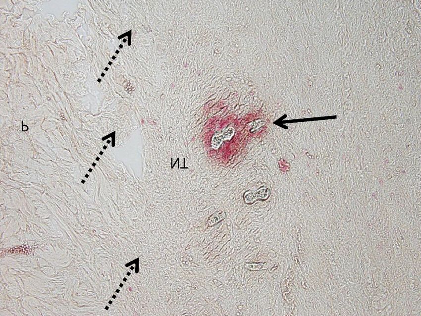
Tlapák et al.: Hyperbaric oxygen therapy and acute wounds - an experimental study

fibroblast proliferation, collagen synthesis (2), epithelialization (3), wound edema reduction (4), the redistribution
of flow to the hypoxic area, the correction of tissue hypoxia (5) and also has an antibacterial effect (6).

    Methods and procedures accelerating the healing process are, in the long term, in the interest of all teams dealing
with traumatology and health care management, especially in terms of economizing treatment costs. The studies
included in the latest systematic review by Smet, et al. (7) show a positive outcome on wound healing when using
HBOT. The economic impact of the use of HBOT in this indication is shown by the results of a systematic review
by Santema, et al. (8). In this systematic review is demonstrated little direct evidence of the cost-effectiveness for HBOT
despite its wide application. Review articles indicate a lack of available strong evidence for both the economic outcome
and effect of HBOT on acute wounds. The application of hyperbaric oxygenation as an adjuvant therapy appears to be
beneficial not only in the civilian field, but also in the military, regarding complex battle wounds (9).

    This randomized controlled experimental study was conducted to evaluate the effectiveness of HBOT
in the treatment of acute open uncomplicated wounds on an animal model using histological and immuno-
histochemical analyses.

Methods and Materials

     This experimental study of histopathological changes in animal wound models treated with hyperbaric oxygen
was analyzed using histological and immunohistochemical techniques. Thirty-six adult female New Zealand White
rabbits (initial weight between 2.4 – 2.9 kg) were randomly divided into three parallel-groups to compare the use
of standard wound management (wound and incision sites were cleansed, desinfected and used sterile dressings)
and HBOT in two different regimens. Four wounds were evaluated. Sampling was performed at 4- (right distal wound),
7- (left distal wound) and 10-day (right proximal wound) time intervals. The left proximal wound (the fourth wound)
was left without a biopsy to compare the average healing time between the groups. Surgery day was defined as day 0
(wound induction). The experiment was terminated after the complete healing of the left proximal wound. The study
was conducted at the Institute of Aviation Medicine, Prague.

Drugs

    The introduction of the subjects to general anesthesia was through a combination of an intramuscular application
of ketamine (50 mg/kg) and xylazine (3 mg/kg), and supplemented with 100% oxygen (flow rate 3 L/minute)
via a face mask. General anesthesia was administered via an inhalation anesthetic with isoflurane. This drug application
was used for both the wound procedure and biopsy.

Animal and tissue preparation

    Four wounds, measuring 2 x 2 cm in the area of the dorsal muscles that penetrated the level of the muscular
fascia, were created. The wound position was chosen to achieve as much tissue tension as possible. The proximal
wound was 6 cm from the blade-bone on both sides of the spine and 1.5 cm from the midline of the spine; distal wounds
were located 11 cm distally from the blade-bone on both sides of the spine, at the same distance from the median
line as the proximal wound. In order to maintain the greatest possible unification of the wounds, the wounds were
formed according to a template with an internal dimension of 2 x 2 cm of sterilizable material. Sampling was again
performed by a unified method using a template with internal dimensions of 3.5 x 3.5 cm. The methodology of wound
procedure and the technical implementation of HBOT had already been used and applied in another experiment
at our Institution (10).

    Bandages were monitored three times a day. Every second day, wounds were cleansed and disinfected
(with a Betadine® solution) and covered with a new sterile dressing.

Hyperbaric oxygen treatment

   A hyperbaric chamber with a volume of 10 cubic meters was slightly modified to comply with our experiment.
Subjects were placed into steel, semi-hermetic, monoplace boxes filled with a continuous flow of oxygen to provide

                                                            3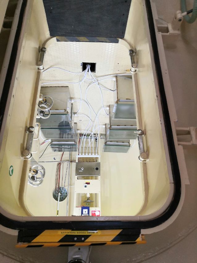
Tlapák et al.: Hyperbaric oxygen therapy and acute wounds - an experimental study

Figure 1. Semi-hermetic monoplace boxes placed in the hyperbaric oxygen chamber and their connection to the oxygen system.

The Institute of Aviation Medicine, Prague, Czech Republic; by Tlapák J.

sufficient fresh gas exchange (Figure 1). The suitability of the materials used was based on the recommendations
of the American Society for Testing and Materials International and WHA International. All of the boxes had been
tested for their resistance to pressure. Welfare monitoring of the animal subjects was done through a transparent
top plate on these boxes by a continuous camera system. A tympanoscopy (Otoscope OX1-USB, Germany)
was performed on sampling days when each animal was anesthetized. HBOT was applied using two different
regimens—Two hours once a day and two hours twice a day, both under 2.5 atmospheres absolute (ATA) of pressure
and a fixed oxygen flow. Slow compression and decompression rates were chosen, with each phase lasting for fifteen
minutes. Length of exposure, including compression and decompression, was 120 minutes without a break. The length
of isocompression was 90 minutes. A 4 L/minute flow rate of oxygen was input into each box. The therapy started
two days after the wound induction. The animals were exposed for eight consecutive days.

Histology analysis

    After the processing of bioptic material (fixation, standard processing, sectioned at 5 µm for stains), the samples
were evaluated under the Olympus BX51 (Olympus, Tokyo, Japan) and Olympus IX71 microscopes, equipped
with MMI software (Olympus) for computer image analysis (ImagePro 5.1.; Media Cybernetics, Bethesda, MD,
USA) by a certified independent histologist blinded to the experimental groups. A microscope magnification of 400x
was used. The histopathological evaluation included epidermal leaf parameters (length and surface, mitotic activity,
and the distance of epidermal leaves), the number of microabscesses, the Histopathological Superficial Epithelium
Healing Score, Connective Tissue Healing Score, Histopathological Acute Inflammation Score (HAIS) and Total
Histopathological Wound Healing Score between the groups. The stains used to evaluate the samples were
hematoxylin-eosin (H-E), Masson's Trichrome, Van Gieson's stain, and naphthol AS-D chloroacetate esterase-positive
cell detection.

                                                             4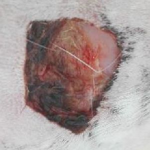
Tlapák et al.: Hyperbaric oxygen therapy and acute wounds - an experimental study

    The Histopathological Superficial Epithelium Healing Score parameters (with H-E stain) included the extent
of epithelialization and differentiation. Epithelialization was scored from 0-4, with 0 = in front of the cut edge; 1 = on
the edge of the cut; 2 = in the wound < 50% of extent; 3 = in the wound > 50% of extent; 4 = complete epithelial
coverage. Differentiation was scored from 0-2, with 0 = none or only spinous; 1 = granular differentiation-keratinization;
2 = adnexa creation. Their total scores were 0-6; a higher number indicating a better degree of wound healing.

    The length, surface, mitotic activity, and distance of the epidermal leaves (H-E stain; ImagePro 5.1. analysis)
were evaluated. To measure length, surface and distance of the epidermal leaves, whole tissue microphotographs
were acquire 400x magnification using Olympus IX71 microscopes equipped with MMI software. The epidermal
leaf length were defined as the shortest distance between the apex of the newly formed epidermal leaf and the nearest
adnexa. The epidermal leaf surface represents the surface between the apex of the newly formed epidermal leaf
and the nearest adnexa. Distance of the epidermal leaves represents the shortest straight distance between the two
apexes of the epidermal leaves. Mitotic activity was assessed occulometrically as the number of mitotic figures
in the epidermal leaf per 100 basal cells starting from the apex. Connective Tissue Healing Score was defined
by five parameters, the criteria being shown in Table 1.

Table 1. Connective Tissue Healing Scoring criteria; scale modified from Sultana et al. (2009), (11)
Scoring criterion                           Scoring scale
used stain
1 – amount of granulation tissue
                                            0 – profound,              1 – moderate,    2 – scanty,   3 – minimal,     4 - absent
Hematoxylin-eosin stain
2 – amount of early collagen
                                            0 – absent,                1 – minimal,     2 – scanty,   3 – moderate,    4 – profound
Masson's Trichrome stain
3 – amount of mature collagen
                                            0 – absent,                1 – minimal,     2 – scanty,   3 – moderate,    4 – profound
Masson's Trichrome stain
4 – collagen fiber orientation
                                            0 – without orientation,   1 – vertical,    2 – mixed,    3 – horizontal
Masson's Trichrome + Van Gieson's stain
5 – pattern of collagen
                                            0 – amorphous,             1 – reticular,   2 – mixed,    3 - fascicle
Masson's Trichrome + Van Gieson's stain

Total healing score of each member was calculated by adding the score of individual criteria and with higher scores indicating
better wound healing. Healing status with values from 0 to 18 points.

    HAIS indicates the amount of naphthol AS-D chloroacetate esterase-positive cells per field of view, which was
defined as the microscopic region in close proximity to the fibrin or epithelial layer. For each sample, ten fields
of view were randomly scored according to a semi-quantitative scale—from 0-4, with 0 = high (> 200); 1 = medium
(50 – 200); 2 = low (20 – 49); 3 = minimal (10 – 19); 4 = level uninjured skin (< 10). A higher number indicates
a higher degree of wound healing and a lower level of inflammatory response. The number of microabscesses was
evaluated in the samples for the detection of naphthol AS-D chloroacetate esterase-positive cells.

   The Total Histopathological Wound Healing Score equals the Histopathological Superficial Epithelium Healing
Score (total score) plus the Connective Tissue Healing Score (total score) plus HAIS.

Immunohistochemistry

    The immunohistochemical detection of CD34+ endothelial cells was made by using the manual peroxidase
technique in the histological laboratory of the Department of Radiobiology (Faculty of Military Health Sciences,
Hradec Kralove, Czech Republic) according to the previously published protocol (11). The samples were incubated
with mouse anti-CD34 monoclonal antibodies (MA1-10202, ThermoFisher Scientific, Waltham, MA, USA) and rat
anti-CD34 antibodies (GTX28158, GeneTex, Irvine, CA, USA) in a phosphate buffer (pH 7.2, Sigma-Aldrich).
The primary mouse antibody was followed by a secondary biotinylated donkey anti-mouse antibody (715-065-151,
Jackson ImmunoResearch Laboratories, Baltimore, PA, USA) using streptavidin peroxidase (DAKO Corporation,
Carpinteria, CA, USA) during incubation. In the case of the primary rat antibody, a secondary goat anti-rat antibody
(ab214882-15ml; Abcam, Cambridge, England) was utilized using bound polymerized peroxidase. During this step

                                                                5
Tlapák et al.: Hyperbaric oxygen therapy and acute wounds - an experimental study

of incubation, streptavidin-peroxidase was omitted and 3,3´-Diaminobenzidine tetrahydrochloride chromogen
(Sigma-Aldrich) was added to visualize bound in situ antibodies. Negative (no primary antibodies) and positive
controls (human spleen samples) were included to support the validity of staining.

Statistical analysis

    To determine the significance of the data difference, the data obtained were analyzed using IBM SPSS Statistics
version 24 (IBM Corporation, Armonk, NY, USA). Data were evaluated using a nonparametric Kruskal-Wallis test,
to determine whether there was a significant difference between all groups, with a post hoc Mann-Whitney test,
to determine the p-value between the groups. The level of statistical significance (p-value) was set at ≤ 0.05. Results
are presented as means ± 2 × Standard Error of the Mean (SEM).

Results

    The physical examinations were physiological in all the rabbits. The animal tolerance of the pressure changes
did not show any abnormalities. We did not record any incidents or suspicion of discomfort during the experiment.
No signs of barotrauma were observed during tympanoscopic examinations. The open wounds healed without
the use of antimicrobial drugs and no signs of infection were observed. The collective weight gain of the rabbits
was 18%. The average healing time of the left proximal open wound (the fourth wound in which no biopsy was taken)
was 18 days in the control group and 16 days in both treatment groups. The macroscopic view of the wounds
in the time intervals between the experimental groups is compared in the Figure 2.

                                   Control         1x2 hours/day       2x2 hours/day
                                    group              HBOT                HBOT

                       0. day

                       4. day

                       7. day

                  10. day

Figure 2. The wound healing progression of all groups in a rabbit model. Images taken during macroscopic size evaluation
among different treated groups at days 0, 4, 7, and 10 after wounding.

                                                           6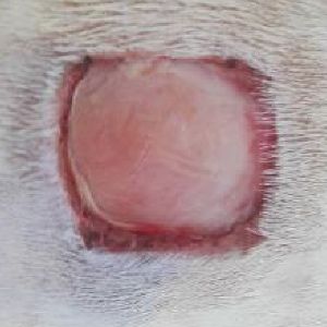
Tlapák et al.: Hyperbaric oxygen therapy and acute wounds - an experimental study
A total of 108 samples were taken (12 samples from each time interval for each group) for histopathological
analysis. Histopathological Superficial Epithelium Healing Scores covering the extent of epithelialization
and differentiation were not significantly affected by HBOT. There were no statistically significant differences
between the groups in the 4-, 7-, or 10-day time intervals of epidermal leaf parameters. Changes in epidermal leaf
length and surface were observed only at time intervals of seven and ten days within groups. Epidermal leaf distance
could only be assessed at 10-day intervals. In previous intervals, the total amount of ligament was low, which could
affect the parameter. Concerning mitotic activity, no differences between the groups were found.
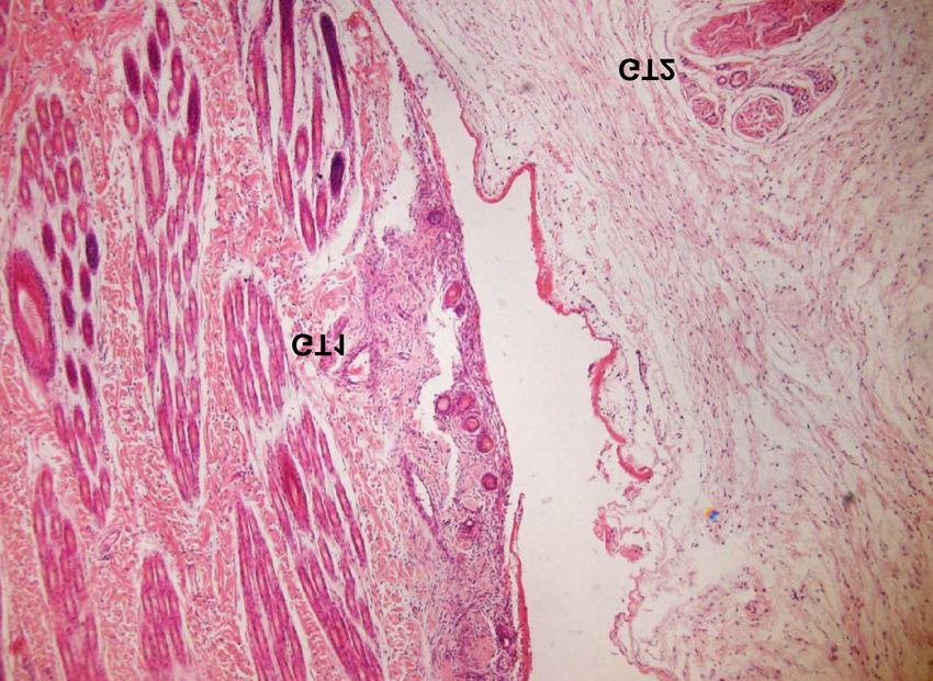
Figure 3. Granulation tissue evaluation. Granulation tissue formation (GT1) is visible at the cut edge of the sample. Another
deposit of granulation tissue is the islet forming from the connective tissue around the nerve fiber (GT2).
GT, granulation tissue; hematoxylin-eosin stain; 400x magnification; 4-day interval; control group
The HAIS and Connective Tissue Healing Scores did not show a significant difference between the groups.
Differences were noted when comparing parameters between time intervals within groups. In the case of the amount
of granulation tissue (Figure 3.), differences were found only between the 7- and 10-day intervals in the control
group and in the HBOT group 2 × 2 hours/day. The amount of early collagen showed changes in all groups between
day intervals. The increase in mature collagen was observed in the control group between the 7- and 10-day
intervals. Collagen fiber orientation showed a significant increase toward a more mature grade in the control
and HBOT 1 × 2 hours/day group between the 4- and 7-day intervals, and in all groups between the 7- and 10-day
intervals. In the collagen fiber pattern, there was a change in all groups only when comparing the 4- and 7-day
intervals. Significant differences in the Connective Tissue Healing total score were noted between all three time
intervals within all groups. The data of the mentioned histopathological parameters above are summarized in Table 2.
Significant differences could only be observed between time intervals within groups.
7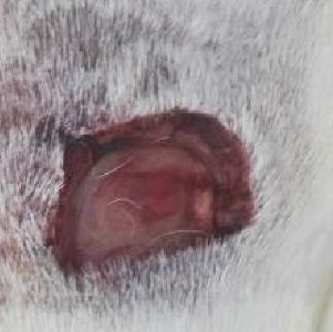
Tlapák et al.: Hyperbaric oxygen therapy and acute wounds - an experimental study
Table 2. Histopathological parameter scores from biopsies of uncomplicated open wounds evaluated on days 4, 7 and 10.
Sup"! ial Epithelium HAIS Epidermal Leaf Number " "ssue Healing S !"
"
!" f MA
Day
E D SE length !
" dis
" mi granula- early mature
"
"
(μm) (μm2) x (μm)
"
" "! "! re
re 1000 sue !"
"!
!
!
4 0.6 ( ( ( 500 20 ( N ( 0.3 ( 0.3 ( 0.0 ( 0.3 ( 0.0 ( 0.3 ( 0.0 (
0.3 0.0 0.3 0.4 0.6
7 1.2 ± 1.0 ± 2.2 ± 2.3 ± 1800 163 ± N ( 0.3 ( 0.2 ( 0.0 1.5 ± ( 0.0 1.1 ± 0.2* 1.0 ± 3.6 ±
0.2* 0.0 * 0.2* 0.4* ±300* 31* 0.3* 0.0* 0.4*
10 2.2 ± 1.4 3.6 ± ( 3300 ± 281 ± 3200 ( 0.3 ( 0.2 1.1 ± 3.3 ± 0.8 ± 1.9 ± 0.5* 1.0 ( 0 8.0 ±
0.3* 0.3 0.5* 0.5 1000* 97* 1900 0.8* 0.5* 0.3* 1.8*
x 2 !
!
4 0.9 0.1 1.0 ( 500( 23 ( N ( 0.2 ( 0.2 ( 0.0 ( 0.3 ( 0.0 ( 0.3 ( 0.0 (
0.2 0.3 100 0.6
7 1.5 ± 0.9 ± 2.5 ± 2.5 ± 1600 ± 134 ± N ( 0.2 ( 0.0 ( 0.0 1.5 ± ( 0.0 1.1 ± 0.2* 1.0 ± 3.5 ±
0.3* 0.2 * 0.4 * 0.3* 500* 45* 0.3* 0.0* 0.4*
10 2.2 ± 1.5 ± 3.6 ± ( 2400 ± 220 ± 2400 ( 0.2 ( 0.2 ( 0.3 2.9 ± ( 0.3 1.8 ± 0.4* ( 0.0 6.5 ±
0.2* 0.3* 0.3* 0.6 600* 51* 600 0.5* 1.1*
x 2 !
!
4 0.9 0.1 1.0 ( 500( 33 ( N ( 0.2 ( 0.4 ( 0.0 ( 0.3 ( 0.0 ( 0.3 ( 0.0 (
0.2 0.2 0.2 0.3 200 0.6
7 1.3 1.0 ± 2.3 ± 2.0 ± 1900 ± 184 ± N ( 0.2 0.2 0.2 0.1 ( 2 1.8 ± ( 0.2 ( 0.2 1.0 ± 4.1 ±
0.3 0.0* 0.3* 0.3* 500* 45* 0.4* 0.0* 0.7*
10 2.3 ± 1.4 3.7 ± 3.0 ± 2900 276( 97 2300 ( 0.2 ( 0.0 0.8 ± 3.0 ± ( 0.4 1.8 ± 0.4* ( 0.0 7.2 ±
0.3* 0.3 0.4* 0.4* 900 900 0.4* 0.5* 1.6*
E, epithelial0/.-*)(; D, '0&%$%)-.-*)# SE, sup%$"!0.l epithelium; HAIS, 0
*
.t *
*0!.
(.!t%(0)..-*) !*$%# MA, mi!$*. !% % ; HBOT,
%$.$0!
*%)(
h%$.
# N, )*
evaluated. Data .$e(%.)( ((EM. *( 0)i"!.)
'0&%$%)!%($*m th%(
$%0* (-me p*0)
(
.
%( 0.05).
No significant differences in the Total Histopathological Wound Healing Score were found between the groups
in the 4-, 7-, and 10-day intervals after wound induction (mean ± SD x SEM, p ≤ 0.05). Results of the control group
2.5 ± 0.5, 8.1 ± 0.6, 14.3 ± 2.4 (4-, 7-, 10-day; n=12), the HBOT 1 x 2 hours/day group 3.1 ± 0.7, 8.5 ± 0.7, 12.8 ± 1.0
(4-, 7-, 10-day; n=12) and the HBOT 2 x 2 hours/day group 3.6 ± 0.6, 8.3 ± 0.8, 13.8 ± 1.9 (4-, 7-, 10-day; n=12).
Summarized in the Figure 4.
Figure 4. Graph of the Total Histopathological Wound Healing Score for individual groups and time intervals.
18
4. day 7. day 10. day
16
14
Total Histological Wound Healing Score
12
(Mean ± SD x SEM)
10
8
6
4
2
0
Control group
HBOT 1 x 2h/day
HBOT 2 x 2h/day
HBOT, hyperbaric oxygen therapy; control group (n=12) versus HBOT 1 x 2 hours per day (n=12) versus 2 x 2 hours per day
(n=12); 4., 7., 10. sampling day; mean ±SD x SEM, p ≤ 0.05
8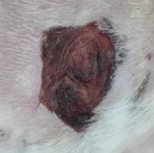
Tlapák et al.: Hyperbaric oxygen therapy and acute wounds - an experimental study

    Because both the mouse and rat antibodies showed a false negativity to the immunohistochemical detection
of CD34+ endothelial cells, the evaluation of the number of CD34 positive structures could not be performed.

Discussion

    This experimental study prospectively evaluates the effects of HBOT on uncomplicated wound healing processes
in animal models. It complements the lack of evidence for acute wounds using this therapy. Despite the fact that pig
models have been shown to have higher correlation to human healing, rabbits are frequently used as animal models
to study wound healing process because of their cost, ease of handling and technical feasibility (12).

    The treatment pressure used is based primarily on the general standard of treatment in the Czech Republic.
2.5 ATA is used not only for wound healing, but also for some otorhinolaryngological diseases (13) and other
indications. However, for some indications, a different protocol may be considered—for example, lower overpressure
for indications related to brain damage (14) or higher for anaerobic infections (15). A conservative approach was
chosen, where 2.5 ATA therapy is commonly used in wound healing (16). HBOT can cause oxygen-induced seizures.
Mammals may be more sensitive to oxygen-induced seizures than humans (17). Moreover, there are species differences
in susceptibility to oxygen toxicity. Pulmonary oxygen toxicity is dependent on the concentration and duration
of exposure to high oxygen concentrations (18). These are other arguments for a conservative therapy. The reason
for comparing more intensive therapy—twice a day—with once daily HBOT is due to the conflicting results
of previous studies. In another hyperbaric oxygen study dealing with lower limb trauma, an increased number
of daily sessions at the beginning of acute wound healing treatment produced better results (19). On the other hand,
other studies on skin graft survival in rats did not confirm this benefit (20). In our study, there was no significant
difference between the individual HBOT regimens.

     If the rate of healing according to histopathological evaluations with published models is compared, it appears
to be slower than it was with punch biopsies on the backs of rabbits in the range of 6 × 10 mm (21). On the other hand,
it corresponds to the healing rate of larger wounds (22). These comparisons are only approximate; the more accurate
comparison corresponds to the results of the study in dogs with the same size wounds (23). Within the groups, it is

Figure 5. Microabscess (solid arrow; red color - stain) forming around the remain of two hairs at the borderline (dotted arrows)
of the original tissue and the newly formed connective tissue.

NT, newly formed connective tissue; P, original tissue; naphthol AS-D chloroacetate esterase-positive cells detection;
200x magnification; 7-day interval; control group.

                                                               9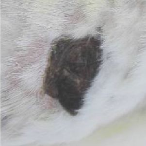
Tlapák et al.: Hyperbaric oxygen therapy and acute wounds - an experimental study

possible to observe a relatively high variance of the values of the Total Histopathological Wound Healing Score.
Although it is not possible to make positive conclusions from the results of the histopathological analysis, factors
that could affect the rate of wound healing are identified. The first factor is the quality of the edge cut. Furthermore,
the variance of HAIS data (especially in the 10-day interval) could, on the one hand, simply reflect slower wound
healing, and on the other hand, indicate the presence of infection in the wound. Macroscopically, the presence
of infection was not observed. Microabscesses were rarely present in the wounds (Figure 5.). There was a cluster
of neutrophilic granulocytes that formed exclusively around the microscopic remnants of hair. The amount was
very low within the groups and individual samples (individual cases) and the size of the microabscesses was small
and did not affect the new formation of the surrounding connective tissue. There is also a question of whether their
presence in a furry animal with a superficial wound can be completely eliminated. In the immunohistochemical
analysis, both mouse and rat antibodies designed to detect CD34 positive structures in a rabbit model showed false
negativity, therefore it would be appropriate to test more species of animal antibodies.

    Wound contraction is one of the most significant limitations of using animals to model human wounds.
The limitations of the present experimental study were the number of subjects and the quality of cutting the edge
of individual samples. The results of the experiment can help increase the level of evidence, which is pointed out
by survey analyses and the consensus of hyperbaric medicine (24). Further large scale studies will be required
to confirm our findings.

Conclusion

     Histopathological analysis did not detect an improvement in the healing process with HBOT both compared
to the control group and between treatment regimens. Although there is literature evidence suggesting the effectiveness
of HBOT, further studies are needed to evaluate the benefit of applying HBOT in acute wound care to treatment
recommendations.

Funding

   This work was financially supported through a grant from the Ministry of Defense of the Czech Republic
(Defense Research and Development OWULZ20160001).

Conflict of Interest

    The authors declare that they have no conflicts of interest regarding the publication of this article.

Adherence to ethical standards

    This article does not contain any studies involving human participants performed by any of the authors.

    The study was approved by the Expert Committee on the Welfare of Experimental Animals at the Faculty
of Military Health Sciences, University of Defence. The experiments were performed in accordance with the guidelines
of EU Directive 2010/63/EU for animal experiments and with the conditions of the National Decree on the Breeding
and Use of Experimental Animals. All workers who manipulated animals are holders of Certificates of Professional
Competence to Design Experiments and Experimental Trials under the Animal Welfare Law Against Cruelty.
The veterinary specialist was part of the research team.

References

1. Sureda A, Batle JM, Martorell M, Capó X, Tejada S, Tur JA, Poms A. Antioxidant Response of Chronic Wounds
   to Hyperbaric Oxygen Therapy. PLoS One. 2016;11(9):e0163371.
2. Francis A, Baynosa RC. Hyperbaric Oxygen Therapy for the Compromised Graft or Flap. Adv Wound Care
   2017;6:23-32.
3. Sander AL, Henrich D, Muth CM, Marzi I, Barker JH, Frank JM. In vivo effect of hyperbaric oxygen on wound
   angiogenesis and epithelialization. Wound Repair Regen 2009;17(2):179-84.

                                                           10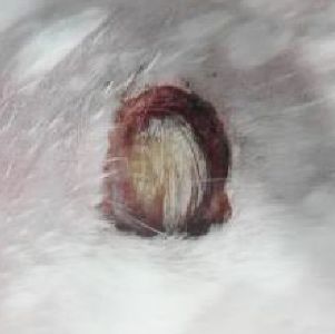
Tlapák et al.: Hyperbaric oxygen therapy and acute wounds - an experimental study

 4. Hatibie MJ, Islam AA, Hatta M, Moenadjat Y, Susilo RH, Rendy L. Hyperbaric Oxygen Therapy for Second-
    Degree Burn Healing: An Experimental Study in Rabbits. Adv Skin Wound Care. 2019;32(3):1-4.
 5. Hadanny A, Efrati S. The Hyperoxic-Hypoxic Paradox. Biomolecules. 2020;10(6):958.
 6. Memar MY, Yekani M, Alizadeh N, Baghi HB. Hyperbaric oxygen therapy: Antimicrobial mechanisms and
    clinical application for infections. Biomed Pharmacother. 2019;109:440-447.
 7. De Smet GHJ, Kroese LF, Menon AG, Jeekel J, van Pelt AWJ, Kleinrensink GJ, Lange JF. Oxygen therapies
    and their effects on wound healing. A systematic review. Wound Repair Regen. 2017;25:591-608.
 8. Santema TJ, Stoekenbroek RM, van Steekelenburg KC, van Hulst RA, Koelemay MJ, Ubbink DT. Economic
    outcomes in clinical studies assessing hyperbaric oxygen in the treatment of acute and chronic wounds. Diving
    Hyperb Med. 2015;45(4):228-34.
 9. Roje Z, Roje Ž, Eterović D, Družijanić N, Petričević, Roje T, Čapkun V. Influence of Adjuvant Hyperbaric
    Oxygen Therapy on Short-term Complications During Surgical Reconstruction of Upper and Lower Extremity
    War Injuries: Retrospective Cohort Study. Croat Med J. 2008;49:224-32.
10. Tlapák J, Chmátal P, Oniščenko B, Pavlík V, Došel P, Páral J, Lochman P. The effect of hyperbaric oxygen
    therapy on gene expression: microarray analysis on wound healing. Undersea Hyperb Med. 2020;47(1):31-37.
11. Sultana J, Molla MR, Kamal M, Shahidullah M, Begum F, Bashar MA. Histological differences in wound
    healing in maxillofacial region in patients with or without risk factors. Bangladesh J Pathol 2009;24:38.
12. Parnell LKS, Volk SW. The Evolution of Animal Models in Wound Healing Research: 1993-2017. Adv Wound
    Care. 2019;8(12):692-702.
13. Holy R, Prazenica P, Stolarikova E, Dosel P, Fundova P, Kovar D, Astl J. Hyperbaric oxygen therapy in tinnitus
    with normal hearing in association with combined treatment. Undersea Hyperb Med. 2016;43(3):201-5.
14. Hadanny A, Abbott S, Suzin G, Bechor Y, Efrati S. Effect of hyperbaric oxygen therapy on chronic
    neurocognitive deficits of post-traumatic brain injury patients: retrospective analysis. BMJ Open
    2018;8(9):e023387.
15. Jain KK. HBO Therapy in Infections. In: Jain, KK, ed. by Textbook of Hyperbaric Medicine. 5th ed. Toronto:
    Hogrefe and Huber Publishers; 2009.p.135-148.
16. Hájek M. Akutní traumatická ischemie a drtivá poranění končetin. In: Hájek M., ed. by Hyperbarická medicína.
    Praha: Mladá fronta; 2017.p.301-313.
17. Crowe DT. Hyperbaric oxygen therapy in veterinary medicine: a case series at Carson-Tahoe veterinary hospital.
    Hyperbaric Med Today. 2000;1(3):13–15.
18. Mensack S, Murtaugh R. Oxygen toxicity. Compend Contin Educ Pract Vet. 1999;21(4):341–351.
19. Millar I, McGinnes R, Williamson O, Lind F, Jansson KA, Hajek M, Smart D, Fernandes T, Miller R, Myles P,
    Cameron P. Hyperbaric oxygen in Lower Limb Trauma (HOLLT); protocol for a randomised controlled trial.
    BMJ Open. 2015;5(6):e008381.
20. Weber R, Silver A, Williams SJ, Stephenson L, Usera PC, Zhang F, Tian H, Yang W, Wang WZ, Fang XH,
    Zamboni WA, Baynosa R. Random flap survival with hyperbaric oxygen: daily versus twice-daily treatments.
    Undersea Hyperb Med. 2018;45(2):157-164.
21. Lemo N, Marignac G, Reyes-Gomez E, Lilin T, Crosaz O, Ehrenfest DMD. Cutaneous reepithelialization and
    wound contraction after skin biopsies in rabbits: a mathematical model for healing and remodelling index.
    Veterinarski Arhiv. 2010;80(5):637-652
22. Valizadeh R, Hemmati AA, Houshmand G, Bayat S, Bahadoram M. Wound healing potential of Althaea
    officinalis flower mucilage in rabbit full thickness wounds. Asian Pac J Trop Biomed 2015;5(11):937–943
23. Latimer CR, Lux CN, Roberts S, Drum MG, Braswell C, Sula MJM. Effects of hyperbaric oxygen therapy on
    uncomplicated incisional and open wound healing in dogs. Veterinary Surgery. 2018;47:827-836.
24. Mathieu D., Marroni A, Kot J. Tenth European Consensus Conference on Hyperbaric Medicine:
    recommendations for accepted and non-accepted clinical indications and practice of hyperbaric oxygen
    treatment. Diving Hyperb Med. 2017;47:24-32.

                                                       11
You can also read