Efficient foot motor control by Neymar's brain
←
→
Page content transcription
If your browser does not render page correctly, please read the page content below
ORIGINAL RESEARCH ARTICLE
HUMAN NEUROSCIENCE
                                                                                                                               published: 01 August 2014
                                                                                                                         doi: 10.3389/fnhum.2014.00594

Efficient foot motor control by Neymar’s brain
Eiichi Naito 1,2 * and Satoshi Hirose 1
1
    Center for Information and Neural Networks (CiNet), National Institute of Information and Communications Technology, Suita, Japan
2
    Graduate School of Medicine and Graduate School of Frontier Biosciences, Osaka University, Suita, Japan

Edited by:                                 How very long-term (over many years) motor skill training shapes internal motor
Sven Bestmann, University College
                                           representation remains poorly understood. We provide valuable evidence that the football
London, UK
                                           brain of Neymar da Silva Santos Júnior (the Brasilian footballer) recruits very limited neural
Reviewed by:
Eva Feredoes, University of                resources in the motor-cortical foot regions during foot movements. We scanned his brain
Reading, UK                                activity with a 3-tesla functional magnetic resonance imaging (fMRI) while he rotated
Joseph M. Galea, University of             his right ankle at 1 Hz. We also scanned brain activity when three other age-controlled
Birmingham, UK
                                           professional footballers, two top-athlete swimmers and one amateur footballer performed
*Correspondence:
                                           the identical task. A comparison was made between Neymar’s brain activity with that
Eiichi Naito, Center for Information
and Neural Networks                        obtained from the others. We found activations in the left medial-wall foot motor regions
(CiNet), National Institute of             during the foot movements consistently across all participants. However, the size and
Information and Communications             intensity of medial-wall activity was smaller in the four professional footballers than in the
Technology, 2A6, 1-4 Yamadaoka,
                                           three other participants, despite no difference in amount of foot movement. Surprisingly,
Suita, 565-0871, Japan
e-mail: eiichi.naito@                      the reduced recruitment of medial-wall foot motor regions became apparent in Neymar.
nict.go.jp                                 His medial-wall activity was smallest among all participants with absolutely no difference
                                           in amount of foot movement. Neymar may efficiently control given foot movements
                                           probably by largely conserving motor-cortical neural resources. We discuss this possibility
                                           in terms of over-years motor skill training effect, use-dependent plasticity, and efficient
                                           motor control.

                                           Keywords: Neymar da Silva Santos Júnior, football brain, foot movement, medial-wall motor region, functional
                                           magnetic resonance imaging, efficient motor control, long-term physical training

INTRODUCTION                                                                        that the football brain of Neymar da Silva Santos Júnior (the
It is well-established that motor practice induces plastic changes                  Brasilian footballer) substantially reduces the recruitment of
in the human central motor system even in adults. For exam-                         foot motor regions during foot movements. We scanned his
ple, when people practice a novel hand motor skill, the central                     brain activity with a 3-tesla fMRI while he rotated his right
motor representation in the hand section of the primary motor                       foot (ankle) at 1 Hz and compared his brain activity with that
cortex (executive locus of voluntary motor control: M1) expands                     obtained from three age-controlled professional footballers, two
when they repeat the practice for days or weeks (Karni et al.,                      top-athlete swimmers, and one amateur footballer. We assumed
1995; Pascual-Leone et al., 1995). Namely, Karni et al. (1995)                      that professional footballers have trained to perform various
demonstrated the expansion of M1 activation associated with the                     types of foot movements, e.g., “manipulating” and kicking a
acquisition of motor skills when the local blood oxygenation level-                 ball in many different ways by controlling ankle joint, through
dependent (BOLD) signal is evaluated with functional magnetic                       their intensive over-years daily training. Even though top-athlete
resonance imaging (fMRI). However, much longer-term (over                           swimmers have also trained their foot movements, these are
years) training suggests a different story.                                         usually highly-patterned movements and less variety is required.
    Recently, a non-human primate study revealed that the over-                     Thus, it is very likely that professional footballers have been
years training of a motor skill with a forelimb substantially                       exposed to richer sensory-motor experiences of foot movements
decreases the 14 C-2-deoxyglucose (2DG) uptake widely in the                        and their brains must have stored a variety of repertoire of foot
forelimb section of M1 even when the monkeys perform the                            motor skills as compared to swimmers and amateur footballers.
acquired motor skill (Picard et al., 2013). 2DG uptake can be an                    We hypothesized that neuronal resource in motor-cortical foot
indicator for the metabolic activity of the brain, which is tightly                 regions allocated to control a given simple foot movement is
coupled with such hemodynamic cerebral activity as BOLD sig-                        smaller in professional footballers than in swimmers and amateur
nals. Thus, a similar training effect should be observed in the                     footballer, because previous studies have shown that musicians
BOLD signals of human participants who have performed over-                         (pianists and keyboard players) who should have richer sensory-
years physical training, e.g., sport training.                                      motor experiences and a variety of repertoire of hand/finger
    In the present study, we focus on the motor-cortical foot                       motor skills reduce recruitment of motor areas during finger
regions of professional footballers and provide valuable evidence                   movements compared with musically naïve control (Jäncke et al.,

Frontiers in Human Neuroscience                                        www.frontiersin.org                                 August 2014 | Volume 8 | Article 594 | 1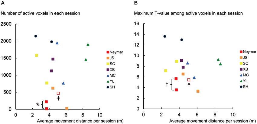
Naito and Hirose Neymar’s football brain
2000; Krings et al., 2000; Haslinger et al., 2004; Koeneke et al., direction was verbally instructed 3 s prior to each run, and the
2004). In particular, we expected that reduced recruitment of directions were altered run by run (rightward run, leftward run,
motor areas could become apparent in an exceptionally skillful rightward run, and so on). Thus, the participants completed
footballer like Neymar who could obviously perform various four rightward runs and four leftward runs in each session. We
repertoire of foot movements. To address these questions, the collected 40 functional volumes (TR = 3 s) during the foot
participants performed the simple foot movement task and movements in each session. For Neymar, we conducted one extra
we tested the hypotheses that professional footballers, espe- (third) session where he generated much larger movements (see
cially Naymar, could perform this movement by recruiting less red open squares in Figure 2).
amount of brain activity in the medial-wall motor-cortical foot For each participant, we recorded the foot movements
regions. (30 frames per second) by a digital video camera (HDR-PJ760,
Sony, Japan) that was located away from the end of the scanner
MATERIALS AND METHODS bed (just outside the five gauss line) at approximately the same
PARTICIPANTS height as the bed. Thus, the movements were recorded from the
Seven healthy male volunteers participated in our experiment view of the bottom of the foot. The bed’s width was also recorded
and their ages ranged from 18–32. They included 22-year-old for the calibration to quantify the foot movements (see below).
Neymar da Silva Santos Júnior, and three other age-controlled
professional footballers who play for the 2nd division in Liga fMRI DATA ANALYSIS
Española (JS, age 19, SC, age 18, XB, age 23), two Spanish We analyzed the fMRI data using Statistical Parametric Mapping
national-training-center level swimmers (MC, age 18, YL, age 22) software (SPM5, Wellcome Department of Cognitive Neurology,
and one amateur footballer (SH, age 32). The following are the London, UK) that was implemented in Matlab 2007a (Math-
years of their football experience (defined as when they joined Works, Inc.). We excluded the first five functional images of each
football clubs): 16 for Neymar, 12 for JS, 13 for SC, 19 for XB, session from our analysis to allow for magnetization equilib-
and 9 for SH. No such football experience was reported by the rium. The remaining functional images were realigned to cor-
swimmers. Except XB and YL, all the participants reported that rect for head movements and co-registered to each participant’s
they prefer to kick and dribble with their right foot. The local anatomical image. Both the functional and anatomical images
ethics committee of the ALOMAR hospital in Barcelona approved were normalized to the Montreal Neurological Institute (MNI)
this study. All participants gave written informed consent, and the template brain using the standard SPM5 defaults. The functional
experiment was carried out based on the principles and guidelines images were smoothed with an isotropic 8-mm full-width at half-
of the Declaration of Helsinki (1975). The public disclosure maximum Gaussian kernel. Finally, the value in each voxel was
of Neymar’s brain activity and the use of his name solely for normalized by converting it into a percentage increase from the
academic purposes were approved by Neymar’s representative. mean of the voxel in each session.
After the preprocessing, we fitted a linear regression (general
MRI MEASUREMENT linear) model to the data obtained from each participant (three
A 3.0-T MRI scanner with a head-coil (Discovery MR750w 3.0T, sessions for Neymar and two sessions for the others). We prepared
General Electric, USA) provided T1-weighted anatomical images a separate regressor for each session. In each regressor, 8 runs
(MP-RAGE) and functional T2*-weighted echo-planar images (4 rightward and 4 leftward) were modeled with boxcar functions
(64 × 64 matrix; pixel size, 3.0 × 3.0 mm2 ; flip angle, 90◦ ; convolved with the canonical hemodynamic response function in
echo time, 35 ms). We collected functional volumes every 3 s SPM5. The regressor specified each run period with a hemody-
(TR = 3000 ms) that comprised 48, 3.0-mm thickness slices namic delay in each session. Since Neymar had three sessions,
without interslice gaps. The whole brain was within the field of we prepared three regressors for him and two for the others.
view (FOV, 192 × 192 mm2 ). We also included the head motion parameters estimated in the
realignment procedure as regressors in each session to minimize
EXPERIMENTAL PROCEDURE the effects of the head motion artifacts. Finally, in the analysis, we
In this fMRI experiment, the participants rested comfortably in a removed the slow signal drifts (frequency > 128 s).
supine position in the scanner. Their heads were immobilized by First, we depicted the brain regions where the BOLD signals
cushions, and their torsos and knees were tightly wrapped with increased during the foot movements by evaluating the beta val-
belts to fixate them to the scanner bed. They were instructed to ues obtained from the two sessions (Figure 1; 2-session analysis).
avoid body movements except for their right foot and to close This was done separately for each participant to describe the
their eyes during the scanning. Auditory instructions about the general features of the individual brain activity during the foot
initiation and cessation timings of their right foot movements movements. For Neymar, we used the data obtained from the
were given to them through an MRI-compatible headphone. first two sessions. In this 2-session analysis, we first generated
Each participant completed two experimental sessions, each an image of active voxels (cluster image) for each participant.
of which consisted of 8 runs. Each run lasted for 15 s, followed To generate this image, consistently across participants, we used
by 15-s resting inter-run-intervals; the first run started 30 s a voxel-wise threshold of p < 0.001 (uncorrected). Since our
after the initiation of the scanning. In each run, the participants main interest was the motor-cortical foot regions, we set the
continuously rotated their right ankle either rightward or leftward region-of-interest (ROI) in the medial-wall foot motor regions
in synchronization with 1-Hz metronome sounds. The rotation (−20 < x < 0, −46 < y < 0, +40 < z in MNI coordinates).
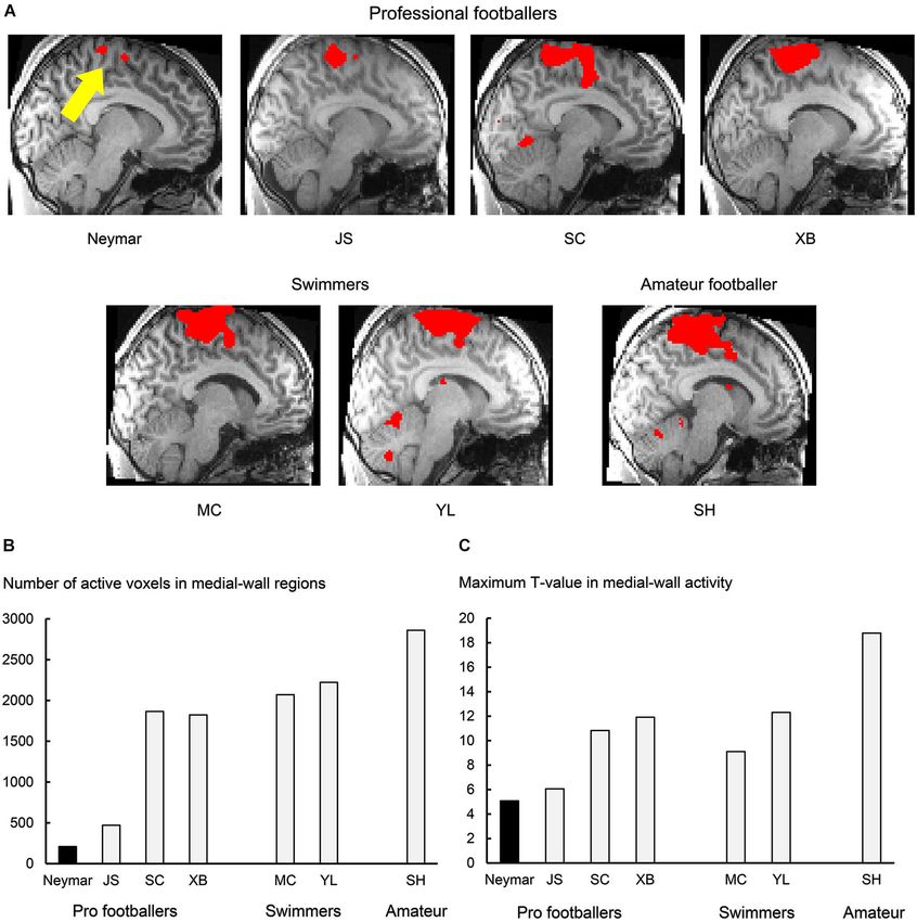
Frontiers in Human Neuroscience www.frontiersin.org August 2014 | Volume 8 | Article 594 | 2Naito and Hirose Neymar’s football brain ROI was defined based on our previous study where we identi- in terms of the spatial extent of activation within the ROI fied the foot sections in the human medial-wall motor regions (Friston et al., 1994). We also counted the number of active (Naito et al., 2007). This ROI likely covers the supplementary voxels (voxel-wise threshold p < 0.001 uncorrected; Figure 1B) motor area (SMA), the cingulate motor area (CMA), the pri- and identified their maximum T-values (Figure 1C) in each mary motor cortex (M1), and the primary somatosensory cortex participant. (SI) in the left hemisphere of an individual normalized brain. Next, we separately analyzed the increase of the BOLD signals When we looked at the individual cluster image, we found that during the foot movements for each session of each participant the largest cluster of brain activation was consistently located (single-session analysis). We also analyzed the data obtained from within this ROI across participants (Figure 1A). The medial- the third session for Neymar. We again consistently used the same wall activation in each participant was significant at cluster- voxel-wise threshold (p < 0.001 uncorrected) across the partici- level (p < 0.05 corrected) when we evaluated its significance pants and counted the number of active voxels (Figure 2A) and FIGURE 1 | Results from 2-session analysis. (A) Activity in medial-wall of medial-wall activity was smallest in Neymar’s brain (yellow arrow). motor regions during foot movements in each participant. Each panel displays (B) Number of active voxels (p < 0.001 uncorrected) in medial-wall regions result obtained from each participant. Voxels with activity greater than during foot movements in each participant. (C) Maximum T -value in voxel-wise threshold p < 0.001 uncorrected (T = 3.12 for Neymar and medial-wall activity in each participant. In both panels (B) and (C), black bars T = 3.14 for the others) are shown in red and superimposed on an individual indicate Neymar’s data. Both number of active voxels and maximum T -value normalized brain. Sagittal section (x = −8) in left hemisphere is shown. Size were smallest in Neymar. Frontiers in Human Neuroscience www.frontiersin.org August 2014 | Volume 8 | Article 594 | 3
Naito and Hirose                                                                                                                        Neymar’s football brain

identified their maximum T-values (Figure 2B) using ROI. We                    STATISTICAL EVALUATION
used this analysis result to evaluate the relationship between the             For the following statistical evaluation, we used statistical software
brain activity and the foot movements (see below and Figure 2).                (PASW Statistics 18, SPSS, Japan). In the 2-session analysis, we
The use of counting the number of active voxels as a measure                   performed a nonparametric correlation analysis between the year
of between-participant variability is still under debate. However,             of football experience and the data (number of active voxels and
many studies suggests that this can be a good measure to evaluate              maximum T-value) across participants by calculating Spearman’s
individual difference (Binder et al., 1996; Carey et al., 2002;                rank correlation coefficient.
Brodtmann et al., 2007; Wang et al., 2012), though it might not                    In the single-session analysis, we performed a nonparametric
be most reliable (Cohen and DuBois, 1999).                                     Mann-Whitney test (two-tailed; Mann and Whitney, 1947) for the
                                                                               data (number of active voxels, maximum T-value, and average
BEHAVIORAL ANALYSIS                                                            movement distance; Figure 2). First, we examined whether the
We performed offline analysis on the size of the foot movements                data obtained from the professional footballers were significantly
using software (DIPP-Motion Pro2D, DITECT, Japan). After cal-                  different from those obtained from the other participants (the
ibrating the sizes of the foot movements using the reference (bed              swimmers and the amateur footballer; group analysis). In the
width), we analyzed them in a two-dimensional plane perpen-                    analysis of each piece of data, the number of samples (n) was eight
dicular to the camera view. The location of the right big toe                  for the pro footballers and six for the other participants, i.e., two
was plotted frame-by-frame (30 frames per second). Then we                     values for each participant.
calculated the toe’s total movement distance in each run and sep-                  Next, we examined whether the data obtained from a partic-
arately averaged the movement distance for each session of each                ular participant were significantly different from those obtained
participant. This piece of data was used in the correlation analysis           from the remaining participants (individual analysis). Here, n
described above (Figure 2). In this analysis, we only quantified               was 2 for a particular participant and 12 for the remaining
the size of the foot movements in the 2D plane; however, this still            6 participants; we performed this individual analysis for all
well described the size of the participant’s foot movements, since             participants. In both the group and individual analyses, as for
the main components were basically the rightward or leftward                   Neymar, we used the data obtained from the first two sessions. To
rotations in the 2D plane.                                                     validate the results of this individual analysis for Neymar’s data,

 FIGURE 2 | Results from single-session analysis. (A) Relationship             that his maximum T -value (red filled) showed significantly smaller trend
 between number of active voxels (y-axis) and average movement distance        than those in the remaining six participants (p = 0.088). Even though
 (x-axis) for each session of each participant. (B) Relationship between       number of active voxels and maximum T -values in pro footballer JS
 maximum T -value in active voxels (y-axis) and average movement distance      (orange) were also small, they did not reach significant level (U = 3,
 (x-axis) for each session of each participant. Note that data in x-axis are   p = 0.13 for both) due to Neymar’s data. In panel (A), the number of active
 identical between panels (A) and (B). Different colors in plotted data        voxels plotted very close to horizontal axis was 13 for Neymar (red filled)
 indicate different participants. Squares indicate data obtained from          and 22 for JS (orange). Even though the number of active voxels was
 professional footballers. Data obtained from an extra third session in        smaller in these cases, this activity should have physiological importance
 Neymar are shown in open red squares with up arrows. Asterisk (*) in          because these voxels in each brain formed a cluster in the ROI, which was
 panel (A) means that number of active voxels in Neymar (red filled) was       actually the only one cluster in the entire brain. And most of these voxels
 significantly smaller than those in remaining six participants (individual    (10/13 voxels for Neymar and 22/22 voxels for JS) in each brain were
 analysis with Mann-Whitney test; p < 0.05). Cross (†) in panel (B) means      consistently active during foot movements between the two sessions.

Frontiers in Human Neuroscience                                      www.frontiersin.org                               August 2014 | Volume 8 | Article 594 | 4
Naito and Hirose Neymar’s football brain
we additionally performed an analysis that included the extra As seen in Figure 2, the individual analysis using the Mann-
third session data (n = 3 for Neymar and 12 for the remaining Whitney test revealed that, only in Neymar, the number of active
6 participants). voxels was significantly smaller than in the remaining six partici-
pants (U = 1, p < 0.05; red filled squares in Figure 2A), with no
RESULTS difference in the movement distance from the others (p = 0.2).
Neymar’s maximum T-values also showed a significantly smaller
All the participants successfully performed their foot movements
trend (U = 2, p = 0.088; see also the squares in Figure 2B).
in synchronization with the 1-Hz metronome sounds. The mean
Even when Neymar made larger movements in the extra third
movement distances across two sessions for the professional foot-
session (red open squares in Figure 2), his brain activity remained
ballers ranged from 3.1 to 5.3 m and those obtained from the
relatively small among the participants (active voxels = 466;
other participants (the swimmers and the amateur footballer)
maximum T-value = 5.46). This strongly indicates the consistency
ranged from 3.3 to 8.5 m. As we describe the details of statistics
of our finding: smaller activity in Neymar’s medial-wall foot
below, no significant difference was observed between the two
motor regions during foot movements. The additional individual
groups in terms of movement distance (see Section Results From
analysis when we included his third session data showed that
Single-Session Analysis).
the number of active voxels and the maximum T-values were
significantly smaller than those in the remaining six participants
RESULTS FROM 2-SESSION ANALYSIS (U = 2, p < 0.05; U = 3, p < 0.05, respectively), with absolutely no
In this analysis, we depicted the general features of the individual difference in movement distance (p = 0.45). Thus, among all par-
brain activity during the foot movements and found that the ticipants, the size of the medial-wall activity and its intensity were
movements activated the medial-wall motor regions in all the smallest in Neymar’s brain even though he generated comparable
participants. The brain activity consistently formed the largest foot movements in size.
cluster in the entire brain across participants (Figure 1A). The If we consider that much greater medial-wall activations were
number of active voxels in the medial-wall ROI (Figure 1B) was observed in participants SC, XB, MC, and SH, all of whom
consistently smaller in the professional footballers (averaged 1092 performed comparable movements in size (see the yellow, purple,
voxels, ranging from 209 to 1866) than in the other partici- blue, and navy symbols in Figure 2), the consistently smaller
pants (the swimmers and the amateur footballer; averaged 2384, medial-wall activity in Neymar’s brain–even when he generated
ranging from 2070 to 2861). The number of active voxels (209) larger foot movements–may represent his individual quality in
was smallest in Neymar’s brain (Figure 1B), and the maximum how he ordinarily uses his brain when controlling his foot
T-value in the medial-wall activity was also smallest in his brain movements.
(T = 5.08; Figure 1C). When we examined the relationship The individual analysis using the Mann-Whitney test also
between the years of football experience and the number of active showed that swimmer YL’s movement distance was greater than
voxels across participants, we found a significant trend of negative the other participants (U = 0, p < 0.05; see above) and that the
correlation [Spearman’s r = −0.7, N = 7, p = 0.078 two-tailed number of active voxels and the maximum T-values in amateur
(not shown in figure). Negative correlation (r = −0.3) was also SH were greater than those in the other participants (U = 0,
observed between the year and the maximum T-value, but this p < 0.05 for both). This means that the amateur footballer
did not reach significant level. recruited the greatest medial-wall activity among all the partici-
pants (see also Figure 1A panel SH).
RESULTS FROM SINGLE-SESSION ANALYSIS Finally, we confirmed that the results for the number of active
In this analysis, we scrutinized the data for each session of each voxels in the single-session analysis are basically reproducible,
participant (Figure 2). First, we confirmed that the movement even when we re-calculated the number of active voxels by adopt-
distance per session was similarly distributed across participants ing different voxel-wise thresholds ranged from p < 10−7 to 10−1 .
(horizontal axis in Figure 2), except the swimmer, YL (see green
triangles in Figure 2 and below for the statistics). We also con- DISCUSSION
firmed that both the number of active voxels and the maximum In the present study, we successfully collected functional brain
T-values had no clear relationship with the movement distance imaging data from Neymar da Silva Santos Júnior and com-
across participants (Figures 2A,B). The number of active voxels pared his brain activity with that obtained from three other age-
and the maximum T-values were relatively smaller in the pro controlled professional footballers, two top-athlete swimmers and
footballers (squares in Figure 2) than in the other participants, one amateur footballer in the same experimental environment
as we observed in the 2-session analysis. This became apparent in (e.g., identical MRI scanner and image acquisition parameters).
Neymar (red filled squares in Figure 2). The relatively smaller number of participants and of experimental
The group analysis using the Mann-Whitney test showed sessions and the lacking of complete non-athlete novice were the
that, despite no significant difference in the movement distance limitation of the current study. However, we found that, among
between the two groups (p = 0.18), the number of active voxels all the present participants, the size of the medial-wall activity
was significantly smaller in the pro footballers than in the other and its intensity were smallest in Neymar’s brain even though
participants (U = 6, p < 0.05; squares in Figure 2A). The maxi- he generated comparable foot movements in size, which was
mum T-values also showed a significantly smaller trend in the pro consistently observed across experimental sessions (Figures 1, 2).
footballers (U = 9, p = 0.059; squares in Figure 2B). Thus, we speculate that the smaller medial-wall activity during the
Frontiers in Human Neuroscience www.frontiersin.org August 2014 | Volume 8 | Article 594 | 5Naito and Hirose Neymar’s football brain
foot movements is reliable and might reflect the characteristic use on motor commands in the control of foot movements, which is
of the brain when he controls his foot. normally the neural source to generate variability in a performed
The relatively small size of the medial-wall activations during motor skill (Churchland et al., 2006); and (2) very efficient motor
foot movements in professional footballers (Figures 1, 2) seems control (presumably highly sophisticated muscle synergy control)
to generally fit with previous findings in musicians (pianists, with less effort is necessary for foot movements per se.
keyboard players and drummers), i.e., reduced recruitment of Neymar may efficiently control foot movements by largely
motor areas during finger movements compared with musically conserving motor-cortical neural resources probably with higher
naïve control (Jäncke et al., 2000; Krings et al., 2000; Haslinger reproducibility and less effort. Ideally, a greater range of foot
et al., 2004; Koeneke et al., 2004; Petrini et al., 2011). Perhaps movements should be assessed to further substantiate this con-
intensive use of their feet through over-years training and expe- clusion. It is intriguing to speculate that conserving motor-
rience that “manipulate” an external ball may cause long-term cortical resources when performing simple foot movement may
plastic changes in human central motor representation. Indeed, in turn expand the possible control capacity for a wide range of
the significant trend of negative correlation between the year of football skills since the remaining resources can be assigned to
football experience and the number of active medial-wall voxels control a variety of movements of lower extremities. We may also
suggests that the longer the football experience, the smaller the assume that this fundamental capability of his football brain could
medial-wall activity in size. This could be true even though the allow him to spend neural resources to focus more on cognitive
present study did not systematically control the year of experience. aspects during a football game, such as anticipating/predicting
We assume that the reduced BOLD effect in M1 is specific to an and detecting the actions of other players (cf. Wright et al.,
effector people are skilled at using. But at the present stage we only 2013). It might be worth testing in future study whether or not
know a reduction in the hand section during finger movements in reduced recruitment of foot motor regions in football expert’s
pianists (Krings et al., 2000) and a reduction in the foot section brain is associated with multi-tasking ability when they perform a
during foot movements in the present study. cognitive task during foot movements.
Among the present professional footballers, Neymar’s medial-
wall activation was the smallest both in size and strength ACKNOWLEDGMENTS
(Figures 1, 2). Together with the correlation result, what we The authors are grateful to NHK’s “Miracle body” staff, in par-
observed in Neymar’s brain might reflect a representative case ticular, Yuma Uchida, Jun Onozawa, Reishi Morishita, Julieta
of the long-term (over years) training effect in the medial-wall Kawaoka, and Jordi Juste for their technical support and also
foot motor regions, though we cannot rule our other possibilities to the staff of the ALOMAR hospital in Barcelona for their
that there might be differences in type, quality and amount of neuroimaging support. We also thank Dr. Daniel Callan for his
current training between Neymar and other pro footballers. But comments on this manuscript. This work was conducted as part
Neymar seems to have richer sensory-motor experiences about of Scientific Research on Innovative Areas “Understanding brain
foot movements because, since childhood, he claims to have used plasticity on body representations to promote their adaptive func-
nearly 50 different types (size and quality of material) of balls and tions” (JSPS KAKENHI No. 26120001) and the work was partially
played with them barefoot. Such experiences could have shaped supported by a Grant-in-Aid for Specially Promoted Research
the characteristic use of his brain when controlling his foot. (No. 24000012).
Anyhow, we speculate how Neymar’s brain controls his foot
movements based on recent findings in non-human primates REFERENCES
(Picard et al., 2013). As briefly mentioned above, when monkeys Binder, J. R., Swanson, S. J., Hammeke, T. A., Morris, G. L., Mueller, W. M., Fischer,
perform acquired motor skills, a substantial reduction in the M., et al. (1996). Determination of language dominance using functional MRI:
a comparison with the Wada test. Neurology 46, 978–984. doi: 10.1212/wnl.46.4.
increase of metabolic activity reflecting synaptic activity can be
978
observed in M1, while a single neuron firing is preserved. This Brodtmann, A., Puce, A., Darby, D., and Donnan, G. (2007). fMRI demonstrates
can be interpreted as the over-years training of a motor skill that diaschisis in the extrastriate visual cortex. Stroke 38, 2360–2363. doi: 10.
is likely to increase the synaptic efficacy in M1. If one supports 1161/strokeaha.106.480574
the view that BOLD signals are well correlated with local field Carey, J. R., Kimberley, T. J., Lewis, S. M., Auerbach, E. J., Dorsey, L., Rundquist,
P., et al. (2002). Analysis of fMRI and finger tracking training in subjects with
potential that reflects synaptic activity (Logothetis et al., 2001),
chronic stroke. Brain 125, 773–788. doi: 10.1093/brain/awf091
the lesser BOLD increase in Neymar’s medial-wall foot regions Churchland, M. M., Afshar, A., and Shenoy, K. V. (2006). A central source of
(Figures 1, 2) implies that synaptic efficacy increases in these movement variability. Neuron 52, 1085–1096. doi: 10.1016/j.neuron.2006.10.
motor regions. If this is the case, since the signal-transmission 034
efficacy should increase in the motor-cortical synapses, motor- Cohen, M. S., and DuBois, R. M. (1999). Stability, repeatability, and the expression
of signal magnitude in functional magnetic resonance imaging. J. Mag. Res.
cortical cells can fire without receiving massive synaptic inputs. Imaging 10, 33–40. doi: 10.1002/(sici)1522-2586(199907)10:13.
Another possibility is that Neymar’s brain recruits only the limited 0.co;2-n
sections in the medial-wall motor regions when he controls the Friston, K. J., Worsley, K. J., Frackowiak, R. S. J., Mazziotta, J. C., and Evans, A. C.
simple foot movements used in this study. If this is true, lesser cells (1994). Assessing the significance of focal activations using their spatial extent.
including cortico-motoneuronal cells (sending motor commands Hum. Brain Mapp. 1, 210–220. doi: 10.1002/hbm.460010306
Haslinger, B., Erhard, P., Altenmüller, E., Hennenlotter, A., Schwaiger, M., Gräfin
to muscles via spinal motoneurons) need to be recruited to con- von Einsiedel, H., et al. (2004). Reduced recruitment of motor association areas
trol foot movements. Both possibilities are likely associated with during bimanual coordination in concert pianists. Hum. Brain Mapp. 22, 206–
the following two factors: (1) reduction of signal-dependent noise 215. doi: 10.1002/hbm.20028
Frontiers in Human Neuroscience www.frontiersin.org August 2014 | Volume 8 | Article 594 | 6Naito and Hirose Neymar’s football brain Jäncke, L., Shah, N. J., and Peters, M. (2000). Cortical activations in primary Petrini, K., Pollick, F. E., Dahl, S., McAleer, P., McKay, L. S., Rocchesso, D., et al. and secondary motor areas for complex bimanual movements in professional (2011). Action expertise reduces brain activity for audiovisual matching actions: pianists. Brain Res. Cogn. Brain Res. 10, 177–183. doi: 10.1016/s0926- an fMRI study with expert drummers. Neuroimage 56, 1480–1492. doi: 10. 6410(00)00028-8 1016/j.neuroimage.2011.03.009 Karni, A., Meyer, G., Jezzard, P., Adams, M. M., Turner, R., and Ungerleider, L. G. Picard, N., Matsuzaka, Y., and Strick, P. L. (2013). Extended practice of a motor skill (1995). Functional MRI evidence for adult motor cortex plasticity during motor is associated with reduced metabolic activity in M1. Nat. Neurosci. 16, 1340– skill learning. Nature 377, 155–158. doi: 10.1038/377155a0 1347. doi: 10.1038/nn.3477 Koeneke, S., Lutz, K., Wüstenberg, T., and Jäncke, L. (2004). Long-term training Wang, X. F., Jiang, Z., Daly, J. J., and Yue, G. H. (2012). A generalized regression affects cerebellar processing in skilled keyboard players. Neuroreport 15, 1279– model for region of interest analysis of fMRI data. Neuroimage 59, 502–510. 1282. doi: 10.1097/01.wnr.0000127463.10147.e7 doi: 10.1016/j.neuroimage.2011.07.079 Krings, T., Töpper, R., Foltys, H., Erberich, S., Sparing, R., Willmes, K., et al. (2000). Wright, M. J., Bishop, D. T., Jackson, R. C., and Abernethy, B. (2013). Brain regions Cortical activation patterns during complex motor tasks in piano players and concerned with the identification of deceptive soccer moves by higher-skilled control subjects. A functional magnetic resonance imaging study. Neurosci. Lett. and lower-skilled players. Front. Hum. Neurosci. 7:851. doi: 10.3389/fnhum. 278, 189–193. doi: 10.1016/s0304-3940(99)00930-1 2013.00851 Logothetis, N. K., Pauls, J., Augath, M., Trinath, T., and Oeltermann, A. (2001). Neurophysiological investigation of the basis of the fMRI signal. Nature 412, Conflict of Interest Statement: The authors declare that the research was conducted 150–157. doi: 10.1038/35084005 in the absence of any commercial or financial relationships that could be construed Mann, H. B., and Whitney, D. R. (1947). On a test of whether one of two random as a potential conflict of interest. variables is stochastically larger than the other. Ann. Math. Stat. 18, 50–60. doi: 10.1214/aoms/1177730491 Received: 14 May 2014; accepted: 16 July 2014; published online: 01 August 2014. Naito, E., Nakashima, T., Kito, T., Aramaki, Y., Okada, T., and Sadato, Citation: Naito E and Hirose S (2014) Efficient foot motor control by Neymar’s brain. N. (2007). Human limb-specific and non limb-specific brain represen- Front. Hum. Neurosci. 8:594. doi: 10.3389/fnhum.2014.00594 tations during kinesthetic illusory movements of the upper and lower This article was submitted to the journal Frontiers in Human Neuroscience. extremities. Eur. J. Neurosci. 25, 3476–3487. doi: 10.1111/j.1460-9568.2007. Copyright © 2014 Naito and Hirose. This is an open-access article distributed under 05587.x the terms of the Creative Commons Attribution License (CC BY). The use, distribution Pascual-Leone, A., Dang, N., Cohen, L. G., Brasil-Neto, J. P., Cammarota, A., and or reproduction in other forums is permitted, provided the original author(s) or licensor Hallett, M. (1995). Modulation of muscle responses evoked by transcranial mag- are credited and that the original publication in this journal is cited, in accordance with netic stimulation during the acquisition of new fine motor skills. J. Neurophysiol. accepted academic practice. No use, distribution or reproduction is permitted which 74, 1037–1045. does not comply with these terms. Frontiers in Human Neuroscience www.frontiersin.org August 2014 | Volume 8 | Article 594 | 7
You can also read