PCR-based genotyping assays to detect germline APC variant associated with hereditary gastrointestinal polyposis in Jack Russell terriers
←
→
Page content transcription
If your browser does not render page correctly, please read the page content below
Yoshizaki et al. BMC Veterinary Research   (2021) 17:32
https://doi.org/10.1186/s12917-020-02731-7

 METHODOLOGY ARTICLE                                                                                                                                  Open Access

PCR-based genotyping assays to detect
germline APC variant associated with
hereditary gastrointestinal polyposis in Jack
Russell terriers
Kyoko Yoshizaki1, Akihiro Hirata1* , Hiroyuki Matsushita1, Naohito Nishii2, Mifumi Kawabe3, Takashi Mori4,5 and
Hiroki Sakai1,5

  Abstract
  Background: The prevalence of gastrointestinal (GI) neoplastic polyps in Jack Russell terriers (JRTs) has increased in
  Japan since the late 2000s. Recently, we demonstrated that JRTs with GI polyps harbor identical germline variant in
  the APC gene (c.[462_463delinsTT]) in the heterozygous state. Thus, this disease is an autosomal dominant
  hereditary disorder. Although the affected JRTs have distinct features, such as the development of multiple GI
  polyps and an early age of disease onset, genetic testing is indispensable for a definitive diagnosis. Here,
  polymerase chain reaction (PCR)-based assays capable of detecting germline APC variant were designed and
  validated using synthetic wild-type and mutant DNAs and genomic DNAs from carrier and non-carrier dogs.
  Result: First, the PCR-restriction fragment length polymorphism (PCR-RFLP) assay was developed by taking
  advantage of the germline APC variant creating a new restriction site for MseI. In the PCR-RFLP assay, the 156-bp
  region containing the variant site was amplified by PCR and subsequently digested with MseI, yielding diagnostic
  51 and 58 bp fragments from the mutant allele and allowing determination of the APC genotypes. It was possible
  to determine the genotypes using genomic DNA extracted from the peripheral blood, buccal swab, or formalin-
  fixed paraffin-embedded tissue. Next, a TaqMan duplex real-time PCR assay was developed, where a 78-bp region
  flanking the variant was amplified in the presence of wild-type allele- and mutant allele-specific fluorescent probes.
  Using blood-derived DNA, altogether 40 cycles of PCR amplification determined the APC genotypes of all examined
  samples by measuring the fluorescence intensities. Importantly, false-positive and false-negative errors were never
  detected in both assays.
  Conclusion: In this study, we developed highly reliable genetic tests for hereditary GI polyposis in JRTs, providing
  accurate assessment of the presence of the causative germline APC variant. The genotyping assays could contribute
  to the diagnosis and prevention of hereditary GI polyposis in dogs.
  Keywords: Hereditary gastrointestinal polyposis, Jack Russell terrier, Genetic testing, Diagnosis

* Correspondence: akatsuki@gifu-u.ac.jp
1
 Laboratory of Veterinary Pathology, Joint Department of Veterinary
Medicine, Faculty of Applied Biological Sciences, Gifu University, 1-1
Yanagido, Gifu 501-1193, Japan
Full list of author information is available at the end of the article

                                          © The Author(s). 2021 Open Access This article is licensed under a Creative Commons Attribution 4.0 International License,
                                          which permits use, sharing, adaptation, distribution and reproduction in any medium or format, as long as you give
                                          appropriate credit to the original author(s) and the source, provide a link to the Creative Commons licence, and indicate if
                                          changes were made. The images or other third party material in this article are included in the article's Creative Commons
                                          licence, unless indicated otherwise in a credit line to the material. If material is not included in the article's Creative Commons
                                          licence and your intended use is not permitted by statutory regulation or exceeds the permitted use, you will need to obtain
                                          permission directly from the copyright holder. To view a copy of this licence, visit http://creativecommons.org/licenses/by/4.0/.
                                          The Creative Commons Public Domain Dedication waiver (http://creativecommons.org/publicdomain/zero/1.0/) applies to the
                                          data made available in this article, unless otherwise stated in a credit line to the data.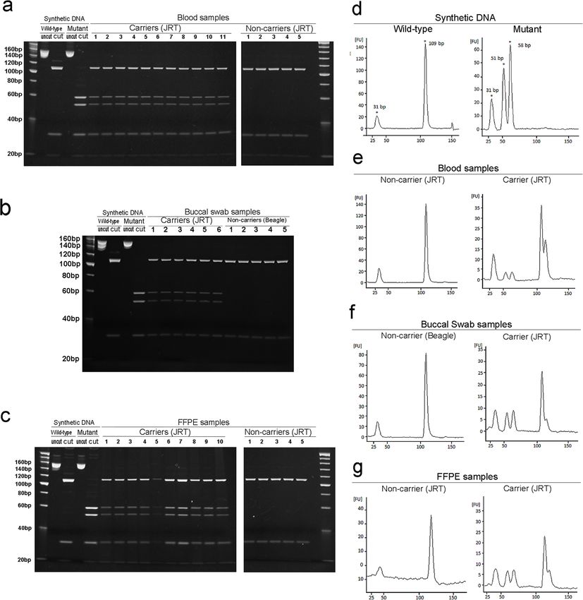
Yoshizaki et al. BMC Veterinary Research   (2021) 17:32                                                         Page 2 of 9

Background                                                      (PCR-RFLP) and TaqMan real-time PCR assays can
Recent advances in clinical veterinary medicine and mo-         be designed, enabling rapid detection of the target
lecular genetics, including availability of the complete ca-    variant in any standard laboratory. These assays are
nine genome sequence [1], have led to the discovery of          available for the detection of single-base changes,
canine and feline hereditary disorders [2–4]. The cases of      such as point mutations and single nucleotide poly-
gastrointestinal (GI) neoplastic polyps in Jack Russell ter-    morphisms (SNPs), and used as genotyping assays for
riers (JRTs) have increased in Japan since the late 2000s,      canine hereditary diseases [12–17].
with the suspicion of it being a novel hereditary disorder        In a recent study of hereditary GI polyposis in JRTs,
[5]. Recently, we demonstrated that JRTs with GI polyps         we developed PCR-RFLP and TaqMan duplex real-time
harbor identical germline variant in the APC gene, c.[462_      PCR assays to investigate the molecular mechanism
463delinsTT] (OMIA ID: 001916–9615), in the heterozy-           underlying tumor development [6]. In particular, using
gous state. Thus, this is an autosomal dominant hereditary      these assays, somatic loss of the wild-type APC allele
disorder [6]. The solitary and multiple polyps were found       was successfully detected in the GI tumors of JRTs with
in either one or both the stomach and colorectum, with a        heterozygous germline APC variant [6]. These assays
predilection for the gastric antrum and rectum, in the af-      would also be applicable to genotyping assays for heredi-
fected dogs, and most of the GI lesions were histopatho-        tary GI polyposis, but further validation is required. In
logically diagnosed as adenomas or adenocarcinomas [6].         this study, we showed that the PCR-RFLP and TaqMan
This cancer-prone disease could possibly be a canine            duplex real-time PCR assays were capable of accurately
counterpart of attenuated familial adenomatous polyposis        determining the germline APC variant status of carrier
(FAP) in humans [7].                                            and non-carrier dogs without any false-positive or false-
  The following features could provide clinical veterinar-      negative errors.
ians with indications to diagnose hereditary GI polyposis
in JRTs: (1) multiple polyps in the stomach and/or color-       Results
ectum, (2) early onset of GI polyposis, and (3) repeated        APC variant status of the samples
recurrence [6]. However, as observed in a recent study,         In this study, genomic DNA samples with heterozygous
this hereditary disease can present as a solitary GI polyp      germline APC variant (c.[462_463delinsTT]) were ob-
[6], with the lesion distribution similar to those of spor-     tained from JRTs with hereditary GI polyposis. These
adic GI cancers [6, 8–10]. Although onset at a young            cases were diagnosed as hereditary GI polyposis based
age is a distinctive feature, initial GI lesions can arise at   on the detection of APC variant, and clinical and patho-
a wide range of ages, even after 10 years in JRTs with          logical characteristic findings in our previous study [6].
germline APC variant [6]. These findings indicate the           Samples without APC variant were obtained from JRTs
necessity of genetic testing for definitive diagnosis of        with other diseases or laboratory beagles. Genomic DNA
hereditary GI polyposis. Furthermore, JRTs with heredi-         samples were extracted from the peripheral blood, buc-
tary GI cancers had a longer survival time than sporadic        cal swab, or formalin-fixed paraffin-embedded (FFPE)
cases but were at a higher risk for disease recurrence [6],     tissue.
emphasizing the need for differential diagnosis for for-           First, the germline APC variant status of all samples
mulating the treatment and follow-up.                           used in this study was validated by PCR-direct sequen-
  Recently, genetic testing for hereditary disorders is         cing. Representative results of PCR-direct sequencing
well-established for the diagnosis of affected animals and      were shown in Fig. 1a. Heterozygous deletion-insertion
the prediction of future risks for breed-specific diseases      variant involving two consecutive nucleotides, c.[462_
in several breeds [2–4]. Since hereditary GI polyposis is       463delinsTT], which spaned codons 154 and 155 of the
an adult-onset disease [6], future risk assessment is im-       canine APC gene, were detected in all the carrier JRTs,
portant for early detection of the onset. Furthermore,          while all non-carrier JRTs and laboratory beagles were
genetic testing is indispensable to prevent the spread of       homozyous for the wild-type allele.
canine hereditary disorders [2, 3, 11]. Hereditary GI
polyposis is spreading among JRTs in Japan since the            PCR-RFLP assay
2000s, and thus, excluding the carriers of the pathogenic       PCR-RFLP assay was designed by taking advantage of
variant from the breeding population is necessary as            the germline APC variant creating a new restriction site
early as possible.                                              for MseI (Fig. 1b). First, the optimal time for enzyme di-
  In a recent study, we discovered a previously unknown         gestion was examined. PCR products amplified from
disease-causing variant in the APC gene using PCR-              blood-derived DNAs of a carrier and a non-carrier,
direct sequencing [6]. Once the disease-causing variant         along with synthetic wild-type and mutant DNAs were
is identified, other PCR-based genetic testing methods          digested for 1, 2, 4, 8, 12, and 24 h. The digested frag-
such as PCR-restriction fragment length polymorphism            ments of the expected sizes were obtained from each
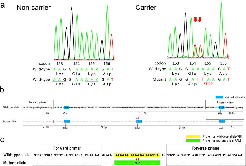
Yoshizaki et al. BMC Veterinary Research        (2021) 17:32                                                                                   Page 3 of 9

 Fig. 1 DNA sequence of codons 153–156 in the canine APC gene determined by PCR-direct sequencing and schemes of PCR-RFLP and TaqMan
 duplex real-time PCR assays for the canine APC genotyping. a PCR-direct sequencing. Representative results of peripheral blood-derived DNAs of
 JRTs with and without the germline APC variants (right and left panels). Note heterozygous deletion-insertion variant at codons 154 and 155 in a
 carrier dog (arrows). b PCR-RFLP assay. Note that, while the 156-bp fragment of the wild-type APC allele contains two restriction sites of MseI,
 there are 3 restriction sites in the fragment of the mutant APC allele. Restriction sites of MseI (T/TAA) are highlighted in blue. Asterisks indicate the
 variant sites. Uppercase and lowercase letters indicate exon 4 and introns, respectively. c TaqMan duplex real-time PCR assay. Probes specific for
 wild-type and mutant alleles are highlighted in yellow and green, respectively. Asterisks indicate variant sites

sample after 1 h of digestion (Supplementary Fig. 1), and                      patterns were observed in the analyses of the buccal
the shortest digestion time was applied to the subse-                          swabs of six carrier JRTs and five non-carrier beagles
quent analyses. In addition, consistent with the low star                      (Fig. 2b) and FFPE samples of ten carrier and five non-
activity of MseI [18], nonspecific digestion was not de-                       carrier JRTs (Fig. 2c), showing the viability of the assay
tected even after 24 h of digestion.                                           using other DNA sources.
  The initial PCR produced a single amplicon of 156 bp                           All digested PCR products were also analyzed by micro-
from all DNA samples, irrespective of the source of the                        chip capillary electrophoresis. An electropherogram with
DNA template (data not shown), and subsequent en-                              different peak patterns was obtained for each genotype.
zymatic digestion yielded distinct fragment patterns, de-                      When the synthetic DNAs were analyzed, two peaks at 31
pending on the APC genotype. The amplicons from                                and 109 bp and three peaks at 31, 51, and 58 bp were
synthetic wild-type and mutant DNAs were cleaved into                          observed for wild-type and mutant DNA, respectively (Fig.
three fragments of 16, 31, and 109 bp, and four frag-                          2d). Representative results of the analysis using genomic
ments of 16, 31, 51, and 58 bp, respectively (Fig. 2a-c                        DNAs extracted from other sources were shown in Fig.
and Supplementary Fig. 1). When analyzing the blood-                           2e-g. For non-carrier dogs, the peak pattern was the same
derived DNA, PCR products from five non-carrier JRTs                           as that for synthetic wild-type DNA (Fig. 2e-g, left panels).
were cleaved into three fragments and those from 11                            For carrier JRTs, a combined pattern of the wild-type and
carrier JRTs into five fragments, including both wild-                         mutant DNAs was observed, with five peaks at 31, 51, 58,
type allele-derived 109 bp and mutant allele-derived 51                        and 109 bp (Fig. 2e-g, right panels). Genotype-specific pat-
and 58 bp fragments (Fig. 2a). The same fragmentation                          terns were observed for all samples obtained from blood
Yoshizaki et al. BMC Veterinary Research (2021) 17:32 Page 4 of 9
Fig. 2 PCR-RFLP assay. a Polyacrylamide gel electrophoresis of MseI-digested PCR products amplified from blood samples of carrier and
non-carrier JRTs (n = 11 and 5, respectively); b buccal swab samples of carrier JRTs and laboratory beagles (n = 6 and 5, respectively); c
FFPE tissue samples of carrier and non-carrier JRTs (n = 10 and 5, respectively). Representative microfluidic capillary gel electropherograms
of MseI-digested PCR products amplified from synthetic wild-type and mutant DNA d, blood e, buccal swab f, and FFPE samples g of
carrier and non-carrier dogs
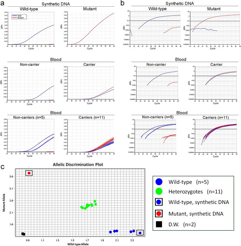
(n = 5 and 11 for non-carrier and carrier dogs), buccal exponential increase in fluorescence intensity of either
swab (n = 5 and 6), and FFPE tissues (n = 5 and 10). VIC or FAM was observed without nonspecific allelic
amplification (Fig. 3a and b, top panels). Consistently,
TaqMan duplex real-time PCR assay when blood-derived DNA samples of 5 non-carrier JRTs
The real-time PCR assay with TaqMan probes deter- were analyzed, only VIC fluorescence intensity increased
mined the genotypes of all samples. When the synthetic without detection of FAM fluorescence, even after 40 cy-
wild-type and mutant DNAs were analyzed, an cles of amplification (Fig. 3a and b, middle and bottomYoshizaki et al. BMC Veterinary Research (2021) 17:32 Page 5 of 9 Fig. 3 TaqMan duplex real-time PCR assay. a, b Amplification plots of wild-type (blue curve) and mutant (red curve) APC gene copies on synthetic wild-type and mutant DNAs (upper panels) and blood-derived DNAs of carrier or non-carrier JRTs (middle and lower panels). Amplification was plotted as fluorescence intensity (ΔRn value) against cycle numbers and shown on linear (a) and log (b) scales. The middle panels show representative results of individual cases of carrier or non-carrier dogs and the lower panels summarize the results of all examined cases (n = 11 and 5 for carrier and non-carrier JRTs, respectively). c Allelic discrimination plot based on the signal intensity ratio of FAM and VIC at the end points of PCR amplification. Synthetic wild-type and mutant DNAs are located at bottom right and top left corners, respectively, and distilled water used as negative control is at bottom left corner. Non-carrier JRTs are clustered together with the synthetic wild-type DNA at bottom right corner. Carrier JRTs are located near the diagonal line forming a distinct cluster panels). When the DNA samples of 11 JRTs with the Figure 3c shows the allelic discrimination plot con- germline APC variant were examined, both FAM and VIC structed based on the signal intensity ratio of FAM and fluorescence intensities increased simultaneously and VIC at the end points of PCR amplification. The exam- exceeded the baseline level at around 30 cycles (Fig. 3a ined samples were divided into the following three clus- and b, middle and bottom panels). ters, predicting their genotypes with 100% accuracy: (1)
Yoshizaki et al. BMC Veterinary Research   (2021) 17:32                                                         Page 6 of 9

wild-type cluster consisting of synthetic wild-type DNA        veterinarians need to differentiate between hereditary
and five non-carrier dogs, (2) heterozygote cluster of 11      and sporadic GI cancers in dogs for the following rea-
carrier dogs, and (3) the mutant synthetic DNA.                sons. First, JRTs affected with the hereditary disease are
                                                               much more likely than sporadic cases to have multiple
Discussion                                                     cancers [6]. Second, in the case of the hereditary GI
In this study, we developed highly reliable genetic tests      polyposis, the affected dogs can be expected to have a
for hereditary GI polyposis in JRTs, providing accurate        longer survival time than sporadic cases although they
assessment of the presence of the causative germline           have an increased lifelong risk of disease recurrence [6].
APC variant, c.[462_463delinsTT] [6]. Importantly, false-      The genetic testing enables the differential diagnosis be-
positive and false-negative errors were never detected in      tween hereditary and sporadic GI cancers. In addition,
both PCR-RFLP and TaqMan real-time PCR assays,                 JRTs with chronic GI symptoms such as vomiting and
demonstrating the high specificity and sensitivity of the      bloody stool can be candidates for genetic testing [6].
established assays. While PCR-RFLP and TaqMan PCR              Considering that endoscopic examination requires gen-
assays are capable of detecting even a single-base differ-     eral anesthesia in dogs, genetic testing could be an op-
ence, the target APC variant is a deletion-insertion vari-     tion to predict the occurrence of GI lesions in JRTs. The
ant involving two consecutive nucleotides [6], providing       positive test results strongly support the need for further
substantial advantage to enhance the specificities of the      examinations such as endoscopy. Furthermore, genetic
established assays.                                            testing would be useful for the assessment of future risk
  Although the PCR-RFLP assay is one of the most com-          for hereditary GI polyps. Many commercial laboratories
mon methods for genotyping, a lack of suitable restric-        offer genetic tests for canine and feline hereditary dis-
tion enzymes hinders its application in some cases. In         eases [4], available to pet owners and veterinarians for
this study, we successfully developed the PCR-RFLP             assessment of lifelong risk. Hereditary GI polyposis is an
assay for hereditary GI polyposis by taking advantage of       adult-onset disease, and initial GI lesions can arise at
the causative germline APC variant creating a new re-          variable ages, reportedly between 2 to over 10 years [6].
striction enzyme site for MseI. In the PCR-RFLP assay,         Therefore, knowing the lifelong risk is necessary for
MseI digestion yielded diagnostic fragments of 51 and          early detection and treatment.
58 bp from the mutant APC allele and differentiated car-          Genetic testing is indispensable for preventing the
rier and non-carrier dogs. In this assay, the PCR ampli-       spread of hereditary diseases in dogs [2, 3, 11]. In adult-
con contained two restriction sites of MseI besides the        onset diseases, there is a risk of the carriers of patho-
variant site; therefore, the 156-bp band disappeared after     genic variant being unintentionally used for breeding be-
the digestion regardless of the genotypes, contributing to     fore the disease onset. If a popular sire, such as a dog
the prevention of false negative errors by incomplete di-      show champion, is a variant carrier, causative variant
gestion. Furthermore, the entire process of this assay can     can spread rapidly within the breed due to their exten-
be completed within half a day using an ordinary ther-         sive use for breeding [2]. Our previous retrospective
mal cycler and electrophoresis apparatus, enabling rapid       study revealed that the incidence of GI polyposis in JRTs
diagnosis in any standard laboratory. In addition, by          increased since the late 2000s, and that all the affected
using the buccal swab as test materials, determination of      dogs were born during the first decade of the 2000s [6].
the genotypes is possible in a less invasive manner.           The spread of hereditary GI polyposis among JRTs in
Moreover, when dogs are suspected to have a hereditary         Japan might be due to the popular sire effect during this
disease on histopathological examination of their GI le-       period. Proper genetic screening of sires and dams be-
sions, it is possible to determine their APC genotype          fore breeding would prevent the transmission of germ-
from FFPE samples without the need for additional              line APC variant to future generations. This approach
blood or swab samples. This approach is also applicable        would substantially reduce the future incidence and
to retrospective studies using archival FFPE samples.          eventually eradicate this hereditary disease in the future.
  TaqMan duplex real-time PCR assay requires dedicated         Therefore, genetic screening should preferably become a
equipment capable of measuring multiple fluorescences          routine for animal breeding [2, 4].
in real time, but this assay enables high-throughput geno-        The genotyping assays established in this study would
typing. Therefore, it is suitable for large-scale molecular    certainly facilitate large-scale epidemiological studies.
epidemiological studies. Another advantage of this assay       There are no reports of JRTs with hereditary GI polyp-
is determination of genotypes in a single PCR without          osis in other countries, except Japan; thus, the germline
any additional post-amplification manipulations, thus          APC variant are possibly prevalent only among JRTs in
minimizing the risk of human errors.                           Japan. However, when examining the pedigree certifi-
  The genetic tests developed in this study would pro-         cates of the JRTs affected by hereditary GI polyposis,
vide significant benefits in small animal practice. Clinical   some ancestor dogs were introduced from Australia
Yoshizaki et al. BMC Veterinary Research (2021) 17:32 Page 7 of 9
(unpublished data), and thus there is a possibility of the FFPE tissue samples
presence of JRTs with the germline APC variant in other Archival FFPE tissue specimens stored in the Laboratory
countries. Furthermore, the prevalence of germline APC of Veterinary Pathology at the Gifu University were
variant in JRTs in Japan remains to be investigated. Fur- used. Genomic DNA was extracted from non-neoplastic
ther studies are needed to clarify the prevalence of germ- tissues on FFPE specimens of 15 JRTs, including 10 dogs
line APC variant in JRTs in Japan and other countries. with hereditary GI polyposis and five with other diseases
Moreover, in future epidemiological studies of healthy (chronic gingivitis, fibrous follicular harmartoma, lip-
JRTs, the carrier dogs could be identified before disease oma, fibrous gingival hyperplasia and cystic dilatation of
onset, indicating the usefulness of genetic testing for fu- mammary duct), using the QIAamp DNA FFPE Tissue
ture risk assessment. Kit (QIAGEN).
Synthetic DNA
Conclusions Obtaining DNA samples homozygous for the mutant
In this study, we established reliable genetic tests for APC alleles from dogs is impossible because of embry-
hereditary GI polyposis in JRTs, enabling definitive diag- onic lethality in animals with germline homozygous APC
nosis, lifelong risk assessment of this novel disease, and variants [19]. To prepare control DNA templates homo-
prevention of transmission of germline APC variant to zygous for wild-type or mutant APC alleles, 309-bp
future generations. Since dogs affected by this hereditary DNA fragments were synthesized based on the predicted
disease developed multifocal and recurrent GI polyps, sequences of the canine APC gene (GenBank accession
development of non-surgical treatment options is desir- No. NC_006585.3 and XM_014111995.2) (Takara Bio,
able. These assays could be incorporated into small vet- Shiga, Japan) and cloned into T-Vector pMD20 (Takara
erinary practices in the near future, laying groundwork Bio). The synthetic DNA fragments contained entire
for the establishment of effective therapy for hereditary exon 4 (109 bp) with the most proximal 100 bp of both
GI polyposis. upstream and downstream introns. In the mutant type,
in accordance with the germline APC variant in the af-
fected JRTs (c.[462_463delinsTT]) [6], adenosine bases
Materials and methods at nucleotides 462 and 463 were replaced with thymine
Samples and DNA extraction bases.
Blood samples
Peripheral blood was collected from 11 JRTs with her- PCR-direct sequencing
editary GI polyposis. Blood samples taken from five JRTs To validate the APC variant status of each sample, PCR-
with other diseases (portosystemic shunt, chronic chole- direct sequencing was conducted as previously reported
cystitis, lymphoma, mast cell tumor and liver tumor) for [6]. PCR was performed with TaKaRa EX Taq Hot Start
clinical blood testing at the Gifu University Teaching Version (Takara Bio) to amplify a 385-bp fragment con-
Animal Hospital served as non-carriers. Genomic DNA taining exon 4 of the canine APC gene from the blood
was extracted from EDTA-anticoagulated blood samples and swab samples and a 156-bp fragment containing nu-
using the Wizard Genomic DNA Purification Kit (Pro- cleotides 462 and 463 in exon 4 from the FFPE samples.
mega, Madison, WI) or the DNeasy Blood & Tissue Kit The primers used are listed in Table 1. After electro-
(QIAGEN, Venlo, Netherlands) according to the manu- phoresis on the 3.0% agarose gel, PCR products were ex-
facturer’s instructions. tracted using the QIAquick Gel Extraction Kit (QIAG
EN). The purified PCR products were subjected to se-
quencing analysis using an ABI Prism 3500 Genetic
Buccal swab samples Analyzer (Applied Biosystems, Foster City, CA) with the
Buccal swabs were collected from six JRTs with her- Big Dye Terminator v3.1 Cycle sequencing Kit (Thermo
editary GI polyposis and five laboratory beagles using Fisher Scientific, Waltham, MA).
cotton-tipped swabs or 4 N6 FLOQ Swabs™ (COPAN
Flock Technologies, Brescia, Italy) and stored at − Protocol for PCR-based diagnostic assays
20 °C until use. Genomic DNA was extracted using PCR-RFLP assay
the DNeasy Blood & Tissue Kit (QIAGEN). Briefly, DNA fragments of 156 bp, containing the variant sites at
the head of the swab was incubated in a mixture con- nucleotides 462 and 463 of the canine APC gene, were
taining 20 μL proteinase K solution (> 600 mAU/mL), amplified by PCR using Takara EX Taq Hot Start Ver-
400 μL Buffer A, and 400 μL PBS at 56 °C for 10 min, sion (Takara Bio). PCR reaction mixtures (20 μL) con-
and the lysate was purified according to the manufac- tained 2 μL of 10× Ex Taq buffer (Mg2+ plus), 2.5 mM of
turer’s instructions. each dNTP, 0.15 μM of each primer, and 0.75 U ofYoshizaki et al. BMC Veterinary Research (2021) 17:32 Page 8 of 9
Table 1 Primers and probes used for PCR-based assays for detection of the germline APC variant
Sequence Product size (bp)
Primers used for PCR-direct sequencing
Primers sense 5′- AGTCCCACCTTCAAAAATCC −3’ 385
antisense 5′- AACTAAAAATGCAATTATCTTGAATG −3’
Primers used for PCR-RFLP and PCR-directsequencing of FFPE samples
Primers sense 5′- TCTTTTGGCATTGTGTAAACTTG −3’ 156
antisense 5′- CTTACATTTTCAGTTAAAGGGAGACT −3’
Primers and probes used for Taq-Man duplex real-time PCR assay
Primers sense 5′- TTTAGTGAGATTCTGAAGTTGAGCATAATA −3’ 78
antisense 5′- TCATTACTTCTTGCTGATCTTGACAA −3’
Probe for Wild-type allele 5′- (VIC)- CAATCTTTTTCCTTTTC-(MGB) −3’
Probe for Mutant allele 5′- (FAM)-CAATCTTAATCCTTTTC-(MGB) −3’
VIC 2-chloro-7’-phenyl-1,4-dichloro-6-carboxy-fluorescein, FAM 6-carboxyfluorescein, NFQ nonfluorescent quencher, MGB minor groove binder
Takara Ex Taq DNA polymerase, and 1 μL of DNA tem- 0.2 μM of the mutant and wild-type probes, and 10 ng of
plate. The primers used are listed in Table 1. The cycling template DNA. The ABI StepOne Plus system (Applied
conditions consisted of an initial denaturation at 94 °C Biosystems) was used to amplify and quantify the PCR
for 5 min, followed by 35 cycles of denaturation at 94 °C products. The cycling conditions were 95 °C for 10 min,
for 30 s, annealing at 58 °C for 30 s, extension at 72 °C followed by 40 cycles of 95 °C for 15 s and 60 °C for 1 min.
for 30 s, and a final extension at 72 °C for 5 min. Before Data analysis was conducted using the StepOne software
enzymatic digestion, 4 μL of the PCR products were ana- v2.3 (Applied Biosystems).
lyzed by electrophoresis on 1.5% agarose gel. The
remaining PCR products were digested with restriction Supplementary Information
enzyme MseI (RspRSII) (Takara Bio) at 60 °C in a 20-μL The online version contains supplementary material available at https://doi.
org/10.1186/s12917-020-02731-7.
reaction mixture containing 10 units of MseI, 2 μL of
10× buffer, 2 μL of 0.1% bovine serum albumin, and
Additional file 1: Figure S1. PCR-RFLP assay conducted to determine
15 μL of the product. The digested products were sepa- the optimal digestion time. Acrylamide gel electrophoresis of MseI-
rated by electrophoresis on 15% polyacrylamide gel digested PCR products amplified from synthetic wild-type and mutant
(SuperSep™ Ace, Wako Pure Chemical Industries, Osaka, DNA (A), and blood-derived DNA samples of a carrier and a non-carrier of
germline APC variant (B). PCR products amplified from each sample were
Japan). DNA bands were visualized with a UV transillu- digested with MseI for 0, 1, 2, 4, 8, 12, and 24 h.
minator after staining with ethidium bromide (0.5 μg/
mL). The digested PCR products were also analyzed by Abbreviations
microfluidic capillary gel electrophoresis using an Agi- GI: Gastrointestinal; JRT: Jack Russell terrier; PCR: Polymerase chain reaction;
lent 2100 bioanalyzer (Agilent Technologies, Santa PCR-RFLP: PCR-restriction fragment length polymorphism; FAP: Familial
adenomatous polyposis
Clara, CA) with an Agilent DNA 1000 kit (Agilent
Technologies). Acknowledgements
The authors thank Prof. Katsuya Kitoh (Gifu University) for providing samples
TaqMan duplex real-time PCR assay from laboratory beagles and Dr. Yosuke Uematsu and Tomohiro Yamaguchi
(Canine-Lab. Inc.) for their helpful advice.
Primers were designed to amplify DNA fragments of 78
bp in size, containing the variant sites at nucleotides 462 Authors’ contributions
and 463 of the canine APC gene. TaqMan probes were AH designed the current study. NN, MK, and TM collected the samples at
veterinary teaching hospital and KY, AH, HM, and HS at laboratory of
designed to bind specifically to the wild-type or mutant
veterinary pathology at Gifu University. KY and AH designed PCR-based as-
sequences (Thermo Fisher Scientific). The location and says. KY, AH, and HM collected and interpreted the data. KY and AH drafted
sequences of the primers and probes are shown in Fig. 1c the manuscript. All authors have read and approved the final manuscript.
and Table 1. The probes were modified with 2-chloro-7′-
Funding
phenyl-1,4-dichloro-6-carboxy-fluorescein (VIC) or 6- This work was supported by JSPS KAKENHI Grant Number 18 K05969. The
carboxyfluorescein (FAM) at the 5′ end and with a non- funding body had no role in the design of the study and collection, analysis,
fluorescent quencher and minor groove binder (MGB) at and interpretation of data and in writing the manuscript.
the 3′ end. Real-time PCR was carried out in a 50-μL re-
Availability of data and materials
action mixture containing TaqMan Genotyping Master The datasets used and/or analysed during the current study are available
Mix (Thermo Fisher Scientific), 0.9 μM of each primer, from the corresponding author on reasonable request.Yoshizaki et al. BMC Veterinary Research (2021) 17:32 Page 9 of 9
Ethics approval and consent to participate 13. Chang HS, Kamishina H, Mizukami K, Momoi Y, Katayama M, Rahman MM,
Samples were collected with informed owner consent and with approval of Uddin MM, Yabuki A, Kohyama M, Yamato O. Genotyping assays for the
the Committee for Animal Research and Welfare of Gifu University (approval canine degenerative myelopathy-associated c.118G>A (p.E40K) mutation of
number: 2019–156). the SOD1 gene using conventional and real-time PCR methods: a high
prevalence in the Pembroke Welsh Corgi breed in Japan. J Vet Med Sci.
2013;75(6):795–8.
Consent for publication
14. Kohyama M, Tada N, Mitsui H, Tomioka H, Tsutsui T, Yabuki A, Rahman MM,
Not Applicable.
Kushida K, Mizukami K, Yamato O. Real-time PCR genotyping assay for
canine progressive rod-cone degeneration and mutant allele frequency in
Competing interests Toy Poodles, Chihuahuas and Miniature Dachshunds in Japan. J Vet Med
The authors declared that they have no competing interests. Sci. 2016;78(3):481–4.
15. Mizukami K, Chang HS, Yabuki A, Kawamichi T, Kawahara N, Hayashi D,
Author details Hossain MA, Rahman MM, Uddin MM, Yamato O. Novel rapid genotyping
1 assays for neuronal ceroid lipofuscinosis in Border Collie dogs and high
Laboratory of Veterinary Pathology, Joint Department of Veterinary
Medicine, Faculty of Applied Biological Sciences, Gifu University, 1-1 frequency of the mutant allele in Japan. J Vet Diagnostic Invest. 2011;23(6):
Yanagido, Gifu 501-1193, Japan. 2Laboratory of Veterinary Internal Medicine, 1131–9.
Joint Department of Veterinary Medicine, Faculty of Applied Biological 16. Mizukami K, Yabuki A, Kawamichi T, Chang HS, Rahman MM, Uddin MM,
Sciences, Gifu University, 1-1 Yanagido, Gifu 501-1193, Japan. 3Laboratory of Kohyama M, Yamato O. Real-time PCR genotyping assay for canine trapped
Veterinary Clinical Radiology, Joint Department of Veterinary Medicine, neutrophil syndrome and high frequency of the mutant allele in Border
Faculty of Applied Biological Sciences, Gifu University, 1-1 Yanagido, Gifu collies. Vet J. 2013;195(2):260–1.
501-1193, Japan. 4Laboratory of Veterinary Clinical Oncology, Joint 17. Rahman MM, Yabuki A, Kohyama M, Mitani S, Mizukami K, Uddin MM,
Department of Veterinary Medicine, Faculty of Applied Biological Sciences, Chang HS, Kushida K, Kishimoto M, Yamabe R, et al. Real-time PCR
Gifu University, 1-1 Yanagido, Gifu 501-1193, Japan. 5Center for Highly genotyping assay for GM2 gangliosidosis variant 0 in toy poodles and the
Advanced Integration of Nano and Life Sciences, Gifu University (G-CHAIN), mutant allele frequency in Japan. J Vet Med Sci. 2014;76(2):295–9.
1-1 Yanagido, Gifu 501-1193, Japan. 18. Wei H, Therrien C, Blanchard A, Guan S, Zhu Z. The Fidelity Index provides a
systematic quantitation of star activity of DNA restriction endonucleases.
Received: 7 October 2020 Accepted: 22 December 2020 Nucleic Acids Res. 2008;36(9):e50.
19. Moser AR, Shoemaker AR, Connelly CS, Clipson L, Gould KA, Luongo C,
Dove WF, Siggers PH, Gardner RL. Homozygosity for the Min allele of Apc
results in disruption of mouse development prior to gastrulation. Dev
References Dynamics. 1995;203(4):422–33.
1. Lindblad-Toh K, Wade CM, Mikkelsen TS, Karlsson EK, Jaffe DB, Kamal M,
Clamp M, Chang JL, Kulbokas EJ 3rd, Zody MC, et al. Genome sequence,
comparative analysis and haplotype structure of the domestic dog. Nature. Publisher’s Note
2005;438(7069):803–19. Springer Nature remains neutral with regard to jurisdictional claims in
2. Giger U, Sargan DR, McNiel EA. Breed-specific Hereditary Diseases and published maps and institutional affiliations.
Genetic Screening. In: Ostrander EA, Giger U, Lindblad-Toh K, editors. The
Dog and Its Genome. New York: Cold Spring Harbor Laboratory Press; 2006.
p. 249–89.
3. Gough A, Thomas A, O'Neill D. Introduction. In: Alex Gough AT, O'Neill D,
editors. Breed predispositions to disease in dogs and cats. 3 edn. Hoboken:
Wiley; 2018. p. 1–16.
4. Slutsky J, Raj K, Yuhnke S, Bell J, Fretwell N, Hedhammar A, Wade C, Giger U.
A web resource on DNA tests for canine and feline hereditary diseases. Vet
J. 2013;197(2):182–7.
5. Saito T, Nibe K, Chambers JK, Uneyama M, Nakashima K, Ohno K, Tsujimoto
H, Uchida K, Nakayama H. A histopathological study on spontaneous
gastrointestinal epithelial tumors in dogs. J Toxicol Pathol. 2020;33(2):105–
13.
6. Yoshizaki K, Hirata A, Nishii N, Kawabe M, Goto M, Mori T, Sakai H. Familial
Adenomatous Polyposis in Dogs: Hereditary Gastrointestinal Polyposis in
Jack Russell Terriers with Germline APC Mutations. Carcinogenesis. 2020.
https://doi.org/10.1093/carcin/bgaa045.
7. Giardiello FM, Burt RW, Järvinin HJ, Offerhaus GJ. Familial adenomatous
polyposis. In: Bosman FT, Carneiro F, Hruban RH, Theise ND, editors. WHO
Classification of Tumours of the Digestive System. 4th Edition edn. Lyon:
World Health Organization; 2010. p. 147–51.
8. Munday John S, LCV, Matti K. Tumors of the Alimentary Tract. In: Meuten DJ,
editor. Tumors in Domestic Animals. 5 edn. Hoboken: Wiley; 2017. p. 499–
601.
9. Patnaik AK, Hurvitz AI, Johnson GF. Canine gastrointestinal neoplasms. Vet
Pathol. 1977;14(6):547–55.
10. Swann HM, Holt DE. Canine gastric adenocarcinoma and leiomyosarcoma: a
retrospective study of 21 cases (1986–1999) and literature review. J Am
Anim Hosp Assoc. 2002;38(2):157–64.
11. Farrell LL, Schoenebeck JJ, Wiener P, Clements DN, Summers KM. The
challenges of pedigree dog health: approaches to combating inherited
disease. Canine Genet Epidemiol. 2015;2:3.
12. Chang HS, Arai T, Yabuki A, Hossain MA, Rahman MM, Mizukami K, Yamato
O. Rapid and reliable genotyping technique for GM1 gangliosidosis in Shiba
dogs by real-time polymerase chain reaction with TaqMan minor groove
binder probes. J Vet Diagnostic Invest. 2010;22(2):234–7.You can also read


