Plant-Parasitic Nematodes Associated with Pineapple (Ananas comosus) in Selected Provinces in Luzon, Philippines
←
→
Page content transcription
If your browser does not render page correctly, please read the page content below
European Journal of Molecular & Clinical Medicine
ISSN 2515-8260 Volume 08, Issue 02, 2021
Plant-Parasitic Nematodes Associated with
Pineapple (Ananas comosus) in Selected
Provinces in Luzon, Philippines
Nathaniel C. Benzonan1, Lackie Carl S. Dalisay2, Karen Criste C. Reponte3,
Carmelita P. Mapanao4, Lourdes V. Alvarez5, Arnel O. Rendon6 and Leilidyn Y.
Zurbano7
1,2,3,4,5,6
Department of Biology, Polytechnic University of the Philippines, Sta.Mesa, Manila
7
Agribusiness Department, Polytechnic University of the Philippines, Lopez Quezon Branch
1
ncbenzonan@pup.edu.ph 2lsdalisay@pup.edu.ph
3
ccreponte@pup.edu.ph 4cpmapanao@pup.edu.ph 5lvalavarez@pup.edu.phg
6
aorendon@pup.edu.ph 7lyzurbano@pup.edu.ph
Abstract: Plant parasitic nematodes are one of the major crop problems affecting various
crop productions worldwide and pineapple is one of the most economically important crops
in the Philippines which is also infected by these plant pathogens. Thus, this paper aimed
to identify the species of plant parasitic nematodes associated with pineapple. In this study,
the nematodes from soil and roots sample were extracted using Baermann tray method and
were identified under the light microscope based on their morphological characteristics.
Four species of plant parasitic nematodes were identified namely, Rotylenchus spp.,
Ditylenchus spp., Helicotylenchus spp., and Rotylenchulus spp. Wherein, Helicotylenchus
spp. was the most frequently occurring species in the soil with a total of 75.8% whilst only
1% in the roots. The occurrence of these plant parasitic nematodes indicated that the soil
environment in the area was favorable for these organisms.
Keywords: Ditylenchus spp., Helicotylenchus spp., pineapple, Rotylenchulus spp.,
Rotylenchus spp.,
1. INTRODUCTION
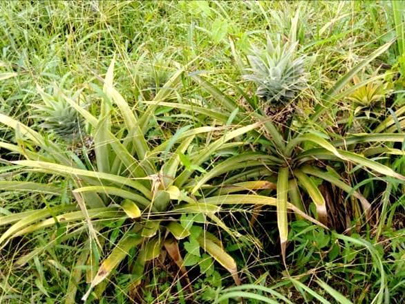
Pineapple is one of the most economically important crops. The country ranks second in
the world, next to Thailand in terms of pineapple production with an estimated of 70,000
hectares planted with the crop. Most are exported thus, contributing to about 17% of the
world supply [1]. Majority of the production is concentrated in Mindanao, with other
provinces in Luzon close behind. However, 85% of the production is controlled by giant
transnational corporations, led by Del Monte and Dole Philippines. However, even if there is
a pedestal for the Philippine pineapple industry, there are still outlying concerns to be
considered. These includes calamities, bug infestations, and infections of the crops by either
fungi or plant parasitic nematodes. Plant parasitic nematodes are one of the major causes of
plant infections and affect the production of crops worldwide [2]. Aside from pineapples,
wheat, rice, maize, potatoes, sweet potatoes and many more are some of reported crops that
have problem with crop losses associated with plant parasitic nematodes [3]. Nematodes are
the most abundant group of multicellular creatures on earth in terms of numbers of
individuals and about 10% of all nematode species are plant parasites. Plant parasitic
nematodes are minute organisms that live underground [4]. Today, plant parasitic nematodes
945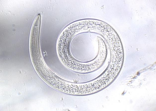
European Journal of Molecular & Clinical Medicine

                                          ISSN 2515-8260               Volume 08, Issue 02, 2021

are apprehended as major rural pathogens and are known to assault plants and cause
misfortunes over the world. They maintain the pedestal of one of the most harmful and hard
to oversee agronomic irritations, and strain to decrease the utilization of exceptionally toxic
nematicides [5]. In 2006, there were already 4,100 species of plant-parasitic nematode that
have been identified (Jones et al., 2013) and yearly, they are causing an estimated annual
crop loss of $78 billion worldwide with 10-15% average crop yield loss [6]. Even in this
modern day, there are still a lot of producers, especially in developing countries that are
ignorant of plant parasitic nematodes.

    The feeding strategy of plant parasitic nematodes in pineapple is usually from the soil
through the roots. Majority of the infections includes lesions of piths and vascular tissues
necessary for water and food transport of the plant. When these tissues are disrupted,
chlorosis will start to occur on the leaves. On the later stage of infection, leaves will facilitate
necrosis, and the roots can be easily pulled from the infected soil. These infections can be
contagious to its relative plants and causes permanent damage to the soil, leaving a permanent
annual loss of production [2].

    With the damage inflicted by PPN’s on pineapples, this study aimed to determine the
PPN’s associated with the crop. Samples were taken from provinces of Cavite, Laguna and
Batangas in Luzon, Philippines where the pineapple plantations are located. This study will
help provide additional information on the occurrence of plant parasitic nematodes on
pineapple crops in the Philippines and sends an awareness to pineapple farmers who are
clueless on the effect of PPN’s on their crops.

   2. METHODOLOGY

     A. Sampling site and sample collection
     The soil and root samples of pineapples were collected from pineapple farms at Tagaytay
City, Cavite, Laguna and Batangas. The surrounding soil of each pineapple plant as well as
its roots was collected as the primary samples. Ensuing the protocol of Coyne et al. [7], the
collection of both roots and soil were done randomly, following a zigzag pattern to ensure
homogeneity. The surface litters at each sampling spot were removed first before drawing out
the soil sample which was done by lashing the soil sample auger to a plough depth of 15 to
20 centimeters. On the other hand, the fine roots were gathered by digging the soil and
cutting it from the main roots using a clean scissor. All the collected soil and root samples
from the two different areas were separately packed in clean polyethylene bags and labeled.
Then, all samples were put in an ice chest to maintain the moisture of the soil while being
transported to the laboratory.

    B. Soil analysis
    One kilogram of soil sample was collected from the sampling site and was sent to Bureau
of Soils and Water Management (BSWM) in Quezon City, Philippines for the analysis of its
nitrogen, phosphorus and potassium contents using a soil test kit.
    C. Extraction of nematodes
    Stones and debris were removed from the soil samples after thoroughly mixed. Five set-
ups were prepared for the extraction of nematodes following the Baermann Tray technique
[7, 8]. After the extraction period, soil samples were sieved wherein the water suspensions
from the five trays were poured together onto three tilted layers of stainless steel sieve (45
μm, 105 μm, and 180 μm) with the smallest size at the bottom. The debris were condensed by

                                               946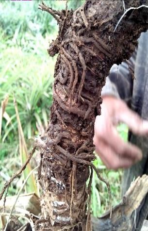
European Journal of Molecular & Clinical Medicine

                                        ISSN 2515-8260               Volume 08, Issue 02, 2021

gently but thoroughly rinsing 180 μm sieve with wash bottle mainly from the back to ensure
that all debris and nematodes from the sieve surface were collected at the bottom point. All
the debris from 180 μm sieve were disregarded while the debris from the remaining 105 μm
and 45 μm were gently washed into a labeled glass bottle. As for the extraction of root
nematodes, incubation technique with hydrogen peroxide (H2O2) was used. The root samples
were gently washed in a running water removing all the associated soils, cut into 1-2
centimeters short and were mixed thoroughly. All the chopped root samples were weighed 15
grams for each of the two 30 milliliter amber bottles which were filled with 1% concentrated
H2O2. The bottles were tightly covered and labeled with the crop name and date of
preparation before incubating for 48 hours at room temperature. After incubation period,
sieving method was done wherein all the contents of the two bottles were poured on to three
tilted layers of stainless steel sieve (40 μm, 80 μm, and 200 μm) with the smallest size at the
bottom. The debris were condensed by gently but thoroughly rinsing the 200 μm sieve with
wash bottle mainly from the back to ensure that all debris and nematodes were collected from
the mesh surface. The same process was done for the 80 μm and 40 μm sieve in where the
nematode suspension began to be collected using a vial.

    D. Killing and fixation of nematodes
    The killing procedure for the nematodes from both soil and root samples were executed
homogenously. Using an electric kettle, water was heated to nearly boiling point then, it was
transferred to a flat container where the glass bottles and vials were immersed and shaken in a
circular manner for about 40 seconds. The glass bottles were removed from the container
then, a double strength (2x) formalin-glycerol fixative with an equal volume of the nematode
suspension was poured into it. After fixation, the glass bottles were left in an undisturbed area
for at least 24 hours to let the suspension settle down as well as to ensure the complete
penetration of the fixative.

    E. Preparation of microscope slides
    Before picking and mounting the nematodes, proper layout of the microscope slides to be
used were primed. By means of a ringer, each microscope slides were painted at the center
with a clear nail polish in a circular manner. A microscope slide was clipped on the stage of a
ringer with the tip of the brush immersed in nail polish slightly touching the portion of the
slide while the stage was being spun [7].

    F. Picking and mounting of nematodes
    Based on the protocol of Coyne et al. [7], part of the nematode suspension was
transferred to a small petri plate then, was observed on a dissecting microscope with under-
stage lighting. The target nematode was located using the lowest magnification, verified at
higher magnification and was picked out using a coconut splinter. Then, the picked out
nematode was placed at the center of a glass slide with nail polish ring containing the single
strength (1x) formalin-glycerol mounting medium. By means of a light microscope with
under-stage lighting, the successful transfer of the nematode was confirmed as well as its
possible position on the glass slide. Afterwards, a cover slip was placed on top of the
specimen which was sealed by painting a clear nail polish on its four sides in which each
microscope slides was labeled with pertinent information about the contained specimen.

   G. Morphological Identification
   The well mounted plant parasitic nematodes were observed under light microscopes in
order to determine its genus identity. Morphological features such as body shape and
stomatostylet structure were first identify to determine which was free living or PPN’s. The

                                              947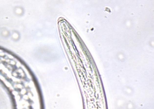
European Journal of Molecular & Clinical Medicine

                                        ISSN 2515-8260              Volume 08, Issue 02, 2021

initial identification on genus level was completed following the taxonomic keys of plant
parasitic nematodes by Stirling et al. [9]. Furthermore, morphometric analysis of each
nematodes were also executed using the ImageJ application following these sets of
parameters from de Man‟s Indices [10]: n (number of specimens on which measurements
were based), L (overall body length) V (length of vulva), S (stylet or spear length), and s =
spicule length. Then, all the pre-identified nematodes were sent to the Department of Plant
Pathology in University of the Philippines – Los Baños for further validation. Lastly, all the
identified plant parasitic nematodes were photographed using the Image Focus 4 application.
Also, appropriate labeling of the nematode body parts were completed which include the
ventral (V) part, dorsal (D) part, stomatostylet, anterior and posterior ends of the body [9].

    H. Frequency Count and Data Analysis of Plant Parasitic Nematodes
    The occurrence of all the extracted plant parasitic nematodes from both soil and root
samples were done by pouring the water suspension from a sample bottle into two petri plates
that were divided into quadrants – this process was done for three trials. Separately, the petri
plates containing water suspension were observed under a light microscope following a
zigzag pattern to ensure homogeneity. This process was performed in order to confirm the
infestation of certain plant parasitic nematode species in pineapple. Moreover, following the
principle by Afolami and Daramola [11], the percentage frequency of occurrence of the
PPN’s was obtained.

   3. RESULTS AND DISCUSSION

    A. Plant Parasitic Nematodes associated with Ananas comosus
    Four species were observed and recorded from the soil and root samples of pineapples. It
includes Rotylenchus spp., Ditylenchus spp., Helicotylenchus spp., and Rotylenchulus spp.,
respectively. The Helicotylenchus spp., was noticeably in higher number in soil samples but
lesser in the root samples. The results obtained coincides with the study of Sipes and Wang
[12] and Afolami and Daramola [11] where all the four PPN’s obtained in the samples were
also the PPN’s they have observed in pineapple.

    Helicotylenchus spp. belongs to the order Tylenchida and family Hoplolaimidae. It is the
most rampant species observed in this study and known for its spiral body shape with body
length that ranges from 438 to 506 μm. However, the shape of the stylet knobs varies from
one individual to another. As shown in Figure 1B, the representative female species of
Helicotylenchus that was picked from the soil samples has rounded knob, conversely,
anteriorly flattened stylet knob was present in the female species found in the root samples
(Figure 2A). Likewise, the stylet length of the extracted species from both soil and root
samples ranged from 12 to 16μm long with 4 μm vulva length, however, the vulva of the
female species from the soil is not prominent. The identification of these organisms under
this genera were based from the dichotomous key of Stirling et al.[9] which were further
supported by a study of Palomares-Rius et al. [13] proving that Helicotylenchus spp. is
characterized by forming a spiral shape when killed and a robust stylet with knobs flattened
and indented anteriorly. Moreover, an extremely inconstant reproduction is extant amongst
species of Helicotylenchus [14]. Some species under this genus have males and females
which sexually reproduced. Others are hermaphrodites that self-fertilize their own eggs
without mating while some species only have females which reproduced through
parthenogenesis since males are either absent or extremely rare [15]. Additionally, spiral
nematodes are generally considered as ectoparasite but, species such as Helicotylenchus
pseudorobustus have an occasional semi-endoparasitic feeding strategy. They infest wide

                                              948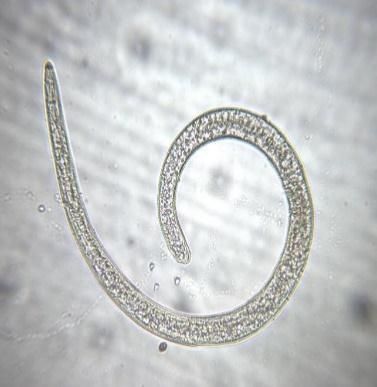
European Journal of Molecular & Clinical Medicine

                                           ISSN 2515-8260                Volume 08, Issue 02, 2021

range of crops such as pineapples, soybean, cotton, corn and banana as well as in turf grasses
like Bermuda grass and Paspalum vaginatum [14].

           A                                                     B
                                       Stomatostylet
                                                                              V

                                                                     D

            11 μm                                                11 μm

           Fig 1 Micrographs of female Helicotylenchus spp. picked from the soil samples.
           (A) Anterior portion of the PPN body displaying the stomatostylet; (B)
           General morphology of the parasitic nematode.

            A                          D                 B
                    Anterior
                                                                               Stomatostylet
                    V

                           Posterior

            11 μm                                        11 μm

           Fig 2 Micrographs of female Helicotylenchus spp. picked from the root samples.
           General morphology of the species; (B) Anterior part of the body showing the
           flattened stylet knob

        Another PPN observed in pineapple’s soil and root samples was Rotylenchulus [16]
belonging to the order Tylenchida and family Hoploloamidae. Further, the extracted
Rotylenchulus spp. from both soil and root samples showed a curved or C-shaped body
situation (Figures 3A and 4B) with immature females having a body length that ranged from
266 to 282 μm long stylet that has rounded basal knobs, not prominent vulva and slightly
tapering to a narrow and rounded terminal tail. On the other hand, males have approximately
299 μm body length, barely notable or weak stylet that was about 15 μm and at least 14 μm
spicule with its curved posterior part of the body. The morphometric results of this study
coincide with the study of several researchers [13, 17].
        Based on the research of [17],,Rotylenchulus spp. have both female and male species.
However, only the female species are infective since the males remain outside of the host’s
roots. The immature female imbeds her head intro the root tissue while the posterior portion
protrudes from the root surface and swells during maturation. The infective stage of its

                                                 949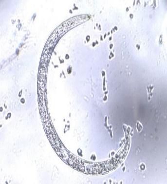
European Journal of Molecular & Clinical Medicine

                                          ISSN 2515-8260           Volume 08, Issue 02, 2021

female species stretched from one to two weeks after hatching and matures after one or two
weeks once they penetrated into the roots of the host plant. Subsequently, species under this
genus are pests of wide range of plants which includes pineapple, cotton, soybean and
cowpea to name a few. Among these crops, pineapple is one of the severely affected by the
species Rotylenchulus spp. The amount and type of damage frequently relies upon the host
species or cultivar as well as the population of nematodes, which causes a reduced root
system, leaf chlorosis, overall stunting of host plants, and reduced yields and plant longevity
[18].

           A       Anterior
                                                     B
                                                                    Spicule

                    Posterior        V

                                      D
          11 μm                                       11 μm

        Fig 3 Micrographs of male Rotylenchulus spp. picked from the soil samples.
        (A) General morphology showing the spicule at the posterior end; (B)
        posterior end with the spicule (14 μm).

           A                                         B
                                                                       Anterior
                                Stylet knob              D

                                                              V

                                                                     Posterior

               11 μm                                 11 μm

         Fig 4 Micrographs of immature female Rotylenchulus spp. picked from the
         root samples. (A) Buccal cavity of the species; (B) general morphology of the
         nematode showing the habitus mortis.
        Ditylenchus spp. [19] belongs to order Tylenchida and family Anguinidae. The
Ditylenchus spp. (Figure 8) collected from the soil samples was observed having a slightly
curved to straight body shape with a body length of 1100 μm long, a stylet of 21μm long and
slightly pointed anterior and posterior ends. The identification of this species using its
morphological features was done following a dichotomous key from Stirling et al., (2014)
and was further supported by the study of Esmaeili et al.[20] which also described the
tapering at both body ends of Ditylenchus spp., short stylet and its rounded knobs.
        Based on some studies, Ditylenchus spp. is well-known plant parasitic nematodes of
garlics, onions and rice that are that are present on the stem and bulb of the crops thereby
being commonly known as stem nematodes. Species under this genera are considered as

                                              950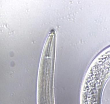
European Journal of Molecular & Clinical Medicine

                                            ISSN 2515-8260                  Volume 08, Issue 02, 2021

migratory endoparasites of plants but they can also be mycophagous or fungivorous that
feeds on fungi [21], which causes stunted growth, discoloration, swellings and distortion of
aerial plant parts (stems, leaves, flowers) and necrosis or rotting of stem bases, bulbs, tubers
and rhizomes. The juveniles of Ditylenchus spp. can survive in a dry condition for several
years and may persist in clay soils. Cool and moist conditions are favorable for the invasion
these plant parasitic nematodes [22].

               A                                         B
                                                                         Buccal cavity

                                           Posterior

                50 μm                                                              10 μm

            Fig 8 Micrographs of female Ditylenchus spp. picked from the soil
            samples. (A) Posterior morphology of the species through high power
            objective; (B) buccal cavity through oil immersion objective.
         Likewise, the identification of Rotylenchus spp., (Figure 5) extracted from the soil
samples was distinctive with their semi-spiral body shape of 518 μm long, a stylet length that
was 13μm, flattened knobs, 9μm vulva and a hemispherical tail which were supported by a
dichotomous key [9]. Its identification was further supported on the study of Palomares-Rius
et al. [13] in where morphometrics of female Rotylenchus spp. were elaborated with having
close C to spiral habitus that varies depending on species, body length of 500–1856 μm long,
and 20 to 51 μm stylet with rounded to hemispherical tail.

       Plant parasitic nematode species of genus Rotylenchus spp. are generally
ectoparasitic, rarely endoparasite that feed on roots of wide range of plants such as
strawberry, sunflowers, carrots, pineapples and boxwood among others [11, 23], which
causes stunting andyellowing of the plant host that have been associated with large
numbers of Rotylenchus spp. [24]. Rotylenchus belongs to the family of Tylenchida and
order Hoplolaimidae.

              A                                         B
                   Anterior

                                                         Stomatostylet

                               Posterior

             11μm                                       11μm

           Fig 5 Micrograph of female Rotylenchus spp. picked from the soil
           samples. (A) General morphology of the species; (B) anterior part of the
           body.

                                                  951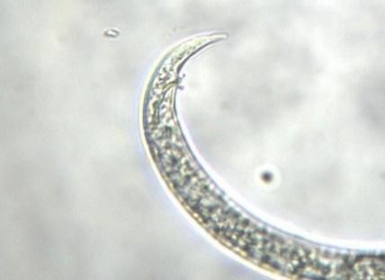
European Journal of Molecular & Clinical Medicine

                                         ISSN 2515-8260              Volume 08, Issue 02, 2021

        All the extracted plant parasitic nematodes in this study are root-feeders and are
considered as herbivores since they feed directly on plants and its tissues thereby being
included at the first tropic level of the soil food web excluding Ditylenchus spp. since species
of this genus have life styles that ranges from being plant parasites to fungal feeders. Thus, its
species belong to different trophic level depending on its life cycle; mycophagous and plant
parasite species belong to the second and first trophic level, respectively [25].

       Identifying plant parasitic nematodes using only the morphological technique could
really be difficult as some species bear resemblance to others in terms of body shape.
Nevertheless, despite the innovating usage of molecular method, conventional taxonomy
using reliable morphological characters is still a vital tool for the identification of nematodes
primarily because it allows a clear link between function and morphological aspects of the
specimen analyzed, it is suitable for quantitative evaluations, it is also used for population
surveys of plant parasitic nematodes to name a few but more importantly, it is cheap [18].

        B. Soil Characteristics of the Sampling Site
        Soil nutrient content such as nitrogen, phosphorus and potassium and soil
characteristics such as pH and texture could influence the proliferation of plant parasitic
nematodes on the sampling site. Among the different PPNs, Helicotylenchus spp. has been
said to thrive in all soil types [26].

        Changes in the soil chemical factors could affect the nematode’s ability to locate plant
roots and reproduction. Presence and distribution of plant parasitic nematodes vary depends
on abiotic characteristics of the soil [26]. As shown on the result of soil analysis (Table 2),
the soil sample collected from the study area have a medium or clay loams to sandy clay
loams texture with a pH level of 4.9. This result concurred with an article by the Bureau of
Plant Industry which indicated that pineapples optimally thrive in sandy loam to clay loam
soils and has a pH of 4.5 to 5.5. Soil texture plays a significant role in nematode distribution
for it has a direct impact to the population density of plant parasitic nematodes which
supports the conclusion of this research [27]; the soil type requirement for the growth of
pineapple is a well-drained sandy loam and has an optimum range of pH level of 4.5 to 5.5,
which is also ideal for nematode reproduction and infectivity [28, 29]. Additionally, it was
indicated that Rotylenchulus spp. has an optimal pH level of approximately 4.8 to 5.2 thereby
[16], supporting the extraction of Rotylenchulus spp. from the soil samples of this study.

        Moreover, the result of physicochemical soil analysis of the soil samples shows that it
has low nitrogen level, medium content of phosphorus and sufficient amount of potassium.
Nitrogen (N) significantly influences the aboveground ecosystem productivity and below-
ground pools. It plays a vital role in biochemical and physiological functions of plant since
nitrogen is a major component of chlorophyll that is used by plants for its food production as
well as amino acids which is the building blocks of protein necessary for proper development
of plants [30]. However, in relation to other nutrients, too much nitrogen could also induce
lateness in flowering or fruit formation, unproductive growth, and prone to disease [31].

                                               952
European Journal of Molecular & Clinical Medicine
ISSN 2515-8260 Volume 08, Issue 02, 2021
Table 2. Physicochemical analysis of the soil sample from the sampling site
Description Test Result
pH N P K
Soil Sample 4.9 Low Medium Sufficient
On the other hand, phosphorus (P) is an essential macro-element used by plants
during photosynthesis, respiration, energy storage and transfer, cell division, cell enlargement
and several other processes in plants. Phosphorous was also taken by plants in relatively
small quantities and shortage of this macro-element results in blue coloration while excess
amount of phosphorous on the plant was extremely difficult to define [31]. Whereas,
potassium (K) in soil is known for its role in regulation of plant metabolic activities as well as
its help in minimizing plant stress. It is present in soils in larger quantities than either
nitrogen or phosphorus [31] which supported the result. It is required in large amount
particularly by those flowering plants or fruit producing plants and shortage of this macro-
element causes marginal leaf scorch while excess amount results in stunted dark blue tinted
growth and sometimes burning of the tips of the leaf. Additionally, there is a close
relationship between the effects of nitrogen and those of potassium, and successful
cultivation of most plants depends on the correct ratio of nitrogen to potassium, while
phosphorus playing little part provided that it is still present in sufficient quantity[31].
In the greenhouse study which evaluated the influence of soil nutrients in the presence
of reniform nematodes, low level of P can suppress plant development and increase the
number of the Rotylenchulus spp. whereas, increase in nitrogen level can suppress plant
parasitic nematodes which means that if the soil has a low amount of N [32]. Then, there will
be an increase in the population of plant parasitic nematodes [7]. On the other hand, unlike
the two minerals, potassium shows no effect on the reproduction of plant parasitic nematodes
[32]. Nonetheless, several studies have proved that the combination of these three minerals
with high amount of availability have a strong correlation with the population decline of plant
parasitic nematodes [32, 33]. Hence, high amount or deficiency of these vital soil macro-
nutrients as well as high population of plant parasitic nematodes can both greatly affect the
development of a plant since low amount of nitrogen in the soil may result to stunted growth
with discoloration on the leaves since, these symptoms are indications of protein deficiency
in the plant and nitrogen deficiency in the soil. However, stunted growth and chlorosis are
also above ground symptoms of disease [26] caused by plant parasitic nematodes such as
Ditylenchus spp. and Rotylenchus spp. [24].
Subsequently, similar symptoms of abnormal discoloration showed when phosphorus
is deficient. In pineapple, aside from chlorosis, phosphorus deficiency also results to the
reduction of plant height, length and number of the leaves which is due to the reduction in the
cell division [29]. Conversely, potassium is not organically bound which means that when the
plant dies and decomposes, it is immediately released back on the soil. Yet, if some crops
have no adequate amounts of potassium they exhibit symptoms such as yellow or white spots
on the margins of the leaflets with symptoms first appearing on the older plant tissue and
reduction in plant growth, root development, as well as fruit development [27]. Consequently,
plant parasitic nematodes that feed on the roots such as Helicotylenchus spp., Rotylenchus and
953European Journal of Molecular & Clinical Medicine
ISSN 2515-8260 Volume 08, Issue 02, 2021
Rotylenchulus spp. also caused above ground symptoms that mostly resulted into many root
injuries such as root necrosis (Figure 10) resulting in severe root pruning [34]. Also,
Rotylenchulus spp. attacked the fibrous roots reducing the absorption ability and other
physiological functions of the plant.
A B
Fig 10 Pineapple plants in the sampling site showing symptoms of plant
parasitic nematode infection. (A) Rotten and impaired root system; (B)
stunted growth and discoloration on the leaf.
Nevertheless, plant diseases are also caused by unfavorable environmental conditions.
Temperature, moisture, light and the properties of the soil are environmental factors which
could affect the plant growth. There is an optimum range for each environmental factors
within which plants grow best, however, growth abnormalities might exhibit if the level of
environmental factor varies greatly from its optimum, or severely impacts on another factor
(Ogle, n.d.). Therefore, biotic and abiotic factors not only greatly affect the growth of plants
but are also considered as factors of plant diseases, which were observed in the sampling
area.
C. Nematode Population
Of the ten set-ups each with 100 g of soil samples, Helicotylenchus spp. was the most
prevalent plant parasitic nematode species encountered. The population density of
Helicotylenchus spp., observed from the soil samples is shown in Table 3.
Table 3. Population Density of Helicotylenchus spp. Associated with Soil and Root Samples of A. comosus
Nematode Population (%)
Collection Schedule Soil Frequency Root Frequency
Sample Percentage Sample Percentage
1st collection 39.54% 51.8% 25% 0.6%
2nd collection 37.18% 99.8% 30.43% 1.4%
Total Percentage 37.95% 75.8% 28.57% 1%
The result showed that Helicotylenchus spp. covered 39.54% of the nematode
population with 51.8% frequency rating for the first collection. Subsequently, comparatively
37.18% of Helicotylenchus spp. manifested on the second sample size with 99.8% of
frequency rating. It is noticeable that the frequency rating of Helicotylenchus spp. on the
second collection was relatively higher than the first collection nevertheless, the attained
combined frequency rating data still suggest that Helicotylenchus spp., favors the clay loams
type of soil; this result corresponds with those of other researchers who have conducted
similar studies [18, 19].
954European Journal of Molecular & Clinical Medicine
ISSN 2515-8260 Volume 08, Issue 02, 2021
On the other hand, Table 4 presented the low occurrence of Helicotylenchus spp., in
the collected root samples with 0.6% frequency rating from the first collection and a fair
increase to 1.4% in the second collection thereby, proposing that not all plant parasitic
nematodes that infest the soil can also be abundant in roots. Nematode occurrence is highly
dependent on the presence of host plants. However, its presence is likewise influenced, more
or less, by most of the biological, chemical and physical components of the soil environment
[24]. Having been observed, the trophic level of the recorded nematode species shows its
dependence on the presence of their host plant. Furthermore, chlorosis, necrosis, and stunted
growth on the pineapples can be inferred that, that is due the presence of the identified plant
parasitic nematodes.
4. CONCLUSIONS AND RECOMMENDATIONS
Plant parasitic nematodes identified in pineapple fields in Cavite, Batangas and Laguna were
Rotylenchus spp., Ditylenchus spp., Helicotylenchus spp., and Rotylenchulus spp. The most prevalent
nematode identified was Helicotylenchus spp that might have affected the crops manifesting
symptoms such as chlorosis, necrosis and stunting. Studies on plant parasitic nematodes in the
Philippines are still limited and so does the knowledge of farmers regarding these parasites, thus,
further studies with wide sampling sites on the occurrence of plant parasitic nematodes in pineapples
is recommended. To further identify the PPN into species level, molecular identification is
recommended.
5. ACKNOWLEDGMENT
The authors are grateful to Asst Prof. Romnick A. Latina of Plant Pathology
Department at University of the Philippines, Los Baños, Laguna, their consultant in the study
and for letting them use the Nematology Laboratory in UPLB. They are also grateful to
Commission on Higher Education (CHED) through K to 12 Research – DARE To Grant for
funding assistance given to this research.
6. REFERENCES
[1] Baroña, M. J. (2005). Our Fruit Industry: Where We Stand. BAR Digest July-September 2005
(Vol. 7 No.3). Retrieved from: https://www.bar.gov.ph/index.php/digest-home/digest-
archives/80-2005-3rd- quarter/4486-julsep05-fruit-industry.
[2] Jones, J.T., Haegeman, A., Danchin, E.G.J., Gaur, H.S., Helder, J., Jones, M.G.K.,…Perry, N.R.
(2013). Top 10 Plant Parasitic Nematodes in Molecular Plant Pathology. 14 (9), 946-961. DOI:
10.1111/mpp.12057.
[3] Bernard, G.C, Egnin, M and Bonsi C. (2017). The Impact of Plant-Parasitic Nematodes on
Agriculture and Methods of Control, Nematology - Concepts, Diagnosis and Control,
Mohammad Manjur Shah and Mohammad Mahamood, IntechOpen, DOI:
10.5772/intechopen.68958.
[4] Lambert, K. and S. Bekal. (2002). Introduction to Plant-Parasitic Nematodes. The Plant Health
Instructor. DOI: 10.1094/PHI-I-2002-1218-01
[5] De Oliveira, C.M.G., Monteiro, A.R., Blok, V.C. (2011). Morphological and molecular
diagnostics for plant-parasitic nematodes: working together to get the identification done.
Tropical Plant Pathology, vol. 36, 2, 065-073.
[6] Carneiro, R.M.D.G., Lima, F.S.O., Correia, V.R. (2017). Methods and Tools Currently Used for
the Identification of Plant Parasitic Nematodes. Http://dx.doi.org/10.5772/intechopen.69403.
[7] Coyne, D.L., Nicol, J.M. and Claudius-Cole, B. (2014). Practical plant nematology: A field and
Laboratory Guide. 2nd edition. SP-IPM Secretariat, International Institute of Tropical
Agriculture (IITA), Cotonou, Benin.
[8] Whitehead, A.G. and Hemming, J.R. (1965). A Comparison of Some Quantitative Methods
Extracting Small Vermiform Nematodes from the Soil. Annals of Applied Biology, 55, 25-38.
http://dx.doi.org/10.1111/j.1744-7348.1965.tb07864.x
955European Journal of Molecular & Clinical Medicine
ISSN 2515-8260 Volume 08, Issue 02, 2021
[9] Stirling GR (2014b) Integrated soil biology management. In: Stirlling G (ed) Biological control
of plant-parasitic nematodes. CABI Publishing, CAB International, Wallingford, pp 304–341
[10] Yongsan, Z., Ye, W., Tredway, L., Martin, S., Martin, M. (2012). Taxonomy and morphology of
plant parasitic nematodes associated with turfgrasses in North and South Carolina, USA.
Zootaxa 3452: 1–46
[11] Afolami, S., and Daramola F. (2014). Studies on the Distribution of Plant parasitic
Nematodes Associated with Pineapple in Delta, Imo and Cross River states of Nigeria.
Australian Journal of Basic and Applied Sciences. Pages: 248- 25
[12] Sipes, B.S. and D.P. Schmitt. (2000). Rotylenchulus reniformis damage threshold on
pineapple. Acta. Horticulturea, 529: 239-246.
[13] Palomares-Rius, J. E., Cantalapiedra-Navarrete, C., Vovlas, N., Subbotin, S. A., Castillo, P.,
Archidona-Yuste, A., Tzortzakakis, E. A., Birmpilis, I. G. (2017). Prevalence and molecular
diversity of reniform nematodes of the genus Rotylenchulus (Nematoda: Rotylenchulinae) in
the Mediterranean Basin. Eur J Plant Pathol.DOI 10.1007/s10658-017-1292-8
[14] Crow, W.T. (2017). Helicotylenchus spp. (Nematoda: Tylenchida: Hoplolaimidae).
Universityof Florida,EENY-544. Retrievedfrom:
http://entnemdept.ufl.edu/creatures/nematode/spiral_nematode.htm
[15] Van Megen, H., Van den Elsen, S., Holterman, M., Karssen, G., Mooyman, P., Bongers, T.,
Holovachov, O., Bakker, J. and Helder, J. (2009). A Phylogenetic Tree of Nematodes Based on
about 1200 full-length Small Subunit Ribosomal DNA Sequences. Nematology, 11, 927–950.
[16] Luc, M., Sikora, R.A., Bridge, J. (2005). Plant Parasitic Nematodes in Subtropical and Tropical
Agriculture. CABI Publishing, Cambridge, M.A.
[17] Wang, K.H. (2007). Rotylenchulusreniformis Linford & Oliveira (Nematoda: Tylenchida:
Tylenchoidea: Hoplolaimidae: Rotylenchulinae). University of Florida, EENY-210. Available
from: http://entnemdept.ufl.edu/creatures/nematode/r_reniformis.htm.
[18] Ferris, V.R. and Bernard, R.L. (1971). Crop Rotation Effects on Population Densities of
Ectoparasitic Nematodes. Retrieved from:
https://www.ncbi.nlm.nih.gov/pmc/articles/PMC2619873/pdf/119.pdf.
[19] Fleming, T.R.,McGowana, N.E.,Maulea, A.G., and Fleming, C.C. (2016). Prevalence and
diversity of plant parasitic nematodes in Northern Ireland grassland and cereals, and the
influence of soils and rainfall. DOI: 10.1111/ppa.12525.
[20] Esmaeili, M., Heydari, R., Castillo, P., Palomares-Rius. J.E. (2017). Molecular and
Morphological Characterisation of Ditylenchus persicus n. sp. (Nematoda: Anguinidae) from
Kermanshah province, western Iran. Nematology 19 (2017) 211-223
[21] Pethybridge, S. J., Gorny, A., Hoogland, T., Jones, L., Hay, F., Smart, C., &Abawi, G. (2016).
Identification and characterization of Ditylenchus spp. populations from garlic in New York
State, USA. Tropical Plant Pathology, 41(3), 193-197.
[22] Data Sheets on Quarantine Pests Ditylenchus dipsaci. (n.d). Retrieved from:
https://gd.eppo.int/download/doc/105_datasheet_DITYDI.pdf
[23] Siddiqi, M.R. (2000). Tylenchida; Parasites of Plants and Insects. Second Edition. CABI
Publishing,275.Retrievedfrom:https://books.google.com.ph/books?id=s9p41TTWLPEC&pg=P
A275&lpg=PA275 dq=rotylenchus+host+plant&source=bl&ots=H8uJhJug9-&sig
[24] Westerdahl, B.B. and Ploeg, A. (2017). Nematodes. UC IPM Pest Management
Guidelines: Lettuce, UC ANR Publication 3450. Available from:
https://www2.ipm.ucanr.edu/agriculture/lettuce/nematodes/.
[25] Ingham, E.R. (2019). Chapter 6: Nematodes. Oregon State University. Available from:
https://extension.illinois.edu/soil/SoilBiology/nematodes.htm.
[26] Cadet, P and Spaull, VW (2001). Nematodes and nutrients: Association between plant parasitic
nematodes and soil chemicals. Proc S AfrSugTechnol Ass 75: 116-117.
[27] Warner, F. (2009). Soil fertility, pH, texture and nematodes. Available from:
https://www.canr.msu.edu/news/soil_fertility_ph_texture_and_nematodes.
[28] Seid, A., Goftishu, M.,Degebassa, L., and Mekete, T. (2015). Occurrence, distribution, and
abundance of plantparasitic nematodes associated with khat (Catha edulis Forsk) in East
Hararghe Zone, Ethiopia. Nematropica 45:208-214.
956European Journal of Molecular & Clinical Medicine
ISSN 2515-8260 Volume 08, Issue 02, 2021
[29] Sipes, B., and Wang, K.H. (2016). Chapter 4: Pests, Diseases and Weeds. Plant and
Environmental Protection Sciences, University of Hawaii at Manoa, Honolulu, HI, 96822,
Hawaii, USA. https://doi.org/10.1002/9781118967355.ch4
[30] Leghari, S. J., Wahocho, N. A., Laghari, G. M., HafeezLaghari, A., MustafaBhabhan, G.,
HussainTalpur, K., ... &Lashari, A. A. (2016). Role of nitrogen for plant growth and
development: A review. Advances in Environmental Biology, 10(9), 209-219.
[31] Pinkney, Dave. (2011). Macro and Micro-Elements in Soil for Plant Nutrition. Available from
http://www.gardeninginfozone.com/macro-elements-in-soil-for-plant-nutrition
[32] Kularathna, M.T., Overstreet, C., McGawley, E.C., Xavier, D.M., and Martin, C.M.(2014).
Influence of Soil Nutrients on Reproduction and Pathogenicity of Rotylenchulus reniformis on
Cotton. Nematropica 44:15-24. Available from:
http://journals.fcla.edu/nematropica/article/viewFile/83312/80184.
[33] Gruzdeva, L.I., Matveeva, E.M., and Kovalenko, T.E. (2007). Changes in Soil Nematode
Communities under the Impact of Fertilizers. ISSN 1064-2293, Eurasian Soil Science, 2007,
Vol. 40, No. 6, pp. 681–693. DOI: 10.1134/S1064229307060105.
[34] Subbotin, S. A., Madani, M., Krall, E., Sturhan, D., and Moens, M. (2005). Molecular
diagnostics, taxonomy, and phylogeny of the stem nematode Ditylenchus dipsaci species
complex based on the sequences of the internal transcribed spacer-rDNA. Phytopathology 95,
1308–1315. doi: 10.1094/PHYTO-95-1308
957You can also read