JBC Papers in Press. Published on July 29, 2020 as Manuscript AC120.014918 The latest version is at ...
←
→
Page content transcription
If your browser does not render page correctly, please read the page content below
JBC Papers in Press. Published on July 29, 2020 as Manuscript AC120.014918
        The latest version is at https://www.jbc.org/cgi/doi/10.1074/jbc.AC120.014918

  Identification of an anti-SARS-CoV-2 receptor binding domain directed
     human monoclonal antibody from a naïve semi-synthetic library

   Hilal Ahmad Parray1£, Adarsh Kumar Chiranjivi1£, Shailendra Asthana1, Naveen Yadav1, Tripti
   Shrivastava1, Shailendra Mani1Chandresh Sharma1, Preeti Vishwakarma1, Supratik Das1, Kamal
            Pindari1, Subrata Sinha 2 Sweety Samal1, Shubbir Ahmed1*, Rajesh Kumar1*

   1
  Translational Health Science & Technology Institute, NCR Biotech Science Cluster, Faridabad,
 Haryana-121001, India. 2Department of Biochemistry, All India Institute of Medical Sciences, New
                                             Delhi

Running Title: Monoclonal antibody (mAb) targeting RBD of SARS-CoV-2

Key words: RBD, scFv, ACE-2, mAb, IgG1

*To whom correspondence may be addressed. E-mail: rajesh@thsti.res.in, sahmed@thsti.res.in

                                                                                                               Downloaded from http://www.jbc.org/ by guest on October 30, 2020
ABSTRACT:
         There is a desperate need for safe and           IgG1). All the three antibody formats showed
effective vaccines, therapies and diagnostics for         high binding specificity to CoV-2 RBD and the
SARS-CoV-2, the development of which will be              spike antigens in different assay systems. Flow
aided by the discovery of potent and selective            cytometry analysis demonstrated specific binding
antibodies against relevant viral epitopes. Human         of the IgG1 format to cells expressing membrane
phage display technology has revolutionized the           bound CoV-2 spike protein. Docking studies
process of identifying and optimizing antibodies,         revealed that the scFv recognizes an epitope that
providing facile entry points for further                 partially overlaps with angiotensin converting
applications. Here in, we use this technology to          enzyme 2 (ACE2)-interacting sites on the CoV-2
search for antibodies targeting the receptor              RBD. Given its high specificity and affinity, we
binding domain (RBD) of CoV-2. Specifically,              anticipate that these anti-CoV-2 antibodies will
we screened a naïve human semi-synthetic phage            be useful as valuable reagents for accessing the
library against RBD, leading to the identification        antigenicity of vaccine candidates, as well as
of a high-affinity single chain fragment variable         developing antibody-based therapeutics and
region (scFv). The scFv was further engineered            diagnostics for CoV-2.
into two other antibody formats (scFv-Fc and

                                                          clinically approved therapeutics are available for
INTRODUCTION                                              CoV-2 and only symptomatic treatment is offered
        The recently identified novel human               (1). One of the approaches is to look for
coronavirus, referred as severe acute respiratory         neutralizing antibodies (NAbs) for CoV-2 either
syndrome coronavirus 2 (CoV-2) is the causative           from convalescent patient samples or from
agent of the ongoing pandemic of COVID-19.                synthetic antibody library sources. The antibodies
After the report of first case of the CoV-2               can work via two different mechanisms i.e. by
infection in December 2019, it has spread all over        direct neutralization of target viral antigen and
the globe. To slow down the spread of COVID-              also by indirect effector mechanisms such as
19, many countries have introduced lockdown               antibody-dependent cell-mediated cytotoxicity
measures. However, these measures will not be             (ADCC) and complement-dependent cytotoxicity
enough to eradicate the coronavirus pandemic              (CDC), wherein, antibody binds to infected cells
from the world and there is an urgent requirement         and potentially clear the viral reservoirs (2, 3).
for some medical intervention to control the                       The spike (S) protein on the surface of
spread of infection either in the form of a vaccine       CoV2 plays the primary role in viral attachment
or other therapeutic options such as small                to the host cell receptor followed by fusion and
molecule or therapeutic antibodies. So far, no            entry. The S protein comprises of two

                                                      1
components: S1, which contains the distinct RBD          was selected for further characterization (Fig.
(residues 318–510); and S2, which contains the           1C).
fusion peptide. The virus gains entry into
permissive host cells through interactions of the        Sequences analysis
RBD with the cell surface receptor                       The complete nucleotide sequence of the heavy
ACE2. Therefore, the RBD of CoV-2 S protein              and light chain variable region was determined
is the most likely target for development of virus       using the immunoglobulin BLAST homology
attachment inhibitors, NAbs, and vaccines (4).           search. The result indicated that the coding
An antibody against RBD is expected to block the         sequences are composed of VH gene (351 bp) and
attachment of the surface spike of virions to the        VL gene (327 bp). The closest germline sequence
ACE2 receptors on the host cell surface and is           for II62 scFv-antibody gene was identified by
thereby supposed to neutralize virus entry. Recent       comparison with the database. The VH gene use
success with plasma therapy from convalescent            the VH3 family-derived germline V3-23, D2-15,
patient samples against CoV-2 is a good indicator        and JH4 genes, while the VL gene uses the VL1
of possible successful antibody based therapy to         (V-kappa subgroup I, V1-39) family-derived
avoid further fatalities (5, 6). However, plasma         germline and Jk1. Also, a high degree of mutation
therapy is a crude method of treatment and a             appeared       in    the    VHCDR2        (CDR:
monoclonal antibody (mAb), the main ingredient           complementarity-determining region). The II62
of plasma therapy, is needed for wide and safe           mAb sequence showed, 70.8% (bases) homology

                                                                                                              Downloaded from http://www.jbc.org/ by guest on October 30, 2020
application (7, 8).                                      to the closest germline match (Fig. 1D & S1).
         Thus, novel tools and reagents for
therapy and diagnosis are urgently needed.               Expression, purification and characterization of
Keeping in view of the need, here, we used a scFv        II62-scFv
phage display library to identify a novel scFv,          The His-tagged II62-scFv was overexpressed in
II62, against RBD of CoV-2 that targets an               bacterial system and purified as soluble protein
epitope immediately adjacent to and slightly             using Ni-NTA affinity column. Overall yield of
overlapping with the ACE2 binding region on              the II62-scFv was 3 mg/L with >90 % purity. The
RBD. We further characterized the binding                purified II62-scFv migrated as a single band on
properties of II62-scFv and its two other formats,       SDS–PAGE with an estimated molecular mass of
scFv-Fc and IgG1 against both RBD and S                  28 kDa (Fig. S2D) and same was confirmed by
protein of CoV-2.                                        Western blot using HRP conjugated anti-His
                                                         antibody (data not shown). The functional
RESULTS                                                  activity and specificity of the purified II62-scFv
Identification of CoV-2 RBD reactive clones              was assessed by its binding to the RBD protein in
from phage display library                               ELISA. The II62-scFv showed specific binding to
The human synthetic Tomlinson I single fold              the RBD and did not bind to unrelated proteins
phage library was used for screening of scFvs            like BSA and the envelope proteins from HIV and
against the purified (>95% pure) RBD protein             Chikungunya (Fig. 2B & 2C).
from mammalian expression system. Three
rounds of panning were done and the titres of            Expression, purification and characterization of
input and output phage were calculated at the end        II62-scFv-Fc format.
of each round to monitor the efficiency of               The scFvs are monovalent in nature and lack the
enrichment. After three rounds of selection, ~           Fc mediated functions. The Fc domain of human
1100-fold enrichment of antigen specific clones          IgG1 was introduced to produce a homodimeric
was observed (Fig. 1A). After the third round of         II62-scFv-Fc chimeric protein which is capable of
panning, 50 clones were randomly picked and              bivalent binding and retain Fc functions (Fig.
assessed for binding to RBD protein by                   2D). The II62-scFv-Fc construct was transiently
monoclonal scFv-phage ELISA (Fig. 1B).                   expressed in Expi293F cells and subsequently
Twenty clones out of the 50 showed five times            purified by using protein G affinity column, with
better binding to RBD in ELISA as compared to            >95% purity and yields of 40-60 mg/L. The
the negative controls (BSA and helper phage) and         purified II62-scFv-Fc migrated as a single protein
hence, were considered as RBD specific binders           band on SDS–PAGE with an estimated molecular
One clone of scFv, II62, was dominantly selected         mass of 55 kDa (Fig. S2E). The scFv-Fc showed
via panning (50% of the selected binders), and           specific binding to RBD in ELISA (Fig. 2E & 2F)

                                                     2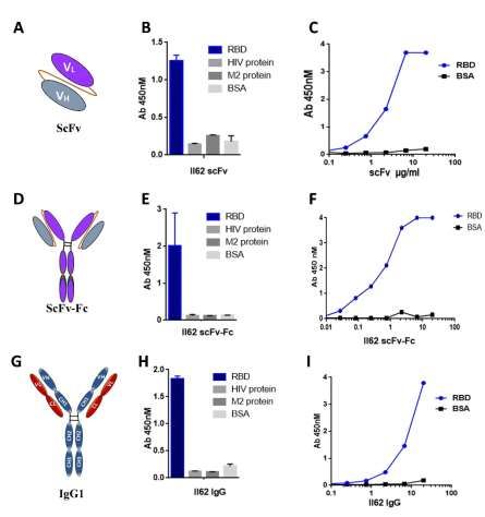
Expression and purification of full length II62-           specificity of II62 mAb was also tested towards
IgG1 mAb                                                   purified soluble spike protein through western
The full-length version of the II62-scFv (II62-            blot analysis and ELISA. Antibody II62
IgG1) was purified from the supernatant of                 specifically recognizes the band of ~180KDa
transiently transfected Expi293F cells using               (uncleaved soluble spike) (Fig. 3E & S5).
protein A/G affinity columns (Fig. 2G). The
purified IgG1 migrated as two protein bands on             Binding specificity of II62 mAbs towards
SDS–PAGE with estimated molecular mass of 50               cleaved and un-cleaved spike S protein
kDa for heavy chain and 25 kDa for light chain             Like other coronaviruses, the S protein of CoV-2
(Fig. S2F). Overall yield of the II62-IgG1 was 80-         contains cleavage sites and the precursor S
100 mg/L with >98 % purity. The purified II62-             protein is cleaved into the S1 and S2 subunits by
IgG1 also showed specific binding to CoV-2                 host cell proteases (9), an important step for
RBD protein (Fig. 2H & 2I)                                 initializing of infection. To further assess the
                                                           effect of the binding of II62-IgG1 to cleaved and
Affinity determination of II62 mAb formats                 un-cleaved forms of the S protein, 293T cells
To evaluate the affinity of scFv-Fc and IgG1               were transiently transfected with pseudo virus
format of II62 against the RBD, the antibodies             expressing CoV-2 S protein plasmid. Cell lysates
were captured on the anti-human Fc biosensor               of these transfected cells were immune-blotted
and RBD was used as analyte in concentrations              with II62-IgG1. Immuno-blotting of the cell

                                                                                                                 Downloaded from http://www.jbc.org/ by guest on October 30, 2020
ranging from 10.0µM to 0.6µM with half-fold                lysate revealed two bands for the CoV-2 S protein
serial dilution. The scFv-Fc format showed an              at about ~110-130 kDa, which might be the un-
affinity of ~700 nM with both slow on [1.5x10²             cleaved S1 protein (Fig. 3F & S6), The molecular
(1/Ms)] and off [1.1x10⁻⁴ (1/s)] rate, suggesting          weight of each monomer of trimeric S protein is
that the scFv-Fc format binds slowly to the RBD            about 180 kDa, and contains two subunits, S1
but once bound it also dissociates slowly (Fig.            (110 kDa) and S2 (70 kDa) (10). The Western
3A). The full-length format, II62-IgG1, showed             blot showed another band at position of 90 kDa
an affinity of ~160 nM, with an on rate of (kon) of        reflecting the presence of cleaved S1 protein (Fig.
3.3x10² (1/Ms) and off rate (koff) of 5.3x10⁻⁵ (1/s)       3F & S6). The band size above 200 kDa likely
(Fig. 3B).                                                 shows the presence of dimeric or trimeric S
                                                           proteins as reported previously (11).
Binding of II62 mAb to cell surface expressed
CoV-2 S protein                                            Molecular Modelling and docking to explore the
We investigated the binding of II62-scFv-Fc and            interaction site and key residues between RBD
IgG1 with the cell surface expressed CoV-2 S               and mAb
protein in native conformation. For this,                  The hot-spot residues involved in mAb-RBD
HEK293T cells were transiently transfected with            interactions have been identified using protein-
plasmid containing the full length S protein gene          protein docking (PPD) approaches. Through
to express the same on the cell surface. The cells         multiple quantitative analysis we found 24
were then incubated with II62 scFv or IgG1 and             residues of RBD interact with II62-scFv. Of these
analysed by flow cytometry. The CoV-2 S                    residues Ser344, Arg341, Phe481, Tyr484,
proteins expressed on the 293T cell surface were           Tyr346, Tyr446 and Tyr488 of RBD were found
readily detected by II62-scFv-Fc and IgG1. The             to contribute significantly (Fig. 4A). While
results suggest that both formats of II62                  comparing the interacting interfaces of the
recognizes CoV-2 S protein on cell surface (Fig.           complex of RBD-scFv with RBD-ACE2, 11
3C, S3 & S4).                                              residues were found common between RBD-scFv
                                                           and      RBD-ACE2.         The      sequence-wise
Specificity of II62 antibody formats to purified           comparisons among CoV/CoV-2/MERS revealed
RBD and spike protein                                      the structurally and sequentially conserved
Specificity of different antibody formats of II62          residues i.e. Tyr346, Tyr446 and Leu487,
(scFv, scFv-Fc and IgG1) were further                      indicating that this mAb is possibly occupying the
individually tested for their specific binding to          RBD-ACE2 interacting site (Fig. 4 and
CoV-2 RBD by immuno-precipitation (IP)                     supporting information).
followed by Western blot analysis. All the three
antibody formats showed specific binding to                DISCUSSION
RBD protein in co-IP (Fig. 3D). Binding

                                                       3
The RBD of SARS-CoV and CoV-2 S gene sequence of II62 reveals only few somatic
protein uses ACE2 on the host cell surface as mutations compared to the closest germline
receptor. The NAbs can block this interaction and genes.
prevent virus entry into the host cell (12). Phage The II62 mAb did not show significant
display technique is a powerful tool that has been reduction in neutralization potential in cytopathic
used for years for the discovery of therapeutic effect (CPE) based and plaque reduction assay
mAbs against infectious diseases (13). The when tested up to 100µg/ml against CoV-2 live
advantage of usage of such libraries is that it virus (Fig. S7 & S8). Docking study suggests that
avoids the direct use of patients’ samples in a the II62-scFv epitope on RBD partially overlaps
pandemic situation and related ethical, safety and with the ACE2-binding site. It was further
handling concerns (14). Biopanning of supported by the ACE2-RBD competition assay
Tomlinson I library against CoV-2 RBD captured (Fig. S9). The II62-scFv-Fc antibody showed a
II62 as predominant clone that showed specific concentration-dependent inhibition of RBD
binding to CoV-2 RBD protein. We further binding to ACE2 (starting from 5 µM to 0.008
engineered and characterized three different µM). The other possible explanation is that the
formats of the selected II62-scFv i.e. scFv, scFv- epitope is inaccessible in the closed prefusion
Fc and IgG1 respectively. We examined the form of S protein and accessible only in open post
binding of different formats to CoV-2 RBD fusion conformation. Pinto et. al. also reported
protein by ELISA and by co-IP followed by similar phenomenon with mAbs S306 and S310
Downloaded from http://www.jbc.org/ by guest on October 30, 2020
Western blot analysis. The results show that all that recognize post-fusion conformation of CoV-
the three formats efficiently and specifically bind 2 S glycoprotein and were found to be poorly
to the CoV-2 RBD protein. Next, we tested neutralizing (16). Similar findings have been
whether these formats bind with native-like cell recently reported for non-neutralizing behaviour
surface expressed S protein by flow cytometry of CR3022 to CoV-2 (17). Human and animal
analysis of transiently transfected HEK293T cells studies for various viral infection model and
expressing membrane bound CoV-2 S protein. vaccine studies clearly suggests that non-NAb in
Our data suggests that the II62 antibody formats vitro may play an important role to block cell
(scFv-Fc and IgG1) bind to a conformational surface infection in vivo without disturbing virus
epitope on the full-length S protein as presented entry (18). The high affinity and specificity of
on the cell surface. Furthermore, we determined II62 mAb makes them an attractive target in
the binding affinity of these mAb formats using mucosal antibody based prevention strategies to
BLI. Both, scFv-Fc and IgG1 effectively bind trap the virus particles even with non-NAb mAbs
with nanomolar affinity to CoV-2 RBD. The before they initiate infection. In the past it has
RBD in the S1 domain of CoV-2 S protein been shown that topical application of mAb was
interacts with the ACE2 receptors expressed on approximately 100 times more effective than
the host cell that triggers a conformational change systemic delivery (19).
of the S2 domain resulting in formation of six Our study also provides a way to compare
helix bundles and insertion of fusion peptide into different engineered versions of a mAb and their
the host cell. It is reported that most of the CoV- suitability for different applications. The one
2 S proteins in pseudovirions are cleaved and our advantage of scFv format is that it is devoid of Fc
result suggests that II62 mAb binds efficiently to mediated functions that sometimes exaggerate the
various forms of S protein. This mAb could be infection through ADE (20). Wan et. al. 2019
potentially used to address various research suggested that ADE events could also happen
questions on CoV-2 S protein biosynthesis, with anti-CoV-2 Abs (21). To overcome the ADE
cleavage, intra-cellular processing and expression related issues one approach is to develop either
and will help to identify non-neutralizing epitopes scFv or llamas antibodies that are devoid of Fc
to facilitate design of conformationally stable related functions (22). Our study opens a path
vaccine candidates. Similar kind of findings has forward towards isolation of scFv molecules with
been previously reported for MERS (10). Due to high affinity and specificity. To the best of our
sequence divergence among MERS, CoV and knowledge this is the first report of isolation and
CoV-2, most of the polyclonal and mAbs raised characterization of scFv and its other formats for
against MERS and CoV poorly cross-react with CoV-2 using human-semi-synthetic Tomlinson I
CoV-2 (15). In future it will be important to phagemid library.
determine if this mAb can also cross-react with The other interesting observation of our
MERS and CoV S proteins. Interestingly, the study is that II62 mAb showed more ELISA
4binding reactivity with baculovirus expressed For expression of soluble II62-scFv HB2151
RBD-His protein as compared to mammalian strain of E. coli bearing scFv plasmid was grown
expressed RBD-His protein (Fig. S10.) Similar in 1 litre of 2xTY medium at 37ºC with shaking
findings have also been recently reported in a at 200 rpm. The culture was induced with 1mM
preprint from Bertoglio et. al., where they have IPTG at OD600 0.4-0.6 and incubated at 18°C for
compared the binding of mammalian cell and 16-20 h. The II62-scFv antibody was purified
high five cells expressed S1 protein to ACE2 (23). from the periplasmic fraction using NiNTA
One probable reason for this might be due to affinity chromatography. The scFv-Fc and IgG1
different protein glycosylation pathways of the formats were expressed by transient transfection
heterologous hosts. of respective plasmids in Expi293F cells and both
the antibody formats were purified using protein
EXPERIMENTAL PROCEDURES G affinity column.
Library amplification and panning
The stock of Tomlinson I library was expanded ELISA:
and amplified in 2xTY (tryptone and yeast For phage ELISA NUNC Maxisorp plates
extract) medium containing 100μg/ml ampicillin (Thermo Scientific) were coated with 100μl of
and 2% glucose. The panning procedure was RBD protein or soluble S protein, 50μl phage-
performed according to the Tomlinson manual rescued supernatants diluted in 50μl of 2% MPBS
(file:///C:/Users/DELL/Downloads/tomlinsonijpr (milk phosphate buffer saline) were added to each
Downloaded from http://www.jbc.org/ by guest on October 30, 2020
otocol%20(1). A detailed methodology for this well and incubated for 1h. HRP conjugated anti-
has been provided in supporting information. M13 (Sigma) was used for developing ELISA
with tetramethylbenzidine (TMB) substrate.
Screening of clones (monoclonal phage ELISA): For CoV-2 antigen specific ELISA, plates were
After third round of biopanning individual coated with equimolar amounts of antigens.
colonies were picked and tested for specificity by Different formats of antibody were added in serial
phage ELISA. Monoclonal Phage ELISA was dilutions starting from 20μg/ml. In case of II62-
carried out as described by Kumar et. al. (24, 25) scFv antibody, HRP conjugated Protein-L was
and detailed in the supporting information. used as secondary antibody. For Fc bearing
Briefly, individual colonies from third round of antibody formats, HRP-conjugated goat anti-
selection were grown in 2xTY medium human secondary antibody (Jackson
containing 100μg/ml ampicillin and 2% glucose Immunoresearch) was used. Standard protocols
and infected with helper phage in 96 well plate of blocking and washing of ELISA plates were
format. The plate was then incubated at 30°C with followed.
shaking overnight. 50μl of the supernatant from
each well was used for phage ELISA. Immuno-precipitation (IP) and Western blot
analysis:
Cloning, expression and purification of proteins For IP, 1µg of purified protein was incubated
and antibody formats: overnight at 4°C with 2µg of mAbs of choice in
His-tagged codon optimized genes for the presence of protein G-agarose (100µl of 50%
mammalian expression were used for transient slurry; G Biosciences) followed by washing of
transfection of CoV-2 RBD and S protein beads with PBS before SDS-PAGE analysis. In
ectodomain in Expi293F cells to produce the case of scFv, Fc tagged RBD protein was
recombinant proteins and purified by NiNTA immobilised to Protein G beads and His-tagged
affinity chromatography following standard scFv was pulled down. For scFv-Fc and IgG1, the
protocol (26). For scFv-Fc format, the II62-scFv antibodies were immobilised to Protein G beads
gene was amplified using gene specific primers and RBD-His was pulled down. For Western blot
and cloned into pCMX2.5-hIgG1-XP vector analysis, proteins were transferred from PAGE to
using Nco1 and Not1 restriction enzymes. For PVDF membrane. The membrane was blocked
II62-IgG1 the variable regions of the heavy and with 5% skim milk and developed with HRP
light chains were cloned into respective conjugated anti-His antibody in 1:3000 dilutions
expression plasmids containing the constant for 2 h at RT.
regions of human IgG1 heavy chain and Ig kappa
light chain (InvivoGen). In each case proper Binding kinetics using Bio-layer interferometry
cloning was confirmed by gene sequencing. (BLI):
5For measuring binding kinetics anti-human Fc Acknowledgment:
sensors (ForteBio Inc.) was used to capture the We thank Dr. Gagandeep Kang, THSTI for
scFv-Fc or the IgG1 antibody formats and the development of the project and Dr. Anna George,
RBD-His was used as analyte. The analytes were for critical inputs. We thank MRC UK for
used in various concentrations with ½ fold serial allowing us to use the Tomlinson libraries, Prof.
dilution in the PBS buffer background S Pöhlmann, Infection Biology Unit, Göttingen,
supplemented with 0.1% BSA. The ligands were Germany for ACE2-Fc plasmids as kind gift.
used at a concentration of 10 µg/ml. Associations SARS-CoV-2-S-RBD-Fc was a gift from Erik
and dissociations were recorded for 1500 s. Data Procko (Addgene plasmid # 141183). We also
were analysed using the ForteBio Data Analysis thank Prof. M Hust for providing pCMX2.5-
software, 10.0 (Forte-Bio Inc). The kinetic hIgG1-XP vector. The RBD-His is proprietary
parameters were calculated using a global fit 1:1 reagents with IP No. 202011018845. We thank Dr
model. B Graham (VRC/NIAID/NIH) for providing us
spike construct (SARS-2-CoV S 2P). The
Molecular modelling and protein–protein following reagent was deposited by the Centers
docking study: for Disease Control and Prevention and obtained
For RBD the crystal structure 6M17 was used. through BEI Resources, NIAID, NIH: SARS-
Based on sequence identity/similarity homology Related Coronavirus 2, Isolate USA-WA1/2020,
model was generated and validated. Protein- NR-52281.
Downloaded from http://www.jbc.org/ by guest on October 30, 2020
protein docking tools were used to identify the
most likely binding interface (27, 28) (see
supporting information). Funding: This work was supported by
Department of Biotechnology (DBT), THSTI
Data availability core grant.
The data supporting the findings of this study are
available within the paper and its supporting Conflict of interest: The authors declare that
information files. they have no conflict of interest.
REFERENCES:
1. Felsenstein, S., Herbert, J. A., McNamara, P. S., and Hedrich, C. M. (2020) COVID-19:
Immunology and treatment options. Clinical Immunology. 10.1016/j.clim.2020.108448
2. Hey, A. (2015) History and Practice: Antibodies in Infectious Diseases. Microbiology Spectrum.
10.1128/microbiolspec.aid-0026-2014
3. Kumar, R., Shrivastava, T., Samal, S., Ahmed, S., and Parray, H. A. (2020) Antibody-based
therapeutic interventions: possible strategy to counter chikungunya viral infection. Applied
Microbiology and Biotechnology. 104, 3209–3228
4. Premkumar, L., Segovia-Chumbez, B., Jadi, R., Martinez, D. R., Raut, R., Markmann, A.,
Cornaby, C., Bartelt, L., Weiss, S., Park, Y., Edwards, C. E., Weimer, E., Scherer, E. M.,
Rouphael, N., Edupuganti, S., Weiskopf, D., Tse, L. V., Hou, Y. J., Margolis, D., Sette, A.,
Collins, M. H., Schmitz, J., Baric, R. S., and Silva, A. M. de (2020) The receptor binding domain
of the viral spike protein is an immunodominant and highly specific target of antibodies in SARS-
CoV-2 patients. Science Immunology. 5, eabc8413
5. Casadevall, A., and Pirofski, L. A. (2020) The convalescent sera option for containing COVID-
19. Journal of Clinical Investigation. 130, 1545–1548
6. Brown, B. L., and McCullough, J. (2020) Treatment for emerging viruses: Convalescent plasma
and COVID-19. Transfusion and apheresis science : official journal of the World Apheresis
Association : official journal of the European Society for Haemapheresis. 59, 102790
7. Burnouf, T., and Seghatchian, J. (2014) Ebola virus convalescent blood products: Where we are
now and where we may need to go. Transfusion and Apheresis Science. 51, 120–125
8. Marano, G., Vaglio, S., Pupella, S., Facco, G., Catalano, L., Liumbruno, G. M., and Grazzini, G.
(2016) Convalescent plasma: New evidence for an old therapeutic tool? Blood Transfusion. 14,
152–157
69. Shang, J., Wan, Y., Luo, C., Ye, G., Geng, Q., Auerbach, A., and Li, F. (2020) Cell entry
mechanisms of SARS-CoV-2. Proceedings of the National Academy of Sciences of the United
States of America. 10.1073/pnas.2003138117
10. Qian, Z., Dominguez, S. R., and Holmes, K. V. (2013) Role of the Spike Glycoprotein of Human
Middle East Respiratory Syndrome Coronavirus (MERS-CoV) in Virus Entry and Syncytia
Formation. PLoS ONE. 8, 1–12
11. Ou, X., Liu, Y., Lei, X., Li, P., Mi, D., Ren, L., Guo, L., Guo, R., Chen, T., Hu, J., Xiang, Z., Mu,
Z., Chen, X., Chen, J., Hu, K., Jin, Q., Wang, J., and Qian, Z. (2020) Characterization of spike
glycoprotein of SARS-CoV-2 on virus entry and its immune cross-reactivity with SARS-CoV.
Nature Communications. 10.1038/s41467-020-15562-9
12. Shi, R., Shan, C., Duan, X., Chen, Z., Liu, P., Song, J., Song, T., Bi, X., Han, C., Wu, L., Gao,
G., Hu, X., Zhang, Y., Tong, Z., Huang, W., Liu, W. J., Wu, G., Zhang, B., Wang, L., Qi, J., Feng,
H., Wang, F., Wang, Q., Gao, G. F., Yuan, Z., and Yan, J. (2020) A human neutralizing antibody
targets the receptor binding site of SARS-CoV-2. Nature. 10.1038/s41586-020-2381-y
13. Kumar, R., Andrabi, R., Tiwari, A., Prakash, S. S., Wig, N., Dutta, D., Sankhyan, A., Khan, L.,
Sinha, S., and Luthra, K. (2012) A novel strategy for efficient production of anti-V3 human scFvs
against HIV-1 clade C. BMC Biotechnology. 10.1186/1472-6750-12-87
14. Kumar, R., Parray, H. A., Shrivastava, T., Sinha, S., and Luthra, K. (2019) Phage display antibody
libraries: A robust approach for generation of recombinant human monoclonal antibodies.
Downloaded from http://www.jbc.org/ by guest on October 30, 2020
International journal of biological macromolecules. 135, 907–918
15. Haynes, B. F., Ma, B., Montefiori, D. C., Wrin, T., Petropoulos, C. J., Sutherland, L. L., Scearce,
R. M., Denton, C., Xia, S. M., Korber, B. T., and Liao, H. X. (2006) Analysis of HIV-1 subtype
B third variable region peptide motifs for induction of neutralizing antibodies against HIV-1
primary isolates. Virology. 345, 44–55
16. Pinto, D., Park, Y.-J., Beltramello, M., Walls, A. C., Tortorici, M. A., Bianchi, S., Jaconi, S.,
Culap, K., Zatta, F., De Marco, A., Peter, A., Guarino, B., Spreafico, R., Cameroni, E., Case, J.
B., Chen, R. E., Havenar-Daughton, C., Snell, G., Telenti, A., Virgin, H. W., Lanzavecchia, A.,
Diamond, M. S., Fink, K., Veesler, D., and Corti, D. (2020) Cross-neutralization of SARS-CoV-
2 by a human monoclonal SARS-CoV antibody. Nature. 10.1038/s41586-020-2349-y
17. Yuan, M., Wu, N. C., Zhu, X., Lee, C.-C. D., So, R. T. Y., Lv, H., Mok, C. K. P., and Wilson, I.
A. (2020) A highly conserved cryptic epitope in the receptor binding domains of SARS-CoV-2
and SARS-CoV. Science. 368, 630–633
18. Excler, J. L., Ake, J., Robb, M. L., Kim, J. H., and Plotkin, S. A. (2014) Nonneutralizing
functional antibodies: A new “old” paradigm for HIV vaccines. Clinical and Vaccine
Immunology. 21, 1023–1036
19. Effectiveness of Topically Administered Neutralizing Antibodies in Experimental
Immunotherapy of Respiratory Syncytial Virus Infection in Cotton Rats - PubMed [online]
https://pubmed.ncbi.nlm.nih.gov/3553614/ (Accessed June 16, 2020)
20. Ngono, A. E., and Shresta, S. (2018) Immune Response to Dengue and Zika. Annual review of
immunology. 36, 279–308
21. Wan, Y., Shang, J., Sun, S., Tai, W., Chen, J., Geng, Q., He, L., Chen, Y., Wu, J., Shi, Z., Zhou,
Y., Du, L., and Li, F. (2019) Molecular Mechanism for Antibody-Dependent Enhancement of
Coronavirus Entry. Journal of Virology. 10.1128/jvi.02015-19
22. Benjathummarak, S., Pipattanaboon, C., Boonha, K., Wongwit, W., Ramasoota, P., and
Pitaksajjakul, P. (2018) Human single-chain variable fragment antibody expressed in E. coli with
optimal in vitro cross-neutralizing and no enhancing activity. Biologicals. 56, 54–62
23. Bertoglio, F., Meier, D., Langreder, N., Steinke, S., Rand, U., Simonelli, L., Heine, P. A.,
Ballmann, R., Schneider, K.-T., Roth, K. D. R., Ruschig, M., Riese, P., Eschke, K., Kim, Y.,
Schäckermann, D., Pedotti, M., Kuhn, P., Zock-Emmenthal, S., Wöhrle, J., Becker, M., Grasshoff,
M., Wenzel, E. V., Russo, G., Kröger, A., Brunotte, L., Ludwig, S., Fühner, V., Krämer, S. D.,
Dübel, S., Varani, L., Roth, G., Čičin-Šain, L., Schubert, M., and Hust, M. (2020) SARS-CoV-2
neutralizing human recombinant antibodies selected from pre-pandemic healthy donors binding
at RBD-ACE2 interface. bioRxiv. 10.1101/2020.06.05.135921
724. Kumar, R., Andrabi, R., Tiwari, A., Prakash, S. S., Wig, N., Dutta, D., Sankhyan, A., Khan, L.,
Sinha, S., and Luthra, K. (2012) A novel strategy for efficient production of anti-V3 human scFvs
against HIV-1 clade C. BMC biotechnology. 12, 87
25. Sankhyan, A., Sharma, C., Dutta, D., Sharma, T., Chosdol, K., Wakita, T., Watashi, K., Awasthi,
A., Acharya, S. K., Khanna, N., Tiwari, A., and Sinha, S. (2016) Inhibition of preS1-hepatocyte
interaction by an array of recombinant human antibodies from naturally recovered individuals.
Scientific Reports. 10.1038/srep21240
26. Spriestersbach, A., Kubicek, J., Schäfer, F., Block, H., and Maertens, B. (2015) Purification of
His-Tagged Proteins. Methods in enzymology. 559, 1–15
27. Kanwal, A., Kasetti, S., Putcha, U. K., Asthana, S., and Banerjee, S. K. (2016) Protein kinase C-
mediated sodium glucose transporter 1 activation in precondition-induced cardioprotection. Drug
Design, Development and Therapy. 10, 2929–2938
28. Mattapally, S., Singh, M., Murthy, K. S., Asthana, S., and Banerjee, S. K. (2018) Computational
modeling suggests impaired interactions between NKX2.5 and GATA4 in individuals carrying a
novel pathogenic D16N NKX2.5 mutation. Oncotarget. 9, 13713–13732
Downloaded from http://www.jbc.org/ by guest on October 30, 2020
Figure 1
Figure 1. A) Table showing enrichment of phage library with each round of panning. In each round the
stringency of selection was increased by reducing the amount of antigen and increasing the number of
washing. B) Phage ELISA of clones screened for binding to RBD after third round of selection. C) The
best binding clone, II62, selected for further characterization D) Table showing the antibody gene locus
as determined by blasting against the IMGT database.
Figure 2
8Downloaded from http://www.jbc.org/ by guest on October 30, 2020
Figure 2. A) Representative image of scFv. The VH and VL chains are connected by a flexible linker.
B) Binding specificity of purified II62-scFv against the target antigen, RBD. Unrelated antigens used
as control showed no binding. C) Purified II62-scFv used in ELISA for titration with increasing
concentration of RBD. D) Representative image of scFv-Fc. The scFv is fused to the Fc region of Heavy
chain. Through dimerization of Fc parts, this format forms a bivalent antibody. E) Binding specificity
of purified II62-scFv-Fc against the target antigen, RBD. Unrelated antigens used as control showed no
binding. F) Purified II62-scFv-Fc used in ELISA for titration with increasing concentration of RBD.
G) Representative image of full length antibody constructed by fusing the light and heavy chain of II62-
scFv to respective constant regions of IgG1 framework. H) Binding specificity of purified II62-IgG1
against the target antigen, RBD. Unrelated antigens used as control showed no binding. I) Purified II62-
IgG1 used in ELISA for titration with increasing concentration of RBD.
Figure 3
9Downloaded from http://www.jbc.org/ by guest on October 30, 2020
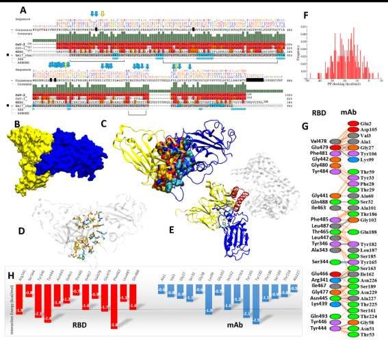
Figure 3. Binding kinetics of A) II62-scFv-Fc and B) II62-IgG1 against RBD. In both cases the
antibody was immobilized on anti-human Fc sensors and RBD used as analyte. C) FACS analysis
showing shift compared to control when II62-IgG1 and II62-scFv-Fc used in staining of cell expressing
membrane bound full-length spike protein. D) Immuno-precipitation (IP) followed by Western blot
analysis using HRP conjugated anti-His antibody. In lane 1 and 2, RBD-His was pulled down with Fc
bearing antibodies (scFv-Fc and IgG1) immobilized to Protein A-agarose resin. In lane 3, RBD-Fc was
immobilized to Protein A agarose resin and scFv-His was pulled down. Non-specific antigens (His
tagged CHIKV envelope protein) used as control. E) His-tagged full length spike is detected on Western
blot by using II62-scFv-Fc antibody using HRP conjugated anti-Fc antibody. F) Cell lysate of
pseudovirus transfected 293T cells was run on SDS-PAGE and probed with II62-IgG1 mAb.
Figure 4
10Downloaded from http://www.jbc.org/ by guest on October 30, 2020 Figure 4. Protein-protein interaction and interface characterization A) sequence alignment of CoV- 2/CoV/ MERS viruses. Conserved residues are shown in yellow and blue arrows. B) The complex of II62-scFv and RBD in surface view (yellow and blue color respectively), C) the interacting interface residues in yellow and blue cartoons, D) the interacting residues are shown in atom-wise coloring, orange (II62) and cyan (RBD) site. E) The overlay of RBD-mAb and RBD-ACE2. The ACE2 interacting secondary structure, alpha-helix, are shown by red color cartoon. F) Histogram of protein- protein docking score in kcal/mol. G) The quantification of interacting residues such as hydrogen bonds (in direct blue line), VdW in dotted orange lines. The residues are mentioned, basic:blue, acidic:red, polar:green, aromatic:purple and hydrophobic contacts in grey. H) The interaction energetics quantification (
Identification of an anti-SARS-CoV-2 receptor binding domain directed human
             monoclonal antibody from a naïve semi-synthetic library
 Hilal Ahmed Parray, Adarsh Kumar Chiranjivi, Shailendra Asthana, Naveen Yadav,
Tripti Shrivastava, Shailendra Mani, Chandresh Sharma, Preeti Vishwakarma, Supratik
Das, Kamal Pindari, Subrata Sinha, Sweety Samal, Shubbir Ahmed and Rajesh Kumar
J. Biol. Chem. published online July 29, 2020

Access the most updated version of this article at doi: 10.1074/jbc.AC120.014918

Alerts:
     • When this article is cited
     • When a correction for this article is posted

Click here to choose from all of JBC's e-mail alerts

                                                                                      Downloaded from http://www.jbc.org/ by guest on October 30, 2020
You can also read