Humoral Immunity against Capsule Polysaccharide Protects the Host
←
→
Page content transcription
If your browser does not render page correctly, please read the page content below
INFECTION AND IMMUNITY, Feb. 2009, p. 615–621                                                                                  Vol. 77, No. 2
0019-9567/09/$08.00⫹0 doi:10.1128/IAI.00931-08
Copyright © 2009, American Society for Microbiology. All Rights Reserved.

  Humoral Immunity against Capsule Polysaccharide Protects the Host
    from magA⫹ Klebsiella pneumoniae-Induced Lethal Disease by
             Evading Toll-Like Receptor 4 Signaling䌤
           Ming-Fang Wu,1 Chih-Ya Yang,1 Tzu-Lung Lin,2 Jin-Town Wang,2,3 Feng-Ling Yang,4
                  Shih-Hsiung Wu,4 Bor-Shen Hu,5 Teh-Ying Chou,6 Ming-Daw Tsai,4,7
                              Chi-Hung Lin,1 and Shie-Liang Hsieh1,7,8*
 Department and Institute of Microbiology and Immunology, National Yang-Ming University, Taipei, Taiwan1; Department of
    Microbiology2 and Department of Internal Medicine,3 National Taiwan University Hospital, Taipei, Taiwan; Institute of
       Biological Chemistry, Academia Sinica, Taipei, Taiwan4; Section of Infectious Diseases, Department of Internal Medicine,

                                                                                                                                                Downloaded from http://iai.asm.org/ on February 17, 2021 by guest
        Taipei City Hospital Heping Branch, Taipei, Taiwan5; Section of Surgical Pathology, Department of Pathology and
            Laboratory Medicine, Taipei Veterans General Hospital, Taipei, Taiwan6; Genomics Research Center,
                 Academia Sinica, Taipei, Taiwan7; and Immunology Research Center, National Yang-Ming University and
                                          Taipei Veterans General Hospital, Taipei, Taiwan8
                        Received 26 July 2008/Returned for modification 27 August 2008/Accepted 3 November 2008

            Klebsiella pneumoniae magA (for mucoviscosity-associated gene A) is linked to the pathogenesis of primary
          pyogenic liver abscess, but the underlying mechanism by which magA increases pathogenicity is not well
          elucidated. In this study, we investigated the role of the capsular polysaccharides (CPS) in the pathogenesis of
          magAⴙ K. pneumoniae by comparing host immunity to magAⴙ K. pneumoniae and a ⌬magA mutant. We found
          that Toll-like receptor 4 recognition by magAⴙ K. pneumoniae was hampered by the mucoviscosity of the magAⴙ
          K. pneumoniae CPS. Interestingly, monoclonal antibodies (MAbs) against magAⴙ K. pneumoniae CPS recog-
          nized all of the K1 strains tested but not the ⌬magA and non-K1 strains. Moreover, the anti-CPS MAbs
          protected mice from magAⴙ K. pneumoniae-induced liver abscess formation and lethality. This indicates that
          the K1 epitope is a promising target for vaccine development, and anti-CPS MAbs has great potential to protect
          host from K1 strain-induced mortality and morbidity in diabetic and other immunocompromised patients in
          the future.

   Klebsiella pneumoniae is an enteric gram-negative bacillus               Even though the K. pneumoniae strains responsible for these
that causes human nosocomial infections in immunocompro-                    infections are sensitive to aminoglycosides and cephalosporins,
mised patients and accounts for a significant proportion of                 the mortality rates of primary liver abscess and metastatic
hospital-acquired infections in neonatal wards. K. pneumoniae               meningitis are 10% (35) and 30 to 40% (11, 32), respectively.
is well known as a major cause of nosocomial bacterial pneu-                This reflects the ineffectiveness of the current antibiotic ther-
monia and urinary tract infection (22, 26).                                 apy alone for this infection-related organ failure.
   The majority of clinical isolates of K. pneumoniae express a                Among the 77 serotypes of Klebsiella, strains expressing the
pronounced capsular polysaccharide that is essential to the                 K1 capsular antigen account for the majority (63.4%) of liver
virulence of Klebsiella (22). It has been demonstrated that                 abscess isolates of K. pneumoniae (13). This suggests that K1
strains with capsule (such as serotype K1 and K2) were virulent             capsular antigens confer survival advantage to bacteria and is
in animal model, whereas serotypes without capsule are less                 an important indicator for the occurrence of liver abscess and
virulent or without virulence (21, 22). R. W. Tsay et al. (33)              endophthalmitis in K. pneumoniae infection. However, the mo-
found that capsular serotype K1 was the most common sero-                   lecular mechanism rendering the K1 strain more invasive has
type (23.4% versus 14%) found in the community-acquired                     not been well elucidated.
and nosocomial K. pneumoniae infections.                                       Recently, a novel gene magA (named for mucoviscosity-
   Recently, a new type of invasive K. pneumoniae (K1 strain)               associated gene A) located in the K1 capsular gene cluster was
has become the main agent causing primary liver abscesses in                identified from a Taiwan K. pneumoniae strain (NTUH-
community-acquired infections (16, 35), and 10 to 12% of
                                                                            K2044), and the presence of magA correlated with the K1
these cases were complicated by either metastatic meningitis
                                                                            serotype of K. pneumoniae (7, 12). Moreover, MagA is essen-
(5) or endophthalmitis (6, 13, 19). Such infections occur not
                                                                            tial for the synthesis of NTUH-K2044 capsular polysaccharide
only in Taiwan but also in Western countries (2, 3, 18, 25).
                                                                            (CPS), which is associated with high mucoviscosity and inter-
                                                                            feres with complement deposition. This feature makes magA⫹
                                                                            K. pneumoniae resistant to complement-mediated lysis when
  * Corresponding author. Mailing address: Department of Microbi-           incubated with nonimmune human serum (NHS) (12). It is
ology and Immunology, National Yang-Ming University, Taipei 11221,
                                                                            interesting that a magA-deficient mutant of NTUH-K2044
Taiwan. Phone: 886-2-28267161. Fax: 886-2-28277933. E-mail: slhsieh
@ym.edu.tw.                                                                 (⌬magA mutant) completely loses mucoviscosity and becomes
  䌤
    Published ahead of print on 17 November 2008.                           susceptible to complement deposition and phagocytosis (12).

                                                                      615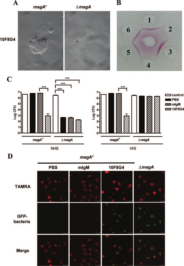
616 WU ET AL. INFECT. IMMUN.
This indicates that magA⫹ K. pneumoniae CPS plays an essen- g/ml) was the positive control. The macrophage cell lines HeNC2 and GG2EE
tial role in pathogen resistance to host immunity. Therefore, (6 ⫻ 105/ml) were incubated with bacteria at an MOI of 0.5 in 24-well plates,
respectively. LPS (1 ng/ml) was used as a positive control. All experiments were
we sought to determine whether the magA⫹ K. pneumoniae performed at least in triplicate.
CPS can mask underlying lipopolysaccharide (LPS) and inter- Preparation of MAbs. BALB/c (8-week-old) mice were immunized subcuta-
feres with host recognition by Toll-like receptor (TLR) (34). neously with K. pneumoniae (2 ⫻ 103 CFU per mice, the 50% lethal dose was 5 ⫻
To further understand the role of CPS in the pathogenesis of 102 CFU in our study) three times, with 2 weeks between each injection before
sacrifice. The spleen cells from survival mice were fused with murine myeloma
K. pneumoniae, we compared the differential immune response
cells (NS-1) using polyethylene glycol. After incubation in hypoxanthine-ami-
to magA⫹ and ⌬magA K. pneumoniae strains and generate nopterin-thymidine for 2 weeks, hybridoma secreting antibodies against immo-
anti-magA⫹ K. pneumoniae CPS monoclonal antibodies bilized CPS (100 ng of CPS from magA⫹ strain per well) were selected by
(MAbs) to test their effect to protect mice for magA⫹ K. enzyme-linked immunosorbent assay (ELISA). The isolation of bacterial CPS
pneumoniae-induced lethality. We report here that magA⫹ K. was as described in a previous study (37), and the LPS level in the CPS samples
(⬍1 EU/ml in the CPS) is determined by the Limulus amebocyte lysate assay.
pneumoniae CPS is sensitive to heat treatment, and disruption ELISA. To determine the tumor necrosis factor alpha (TNF-␣) levels in the
of CPS structure increases the host response to magA⫹ K. culture supernatants of macrophages (hMDMs or mouse macrophage) incu-
pneumoniae to a level similar to that of the ⌬magA strain. bated with UV or heat-inactivated magA⫹ or ⌬magA K. pneumoniae strains, 200
Downloaded from http://iai.asm.org/ on February 17, 2021 by guest
Moreover, anti-CPS MAbs could agglutinate all of the K1 l of supernatant was harvested at 24 h after incubation, and the concentrations
of TNF-␣ were determined by using a ELISA kit (R&D Systems).
strains tested, enhance phagocytosis, and protect mice from
Rapid agglutination assay. K. pneumoniae was diluted to an optical density at
magA⫹ K. pneumoniae-induced lethality. This suggests that 600 nm of 0.1 with PBS. An aliquot (25 l, 3 ⫻ 106 to 6 ⫻ 106 CFU) was
CPS is a promising target for vaccine development, and anti- incubated with anti-CPS MAb (7.5 g in 25 l) and shaken on a rotary platform
CPS MAbs are the potential therapeutic agents to protect host (130 rpm) for 1 h at room temperature. Agglutination was observed and photo-
against the complications induced by K. pneumoniae of K1 graphed. The original magnification under the microscope was ⫻200.
Double immunodiffusion. A double immunodiffusion test was performed as
serotype. described previously (7). Briefly, the anti-CPS ascites (10F8G4 clone) (1 l) was
loaded into the central well, while the CPS extract (20 g) was loaded into
peripheral wells. After an overnight incubation at 37 °C, the gels were incubated
MATERIALS AND METHODS with 1% azocarmine (dissolved in 2% glacial acid; Chroma) for 2 h, followed by
Reagents. Human macrophage–colony-stimulating factor was purchased from incubation with 2% glacial acid for destaining.
R&D Systems. The other chemicals were purchased from Sigma Chemical, Phagocytosis assay. To observe the ability of hMDMs to uptake anti-CPS
including LPS (isolated from Escherichia coli serotype O111:B4), 5(6)-car- MAb-coated bacteria, cells (2 ⫻ 105) were labeled with TAMRA [5(6)-car-
boxytetramethyl-rhodamine N-hydroxy-succinimide ester [5(6)-TAMRA], and boxytetramethyl-rhodamine N-hydroxy-succinimide ester] and incubated with K.
other chemicals. pneumoniae strains (magA⫹ and ⌬magA) stably expressing green fluorescent
Cell cultures. Human monocyte-derived macrophages (hMDMs) were cul- protein at 37°C for 1 h (MOI ⫽ 5) in glass-bottom culture plates. The cells were
tured as previous described (4). Briefly, human peripheral blood samples were washed with PBS and then fixed in 1% paraformaldehyde at room temperature
isolated from the blood of normal individuals by standard density gradient for 1 h before observation under a confocal microscope (Leica TCS-SP5).
centrifugation with Ficoll-Paque (Amersham Biosciences, Piscataway, NJ). Serum resistance assay. A serum resistance assay was performed as described
CD14⫹ cells were subsequently purified from human peripheral blood cells by previously (12). The bacteria (3 ⫻ 106 to 6 ⫻ 106 CFU) were incubated with
high-gradient magnetic sorting using the VARIOMACS technique with anti- anti-CPS MAb (clone 10F8G4, 7.5 g) at room temperature for 1 h, followed by
CD14 microbeads (Miltenyi Biotec, Bergisch Gladbach, Germany). CD14⫹ addition of NHS or heat-inactivated serum (HIS; 56°C for 30 min) to a final
monocytes were cultured in complete RPMI 1640 medium (JRH) supple- concentration of 25%, and were incubated at 37°C for another 1 h. Samples were
mented with 10 ng of human macrophage–colony-stimulating factor/ml at diluted serially and plated on LB plates to determine the colony numbers after
37°C in 5% CO2. incubation at 37°C overnight.
HeNC2 (with functional TLR4) and GG2EE cells (lacking functional TLR4) Protection assays. For the protection assay, groups of 8-week-old mice (12
are bone marrow-derived J2 virus-transformed macrophage cell lines as de- mice for each group) were injected intraperitoneally with 100 g of purified
scribed previously (1), were propagated in RPMI 1640 medium (JRH) supple- MAbs (9E9F11 and 10F8G4) or isotype control antibody (mouse immunoglob-
mented with 10% heat-inactivated fetal calf serum, and cultured in a 37°C, 5% ulin M [IgM]) or PBS. After 24 h, mice were inoculated with magA⫹ K. pneu-
CO2 incubator. moniae strain (2 ⫻ 103 CFU) intraperitoneally and were observed for 1 month to
Bacterial strains and growth conditions. The parental strain of the ⌬magA determine the mortality rate. The surviving mice were sacrificed at the end of the
strain, NTUH-K2044 (magA⫹ K. pneumoniae), was a clinical isolate; the con- fourth week. For histochemical staining, livers were removed from infected mice
struction of ⌬magA strain was described in a previous study (12). Bacteria strains at day 12 postinfection. The weights of the spleens were also measured at day 12
were grown in Luria-Bertani (LB) medium at 37°C. Selected antibiotics were after infection.
added for the culture of the ⌬magA strain (kanamycin, 50 g/ml) (12) and K. Statistical analysis. A Student t test was used to analyze the statistical signif-
pneumoniae strains carrying plasmid GFPuv gene (chloramphenicol, 100 g/ icance of differences using the Prism software package (GraphPad), and a P
ml), respectively (12). Other clinical bacterial strains were obtained from the value of ⬍0.05 was considered significant. The survival rate was determined by
Department of Internal Medicine Taipei City Hospital (Heping Branch, Kaplan-Meier analysis with a log-rank test, and statistical significance was ac-
Taipei, Taiwan). cepted at a P value of ⬍0.05.
String test for hypermucoviscosity. The string test was performed as described
previously (12). Hypermucoviscosity was defined by the formation of viscous
strings ⬎5 mm in length when a loop was used to stretch the colony on an agar RESULTS
plate (positive string test).
Negative staining for bacterial capsule. The capsule of K. pneumoniae is Differential effects of heat and UV on the CPS of magAⴙ K.
detected by negative staining. Briefly, the bacterial suspension is mixed with an
pneumoniae. Before incubation of hMDMs with magA⫹ and
equal volume of nigrosin (10%), spread onto a glass slide, and heat fixed for
several seconds. The slide was further incubated with 1% crystal violet for 2 min. ⌬magA K. pneumoniae, bacteria were inactivated by heat (95°C
The expression of capsule was observed as the exclusion of nigrosin and crystal for 30 min) or UV (20 J/cm2). The capsule of the ⌬magA
violet around the bacteria. mutant was thin without obvious mucoviscosity and was easily
Stimulation of hMDMs. The hMDMs were seeded in 24-well plates at a pelleted by centrifugation. In contrast, the capsule of magA⫹
density of 6 ⫻ 105/ml. The bacteria were either inactivated by exposure to UV
light at 20 J/cm2 or to heat at 95°C for 30 min. After three washes with phos-
K. pneumoniae was thick with high mucoviscosity and could not
phate-buffered saline (PBS), the bacteria were resuspended in PBS and added to be pelleted by centrifugation. Heat treatment reduced the mu-
hMDMs at a multiplicity of infection (MOI) of 5 and incubated for 24 h. LPS (1 coviscosity as determined by the string test (Fig. 1A), while UVVOL. 77, 2009 HUMORAL IMMUNITY TO magA⫹ K. PNEUMONIAE 617
Downloaded from http://iai.asm.org/ on February 17, 2021 by guest
FIG. 1. Differential effect of heat and UV on the CPS of magA⫹ and ⌬magA K. pneumoniae strains. (A) After heat (95°C for 30 min) and UV
irradiation (20 J/cm2) treatment (TX), bacterial colonies were subjected to the string test determine the viscosity. The string length of magA⫹ K.
pneumoniae is about 50 mm, and the y axis denotes the fold change after either heat or UV treatment compared to untreated magA⫹ K.
pneumoniae. Samples 1 to 3, magA⫹ K. pneumoniae; samples 4 to 6, ⌬magA K. pneumoniae. n/d, Not detectable. The significance of the coupled
difference was determined by using the Student t test. **, P ⬍ 0.01. (B) The magA⫹ K. pneumoniae (5 ⫻ 107 to 1 ⫻ 108 CFU in 10 l) was either
untreated or inactivated by UV or heat inactivated. The capsule structure was visualized by negative staining. Panels: 1, untreated; 2, UV
inactivated; 3, heat inactivated; 4, ⌬magA strain. Scale bar, 1 m. (C) hMDMs (6 ⫻ 105/ml) were incubated with UV-irradiated or heat-treated
magA⫹ and ⌬magA strains (MOI ⫽ 5) for 24 h. Culture supernatants were harvested and subjected to ELISA to measure the secretion of TNF-␣.
LPS (1 g/ml) was used as a positive control. (D) HeNC2 and its TLR4-defective mutant GG2EE were incubated with UV-inactivated or
heat-inactivated magA⫹ bacteria (MOI ⫽ 0.5) for 24 h to measure TNF-␣ secretion. LPS (1 ng/ml) is used as a positive control. The data are
expressed as the means ⫾ the standard deviations from three independent experiments. **, P ⬍ 0.01; ***, P ⬍ 0.001 (Student t test).
treatment still maintained the mucoviscosity of magA⫹ K. tlr-4 gene impede LPS signal transduction, and the mice be-
pneumoniae, suggesting that UV treatment did not disrupt come resistant to LPS and yet are highly susceptible to gram-
magA⫹ K. pneumoniae CPS structure. This argument is sup- negative bacterial infection (23). Two mouse macrophage cell
ported by the observation that the capsule of magA⫹ K. pneu- lines (HeNC2 and GG2EE) containing the functional TLR4
moniae is maintained after UV treatment (Fig. 1B2), whereas and nonfunctional TLR4, respectively, were used to investigate
heat inactivation disrupted the capsule (Fig. 1B3). We therefore whether TLR4 is involved in host recognition to K. pneu-
decided to use UV for bacteria inactivation in the subsequent moniae. As expected, magA⫹ K. pneumoniae only has a weak
experiments. stimulatory effect on TNF-␣ production from GG2EE. Com-
CPS of magAⴙ K. pneumoniae attenuates macrophage re- pared to UV-treated magA⫹ K. pneumoniae, heat-treated
sponses by hindering recognition of LPS by TLR4. After in- magA⫹ K. pneumoniae more strongly stimulates TNF-␣ pro-
cubation with hMDMs, UV-inactivated ⌬magA K. pneumoniae duction from HeNC2 cells (Fig. 1D). This suggests that TNF-␣
was more potent than the UV-inactivated magA⫹ strain to secretion is via interaction of TLR4 and underlying LPS, and
stimulate TNF-␣ production (Fig. 1C). In contrast, the ability disruption of CPS by heat exposes the underlying LPS to stim-
of heat-inactivated magA⫹ K. pneumoniae was similar to that ulate TNF-␣ secretion via interaction with TLR4 on HeNC2
of the heat-inactivated ⌬magA strain to stimulate TNF-␣ pro- cells. It has been shown that OmpA can bind and activate
duction from hMDMs (Fig. 1C). That heat treatment disrupts macrophages and synergize with LPS, leading to maximal
the capsule integrity of the magA⫹ strain (Fig. 1B3) and re- TNF-␣ production from macrophages (30). Therefore, we can-
stores its ability to stimulate TNF-␣ production (Fig. 1C) sug- not rule out the possibility that CPS also reduces TNF-␣ se-
gests that the integrity of CPS decides the stimulatory effect of cretion by interfering with the interaction of OmpA with a
magA⫹ K. pneumoniae, and CPS may function as a barrier to not-yet-defined pattern recognition receptor on host cells.
hinder hMDM recognition to other underlying bacterial com- Agglutination of K1 strains by anti-magAⴙ K. pneumoniae
ponents, such as LPS. A critical role of TLR4 in the recogni- CPS MAbs. We further sought to determine whether CPS is
tion of the microbial component LPS was initially character- the target for the development of vaccine against the invasive
ized by the evidence that C3H/HeJ mice with mutation in the magA⫹ K. pneumoniae strains. To address this question, hy-618 WU ET AL. INFECT. IMMUN.
Downloaded from http://iai.asm.org/ on February 17, 2021 by guest
FIG. 2. Anti-CPS MAb inhibits the growth and enhances phagocytosis of magA⫹ K. pneumoniae (A) Rapid agglutination assay. magA⫹ and
⌬magA strains were incubated with anti-CPS MAb (10F8G4) as described in Materials and Methods. Magnification, ⫻200. (B) Double immu-
nodiffusion assay. An anti-CPS MAb (clone 10F8G4) was loaded into the central well, while CPS was loaded into peripheral wells of agar. The
agar was incubated at 37°C overnight and dried in air before incubation with 1% azocarmine (dissolved in 2% glacial acid) for 2 h. The precipitation
lines were visualized after incubation with 5% glacial acid for destaining. Spots: 1, magA⫹ strain; 2, ⌬magA strain; 3, 5, and 6, clinical K1 strains
randomly selected from Table 2; 4, K62 from Table 3. (C) Bacteria (3 ⫻ 106 to 6 ⫻ 106 CFU, control) as described in panel A were incubated
with anti-CPS MAb (7.5 g/sample) for 1 h at room temperature, followed by the addition of NHS or HIS to a final concentration of 25%. The
bacteria were then diluted (10-fold serial dilutions) and plated onto LB agar plates overnight to observe colony formation. The y axis of colony
numbers was expressed as the log CFU. The data are expressed as the means ⫾ the standard deviations from three independent experiments. ***,
P ⬍ 0.001 (Student t test). (D) Phagocytosis was performed by incubating TAMRA-labeled hMDMs with bacteria stably expressing green
fluorescent protein as described in Materials and Methods, followed by observation under a confocal microscope.
bridomas were generated by fusing myeloma cells with spleno- tional double-immunodiffusion assay (Fig. 2B). Furthermore, all
cytes isolated from mice immunized with magA⫹ K. pneu- seven MAbs agglutinated all of the K1 strains of the clinical
moniae. All seven MAbs (IgM) recognized only the CPS of isolates (Table 2) and the reference strain but not the other
magA⫹ K. pneumoniae but not that of the ⌬magA mutant by serotypes (Table 3). This suggests that the anti-CPS MAbs rec-
agglutination assay (Fig. 2A). Moreover, all seven clones agglu- ognize the K1 epitope(s) of all of the K1 strains tested, and the
tinated magA⫹ K. pneumoniae but not the ⌬magA strain, as de- rapid serotyping without the need to extract CPS provided an
termined by both rapid agglutination assay (Table 1) and conven- alternative to the double-immunodiffusion assay.VOL. 77, 2009 HUMORAL IMMUNITY TO magA⫹ K. PNEUMONIAE 619
TABLE 1. MAbs to magA⫹ strain TABLE 3. Reference K. pneumoniae strainsa
Resulta determined by: Bacterial strain Serotype Agglutination
Clone Isotype ELISA Agglutination ATCC 8045 K1 ⫹
magA⫹
MGH78578 K52 –
⌬magA magA⫹ ⌬magA
K5 K5 –
9E9C7 IgM ⫹ – ⫹ – K9 K9 –
9E9D8 IgM ⫹ – ⫹ – K14 K14 –
9E9F11 IgM ⫹⫹ – ⫹⫹ – K62 K62 –
10F8C3 IgM ⫹ – ⫹ – a
ATCC, American Type Culture Collection. K5, K9, K14, and K62 were
10F8E3 IgM ⫹ – ⫹ – isolated from patients with primary liver abscess; “⫹” means that there was a
10F8F4 IgM ⫹ – ⫹ – positive result in the agglutination reactions; “–” means that there was a negative
10F8G4 IgM ⫹⫹⫹ – ⫹⫹⫹ – result in the agglutination reactions.
a
“⫹” or “–” indicates that there was a positive or negative result, respectively,
in the agglutination reactions and ELISA, “⫹⫹⫹” indicates the strongest reac-
Downloaded from http://iai.asm.org/ on February 17, 2021 by guest
tion, and “⫹⫹” indicates an intermediate reaction.
respectively (Fig. 3C). These data suggest that the K1 epitope
of CPS is a promising target for vaccine development and that
Complement-independent killing effects of anti-CPS MAbs. anti-CPS MAbs have great potential as therapeutic agents to
It has been reported that magA⫹ K. pneumoniae CPS interferes protect against K1-induced lethality.
with complement deposition, and magA⫹ K. pneumoniae is
resistant to NHS-mediated lysis (12). Therefore, we sought to DISCUSSION
determine whether anti-CPS MAbs have a killing effect on
magA⫹ and ⌬magA K. pneumoniae in the presence or absence We report here that the integrity of magA⫹ K. pneumoniae
of NHS or HIS. As previously observed, the ⌬magA strain was CPS is not only essential to interfere with complement depo-
sensitive, whereas the magA⫹ K. pneumoniae strain was resis- sition (12) but also to determine the hMDM response to
tant, to NHS-mediated lysis (Fig. 2C, left panel). Interestingly, magA⫹ K. pneumoniae. Furthermore, anti-magA⫹ K. pneu-
magA⫹ K. pneumoniae was killed by anti-CPS MAb (clone moniae CPS MAbs could enhance the phagocytosis of magA⫹
10F8G4), whether coincubated with NHS or HIS (Fig. 2C). K. pneumoniae, prevent liver abscess formation, and protect
This indicates that the anti-CPS MAb itself is enough to kill mice from magA⫹ K. pneumoniae-induced lethality. Since the
magA⫹ K. pneumoniae. In addition, anti-CPS MAb also en- anti-CPS MAbs can recognize all of the K1 strains tested, this
hanced the uptake of magA⫹ K. pneumoniae by MDMs (Fig. suggests K1 epitope is a promising target for vaccine develop-
2D). This suggests that anti-CPS MAbs may be useful in re- ment against all of the K1 strains of K. pneumoniae.
stricting bacterial survival and spread in vivo. Multiple Klebsiella components (e.g., fimbriae, siderophores,
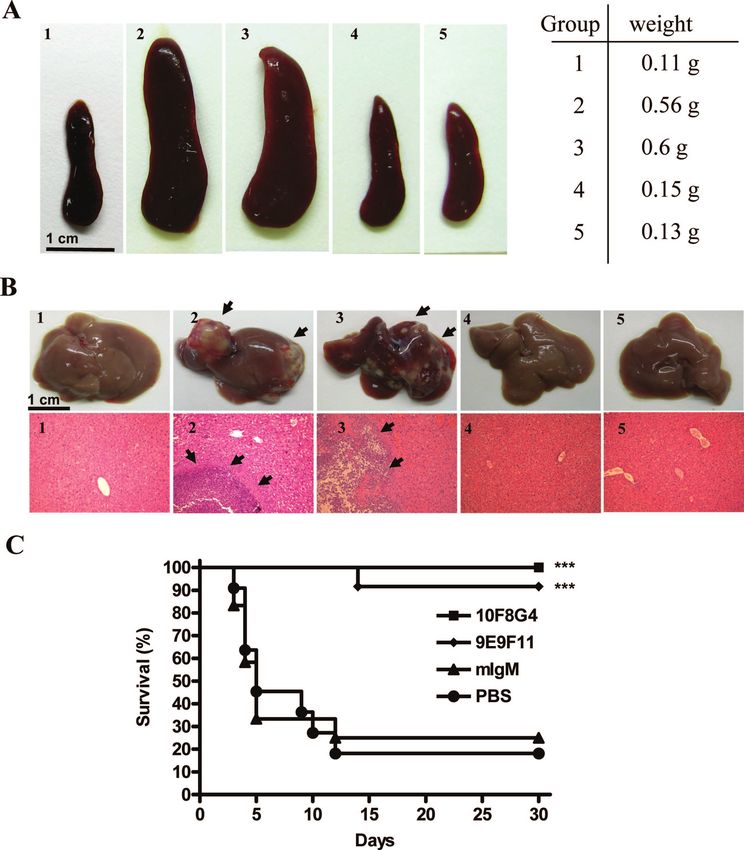
Anti-CPS MAbs protected mice from magAⴙ K. pneumoniae- LPS, and capsules) have been considered to be potential vir-
induced lethality. To investigate the potential therapeutic ef- ulence factors (22). Among these factors, LPS induces TNF-␣
fects of anti-CPS MAbs in vivo, mice were injected with (15). Although the CPS has been shown to play a major role in
anti-CPS MAbs 24 h before intraperitoneal inoculation with increasing pathogenicity, the mechanism(s) by which CPS in-
magA⫹ K. pneumoniae (2 ⫻ 103 CFU). At day 12 after infec- creases the virulence of K. pneumoniae, in terms of its ability to
tion, splenomegaly (Fig. 3A) and multiple liver abscesses with induce liver abscess, is not well elucidated (9, 10).
leukocyte infiltration (Fig. 3B) were observed in mice pre- Capsules can increase the virulence of K. pneumoniae by
treated with PBS (Fig. 3A2) or isotype control antibodies (Fig. acting as a physical barrier between immunostimulatory bac-
3A3). In contrast, anti-CPS MAbs (clones 9E9F11 and terial products (such as fimbriae and LPS) and the host’s im-
10F8G4) prevented splenomegaly (Fig. 3A4 and 5) and abscess mune system. The capsule, through a physical shielding mech-
formation (Fig. 3B4 and 5). The survival rates of mice treated anism, can inhibit fimbrial function (28) and impede the
with PBS or isotype control antibodies were 20 and 25%, adhesion to invade epithelial cells by K. pneumoniae (27). This
respectively, while the survival rates in mice treated with anti- shielding of adhesion by the capsule has also been observed in
CPS MAbs (clones 10F8G4 and 9E9F11) were 100 and 95%, other organisms, such as E. coli (29) and Haemophilus influen-
zae (31). The capsule can also mask the LPS molecules to
prevent complement deposition (20). In the present study, we
TABLE 2. Clinical K. pneumoniae strainsa further demonstrated that, in addition to preventing comple-
ment-mediated lysis and uptake by macrophages, CPS plays an
Total K1 strains Non-K1 isolates
Clinical diagnosis important role in attenuating hMDM responses via interfer-
no. of No. of No. of
of patients Agglutination Agglutination ence with the interaction of TLR4 with the underlying LPS,
isolates strains isolates
thus decreasing the induction of TNF-␣, which is essential for
PLA 23 21 ⫹ 2 – the prevention of bacterial spreading and invasion (Fig. 1C).
PLA plus fasciitis 6 5 ⫹ 1 –
Recurrent liver 4 2 ⫹ 2 – This elucidates a novel mechanism of CPS-mediated bacterial
abscess virulence.
PLA plus 7 6 ⫹ 1 –
endophthalmitis In addition to the magA⫹ K. pneumoniae CPS, the produc-
Meningitis 4 3 ⫹ 1 – tion of TNF-␣ was also suppressed by the Salmonella enterica
a
PLA, primary pyogenic liver abscess. “⫹” or “–” means that there was serovar Typhi Vi capsular antigen (24, 36) and Neisseria men-
positive or negative result, respectively, in the agglutination reactions. ingitidis type C polysaccharide (17). The Vi capsule in the620 WU ET AL. INFECT. IMMUN.
Downloaded from http://iai.asm.org/ on February 17, 2021 by guest
FIG. 3. Effects of anti-CPS MAbs on magA⫹ strains in inducing pathological changes and lethality. At 24 h before inoculation with magA⫹
strains (2 ⫻ 103 CFU), mice were injected with anti-CPS MAbs (100 g/mouse), PBS, or isotype control (mIgM). At day 12 postinfection, mice
were sacrificed, and the spleens (A) and livers (B) were removed and examined grossly, while liver abscesses were examined by hematoxylin and
eosin staining and observed under a microscope (magnification, ⫻200). Panels: 1, no infection; 2, PBS; 3, mIgM; 4, anti-CPS MAb (clone 9E9F11);
5, anti-CPS MAb (clone 10F8G4). (C) The survival rate of each group was determined at 30 days after inoculation. ***, P ⬍ 0.001 (as determined
by using Kaplan-Meier survival analysis and compared by log-rank test [n ⫽ 12 for each group]).
serovar Typhi can lower the production of TNF-␣ and inhibit that the K1 epitope is a promising target for vaccine develop-
neutrophil infiltration by preventing the recognition of TLR4 ment and that anti-K1 MAbs have the potential to prevent
to the serovar Typhi LPS (24, 36). Similarly, the capsule on N. liver abscess induced by all of the K1 strains.
meningitidis can inhibit TLR4 activation by interfering with the
binding of the soluble components (CD14 and lipoprotein ACKNOWLEDGMENTS
binding protein, the two members of the LOS receptor com- We thank BioLegend for antibody preparation and Hsien-Yeh Hsu
plex) to TLR4 binding (17). From these observations, evasion for providing the HeNC2 and GG2EE cell lines. We are grateful to
of innate immunity via inhibiting TLR4-mediated signaling Chang-Phone Fung for providing the clinical strains of K. pneumoniae
isolated from liver abscesses.
may be a general feature of bacterial CPS. This study was supported mainly by the Department of Health,
Over the past two decades, CPS has been the obvious vac- Executive Yuan, Republic of China (Taiwan) (grant DOH 94-TD-G-
cine candidate for Klebsiella-induced pneumonia studies. Cryz 111-039), grant V97S5-005 from the Taipei Veterans General Hospi-
et al. have demonstrated that active immunization with puri- tal, and grant 94F008-5 from Academia Sinica.
fied CPS protected rats against lethal Klebsiella-induced pneu- REFERENCES
monia (8). MAbs against K2 CPS reduced the inflammatory 1. Blasi, E., D. Radzioch, S. K. Durum, and L. Varesio. 1987. A murine mac-
response in the lung and eliminated bacteria in a lung infection rophage cell line, immortalized by v-raf and v-myc oncogenes, exhibits nor-
mal macrophage functions. Eur. J. Immunol. 17:1491–1498.
model (14). However, the ability of anti-CPS to prevent liver 2. Cahill, M., B. Chang, and A. Murray. 2000. Bilateral endogenous bacterial
abscess formation has never been addressed. Our study dem- endophthalmitis associated with pyogenic hepatic abscess. Br. J. Ophthal-
onstrates that anti-magA⫹ K. pneumoniae CPS antibodies can mol. 84:1436.
3. Casanova, C., J. A. Lorente, F. Carrillo, E. Perez-Rodriguez, and N. Nunez.
agglutinate all of the K1 strains tested and prevent liver abscess 1989. Klebsiella pneumoniae liver abscess associated with septic endoph-
formation induced by magA⫹ K. pneumoniae. This indicates thalmitis. Arch. Intern. Med. 149:1467.VOL. 77, 2009 HUMORAL IMMUNITY TO magA⫹ K. PNEUMONIAE 621
4. Chang, Y. C., T. L. Hsu, H. H. Lin, C. C. Chio, A. W. Chiu, N. J. Chen, C. H. Mechanisms of Klebsiella pneumoniae resistance to complement-mediated
Lin, and S. L. Hsieh. 2004. Modulation of macrophage differentiation and killing. Infect. Immun. 60:2529–2535.
activation by decoy receptor 3. J. Leukoc. Biol. 75:486–494. 21. Mizuta, K., M. Ohta, M. Mori, T. Hasegawa, I. Nakashima, and N. Kato.
5. Cheng, D. L., Y. C. Liu, M. Y. Yen, C. Y. Liu, and R. S. Wang. 1991. Septic 1983. Virulence for mice of Klebsiella strains belonging to the O1 group:
metastatic lesions of pyogenic liver abscess. Their association with Klebsiella relationship to their capsular (K) types. Infect. Immun. 40:56–61.
pneumoniae bacteremia in diabetic patients. Arch. Intern. Med. 151:1557– 22. Podschun, R., and U. Ullmann. 1998. Klebsiella spp. as nosocomial patho-
1559. gens: epidemiology, taxonomy, typing methods, and pathogenicity factors.
6. Chiu, C. T., D. Y. Lin, and Y. F. Liaw. 1988. Metastatic septic endophthalmi- Clin. Microbiol. Rev. 11:589–603.
tis in pyogenic liver abscess. J. Clin. Gastroenterol. 10:524–527. 23. Poltorak, A., X. He, I. Smirnova, M. Y. Liu, C. Van Huffel, X. Du, D.
7. Chuang, Y. P., C. T. Fang, S. Y. Lai, S. C. Chang, J. T. Wang, C. Struve, M. Birdwell, E. Alejos, M. Silva, C. Galanos, M. Freudenberg, P. Ricciardi-
Bojer, E. M. Nielsen, D. S. Hansen, K. A. Krogfelt, R. Gierczynski, S. Castagnoli, B. Layton, and B. Beutler. 1998. Defective LPS signaling in
Kaluzewski, A. A. Zasada, W. Rastawicki, M. Jagielski, L. C. Ma, C. Z. Lee, C3H/HeJ and C57BL/10ScCr mice: mutations in Tlr4 gene. Science 282:
C. T. Shun, F. C. Fang, N. Sandler, and S. J. Libby. 2006. Genetic determi- 2085–2088.
nants of capsular serotype K1 of Klebsiella pneumoniae causing primary 24. Raffatellu, M., D. Chessa, R. P. Wilson, R. Dusold, S. Rubino, and A. J.
pyogenic liver abscess. J. Infect. Dis. 193:645–654. Baumler. 2005. The Vi capsular antigen of Salmonella enterica serotype
8. Cryz, S. J., Jr., E. Furer, and R. Germanier. 1986. Immunization against fatal Typhi reduces Toll-like receptor-dependent interleukin-8 expression in the
experimental Klebsiella pneumoniae pneumonia. Infect. Immun. 54:403–407. intestinal mucosa. Infect. Immun. 73:3367–3374.
9. Cryz, S. J., Jr., F. Furer, and R. Germanier. 1984. Experimental Klebsiella 25. Saccente, M. 1999. Klebsiella pneumoniae liver abscess, endophthalmitis, and
Downloaded from http://iai.asm.org/ on February 17, 2021 by guest
pneumoniae burn wound sepsis: role of capsular polysaccharide. Infect. Im- meningitis in a man with newly recognized diabetes mellitus. Clin. Infect.
mun. 43:440–441. Dis. 29:1570–1571.
10. Domenico, P., W. G. Johanson, Jr., and D. C. Straus. 1982. Lobar pneumo- 26. Sahly, H., and R. Podschun. 1997. Clinical, bacteriological, and serological
nia in rats produced by clinical isolates of Klebsiella pneumoniae. Infect. aspects of Klebsiella infections and their spondylarthropathic sequelae. Clin.
Immun. 37:327–335. Diagn. Lab. Immunol. 4:393–399.
11. Fang, C. T., Y. C. Chen, S. C. Chang, W. Y. Sau, and K. T. Luh. 2000. 27. Sahly, H., R. Podschun, T. A. Oelschlaeger, M. Greiwe, H. Parolis, D. Hasty,
Klebsiella pneumoniae meningitis: timing of antimicrobial therapy and prog- J. Kekow, U. Ullmann, I. Ofek, and S. Sela. 2000. Capsule impedes adhesion
nosis. QJM 93:45–53. to and invasion of epithelial cells by Klebsiella pneumoniae. Infect. Immun.
68:6744–6749.
12. Fang, C. T., Y. P. Chuang, C. T. Shun, S. C. Chang, and J. T. Wang. 2004.
28. Schembri, M. A., J. Blom, K. A. Krogfelt, and P. Klemm. 2005. Capsule and
A novel virulence gene in Klebsiella pneumoniae strains causing primary liver
fimbria interaction in Klebsiella pneumoniae. Infect. Immun. 73:4626–4633.
abscess and septic metastatic complications. J. Exp. Med. 199:697–705.
29. Schembri, M. A., D. Dalsgaard, and P. Klemm. 2004. Capsule shields the
13. Fung, C. P., F. Y. Chang, S. C. Lee, B. S. Hu, B. I. Kuo, C. Y. Liu, M. Ho, and
function of short bacterial adhesins. J. Bacteriol. 186:1249–1257.
L. K. Siu. 2002. A global emerging disease of Klebsiella pneumoniae liver
30. Soulas, C., T. Baussant, J. P. Aubry, Y. Delneste, N. Barillat, G. Caron, T.
abscess: is serotype K1 an important factor for complicated endophthalmi-
Renno, J. Y. Bonnefoy, and P. Jeannin. 2000. Outer membrane protein A
tis? Gut 50:420–424.
(OmpA) binds to and activates human macrophages. J. Immunol. 165:2335–
14. Held, T. K., M. Trautmann, M. E. Mielke, H. Neudeck, S. J. Cryz, Jr., and 2340.
A. S. Cross. 1992. Monoclonal antibody against Klebsiella capsular polysac- 31. St. Geme, J. W., III, and S. Falkow. 1991. Loss of capsule expression by
charide reduces severity and hematogenic spread of experimental Klebsiella Haemophilus influenzae type b results in enhanced adherence to and invasion
pneumoniae pneumonia. Infect. Immun. 60:1771–1778. of human cells. Infect. Immun. 59:1325–1333.
15. Henderson, B., S. Poole, and M. Wilson. 1996. Bacterial modulins: a novel 32. Tang, L. M., and S. T. Chen. 1994. Klebsiella pneumoniae meningitis: prog-
class of virulence factors which cause host tissue pathology by inducing nostic factors. Scand. J. Infect. Dis. 26:95–102.
cytokine synthesis. Microbiol. Rev. 60:316–341. 33. Tsay, R. W., L. K. Siu, C. P. Fung, and F. Y. Chang. 2002. Characteristics of
16. Ko, W. C., D. L. Paterson, A. J. Sagnimeni, D. S. Hansen, A. Von Gottberg, bacteremia between community-acquired and nosocomial Klebsiella pneu-
S. Mohapatra, J. M. Casellas, H. Goossens, L. Mulazimoglu, G. Trenholme, moniae infection: risk factor for mortality and the impact of capsular sero-
K. P. Klugman, J. G. McCormack, and V. L. Yu. 2002. Community-acquired types as a herald for community-acquired infection. Arch. Intern. Med.
Klebsiella pneumoniae bacteremia: global differences in clinical patterns. 162:1021–1027.
Emerg. Infect. Dis. 8:160–166. 34. Uematsu, S., and S. Akira. 2006. Toll-like receptors and innate immunity. J.
17. Kocabas, C., N. Katsenelson, S. Kanswal, M. N. Kennedy, X. Cui, M. S. Mol. Med. 84:712–725.
Blake, D. M. Segal, and M. Akkoyunlu. 2007. Neisseria meningitidis type C 35. Wang, J. H., Y. C. Liu, S. S. Lee, M. Y. Yen, Y. S. Chen, J. H. Wang, S. R.
capsular polysaccharide inhibits lipooligosaccharide-induced cell activation Wann, and H. H. Lin. 1998. Primary liver abscess due to Klebsiella pneu-
by binding to CD14. Cell Microbiol. 9:1297–1310. moniae in Taiwan. Clin. Infect. Dis. 26:1434–1438.
18. Lederman, E. R., and N. F. Crum. 2005. Pyogenic liver abscess with a focus 36. Wilson, R. P., M. Raffatellu, D. Chessa, S. E. Winter, C. Tukel, and A. J.
on Klebsiella pneumoniae as a primary pathogen: an emerging disease with Baumler. 2008. The Vi-capsule prevents Toll-like receptor 4 recognition of
unique clinical characteristics. Am. J. Gastroenterol. 100:322–331. Salmonella. Cell Microbiol. 10:876–890.
19. Liu, Y. C., D. L. Cheng, and C. L. Lin. 1986. Klebsiella pneumoniae liver 37. Zamze, S., L. Martinez-Pomares, H. Jones, P. R. Taylor, R. J. Stillion, S.
abscess associated with septic endophthalmitis. Arch. Intern. Med. 146:1913– Gordon, and S. Y. Wong. 2002. Recognition of bacterial capsular polysac-
1916. charides and lipopolysaccharides by the macrophage mannose receptor.
20. Merino, S., S. Camprubi, S. Alberti, V. J. Benedi, and J. M. Tomas. 1992. J. Biol. Chem. 277:41613–41623.
Editor: A. J. BäumlerYou can also read