89Zr-DFO-J591 for ImmunoPET of Prostate-Specific Membrane Antigen Expression In Vivo
←
→
Page content transcription
If your browser does not render page correctly, please read the page content below
89Zr-DFO-J591   for ImmunoPET
  of Prostate-Specific Membrane Antigen
  Expression In Vivo
  Jason P. Holland1, Vadim Divilov1, Neil H. Bander 2,3, Peter M. Smith-Jones1,4, Steven M. Larson4–6, and Jason S. Lewis1,6
  1Radiochemistry Service, Department of Radiology, Memorial Sloan-Kettering Cancer Center, New York, New York; 2Laboratory of
  Urological Oncology, Department of Urology, New York-Presbyterian Hospital, Weill Medical College of Cornell University, New
  York, New York; 3Department of Urology, Memorial Sloan-Kettering Cancer Center, New York, New York; 4Ludwig Center for
  Cancer Immunotherapy, Sloan-Kettering Institute, Memorial Sloan-Kettering Cancer Center, New York, New York; 5Nuclear
  Medicine Service, Department of Radiology, Memorial Sloan-Kettering Cancer Center, New York, New York; and 6Program in
  Molecular Pharmacology and Chemistry, Memorial Sloan-Kettering Cancer Center, New York, New York

89Zr (half-life, 78.41 h) is a positron-emitting radionuclide that   89Zr-DFO-J591 provides excellent image contrast, with tumor-

displays excellent potential for use in the design and synthesis     to-muscle ratios greater than 20, for the delineation of LNCaP
of radioimmunoconjugates for immunoPET. In the current               xenografts between 48 and 144 h after administration. Conclu-
study, we report the preparation of 89Zr-desferrioxamine B           sion: These studies demonstrate that 89Zr-DFO–labeled mAbs
(DFO)-J591, a novel 89Zr-labeled monoclonal antibody (mAb)           show exceptional promise as radiotracers for immunoPET of
construct for targeted immunoPET and quantification of pros-         human cancers. 89Zr-DFO-J591 displays high tumor–to–back-
tate-specific membrane antigen (PSMA) expression in vivo.            ground tissue contrast in immunoPET and can be used to delin-
Methods: The in vivo behavior of 89Zr-chloride, 89Zr-oxalate,        eate and quantify PSMA-positive prostate tumors in vivo.
and 89Zr-DFO was studied using PET. High-level computational         Key Words: immunoPET; 89Zr; prostate-specific membrane
studies using density functional theory calculations have been       antigen (PSMA); J591; monoclonal antibodies; density functional
used to investigate the electronic structure of 89Zr-DFO and         theory
probe the nature of the complex in aqueous conditions.
89Zr-DFO-J591 was characterized both in vitro and in vivo.           J Nucl Med 2010; 51:1293–1300
                                                                     DOI: 10.2967/jnumed.110.076174
ImmunoPET in male athymic nu/nu mice bearing subcutaneous
LNCaP (PSMA-positive) or PC-3 (PSMA-negative) tumors was
conducted. The change in 89Zr-DFO-J591 tissue uptake in

                                                                     T
response to high- and low-specific-activity formulations in the
2 tumor models was measured using acute biodistribution stud-
ies and immunoPET. Results: The basic characterization of                 he National Cancer Institute estimated that in the
3 important reagents—89Zr-chloride, 89Zr-oxalate, and the            United States during 2009, approximately 192,000 cases
complex 89Zr-DFO—demonstrated that the nature of the 89Zr            of prostate cancer (PC) would be diagnosed, with a pro-
species dramatically affects the biodistribution and pharmaco-       jected mortality rate of over 27,000 men (.14%). Despite
kinetics. Density functional theory calculations provide a ration-   the high incidence of PC, standard diagnostic imaging tech-
ale for the observed high in vivo stability of 89Zr-DFO–labeled
                                                                     niques used for the detection and monitoring of therapy
mAbs and suggest that in aqueous conditions, 89Zr-DFO forms
a thermodynamically stable, 8-coordinate complex by coordi-          remain inadequate. For example, early-stage, hormonally
nation of 2 water molecules. 89Zr-DFO-J591 was produced in           sensitive tumors on treatment and noncastrate PCs often
high radiochemical yield (.77%) and purity (.99%), with a spe-       appear negative on PET scans using either the metabolic
cific activity of 181.7 6 1.1 MBq/mg (4.91 6 0.03 mCi/mg). In        radiotracer 18F-FDG or the hormone-based radiopharma-
vitro assays demonstrated that 89Zr-DFO-J591 had an initial          ceutical 16b-18F-dihydrotestosterone (18F-FDHT) for imag-
immunoreactive fraction of 0.95 6 0.03 and remained active           ing the overexpression of androgen receptors (1). Therefore,
for up to 7 d. In vivo biodistribution experiments revealed high,
                                                                     there is an urgent requirement to develop new tools for the
target-specific uptake of 89Zr-DFO-J591 in LNCaP tumors after
24, 48, 96, and 144 h (34.4 6 3.2 percentage injected dose per       noninvasive delineation and staging of PC in vivo.
gram [%ID/g], 38.0 6 6.2 %ID/g, 40.4 6 4.8 %ID/g, and 45.8 6            Prostate-specific membrane antigen (PSMA) is a 100-
3.2 %ID/g, respectively). ImmunoPET studies also showed that         kDa, type II transmembrane glycoprotein and is one of the
                                                                     best characterized oncogenic markers or targets (2,3).
   Received Feb. 10, 2010; revision accepted Mar. 30, 2010.          PSMA expression has been detected in a limited range of
   For correspondence or reprints contact: Jason S. Lewis,           normal tissues including benign prostatic epithelium, renal
 Radiochemistry Service, Department of Radiology, Memorial Sloan-
 Kettering Cancer Center, 1275 York Ave., New York, NY 10065.        proximal tubule, small bowel, and brain (a subset of astro-
   E-mail: lewisj2@mskcc.org                                         cytes). However, these normal sites express PSMA at levels
   COPYRIGHT ª 2010 by the Society of Nuclear Medicine, Inc.
                                                                     2–3 orders of magnitude lower than that observed in more

                                                           89ZR-DFO-J591 IMMUNOPET OF         PSMA • Holland et al.           1293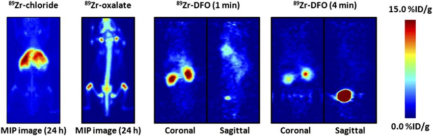
than 95% of clinical PC specimens (4,5). In addition, these     MATERIALS AND METHODS
normal-tissue PSMA sites are highly polarized to the apical        Full details of all methods and equipment used are presented in
or luminal aspect of the benign prostatic glands, renal         the supplemental materials (available online only at http://jnm.
tubules and small bowel, basement membrane, and epithe-         snmjournals.org).
lial tight junctions, which form substantial barriers to cir-
                                                                Density Functional Theory (DFT) Calculations
culating monoclonal antibodies (mAbs). PSMA expression
                                                                   All calculations were conducted using DFT as implemented in
by astrocytes is similarly sequestered behind the blood–        the Gaussian03 suite of ab initio quantum chemistry programs
brain barrier. As a result, antibodies to PSMA are function-    (21). Full computational details and Cartesian coordinates of the
ally tumor-specific, whereas small-molecule PSMA ligands        optimized structures are presented in the supplemental materials.
excreted via the renal tubular lumen are not.                   Energetic values are reported in units of kJ mol21.
   PSMA expression levels have been shown to exhibit a
                                                                Antibody Conjugation and Radiolabeling
positive correlation with increased tumor aggression,
                                                                   The humanized IgG1 mAb J591 was conjugated to the tris-
metastatic spread, and the development of castrate resist-      hydroxamate hexadentate chelate, desferrioxamine B (DFO) (Cal-
ance, or resistance to hormone-based therapies. PSMA            biochem), using a 6-step procedure modified (22) from the
expression has also been reported in the neovasculature of      approach described by Verel et al. (23) (supplemental material).
most solid tumors (6). The failure of 18F-FDG PET for              89Zr was produced via the 89Y(p,n)89Zr transmutation reaction

detecting early and treated PC and the acquired resistance      on a TR19/9 variable-beam-energy cyclotron (Ebco Industries
of many advanced PCs to androgen-based agents have              Inc.) in accordance with previously reported methods (23,24).
been the driving force behind recent efforts toward devel-      The 89Zr-oxalate was isolated in high radionuclidic and radio-
oping novel chemo- and radioimmunoconjugate-based               chemical purity (RCP) greater than 99.9%, with an effective spe-
drugs and imaging agents. In particular, in 1996 the U.S.       cific activity of 195–497 MBq/mg, (5.28–13.43 mCi/mg) (24).
Food and Drug Administration approved the use of 111In-         Stability Studies
capromab pendetide or 111In-7E11 (111In-ProstaScint;               The stability of 89Zr-DFO-J591 with respect to change in RCP,
Cytogen Corp.), a murine–mAb specific for an intracellu-        loss of radioactivity from the mAb, or change in immunoreactivity
lar epitope of PSMA, for SPECT of PC soft-tissue meta-          was investigated in vitro by incubation in solutions of 0.9% saline
stases. However, 111In-capromab pendetide for clinical          and 1% bovine serum albumin for 7 d at 37C. The RCP was
diagnosis is suboptimal because of low sensitivity for via-     determined by radio–instant thin-layer chromatography and
ble tumor sites (62% for lymph node metastases, 50% for         g-counting, and the immunoreactive fraction was measured using
                                                                the LNCaP cellular binding assay.
prostate bed recurrence), which is probably because the
number of available targets (presented in dead or dying         Xenograft Models
tissue) is limited. Furthermore, 111In-capromab pendetide          All animal experiments were conducted in compliance with
does not bind to viable PC sites in bone (the most common       Institutional Animal Care and Use Committee guidelines. Male
site of metastatic disease), and in contrast to PET, SPECT      athymic nu/nu mice (NCRNU-M, 20–22 g, 6–8 wk old) were
remains only semiquantitative in the clinical setting.          obtained from Taconic Farms Inc. and were allowed to acclimatize
Despite these limitations, the National Comprehensive           at the Memorial Sloan-Kettering Cancer Center vivarium for 1 wk
                                                                before tumors were implanted. Mice were provided with food and
Cancer Network Clinical Practice Guidelines (5)—which
                                                                water ad libitum. In separate animals, LNCaP (PSMA-positive)
propose using 111In-capromab pendetide before salvage           and PC-3 (PSMA-negative) tumors were induced on the left and
therapy after radiotherapy or prostatectomy—still recom-        right shoulders, respectively. Full details are provided in the sup-
mend the mAb as being useful for specific clinical situa-       plemental material.
tions. This fact is testament to the relative lack of better
imaging methods for the detection of metastatic prostate        Acute Biodistribution Studies
cancer, especially in soft tissue.                                 LNCaP and PC-3 tumor–bearing mice were randomized before
                                                                the study and were warmed gently with a heat lamp 5 min before
   In 1997, Liu et al. produced 4 mAbs (IgG1: J415, J533,
                                                                administration of 89Zr-DFO-J591 (0.55–0.74 MBq [15–20 mCi],
and J591; and IgG3: E99) specific for binding to 2 distinct     3–4 mg of mAb, in 200 mL of sterile saline for injection) via
epitopes on the extracellular domain of PSMA (7,8). Sub-        injection into the tail vein (0 h). Animals (n 5 3–5, per group)
sequent in vitro and in vivo studies identified J591 as the     were euthanized by CO2 gas asphyxiation at 24, 48, 96, and 144 h
most promising candidate for developing diagnostic and          after injection, and 12 organs (including the tumor) were removed,
therapeutic immunoconjugates for the targeting of extrac-       rinsed in water, dried in air for 5 min, weighed, and counted on a
ellular PSMA in viable tissue (9–11). Since these initial       g-counter for accumulation of 89Zr radioactivity. Full details are
studies, J591 has been humanized, and a range of preclin-       presented in the supplemental material.
ical and clinical studies using J591 radiolabeled with 90Y,     Small-Animal immunoPET
177Lu, or 131I for b-therapy; 213Bi and 225Ac for a-therapy;
                                                                   PET experiments were conducted on a microPET Focus 120
and 111In for SPECT have been reported (9–20).                  scanner (Concorde Microsystems) (25). Mice were administered
   The work presented here describes the production and         89Zr-DFO-J591 formulations (10.9–11.3 MBq [295–305 mCi], 60–

preclinical evaluation of 89Zr-radiolabeled humanized-J591      62 mg of mAb, in 200 mL of 0.9% sterile saline for injection) via
for targeted immunoPET of PSMA-positive tumors in vivo.         injection into the tail vein. Approximately 5 min before PET

1294     THE JOURNAL    OF   NUCLEAR MEDICINE • Vol. 51 • No. 8 • August 2010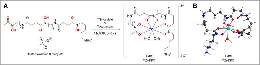
images were recorded, mice were anesthetized by inhalation of a Natural bond-order charge analysis (Supplemental Table 7)
1% isoflurane (Baxter Healthcare)/oxygen gas mixture and placed also supports the conclusion that thermodynamic stabiliza-
on the scanner bed. PET images were recorded at various times tion of complex 3-cis arises from increased electrostatic
between 3 and 144 h after injection (supplemental material). interaction between the Zr41 ion and axial H2O ligand
Statistical Analysis (ligand-to-metal charge transfer). We expect that the coor-
Data were analyzed using the unpaired, 2-tailed Student t test. dinated H2O ligands would be kinetically labile and that
Differences at the 95% confidence level (P , 0.05) were consid- species 1–3 were likely in rapid equilibrium at physiologic
ered to be statistically significant. temperatures.
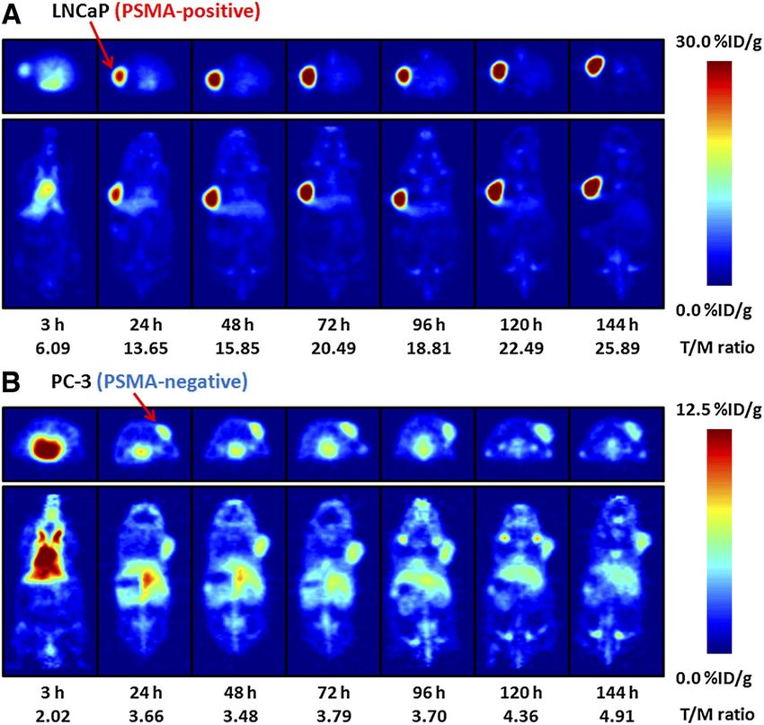
Basic Characterization of 89Zr Species In Vivo
RESULTS 89Zr-radiolabeled
Before full studies on mAbs are begun,
DFT Calculations it is important to understand the in vivo behavior of various
The complexation reaction between 89Zr-chloride or 89Zr-labeled species, including the starting reagents, and
89Zr-oxalate and the hexadentate, tris-hydroxamate chelate potential impurities or metabolites. Therefore, we exam-
DFO is shown in Figure 1A. Structures of the octahedral ined the biodistribution of 89Zr-chloride and 89Zr-oxalate
complex [89Zr(HDFO)]21 (1), the 7-coordinate complexes and the complex 89Zr-DFO using PET (Fig. 2). Maxi-
with mono-H2O coordination in the axial and equatorial mum-intensity-projection images of 89Zr-chloride and
sites [89Zr(HDFO)-ax-(H2O)]21 (2-ax) and [89Zr(HDFO)- 89Zr-oxalate (11.1 MBq [300 mCi], 200 mL of sterile saline)
eq-(H2O)]21 (2-eq), and the 8-coordinate complex [89Zr were recorded at 24 h after injection in the tail vein of male
(HDFO)-cis-(H2O)2]21 (3-cis) were fully optimized using athymic nu/nu mice (24). 89Zr-chloride was found to be
DFT. The optimized structure of the 8-coordinate complex sequestered in the liver, with little excretion (Supplemental
3-cis is shown in Figure 1B (Supplemental Tables 1–7; Fig. 3). In contrast, administration of 89Zr-oxalate (most
Supplemental Figs. 1 and 2). likely present as the thermodynamically stable species
The calculations revealed that expansion of the coordi- [89Zr(C2O4)4]42) showed a high accumulation of 89Zr
nation sphere to either 7 or 8 coordinates by the addition of radioactivity in bones, joints, and potentially cartilage (Sup-
1 or 2 water molecules is thermodynamically favorable. plemental Fig. 4). Previous dynamic PET studies on 89Zr-
Interestingly, in the 7-coordinate species the axial and DFO demonstrated that this complex is excreted rapidly
equatorial coordination sites, 2-ax and 2-eq, are energeti- within 20 min via a renal pathway, with a measured bio-
cally inequivalent. Axial coordination (2-ax) is thermody- logic half-life of 305 6 6 s (Supplemental Figs. 5 and 6).
namically more favorable than equatorial coordination
(2-eq) by around 241 kJ mol21. The DFT calculations also Radiochemistry
suggest that the 8-coordinate complex with cis-coordination J591 was functionalized with DFO using bioconjugation
geometry with respect to the orientation of the H2O mole- methods modified from the pioneering work of Verel et al.
cules (3-cis) is 95 kJ mol21 more stable than the parent (23). The conjugation and purification chemistry was found
octahedral complex (1). Furthermore, complex 3-cis is 14 to proceed in a moderate to high yield (65% 6 5%), with
kJ mol21 more stable than the sum of the thermodynamic high chemical purity (.95%). Radiolabeling of DFO-J591
stabilization achieved by complexes 2-ax and 2-eq (sum 5 with 89Zr-oxalate was achieved at room temperature, in
281 kJ mol21). This additional stability of complex 3-cis slightly alkaline solutions (pH 7.7–8.1), with crude radio-
arises because of structural relaxation from the cooperativ- chemical yields (.95%, n 5 6). Facile purification of
ity of the 2-coordinated H2O ligands, which allows the 89Zr-DFO-J591 from small-molecule radiolabeled im-
r(Zr-OH2(ax)) to decrease from 0.236 nm in complex purities was achieved using either size-exclusion chro-
2-ax to 0.233 nm in complex 3-cis (Supplemental Table 6). matography or spin-column centrifugation. The final
RGB
FIGURE 1. (A) Complexation reaction between [89Zr(C2O4)4]42 and DFO. (B) DFT-optimized structure of 8-coordinate complex
[89Zr(HDFO)-cis-(H2O)2]21 (3-cis).
89ZR-DFO-J591 IMMUNOPET OF PSMA • Holland et al. 1295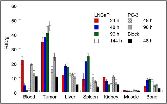
FIGURE 2. PET images showing
 maximum intensity projection of 89Zr-
                                                                                                                              RGB
 chloride and 89Zr-oxalate at 24 h after
 intravenous administration and dynamic
 PET images of 89Zr-DFO at 1 and 4 min
 after injection. For maximum-intensity-
 projection images, upper and lower
 intensity thresholds were set at 100%
 and 0%, respectively. Further details
 are presented in Supplemental Figures
 3–6. MIP 5 maximum intensity projection.

radiochemical yield of the purified 89Zr-DFO-J591 was         receptor in vivo was initially assessed by conducting acute
greater than 77%, and the product was formulated in           biodistribution studies in LNCaP tumor–bearing mice at 24,
0.9% sterile saline with an RCP greater than 99% (n 5         48, 96, and 144 h after intravenous administration (Table 1;
6) and a specific activity of 181.7 6 1.1 MBq/mg (4.91 6      Supplemental Table 8; Fig. 3). The data reveal that high
0.03 mCi/mg) of mAb (Supplemental Figs. 7 and 8). The         LNCaP tumor uptake was observed 24 h after injection
specific activity obtained in these studies compares favor-   (34.4 6 3.2 percentage injected dose per gram [%ID/g]),
ably with the previously reported specific activities of      with a steady increase through 48 (38.0 6 6.2 %ID/g) and
other 89Zr-radiolabeled mAbs (22,26–32). Isotopic dilu-       96 h (40.4 6 4.8 %ID/g) and reaching 45.8 6 3.2 %ID/g at
tion assays revealed an average of 3.9 6 0.3 accessible       144 h (P 5 0.01 between LNCaP uptake at 24 and 144 h).
chelates per mAb.                                             This high accumulation of 89Zr-DFO-J591 is consistent
                                                              with extraction of the activity from the blood pool (24 h,
89Zr-DFO-J591   Immunoreactivity and Stability                21.8 6 2.8 %ID/g; 48 h, 4.4 6 1.9 %ID/g; and 96 h, 1.4 6
Studies In Vitro                                              0.8 %ID/g) and rapid internalization of the J591–PSMA
   The immunoreactive fraction of the 89 Zr-DFO-J591          complex, followed by sequestration of the 89Zr radioactiv-
formulations was measured by specific in vitro cellular       ity inside the cell. In contrast, 89Zr-DFO-J591 uptake in the
association assays using LNCaP (PSMA-positive) cells          PC-3 (PSMA-negative) tumors at 48 (15.6 6 2.1 %ID/g,
before each in vivo experiment (Supplemental Fig. 9)          P 5 0.0025) and 96 h (24.0 6 2.6 %ID/g, P 5 0.0017)
(33). In studies using 213Bi-labeled J591, McDevitt           showed a statistically significant decrease in 89Zr accumu-
et al. reported that the LNCaP cell line had an estimated     lation, compared with uptake in PSMA-positive tumors.
                                                              89Zr-DFO-J591 activity in the blood remained 4-fold higher
180,000 PSMA molecules per cell (11). However, in
other studies using 111In- and 131I-labeled mAbs              at 48 h (19.0 6 1.1 %ID/g, P 5 0.001) and 10-fold higher
(including J415, J533, J591, and 7E11), we found              at 96 h (13.0 6 1.8 %ID/g, P , 0.05) in mice bearing PC-3
higher PSMA copy numbers (600,000–800,000 sites/              tumors, compared with the corresponding LNCaP tumor–
cell) for viable LNCaP cells (9). The average immunor-        bearing mice (48 h, 4.4 6 1.9 %ID/g; 96 h, 1.4 6 0.8 %ID/g).
eactive fraction of 89Zr-DFO-J591 was 0.95 6 0.03                Competitive inhibition (blocking) studies using low-spe-
(n 5 4). Control experiments (n 5 4) using the PC-3           cific-activity formulations (60-fold decrease, 3.04 MBq/mg
(PSMA-negative) cell line showed no binding, further          [0.082 mCi/mg]), compared with high-specific-activity for-
demonstrating the specificity of 89Zr-DFO-J591 for            mulations, revealed only 10.3 6 0.8 %ID/g tumor uptake at 48
PSMA-expressing cells.                                        h after injection, an approximate 4-fold decrease (P , 0.002)
   Incubation of 89Zr-DFO-J591 in either saline or 1%         (Tables 1 and 2; Fig. 3). Furthermore, in the low-specific-
bovine serum albumin for 7 d at 37C revealed a less than     activity experiments, 89Zr-DFO-J591 activity in the blood re-
2% decrease in RCP (via demetalation), with an observed       mained high (2- to 3-fold higher at 48 h, 10.7 6 0.4 %ID/g,
approximate 17% decrease in the immunoreactive fraction       P 5 0.026), but 89Zr-accumulation in the liver showed a stat-
for the 1% bovine serum albumin experiment (0.78 6            istically significant decrease from 17.7 6 1.6 %ID/g to 5.1 6
0.03, Supplemental Fig. 10). Therefore, in the absence        0.4 %ID/g (P , 0.004). The competitive inhibition experi-
of specific proteolysis or reductive or oxidative metabo-     ments concur with the in vitro data and further demonstrate
lism, 89Zr-DFO-J591 is expected to remain intact and          the specificity of 89Zr-DFO-J591 for the PSMA in vivo.
immunoreactive in vivo on a time scale suitable for immu-        Interestingly, in the LNCaP tumor–bearing mice, 89Zr
noPET.                                                        uptake in the bone was relatively high and increased
                                                              between 24 and 96 h (24 h, 4.0 6 0.8 %ID/g; 48 h, 8.2
Biodistribution Studies                                       6 1.2 %ID/g; and 96 h, 8.7 6 1.5 %ID/g) before decreasing
  The ability of 89Zr-DFO-J591 to target an extracellular     slightly to 7.4 6 1.3 %ID/g at 144 h. In contrast, bone
epitope of the PSMA type II transmembrane glycoprotein        accumulation of 89Zr activity in the PC-3 tumor–bearing

1296     THE JOURNAL     OF   NUCLEAR MEDICINE • Vol. 51 • No. 8 • August 2010
TABLE 1. Biodistribution Data of 89Zr-DFO-J591, Administered Intravenously to Mice Bearing Subcutaneous
LNCaP Tumors
Block
(300 mg of mAb)
Organ 24 h (n 5 4) 48 h (n 5 5) 96 h (n 5 5) 144 h (n 5 4) at 48 h (n 5 4)
Blood 21.8 6 2.8 4.4 6 1.9 1.4 6 0.8 2.6 6 1.5 10.7 6 0.4
Tumor 34.4 6 3.2 38.0 6 6.2 40.4 6 4.8 45.8 6 3.2 10.3 6 0.8
Heart 7.4 6 2.2 4.0 6 1.3 1.7 6 0.6 1.4 6 0.5 3.8 6 0.7
Lung 11.7 6 1.9 5.7 6 3.1 2.2 6 0.9 2.5 6 0.9 5.7 6 0.3
Liver 11.7 6 1.5 17.7 6 1.6 17.2 6 2.7 11.2 6 1.6 5.1 6 0.4
Spleen 8.8 6 4.3 21.1 6 0.3 24.6 6 1.8 4.6 6 2.4 3.1 6 0.7
Kidney 10.1 6 1.0 7.5 6 1.5 5.1 6 0.5 5.3 6 0.5 5.1 6 0.2
Muscle 1.1 6 0.1 0.6 6 0.3 0.4 6 0.4 0.2 6 0.2 0.8 6 0.2
Bone 4.0 6 0.8 8.2 6 1.2 8.7 6 1.5 7.4 6 1.3 2.4 6 0.3
Tumor/blood 1.6 6 0.2 8.7 6 4.1 29.7 6 17.1 18.0 6 10.5 1.0 6 0.1
Tumor/heart 4.7 6 1.5 9.6 6 3.5 23.4 6 9.0 31.9 6 10.7 2.7 6 0.5
Tumor/lung 2.9 6 0.5 6.7 6 3.7 18.4 6 7.7 18.5 6 6.8 1.8 6 0.2
Tumor/liver 2.9 6 0.5 2.1 6 0.4 2.3 6 0.5 4.1 6 0.7 2.0 6 0.2
Tumor/spleen 3.9 6 1.9 1.8 6 0.3 1.6 6 0.2 9.9 6 5.2 3.3 6 0.8
Tumor/kidney 3.4 6 0.5 5.1 6 1.3 7.9 6 1.2 8.6 6 1.1 2.0 6 0.2
Tumor/muscle 32.4 6 4.6 59.2 6 28.8 95.9 6 95.3 306.4 6 432.2 13.3 6 3.1
Tumor/bone 8.7 6 1.9 4.7 6 1.0 4.6 6 1.0 6.2 6 1.2 4.3 6 0.6
Complete biodistribution data are presented in Supplemental Table 7. Data are expressed as mean %ID/g 6 SD. Errors for tumor-to-
tissue ratios are calculated as geometric mean of SD. LNCaP tumors: 3–4 mg mAb; PSMA-positive, 50–250 mm3.
mice was reduced by approximately 45% at 48 and 96 h images showing the mean %ID/g radiotracer uptake in var-
(4.3 6 0.6 %ID/g and 5.1 6 0.5 %ID/g, respectively). ious tissues including the heart and blood pool, liver, and
89Zr-DFO-J591
muscle in mice bearing LNCaP (n 5 3) or PC-3 (n 5 3)
ImmunoPET with
tumors are given in Figure 5. Radiotracer uptake in LNCaP
Temporal immunoPET images of 89Zr-DFO-J591 (10.9–
tumors was observed less than 24 h after injection of
11.3 MBq [295–305 mCi], 60–62 mg of mAb, in sterile 89Zr-DFO-J591, and high tumor-to-muscle (T/M) ratios (cal-
saline [200 mL]) recorded in LNCaP and PC-3 tumor–bear-
culated using the mean %ID/g values derived from volume-
ing mice between 3 to 144 h are presented in Figure 4.
of-interest analysis of the immunoPET images) were
Time–activity curves generated from the immunoPET
observed. At 48 h after injection, the immunoPET-measured
mean and maximum %ID/g for radiotracer uptake in LNCaP
tumor–bearing mice was 21.9 6 0.6 and 38.2 6 4.9 %ID/g,
respectively, with a mean T/M ratio of 15.85 (Supplemental
Table 8). By 120 and 144 h, the mean T/M ratio in LNCaP
tumors increased to 22.49 and 25.89, respectively. The lower
uptake observed in the quantitative immunoPET studies,
compared with the biodistribution studies, is likely due to
the different total masses of mAb administered (22).
In contrast to the high absolute tumor uptake and tumor-
to-background contrast ratios observed in the LNCaP model,
low accumulation and immunoPET contrast ratios for 89Zr-
DFO-J591 uptake in PC-3 (PSMA-negative) tumors (Supple-
mental Tables 9 and 10; mean T/M ratios of 3.48, 4.36, and
4.19 at 48, 120, and 144 h, respectively) were observed.
RGB FIGURE 3. Bar chart showing selected tissue Uptake in these PC-3 tumors is in accordance with the
biodistribution data (%ID/g) for uptake of either high- enhanced permeation and retention mechanism (Supplemen-
(181.7 6 1.1 MBq/mg [4.91 6 0.03 mCi/mg]; 3–4 mg of tal Tables 9 and 10; Supplemental Figs. 11–13).
mAb per mouse) or low-specific-activity (60-fold decrease,
3.04 MBq/mg [0.082 mCi/mg]; 300 mg of mAb per mouse)
formulations of 89Zr-DFO-J591 (0.55–0.74 MBq [15–20 mCi], DISCUSSION
in 200 mL of sterile saline for injection) in male athymic nu/nu PET has distinct advantages over SPECT in terms of
mice bearing subcutaneous LNCaP (PSMA-positive) or PC-3 sensitivity and contrast resolution, especially for deep
(PSMA-negative) tumors.
tissues, and these improved imaging characteristics are
89ZR-DFO-J591 IMMUNOPET OF PSMA • Holland et al. 129789Zr–DFO complex due to strong electrostatic interactions,
TABLE 2. Biodistribution Data of 89Zr-DFO-J591,
Administered Intravenously to Mice Bearing
coupled with the enhancement in thermodynamic stability
Subcutaneous PC-3 Tumors (3–4 mg of mAb) induced by expansion of the first coordination sphere and
geometry relaxation to give an 8-coordinate species.
Organ 48 h (n 5 4) 96 h (n 5 3)
Indeed, in the case of 89Zr-DFO—and in contrast to the
Blood 19.0 6 1.1 13.0 6 1.8
Tumor 15.6 6 2.1 24.0 6 2.6
more familiar complexes with radionuclides of copper, gal-
Heart 6.8 6 0.1 4.3 6 0.9 lium, indium, and yttrium—these calculations indicate that
Lung 12.6 6 1.9 7.0 6 2.3 the presence of water or, for example, coordinating anions
Liver 12.4 6 0.9 11.0 6 1.6 such as chloride, may actually increase the thermodynamic
Spleen 10.2 6 2.0 7.2 6 0.7 stability of the 89Zr–DFO complex in vivo.
Kidney 10.5 6 0.9 6.9 6 1.6
Muscle 1.5 6 0.4 0.9 6 0.2
The ability of 89Zr-DFO-J591 to target PSMA-expressing
Bone 4.3 6 0.6 5.1 6 0.5 tissue was examined using acute biodistribution studies and
Tumor/blood 0.8 6 0.1 1.8 6 0.3 immunoPET in vivo. The results demonstrate that 89Zr-
Tumor/heart 2.3 6 0.3 5.6 6 1.4 DFO-J591 shows high specific uptake in LNCaP (PSMA-
Tumor/lung 1.2 6 0.3 3.4 6 1.2 positive) tumors. Although a direct comparison with earlier
Tumor/liver 1.3 6 0.2 2.2 6 0.4
Tumor/spleen 1.5 6 0.4 3.4 6 0.5
work is made difficult because of the use of different mod-
Tumor/kidney 1.5 6 0.2 3.5 6 0.9 els and murine-J591, the absolute tissue uptake values of
89Zr-DFO-J591 (humanized mAb) in most organs at various
Tumor/muscle 10.4 6 3.3 25.4 6 5.8
Tumor/bone 3.6 6 0.7 4.7 6 0.7 time points were found to be higher than those observed for
either 111In-DOTA-labeled or 131I-labeled J591 (10). For
Complete biodistribution data are presented in Supplemental example, at 48 h after administration the tumor uptake
Table 7. Data are expressed as mean %ID/g 6 SD. Errors for value was 13.6 6 2.8 for 111In-DOTA-J591 and 11.2 6
tumor-to-tissue ratios are calculated as geometric mean of SD. 2.9 %ID/g for 131I-J591, with corresponding blood-pool
PC-3 tumors: PSMA-negative, 70–90 mm3.
activities of 8.98 6 2.10 and 8.57 6 2.04 %ID/g, respec-
tively. In contrast, tumor uptake and concordant blood-pool
particularly important for radioimmunoimaging. Basic activity of 89Zr-DFO-J591 at 48 h were 38.0 6 6.2 and
characterization of the in vivo behavior of several important 4.4 6 1.9 %ID/g, respectively. As revealed in the biodis-
89Zr-labeled species, including the starting reagents 89Zr- tribution data, the high immunoreactivity and specificity of
chloride and 89Zr-oxalate and the key complex 89Zr-DFO, 89Zr-DFO-J591 (Tables 1 and 2; Supplemental Table 8) led
are reported. The nature of the aqueous-phase 89Zr species to a high uptake in the PSMA-positive tumors.
using PET was shown to dramatically affect the in vivo The degree of bone uptake is consistent with previously
biodistribution, with 89Zr-chloride and 89Zr-oxalate seques- reported studies using various other 89Zr-labeled mAbs—
tering for over 24 h in the liver and bones, respectively. In including 89Zr-DFO-trastuzumab (22,32), for imaging
contrast, 89Zr-DFO is first-pass excreted through the kid- HER2/neu expression, and 89Zr-DFO-bevacizumab, for
neys and accumulates in the bladder, with a biologic half- imaging vascular endothelial growth factor (36). The nature
life of 305 6 6 s. of the radioactive species accumulating in the bone remains
In this work, the novel radiopharmaceutical 89Zr-DFO- uncertain, but it is plausible that slow intratumoral metab-
J591 has been characterized by a range of stability and olism and subsequent recirculation of 89Zr-labeled metab-
cellular association assays in vitro. Previous studies have olites may occur. Full metabolic studies are beyond the
shown that although diethylenetriaminepentaacetic acid can scope of the current study and will be the subject of further
be used for chelation and radiolabeling of mAbs with investigations. However, in a recent clinical trial investigat-
89Zr41 ions, demetalation occurs in vivo, and until new ing the radiation dosimetry of 89Zr-DFO-U36 (a chimeric
ligands are produced DFO remains the chelate of choice mAb directed against CD44v6) in 20 patients with head and
(24,34,35). Experiments on 89Zr-DFO mAbs have reported neck squamous cell carcinoma, the liver was identified as
high in vivo stability with respect to demetalation or ligand the dose-limiting organ (28,29). Dosimetry studies based on
dissociation and relatively low levels of radiotracer accu- the biodistribution data presented in this work suggest that
mulation in background tissue in both animals and humans for clinical patient studies, kidney uptake of 89Zr-DFO-
(26–28,30–32). J591 is the dose-limiting factor.
The nature of the electronic structure of the 89Zr–DFO The immunoPET data demonstrate that 89Zr-DFO-J591
complex has been explored using high-level DFT calcula- imaging provides high tumor–to–background tissue ratios
tions. The computational results provide a rationale for the and that this high uptake is specific for PSMA expression in
high experimentally observed in vitro and in vivo stability tissue. Overall, the novel radiotracer 89Zr-DFO-J591 repre-
of the 89Zr-DFO–labeled radioimmunoconjugates. DFT sents a promising candidate for translation to the clinic as
studies suggest that the origin of the observed in vivo stabil- an immunoPET agent for the noninvasive delineation of
ity of 89Zr-DFO mAbs lies in a combination of the inher- PSMA-positive primary and metastatic prostate cancers in
ently high thermodynamic and kinetic stability of the vivo.
1298 THE JOURNAL OF NUCLEAR MEDICINE • Vol. 51 • No. 8 • August 2010FIGURE 4. Temporal immunoPET
images of 89Zr-DFO-J591 (10.9–11.3
MBq [295–305 mCi], 60–62 mg of mAb,
in 200 mL of sterile saline) recorded in
LNCaP tumor–bearing (PSMA-positive,
left shoulder) (A) and PC-3 tumor–
bearing (PSMA-negative, right shoulder)
(B) mice between 3 and 144 h after
RGB injection. Transverse and coronal planar
images intersect center of tumors, and
mean tumor-to-muscle ratios derived
from volume-of-interest analysis of
immunoPET images are given. Upper
thresholds of immunoPET have been
adjusted for visual clarity, as indicated
by scale bars.
CONCLUSION subsequent radiolabeling do not compromise the immuno-
89Zr-DFO-J591 has been prepared with a high RCP reactivity, and radiolabeled immunoconjugate remains
(.99%) and specific activity (181.7 6 1.1 MBq/mg). In active for up to 7 d at 37C. Biodistribution and immuno-
vitro stability studies demonstrated that functionalization of PET experiments indicated that 89Zr-DFO-J591 shows high
J591 with 3.9 6 0.3 accessible DFO chelates per mAb and potential as a radiotracer for specific, noninvasive delinea-
tion of PSMA-positive PCs in vivo. Work toward the clin-
ical translation of 89Zr-DFO-J591 and other 89Zr-labeled
mAbs is under way.
ACKNOWLEDGMENTS
We thank Drs. NagaVaraKishore Pillarsetty and Pat
Zanzonico for informative discussions, Valerie M. Longo
for assistance with the biodistribution experiments, Thomas
Ku for advice with in vitro experiments, and Bradley
Beattie for assistance with PET. We thank Professor
Jennifer C. Green (Department of Chemistry, University
of Oxford, United Kingdom) for access to computational
facilities. We also thank the staff of the Radiochemistry/
Cyclotron Core at the Memorial Sloan-Kettering Cancer
Center (MSKCC). This study was funded in part by the
Geoffrey Beene Cancer Research Center of Memorial
FIGURE 5. Time–activity curves derived by volume-of- Sloan-Kettering Cancer Center; the Office of Science
interest analysis of immunoPET images showing mean
%ID/g tissue uptake vs. time/h for 89Zr-DFO-J591 (BER), U.S. Department of Energy (award DE-SC0002456);
radiotracer accumulation in mice bearing subcutaneous the Ludwig Center for Cancer Immunotherapy of the
LNCaP (PSMA-positive) or PC-3 (PSMA-negative) tumors. Sloan-Kettering Institute; and the Starr Cancer Consor-
Complete time–activity curve data for 89Zr-DFO-J591 tium. Technical services provided by the MSKCC Small-
immunoPET imaging is given in supplemental materials Animal Imaging Core Facility were supported in part by
(Supplemental Tables 9 and 10; Supplemental Figs. 11–13).
NIH grants R24 CA83084 and P30 CA08748. Dr. Neil
89ZR-DFO-J591 IMMUNOPET OF PSMA • Holland et al. 1299Bander is the inventor on patents that are owned by 17. Bander NH, Milowsky MI, Nanus DM, Kostakoglu L, Vallabhajosula S,
Goldsmith SJ. Phase I trial of 177Lu-labeled J591, a monoclonal antibody to
Cornell Research Foundation (CRF) for the J591 antibody prostate-specific membrane antigen, in patients with androgen-independent
described in this manuscript. Dr. Neil Bander is a paid prostate cancer. J Clin Oncol. 2005;23:4591–4601.
consultant to BZL Biologics, the company to which the 18. Milowsky MI, Nanus DM, Kostakoglu L, et al. Vascular targeted therapy with
anti-prostate-specific membrane antigen monoclonal antibody J591 in advanced
patents were licensed by CRF for further research and solid tumors. J Clin Oncol. 2007;25:540–547.
development. 19. Pandit-Taskar N, O’Donoghue JA, Morris MJ, et al. Antibody mass escalation
study in patients with castration-resistant prostate cancer using 111In-J591: lesion
detectability and dosimetric projections for 90Y radioimmunotherapy. J Nucl
Med. 2008;49:1066–1074.
REFERENCES
20. McDevitt MR, Ma D, Lai LT, et al. Tumor therapy with targeted atomic
1. Apolo AB, Pandit-Taskar N, Morris MJ. Novel tracers and their development for nanogenerators. Science. 2001;294:1537–1540.
the imaging of metastatic prostate cancer. J Nucl Med. 2008;49:2031–2041. 21. Frisch MJ, Trucks GW, Schlegel HB, et al. Gaussian 03, Revision-C.02.
2. Olson WC, Heston WDW, Rajasekaran AK. Clinical trials of cancer therapies Wallingford, CT: Gaussian, Inc.; 2004.
targeting prostate-specific membrane antigen. Rev Recent Clin Trials. 22. Holland JP, Caldas-Lopes E, Divilov V, et al. Measuring the pharmacokinetic
2007;2:182–190. effects of a novel Hsp90 inhibitor on HER2/neu expression in mice using 89Zr-
3. Jhanwar YS, Divgi C. Current status of therapy of solid tumors. J Nucl Med. DFO-trastuzumab. PLoS ONE. 2010;5:e8859.
2005;46(1 suppl):141S–150S. 23. Verel I, Visser GWM, Boellaard R, Stigter-van Walsum M, Snow GB, van
4. Sokoloff RL, Norton KC, Gasior CL, Marker KM, Grauer LS. A dual- Dongen GAMS. 89Zr immuno-PET: comprehensive procedures for the
monoclonal sandwich assay for prostate-specific membrane antigen: levels in production of 89Zr-labeled monoclonal antibodies. J Nucl Med. 2003;44:1271–
tissues, seminal fluid and urine. Prostate. 2000;43:150–157. 1281.
5. Manyak MJ. Indium-111 capromab pendetide in the management of recurrent 24. Holland JP, Sheh Y, Lewis JS. Standardized methods for the production of high
prostate cancer. Expert Rev Anticancer Ther. 2008;8:175–181. specific-activity zirconium-89. Nucl Med Biol. 2009;36:729–739.
6. Morris MJ, Pandit-Taskar N, Divgi CR, et al. Phase I evaluation of J591 as a 25. Kim JS, Lee JS, Im KC, et al. Performance measurement of the microPET Focus
vascular targeting agent in progressive solid tumors. Clin Cancer Res. 120 scanner. J Nucl Med. 2007;48:1527–1535.
2007;13:2707–2713. 26. Verel I, Visser GWM, Boellaard R, et al. Quantitative 89Zr immuno-PET for in
7. Liu H, Moy P, Kim S, et al. Monoclonal antibodies to the extracellular domain of vivo scouting of 90Y-labeled monoclonal antibodies in xenograft-bearing nude
prostate-specific membrane antigen also react with tumor endothelium. Cancer mice. J Nucl Med. 2003;44:1663–1670.
Res. 1997;57:3629–3634. 27. Perk LR, Visser OJ, Stigter-van Walsum M, et al. Preparation and evaluation of
8. Liu H, Rajasekaran AK, Moy P, et al. Constitutive and antibody-induced 89Zr-Zevalin for monitoring of 90Y-Zevalin biodistribution with positron
internalization of prostate-specific membrane antigen. Cancer Res. 1998; emission tomography. Eur J Nucl Med Mol Imaging. 2006;33:1337–1345.
58:4055–4060. 28. Börjesson PKE, Jauw YWS, Boellaard R, et al. Performance of immuno-positron
9. Smith-Jones PM, Vallabahajosula S, Goldsmith SJ, et al. In vitro characterization emission tomography with zirconium-89-labeled chimeric monoclonal antibody
of radiolabeled monoclonal antibodies specific for the extracellular domain of U36 in the detection of lymph node metastases in head and neck cancer patients.
prostate-specific membrane antigen. Cancer Res. 2000;60:5237–5243. Clin Cancer Res. 2006;12:2133–2140.
10. Smith-Jones PM, Vallabhajosula S, Navarro V, Bastidas D, Goldsmith SJ, 29. Borjesson PKE, Jauw YWS, de Bree R, et al. Radiation dosimetry of 89Zr-
Bander NH. Radiolabeled monoclonal antibodies specific to the extracellular labeled chimeric monoclonal antibody U36 as used for immuno-PET in head
domain of prostate-specific membrane antigen: preclinical studies in nude and neck cancer patients. J Nucl Med. 2009;50:1828–1836.
mice bearing LNCaP human prostate tumor. J Nucl Med. 2003;44:610–617. 30. Perk LR, Stigter-van Walsum M, Visser GWM, et al. Quantitative PET
11. McDevitt MR, Barendswaard E, Ma D, et al. An a-particle emitting antibody imaging of Met-expressing human cancer xenografts with 89Zr-labelled
([213Bi]J591) for radioimmunotherapy of prostate cancer. Cancer Res. monoclonal antibody DN30. Eur J Nucl Med Mol Imaging. 2008;35:
2000;60:6095–6100. 1857–1867.
12. Vallabhajosula S, Smith-Jones PM, Navarro V, Goldsmith SJ, Bander NH. 31. Aerts HJWL, Dubois L, Perk L, et al. Disparity between in vivo EGFR
Radioimmunotherapy of prostate cancer in human xenografts using expression and 89Zr-labeled cetuximab uptake assessed with PET. J Nucl Med.
monoclonal antibodies specific to prostate specific membrane antigen (PSMA): 2009;50:123–131.
studies in nude mice. Prostate. 2004;58:145–155. 32. Dijkers ECF, Kosterink JGW, Rademaker AP, et al. Development and
13. Vallabhajosula S, Kuji I, Hamacher KA, et al. Pharmacokinetics and characterization of clinical-grade 89Zr-trastuzumab for HER2/neu immunoPET
biodistribution of 111In- and 177Lu-labeled J591 antibody specific for prostate- imaging. J Nucl Med. 2009;50:974–981.
specific membrane antigen: prediction of 90Y-J591 radiation dosimetry based on 33. Lindmo T, Boven E, Cuttitta F, Fedorko J, Bunn PA Jr. Determination of the
111In or 177Lu? J Nucl Med. 2005;46:634–641. immunoreactive fraction of radiolabeled monoclonal antibodies by linear
14. Bander NH, Trabulsi EJ, Kostakoglu L, et al. Targeting metastatic prostate extrapolation to binding at infinite antigen excess. J Immunol Methods.
cancer with radiolabeled monoclonal antibody J591 to the extracellular 1984;72:77–89.
domain of prostate specific membrane antigen. J Urol. 2003;170:1717–1721. 34. Meijs WE, Herscheid JDM, Haisma HJ, Pinedo HM. Evaluation of desferal as a
15. Vallabhajosula S, Goldsmith SJ, Hamacher KA, et al. Prediction of myelotoxicity bifunctional chelating agent for labeling antibodies with Zr-89. Int J Rad Appl
based on bone marrow radiation-absorbed dose: radioimmunotherapy studies using Instrum A. 1992;43:1443–1447.
90Y- and 177Lu-labeled J591 antibodies specific for prostate-specific membrane 35. Meijs WE, Haisma HJ, Klok RP, et al. Zirconium-labeled monoclonal
antigen. J Nucl Med. 2005;46:850–858. antibodies and their distribution in tumor-bearing nude mice. J Nucl Med.
16. Vallabhajosula S, Goldsmith SJ, Kostakoglu L, Milowsky M, Nanus DM, 1997;38:112–118.
Bander NH. Radioimmunotherapy of prostate cancer using 90Y- and 177Lu- 36. Nagengast WB, de Vries EG, Hospers GA, et al. In vivo VEGF imaging with
labeled J591 monoclonal antibodies: effect of multiple treatments on radiolabeled bevacizumab in a human ovarian tumor xenograft. J Nucl Med.
myelotoxicity. Clin Cancer Res. 2005;11(19, suppl):7195s–7200s. 2007;48:1313–1319.
1300 THE JOURNAL OF NUCLEAR MEDICINE • Vol. 51 • No. 8 • August 2010You can also read