Disorders of the Skin - Deena Garner, DNP, APRN, CPNP PC
←
→
Page content transcription
If your browser does not render page correctly, please read the page content below
Disorders of the Skin
   Deena Garner, DNP, APRN, CPNP‐PC

                                      ©2020
Disclosures
  Deena Garner, DNP, APRN, CPNP‐PC

  • Has no financial relationship with commercial interests
  • This presentation contains no reference to unlabeled/unapproved uses of drugs or
    products

                                                                                       ©2020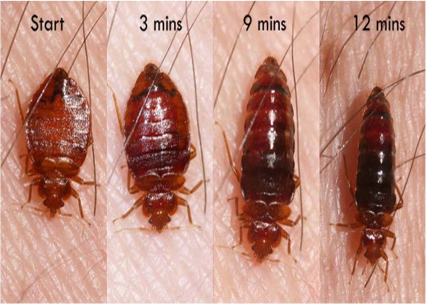
Learning Objectives
• Describe systematic approach to evaluate skin disorders.
• Identify primary, secondary, and special skin lesions.
• Discuss clinical presentation and management of common newborn skin
  conditions.
• Discuss clinical presentation and management of common pediatric
  systemic and local bacterial conditions, fungal infections, inflammatory
  conditions, and systemic and local viral infections.
• Discuss clinical presentation and management of common skin infestations
  and insect bites.

                                                                             ©2020
Evaluation of Skin Disorders
• History
   •   Onset, duration
   •   Original appearance of lesions/treatments
   •   Associated symptoms
   •   Exposures, medications, allergies
• Physical examination
   • Examination of entire body
   • Good light/Wood’s lamp
   • Location, type, color, pattern, distribution
• Diagnostic studies
   • Scrapings of skin for microscopic examination
   • Microbial cultures
   • Biopsies/patch testing

                                                     ©2020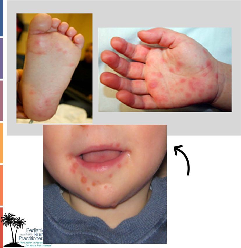
Primary Skin Lesions
          • Flat, circumscribed change of the skin. It may be of any size, although this term is often used for lesions
            1 cm in size.
          • Mongolian Spot, Vitiligo, Larger Café au lair spot
 Patch

          • Circumscribed, nonvesicular, nonpustular, elevated lesion that measures 1 cm. It is commonly formed by a
            confluence of papules.
Plaque    • Tinea corporis, Eczema, Psoriasis

                                                                                                                          ©2020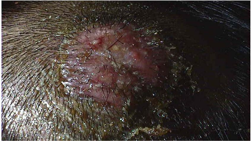
Primary Skin Lesions
          • Circumscribed, elevated, usually solid lesion that measures 0.5 to 2 cm in diameter. It involves the dermis and may
            extend into the subcutaneous tissue with its greatest mass below the surface of the skin; a large nodule (greater
            than 2 cm in diameter) is referred to as a tumor
Nodule    • Furuncle, Melanoma

          • Circumscribed elevation 1 cm in diameter, often with a deeper component and filled with purulent material.
          • Staphylococcal Abscess
Abscess

          • Sharply circumscribed, elevated, fluid‐containing lesion that measures ≤1 cm in diameter.
          • Chickenpox, Impetigo, Herpes Simplex
Vesicle

                                                                                                                                  ©2020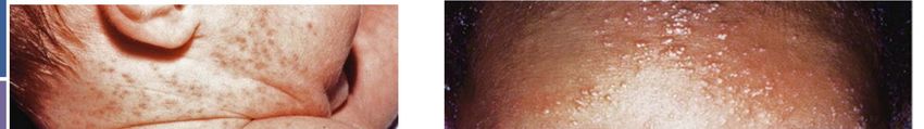
Primary Skin Lesions

          • Circumscribed, elevated, fluid‐containing lesion that measures >1 cm in
            diameter.
  Bulla   • Fixed drug eruption

          • A firm, edematous plaque resulting from infiltration of the dermis with fluid;
            White to pink or pale red, compressible, and evanescent, they often
            disappear within a period of hours. They vary in size and shape.
  Wheal   • Hives, Dermographism

                                                                                             ©2020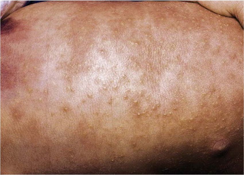
Other Skin Lesions

  Secondary Skin Lesions             • Excoriation
                                     • Comedone
  •   Scales                         • Milia
  •   Crust                          • Cyst
                           Special
  •   Erosion                 Skin   • Petechia
  •   Ulcer                Lesions   • Purpura
  •   Fissure                        • Burrow
  •   Atrophy                        • Lichenification
  •   Scar                           • Telangiectasia

                                                         ©2020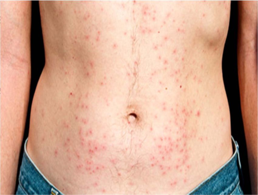
Petechiae & Purpura                                           •Purpura
                                                                      •4‐10 mm petechiae
                                                                      •Common vasculi s
                                                                  •Bruising
      Petechiae
                                                                      •> 10 mm bleeding
                                                                      under the skin

                                              Click to add text

•Petechiae
    •< 4mm spots of bleeding under the skin
    •capillary instability
    •Many causes‐ infectious most worrisome

                                                                                           ©2020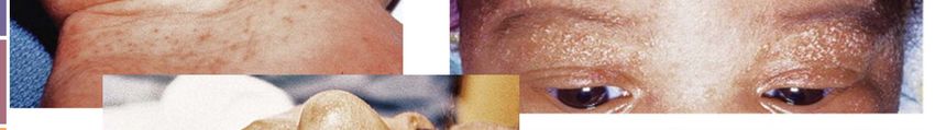
Skin Conditions of the Newborn and Infant

                                            ©2020
Milia Millaria Rubra Sebaceous Gland
Hyperplasia
Key pearly, yellow, 1–3 mm erythematous, 1‐2 mm multiple 1–2 mm diameter
Characteristics diameter papules papules and pustules. Also yellow papules
called "Prickly Heat" or
"Heat Rash"
Initial Eruption face, chin, and Can occur anywhere, but Clusters around the nose,
forehead has a predilection for the may also appear on cheeks
forehead, upper trunk,
and flexural or covered
surfaces.
Onset Shortly after birth After the first week of life At birth or shortly after
Resolves during the first month of may come and go within 4–6 months
life without treatment, throughout infancy. Cooling
they may persist for skin and loosening clothes
several months may cause resolution
©2020©2020
Erythema Toxicum                       Neonatal Acne
Key             barely elevated yellowish papules or multiple, tiny, monomorphous
Characteristics pustules measuring 1–3 mm in           papulopustules on an
                diameter, with a surrounding irregular erythematous base
                macular flare or wheal of erythema
                measuring 1–3 cm; ‘flea‐bitten’
                appearance.
Location       appear first on the face and spread to located primarily on the cheeks,
               the trunk and extremities, but may     but scattered over the face and
               appear anywhere on the body except often extending onto the scalp
               on the palms and soles
Onset          between 24 and 48 h of life            average onset at 3 weeks of age
Resolves       usually fade over 5–7 days, may        spontaneously within 1–3 months
               reoccur for several weeks.

                                                                                         ©2020
Erythema Toxicum and Neonatal Acne
©2020Diaper Dermatitis
Irritant Contact Candidiasis Bacterial Dermatitis
Dermatitis
Cause Contact with urine/feces; Candida Staphylococcal or
wearing diaper streptococcal
Key Characteristics Chapped, shiny, Shallow pustules, fiery Erythematous, denuded
erythematous, red plaques on CONVEX areas or fragile blisters,
parchment‐like skin with surfaces, inguinal folds, crusting, pustules in
possible erosions on labia, scrotum suprapubic areas and
CONVEX surfaces, creases periumbilicus
are spared
Timing Peak occurrences at 9‐12 Satellite lesions; recent Usually occurs in
months, may progress to antibiotic or diarrhea newborns
include creases
Treatment Frequent diaper change, Antifungal cream, Nystatin if yeast is present
gentle cleansing, barrier frequent diaper as well, mupirocin in
cream, air dry, 0.5%‐1% change, gentle cleansing, minimal, Augmentin or
hydrocortisone barrier cream, air dry cephalexin if severe
©2020Fungal Diaper Rash
• + located in folds
• + Satellite lesions
Contact Dermatitis
• Not located in folds
• No satellite lesions
• +Shiny and Erythematous
©2020Seborrheic Dermatitis
• Key Characteristics:
• Chronic inflammatory dermatitis
• Cradle cap in infants; dandruff in adolescents
• Overproduction of sebum and perhaps a saprophytic yeast
• S/S: erythematous, flaky crusts of yellow, greasy scales on scalp, face,
diaper area; mild flakes with dandruff; not pruritic
• Management:
• Antifungal agents: azoles, selenium sulfide
• Anti‐inflammatory agents: topical steroids, calcineurin inhibitors
• Keratolytic agents: salicylic acid, urea
• Facial dermatitis: ketoconazole topical preparation
• Scalp dermatitis: medicated shampoos/steroids
©2020Seborrheic Dermatitis
©2020Systemic Bacterial Skin Infections
©2020Scarlet Fever
• Key Characteristics:
• Scarlet fever is caused by group A β‐hemolytic Streptococcus.
• Illness begins with fever and pharyngitis followed by enanthem and exanthem in 24 to 48 hours.
• S/S
• Face appears flushed, except for circumoral pallor.
• Tongue initially has a white coating (white strawberry tongue) that fades by the fourth
day, leaving a very erythematous tongue with prominent papillae (red strawberry tongue).
• Cervical and submandibular lymphadenopathies are noted.
• diffuse erythema; small fine papules give it a sandpaper like quality.
• Begins on the neck and spreads rapidly to the trunk and extremities.
• Greater intensity of erythema in the axillae and antecubital, inguinal, and popliteal creases.
• The palms and soles are spared.
• The rash resolves in 4 to 5 days with fine peeling of the skin.
• Evaluation and Management
• Culture or rapid test of a pharyngeal swab
• Same as Strep Pharyngitis
©2020Tick Borne Illnesses
• Three most common: Rocky Mountain Spotted Tick Fever, Lyme Disease, Erlichiosis
RMSF Lyme Disease Erlichiosis
Location Throughout most of the Upper Midwestern and Southeastern and south‐
contiguous United States, five northeastern United States. central United States, from the
states (North Carolina, East Coast extending westward
Oklahoma, Arkansas, to Texas
Tennessee, and Missouri)
account for over 60% of
RMSF cases.
Incubation 3–12 days 3‐30 days 5–14 days
Early common Signs and • High fever • Erythema migrans (EM) • Fever, chills
Symptoms • Severe headache • Flu‐like symptoms— • Headache
• Malaise malaise, headache, fever, • Malaise
• Myalgia myalgia, arthralgia • Muscle pain
• Edema around eyes and • Lymphadenopathy • Gastrointestinal symptoms
on the back of hands nausea, vomiting, diarrhea,
• Gastrointestinal symptoms anorexia)
(nausea, vomiting, • Altered mental status
anorexia) • Rash (more commonly
reported among children)
©2020RMSF Lyme Ehrlichiosis
Key • A fever followed by a rash on the fourth day. • Erythema migrans (EM)—red ring‐like or • Tick bites or exposure, fever,
Characteristics • Early Rash homogenous expanding rash; classic severe headache, malaise,
• Small, flat, pink, non‐itchy spots (macules) rash not present in all cases. myalgia.
initially appear on the wrists, forearms, • "Bull's Eye" • Skin rash is not considered a
and ankles then spread to the trunk and • Flu‐like symptoms—malaise, headache, common feature of ehrlichiosis
within 2 days is generalized with fever, myalgia, arthralgia and should not be used to rule in
involvement of the palms and soles. or rule out an infection. E
• Lymphadenopathy chaffeensis infection can cause
• Late Rash rash in up to 60% of children
• Red to purple spots (petechiae) are usually • Physical are minimal.
not seen until day 6 or later after onset of • Splenomegaly is not
symptoms.Petechial rash is considered a uncommon, but some
sign of progression to severe disease. patients develop
• generalized periorbital edema, severe muscle hepatomegaly.
tenderness, GI symptoms and hyponatremia. Lymphadenopathy is very
uncommon.
Evaluation RMSF IgG ‐The first sample should be taken within the Sensitive enzyme immunoassay (EIA) or Detection of DNA by PCR of whole
first week of illness and the second should be taken 2 immunofluorescence assay (IFA) should be blood most sensitive during the first
to 4 weeks later. performed first; if positive or equivocal, it is week of illness
followed by a Western blot*
Management • Doxycycline: • Amoxicillin: 50 mg/kg/day orally TID for Same as RMSF; Pt should be treated
• Under 45 kg (100 lbs.): 2.2 mg/kg body weight 14‐21 days; Max Dose 500 mg/dose for 3 days after fever subsides.
given twice a day • Doxycycline: 4 mg/kg/day orally BID for Minimal course is 5‐7 days
• Over 45 kg: 100 mg every 12 hours 10‐21 days; Max Dose 100 mg/dose
• Maximum dose 100mg/dose
©2020Question 1
A school‐age child has an abrupt onset of sore throat, nausea, headache,
and a temperature of 102.3°F. An examination reveals petechiae on the
soft palate, beefy‐red tonsils with yellow exudate, and fine erythematous
papules that are sandpaper like. A Rapid Antigen Detection Test (RADT) is
negative. What is the next step in management for this child?
1. Obtain an anti‐streptococcal antibody titer
2. Perform a follow‐up throat culture
3. Prescribe amoxicillin for 10 days
4. Send to the ED for further evaluation
5Question 1 A school‐age child has an abrupt onset of sore throat, nausea, headache, and a temperature of 102.3°F. An examination reveals petechiae on the soft palate, beefy‐red tonsils with yellow exudate, and fine erythematous papules that are sandpaper like. A Rapid Antigen Detection Test (RADT) is negative. What is the next step in management for this child? Answer: Perform a follow‐up throat culture
Question 2
A school‐age child has an abrupt onset of headache and fatigue 4 days
ago. An examination reveals small erythematous macules on the wrists
and ankles which is reported to have to developed today. How will the PNP
proceed?

1.   Encourage systematic treatment is needed as the illness is most likely
     viral
2.   Order RMSF IgG titer
3.   Prescribe Amoxicillin
4.   Prescribe Erythromycin

                                                                          5
Question 2 A school‐age child has an abrupt onset of headache and fatigue 4 days ago. An examination reveals small erythematous macules on the wrists and ankles which is reported to have to developed today. How will the PNP proceed? Answer: Order RMSF IgG titer
Localized Bacterial Skin Infections

                                      ©2020
Impetigo
• Key Characteristics:
• Caused by Staphylococcus aureus and/or Group A beta hemolytic streptococcus;
MRSA
• Non‐Bullous vs. Bullous
• Often starts as a bug bite or skin injury
• Spread through autoinoculation via hands, towels, clothing, nasal discharge,
droplets
• "Honey Colored Crusts"
©2020Impetigo
• S/S
• Pruritus; spread of lesion to surrounding skin
• Non‐bullous: 1‐2 mm erythematous papules or pustules, progress to vesicles or
bullae which rupture – honey‐colored crusts
• Bullous: large, flaccid, thin‐wall, superficial, annular or oval blisters/bullae
• Weakness, fever, diarrhea
• Lesions common on face, hands, neck, extremities, perineum
• Regional lymphadenopathy
©2020Impetigo
Bullous Impetigo
Non Bullous Impetigo
©2020Impetigo
• Management
• Topical antibiotics if superficial, nonbullous, localized
• topical mupirocin
• Oral antibiotics for multiple, nonbullous lesions and widespread infections
• Augmentin, cephalexin, clindamycin, or dicloxacillin
• Obtain culture if no response in 7 days
• clindamycin, trimethoprim‐sulfamethoxazole
• Follow‐up in 2‐3 days if no improvement
• Educate about hygiene
• Exclude from school/daycare until treated for 24 hours
©2020Folliculitis
Key Characteristics:
• Superficial bacterial inflammation of hair follicle
– S. aureus‐ scalp and face
– P aeruginosa‐ usually below the neck
– Hot tub exposure
• S/S
• Pruritus
• Follicular pustules & follicular erythematous papules
– Discrete, erythematous 1‐2 mm papules or pustules on inflamed base near follicle
– Face, scalp, extremities, buttocks, back
– Pruritus papules, pustules, deep red/purple nodules in areas under swimsuit
©2020Folliculitis
• Management
• May not require any antibiotics
• Topical antibiotic therapy is
usually sufficient
• mupirocin or clindamycin
• Extensive disease or moderate
illness
• Cephalexin
• SMP‐TMX
• Clindamycin
©2020Abscess (Furuncle)
Key Characteristics:
• Deeper infection of base of follicle and deep dermis (boil)
• Collection of pus within the dermis and surrounding soft tissues
• S. aureus monoinfection (either MSSA or MRSA) in up to 75% of cases
• S/S
• Painful, tender, fluctuant, and erythematous nodules that eventually will have a
pustule
• Spontaneous drainage may occur
• Regional lymphadenopathy possible
• Rare systemic symptoms
• Deep red/purple nodules, painful
©2020Abscess (Furuncle)
• Management
• I & D alone is treatment of choice for deep abscess
• Antibiotics
• MSSA‐ cephalexin
• MRSA‐ SMX‐TMP or clindamycin
©2020Question 3
A child has developed honey colored crusts around his nose, mouth, and
buttocks that are not getting any better. The best treatment would be:
1. Cephalexin
2. Hydrocortisone
3. Mupirocin
4. Triple antibiotic ointment
5Question 3 A child has developed honey colored crusts around his nose, mouth, and buttocks that are not getting any better. The best treatment would be: Answer: Cephalexin
Fungal Skin Infection

                        ©2020
Tinea Capitis/Corporis
• Key Characteristics:
• Complaint of a "Ringworm" of "Wingworm"
• Recent hair cut
• Erythematous, defined borders with central clearing
• S/S
• Annular, oval, circinate lesions with red, scaly borders
• Lesions spread peripherally; clear centrally
• Often prominent over hair follicles
• Hair loss
• Multiple secondary lesions may merge
©2020Tinea Capitis • Evaluation
– KOH‐treated scrapings:
hyphae/spores
– Fungal culture
– Wood’s lamp does not
fluoresce most tinea
• Kerion occurs during the
Kerion inflammatory stage
– Pustular, boggy mass (pus is
sterile)
• Diffuse scaling to the scalp
without much hair breakage
around the kerion
– NO STEROIDS OR ANTIBIOTICS
FOR KERION
©2020Tinea Capitis/Corporis
• Management
• Topical antifungals (skin surface outside of hair line)
• Use until lesion has resolved + 2‐3 days
• Treat 1 inch beyond edge (Do not cover lesion)
• Griseofulvin: tinea capitis, tinea faciei, extensive infection, immunosuppression
• Typical treatment time is at least 4 weeks
• Eat fatty foods
• Check CBC, LFTs every 4 weeks on therapy
• Identify/treat contacts
• Exclude from day care/school until 24 hours of treatment
©2020Tinea Versicolor
• Key Characteristics
• Superficial fungal infection; predominantly on the trunk
• Caused by yeast‐like organism: Malassezia furfur
• warm, humid weather
• Occurs mostly on back and upper shoulders
• S/S
• Multiple annular, scaling macules/patches
• Hypopigmented in dark‐skinned
• Hyperpigmented in light‐skinned
• Raindrop pattern
©2020Tinea Versicolor
• Evaluation: KOH scraping
• Management
• Selenium sulfide lotion or shampoo
• Oral antifungal if resistant
©2020Inflammatory Conditions of the Skin
©2020Eczema (atopic dermatitis)
• Key Characteristics:
• Chronic, pruritic, inflammatory skin disorder
• “THE ITCH THAT RASHES”
• S/S:
• Pruritus/eczematous changes
• Dry skin
• Acute manifestations (more common in
infants)
• Intense itching/redness
• Papules, vesicles, edema, serous discharge/crusts
• Generalized dry skin
• Chronic manifestations (in older children)
• Lichenification
• Scratch marks
• Generalized xerosis
©2020Eczema
• Management:
– Avoid known irritants
– MOISTURIZE, MOISTURIZE, MOISTURIZE
• Vaseline, Cetaphil, Crisco, Aquaphor,
Eucerin
• Mild or mild‐moderate topical
corticosteroids
• Antihistamines
• Wet wrap therapy
• No topical antibiotics
– Unless secondary bacterial infection
• No systemic steroids
©2020Eczema
©2020Allergic Rashes
• Key Characteristics: • Management:
• Allergic/ Contact Dermatitis • Treatment for allergic or contact
• Erythema, vesicles dermatitis is the same as eczema
• Oozing in the area of contact
• Avoid the cause
• Distribution may be a clue to what caused it
• Break the habit
• Nickel dermatitis; lip‐licker; poison ivy
• id reaction • Stop the itching
• Widespread papulovesicular rash • Moisturizer
• From repeat exposures to a substance • Mild to mild‐moderate topical
the child is already sensitized steroids
• Once the original rash is gone the
rest will clear also.
©2020Allergic Rashes
©2020Acne Vulgaris
Key Characteristics
• Inflammatory disorder – excess sebum, keratinous debris, bacteria accumulate
• Produce inflamed or noninflamed microcomedones
• May cause permanent scarring/decreased self‐esteem
©2020Acne Vulgaris
• S/S
• Noninflammatory lesions
• Microcomedone: follicular plug; localized on face and trunk
• Open comedone (blackhead): papule; blockage at mouth of follicle; face, upper back, shoulders,
chest
• Closed comedone (whitehead): semisoft; precursor to inflammatory acne
• Inflammatory lesions
• Secondary to rupture of noninflamed lesions
• Papules, pustules, excoriation, crusting, nodules, cysts, scars, sinus tracts
©2020Acne Vulgaris
Management:
• Oral antibiotics: to decrease bacteria;
• Education use for 3‐6 months
• Wash face BID with mild soap • Tetracycline
• Only use noncomedogenic products • Erythromycin
• *Minocycline
• Identify aggravating causes • Doxycycline
• Medications • Oral retinoids: severe, recalcitrant acne;
contraindicated in pregnancy; refer to
• Topical keratolytic/comedolytic agents: dermatologist.
minimize follicular obstruction • Hormonal/other therapies: in females to
• Topical retinoids – tretinoin, adapalene, oppose effects of androgens on sebaceous
tazarotene glands
• Antibacterial/keratolytics – benzoyl • Noncomedogenic moisturizers: for
peroxide (BPO), azelaic acid dryness common with treatment
• Topical antibiotics: control Follow up:
inflammatory process • Every 4‐6 weeks until control is
• Topical *clindamycin, erythromycin, established
sulfacetamide Refer for severe or non responsive
• Topical erythromycin or *clindamycin cases
with BPO
©2020Treatment Based on Severity
Mild Moderate Severe
Description Fewer than 20 whiteheads Between 20 to 100 multiple inflamed cysts
or blackheads, fewer than whiteheads or blackheads, and nodules. The acne may
15 inflamed bumps, or 15 to 50 inflamed bumps, turn deep red or purple. It
fewer than 30 total lesions. or 30 to 125 total lesions often leaves scars.
Treatment Benzoyl peroxide (BP) or Topical combination Oral antibiotic plus topical
topical retinoid therapy: combination therapy:
~OR~ a topical 1. BP + antibiotic 1. BP + antibiotic
combination therapy: 2. Retinoid + BP 2. Retinoid + BP
1. BP + antibiotic 3. Retinoid + BP + 3. Retinoid + BP +
2. Retinoid + BP antibiotic antibiotic
3. Retinoid + BP + ~OR~ ~OR~
antibiotic 1. Oral antibiotic + topical Oral isotretinoin
retinoid + BP
2. Oral antibiotic + topical
retinoid + BP + topical
antibiotic
©2020Topical Keratolytic or Topical Antibiotics Oral Antibiotics Combination Oral Contraceptions
Comedolytic Agents
• Retinoids: • Clindamycin: 1% • Tetracycline: 250‐500 • Ethinyl estradiol/norgestimate
• Tretinoin: 0.01%‐ solution, lotion, gel, mg per dose twice a day • Ortho Tri‐Cyclen Lo
0.025% gel; 0.025%‐ pledget, foam • Minocycline: 50‐100 mg • Ethinyl estradiol/norethindrone
0.1% cream; 0.1% • Clindamycin: 1% with per dose twice a day acetate/ferrous fumarate
microgel 5% benzoyl peroxide (associated with more • Lo Loestrin Fe
• Tretinoin/clindamycin • Erythromycin: 1.5% to side effects) • Ethinyl estradiol/drospirenone
(combination topical) 2% solution, 3% gel or • Doxycycline: 50‐100 mg • Yasmin, Yaz
• Tazarotene: 0.05%‐ swabs per dose twice a day • Ethinyl estradiol/ drospirenone/
0.1% cream; 0.05%‐ • Erythromycin: 3% with • Erythromycin: 250‐500 levomefolate
0.1% gel benzoyl peroxide 5% mg per dose twice a day • Beyaz
• Adapalene: 0.1% gel gel Ethinyl
or cream; 0.3% gel;
0.1% with 2.5% BP gel
• Benzoyl peroxide: 2.5%‐
20% gel; 5% and 10%
cream; 5%‐20% lotion or
wash
• Azelaic acid: 20% cream;
15% gel
©2020Question 4
A child with boggy nasal mucosa has voluminous clear discharge, dark
circles under his eyes and a very itchy erythematous papular red rash
behind his knees, on his wrists and in his antecubital areas. The diagnosis
is
1. Psoriasis
2. Atopic dermatitis
3. Tinea corporis
4. Poison ivy
5Question 4
A child with boggy nasal mucosa has voluminous clear discharge, dark
circles under his eyes and a very itchy erythematous papular red rash
behind his knees, on his wrists and in his antecubital areas. The diagnosis
is
Answer: Atopic dermatitisSystemic Viral Skin Infections
©2020• Rash preceded by fever, cough, red eyes, Koplik's spots
Rubeola: Measles • Begins as pink then evolved to erythematous. First face,
then chest and abdomen; then arms and legs
Rubella: Three‐ • Rose‐pink, maculopapular rash begins on face, spreads
Day Measles, to trunk and extremities lasting less than 72 hours
• Malaise, joint pain, lymphadenopathy
German Measles
Roseola Infantum: • 3 days of high fever with rapid decline
Sixth Disease, • After the fever abates, a diffuse, faint, blanchable,
erythematous reticulated rash appears
Herpesvirus 6
©2020Koplick Spots
Rash of Rubella on skin
Rubeola (Measles)
of child's back.
3‐days after the Distribution is similar to
onset of a measles that of measles, but the
lesions are less intensely
infection
red.
©2020Erythema
• Fever, pharyngitis, malaise, coryza
Infectiosum: • Then: “slapped cheek” erythema then lacy, reticulated, erythematous
exanthem
Fifth Disease
Coxsackie Virus: • Fever, malaise, headache, pharyngitis, or diarrhea
• Small gray‐white vesicles and erosions with an erythematous
Hand‐Foot‐ ring on the hard palate, buccal mucosa, tongue, and gingiva
• Small oval vesicles with an erythematous ring are seen on the lateral
Mouth aspects of the hands and feet, as well as on the palms and soles.
Varicella: • Progression of lesions from erythematous macules, to papules, to fluid‐
filled vesicles, and to crusted lesions, and fever/malaise
Chicken Pox • Pruritic crops of lesions appear on the face, trunk, and scalp, with
minimal involvement of the distal extremities
©2020Roseola
Hand Foot Mouth
Disease
©2020Lacy rash on arm
Erythema Infectiosum
©2020Pityriasis Rosea
• Key Characteristics:
• Common, mild, self‐limiting • S/S
• Isn't' well understood but thought • Prodrome of mild symptoms
to be triggered by a virus • Herald patch – 2‐5 cm ovoid lesion
• Symmetric, small macular/papular,
• Herald Patch pale pink lesions
• Christmas tree pattern, itching
• Management
• Calamine lotions; Aveeno,
antihistamines, emollients
• Minimal sun exposure
• Oral erythromycin may hasten
resolution of rash
©2020Herpes Simplex
• Key Characteristics
• Primary herpetic gingivostomatitis begins with extensive perioral vesicles and
pustules, and intraoral vesicles and erosions
• S/S:
• Gingivae become edematous, red, friable, and bleed easily.
• Fever, irritability, and cervical adenopathy
• Lesions may also be scattered on the face and upper trunk.
• Finger: Herpes whitlow
©2020Herpes Simplex
• Evaluation:
• Diagnosis initially made clinically • Management
• Tzanck smear • Cool compresses
• Viral cultures (Gold Standard) • Oral analgesics
• ELISA serology • Acyclovir if severe or
• PCR tests (highly effective and immunocompromised
specific) • Oral anesthetics for comfort
• Diphenhydramine/magnesium
hydroxide 1:1 rinse
• Exclude from day care if child
cannot control secretions
©2020Herpes Zoster
• Key Characteristics
• Burning, stinging pain, hyperesthesia, tingling
• Children report more itching than burning
• Commonly follow dermatomes; does not cross midline
• S/S
• 2‐3 clustered groups of macules/papules progressing to vesicles
• Develop over 3‐5 days; last 7‐10 days
©2020Herpes Zoster
• Evaluation:
• Clinical diagnosis
• Viral culture if needed
• Management
• Warm, soothing baths
• Antihistamines/analgesics for comfort
• Moisturizing ointment
• Antiviral medications not recommended unless immunosuppressed
• Refer if eyes, forehead, nose involved for ophthalmologic exam
©2020Question 5
An 18 month old presents a rash faint but covering the face, trunk, and
extremities. Prior to getting the rash, the child had a 103. F temperature
for a "a few days" that "all of a sudden went away. The most likely
diagnosis is:
1. Hand‐foot‐and‐mouth disease
2. Erythema Infectiosum
3. Roseola Infantum
4. Scarlet fever
5Question 5 An 18 month old presents a rash faint but covering the face, trunk, and extremities. Prior to getting the rash, the child had a 103. F temperature for a "a few days" that "all of a sudden went away. The most likely diagnosis is: Answer: Roseola Infantum
Question 6
A 6‐year old boy presents a rash that started on his face then appeared on
his arms. The rash on his arms is lacy in appearance. The child is well‐
hydrated, and afebrile. The most likely diagnosis is:

1.   Hand‐foot‐and‐mouth disease
2.   Erythema Infectiosum
3.   Herpetic gingivostomatitis
4.   Scarlet fever

                                                                         5
Question 6 A 6‐year old boy presents a rash that started on his face then appeared on his arms. The rash on his arms is lacy in appearance. The child is well‐ hydrated, and afebrile. The most likely diagnosis is: Answer: Erythema Infectiosum
Question 7
A 2‐year old girl presents with erythematous, macules and papules on
her hands and feet in addition to oral ulcerations with erythematous
bases. The child is irritable, well‐hydrated, and afebrile. The most likely
diagnosis is:

1.   Hand‐foot‐and‐mouth disease
2.   Aphthous stomatitis
3.   Herpetic gingivostomatitis
4.   Scarlet fever

                                                                              5
Question 7 A 2‐year old girl presents with erythematous, macules and papules on her hands and feet in addition to oral ulcerations with erythematous bases. The child is irritable, well‐hydrated, and afebrile. The most likely diagnosis is: Answer: Hand‐foot‐and‐mouth disease
Localized Viral Skin Infections

                                  ©2020
Molluscum Contagiosum
• Key Characteristics:
• umbilicated with cheesy core/surrounding dermatitis
• S/S
• Small, firm, pink‐flesh‐colored papules
• Become umbilicated with cheesy core/surrounding dermatitis
• Single papule to numerous, clustered papules
• Can be severe in children with eczema, HIV
• Itching at site
©2020Molluscum Contagiosum
• Management
• Lesions resolve over time
• Therapy for comfort, to reduce itching, minimize autoinoculation, cosmetic reasons
• Mechanical removal of central core
• Irritants (surgical tape) may cause resolution
• Topical medications may be beneficial
• Cimetidine orally if treatment fails
• Evaluate for HIV if hundreds of lesions
©2020Warts
• Key Characteristics:
• Human papillomavirus lesions
• Trauma promotes inoculation
• Incubation 1‐3 months, up to several years
• Lesions disappear within 3‐5 years
• Most warts on hands, fingers, elbows, plantar surfaces of feet
• S/S
• Verruca vulgaris: common warts – elevated, flesh‐colored papules
• Plantar warts: weight‐bearing surfaces; grow inward
• Flat warts: face, neck, extremities
• Condylomata acuminata: genital mucosa
©2020Warts
• Management
• Watchful waiting
• No treatment necessary if asymptomatic
• Avoid harm/scarring if treating
• Topical irritants
• salicylic acid and lactic acid
• Liquid nitrogen and electrocautery
©2020WART OTHER NAME DESCRIPTION
Common Verruca vulgarus Solitary papule, irregular, rough, can be anywhere
Periungual Verruca vulgarus Around the cuticles of fingers and toes; spread by trauma.
Refer to dermatologist
Filiform Spiny projections from the skin surface with a stalk. Usually
eyes, lips, nose, or eyelids
Flat Flat‐topped, smooth surface, usually many, skin or tan
colored. Common in sites of trauma
Plantar Weight‐bearing Rough papule that disrupt the dermal ridges; painful, may be
warts grouped together (mosaic)
Venereal Condylemata Discrete or confluent papules with a rough surface that can
acuminate be on the genitals, oral mucosa, respiratory tract. Cauliflower
like lesions.
©2020Infestations and Insect Bites
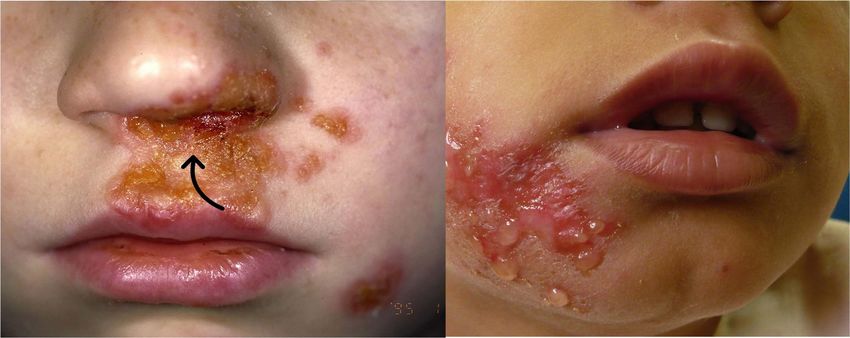
©2020Bed Bug Bites
• Key Characteristics:
• Most commonly occur on exposed areas of the face, neck, arms, or hands.
• Breakfast, Lunch, and Dinner Sign
• 3 linear, erythematous papules in a row
• S/S
• pruritic, erythematous‐edematous papules in a linear array
• Management:
• Antihistamines to control itching
• Exterminator to control pests
©2020Bed Bugs
©2020Pediculosis
• Key Characteristics:
• can affect scalp, body, pubic area
• Head lice can live 30 days on a single host and lay over a hundred nits.
• Transmission is by direct or indirect contact: DOES NOT JUMP
• May cause intense itching behind ears and at neck
• “Flakes” that DO NOT wipe away easily!
• S/S
• Lice can be visualized; nits are small white (...not always) oval cases attached to hair shaft
• Common sites: back of head, nape of neck, behind ears
• Body lice: excoriated macules/papules; belt line, collar, underwear
areas/regional lymphadenopathy
©2020Pediculosis
• Management
• Pediculicides are first‐line treatments
• Permethrin – first choice
• Then remove nits – use special comb
• Then cleanse environment
• Wash sheets, towels, clothing, headgear
• Place items that cannot be washed/dry‐cleaned in plastic bag for 2 weeks
• Vacuum carpeted play areas
• Soak brushes, combs, hair accessories in pediculicide
©2020Pediculosis
©2020Scabies
• Key Characteristics:
• Sarcoptes scabii
• Mites burrow into epidermis, feed off human blood, and there is intense itching
• Itching is caused by antibody sensitization that occurs in about 3 weeks
• Itching; worse at night; progressively intense
• Multiple erythematous papules
• S/S
• Itching; worse at night; progressively intense
• Fitful sleep, crankiness
• S‐shaped burrows; webs of fingers; sides of hands; folds of wrists, armpits
• Vesiculopustular lesions in infants/young children
• Secondary lesions – itchy papules, red‐brown nodules
• Evaluation
• Microscopic exam of scrapings; do not use KOH
• Burrow ink test to stain burrow‐very quick and easy
©2020Scabies
©2020Scabies
• Management
• Pharmacological treatment: scabicide
• Permethrin (5%) – apply from neck down; rinse off in 8‐14
hours
• Repeat in 1 week
• Antihistamines PRN
• Simultaneous treatment of close contacts
• Wash linens, clothing in hot water; vacuum house
• Store non‐washable items in sealed plastic bags
©2020You can also read