Management of Laryngeal Trauma - Nadir Elias, DMDa, James Thomas, MDb, Allen Cheng, MD, DDSc,* - BINASSS
←
→
Page content transcription
If your browser does not render page correctly, please read the page content below
Ma na gement of L ar yng eal
Tr auma
Nadir Elias, DMDa, James Thomas, MDb, Allen Cheng, MD, DDSc,*

  KEYWORDS
   Laryngeal trauma  Laryngotracheal injury  Laryngofissure

  KEY POINTS
   The key step in treatment of any laryngeal injury is the establishment of a secure airway.
   Early intervention (within 24–48 hours) is an important factor for improved patient outcomes (func-
    tional speech, swallowing, and airway patency).
   An awake tracheostomy is the airway of choice with grade II or higher laryngeal injuries.

INTRODUCTION                                                               Whereas blunt injuries have been described as be-
                                                                           ing associated with greater length of hospitaliza-
The larynx is a complex anatomic structure and a                           tion,5 our experience has been that penetrating
properly functioning larynx is essential for breath-                       airway injuries, often associated with ballistic
ing, voice, and swallowing. Injuries to the larynx                         wounds, are much more likely to be associated
and trachea can result in significant and potentially                      with greater endolaryngeal disruption. The types
fatal consequences. Laryngeal trauma is often                              of tissues involved have been divided into hard
associated with other injuries, including intracra-                        and soft tissue injuries. Locations of injuries have
nial injuries (17%), penetrating neck injuries                             been classified as injuries that affect the supraglot-
(18%), cervical spine fractures (13%), and facial                          tic larynx, the glottis, and subglottic larynx.
fractures (9%).1 Laryngeal injuries are rare, occur-                          Lynch5 was the first to classify traumatic injuries
ring in only 1 of 5000 to 137,000 emergency room                           based on location. In 1969, Nahum6 described
visits1–3 and among only 1 in 445 patients with se-                        laryngeal injuries based on injury location and likeli-
vere injuries.4 Because of this, even surgeons with                        hood of recovery with and without intervention. In
a great deal of experience in managing maxillofa-                          1980, Schaefer and colleagues developed what
cial trauma have limited exposure to management                            has become the most popular classification system
of laryngeal and tracheal injury. This article dis-                        to assess the severity of such injuries.7 This classi-
cusses the evaluation, diagnosis, and manage-                              fication describes laryngeal injuries on a scale of
ment of patients with laryngeal and tracheal injury.                       I-IV. Schaefer’s classification was later modified
                                                                           by Fuhrman and colleagues8 to include laryngotra-
                                                                           cheal separation (Table 1) and again by Verschue-
CLASSIFICATION OF LARYNGEAL INJURIES
                                                                           ren and colleagues4 in 2006 to include the use of
Several classification systems have been                                   computed tomography (CT) imaging in staging (Ta-
described to assist in developing an algorithmic                           ble 2). In this article, the discussion of the initial
approach to managing these difficult and rare in-                          evaluation and management of a patient with laryn-
juries. These classification systems have been                             geal trauma is within the framework of the Legacy
based on mode of injury, types of tissues involved                         Emanuel Classification, as outlined by the algorithm
                                                                                                                                                           oralmaxsurgery.theclinics.com

in the injury, anatomic locations of injury, and                           in Fig. 1. However, the principles are generalizable
severity of the injury. Modes of injury have been                          and can be applied to whichever system the reader
divided into blunt and penetrating injuries.                               finds most helpful in their practice.

  a
    Advanced Craniomaxillofacial and Trauma Surgery, Legacy Emanuel Medical Center, 1849 NW Kearney
  Street, Suite 300, Portland, OR 97209, USA; b Private Practice, Voicedoctor.net, 909 NW 18th Avenue, Portland,
  OR 97209, USA; c Oral/Head and Neck Oncology, Legacy Good Samaritan Cancer Center, Portland, OR, USA
  * Corresponding author. 1849 Northwest Kearney, Suite 300, Portland, OR 97209.
  E-mail address: chenga@head-neck.com

  Oral Maxillofacial Surg Clin N Am 33 (2021) 417–427
  https://doi.org/10.1016/j.coms.2021.04.007
  1042-3699/21/Ó 2021 Elsevier Inc. All rights reserved.
Descargado para BINASSS Circulaci (binas@ns.binasss.sa.cr) en National Library of Health and Social Security de ClinicalKey.es por Elsevier en julio 15,
   2021. Para uso personal exclusivamente. No se permiten otros usos sin autorización. Copyright ©2021. Elsevier Inc. Todos los derechos reservados.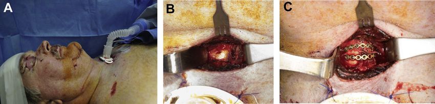
418                  Elias et al

               Table 1
               Fuhrman-Schaefer classification of laryngeal injuries

               Stage                                                            Injury
               I                                                                Minor laryngeal hematoma, edema, laceration; no
                                                                                  detectable fracture
               II                                                               Edema, hematoma, mucosal disruption with no
                                                                                  exposed cartilage, nondisplaced fractures
               III                                                              Significant edema, noted mucosal disruption, exposed
                                                                                  cartilage with or without cord immobility, displaced
                                                                                  fractures
               IV                                                               Significant edema, noted mucosal disruption, exposed
                                                                                  cartilage with or without cord immobility, displaced
                                                                                  fractures with 2 or more fracture lines, skeletal
                                                                                  instability/anterior commissure trauma
               V                                                                Complete laryngotracheal separation

             INITIAL EVALUATION AND INITIAL                                             begins with the primary survey as outlined in
             MANAGEMENT                                                                 Advanced Trauma Life Support algorithms.
                                                                                        Because the larynx and trachea are critical com-
             The initial evaluation of a patient suspected of hav-                      ponents of the airway, prompt identification and
             ing laryngeal or tracheal injury, as with any trauma,                      management of these injuries are prioritized. This

               Table 2
               Legacy Emanuel Medical Center laryngeal injury classification

               Stage                            Diagnostic Findings                                               Management
               I                                Minor airway symptoms                                             Observation
                                                 Voice changes                                                   Humidified air
                                                No fractures                                                      Head of bed elevation
                                                Small lacerations
               II                               Airway compromise                                                 Immediate awake
                                                Nondisplaced fractures                                              tracheostomy if airway not
                                                No cartilage exposure                                               already secured in the field
                                                Voice changes                                                     Humidified air
                                                 Subcutaneous emphysema                                          Head of bed elevation
                                                                                                                   ORIF
               III                              Airway compromise                                                 Immediate awake
                                                Edema                                                               tracheostomy if airway not
                                                Mucosal lacerations                                                 already secured in the field
                                                Palpable laryngeal fractures                                      Direct laryngoscopy
                                                Exposed cartilage                                                 Exploration and ORIF
                                                Subcutaneous emphysemas
                                                Voice changes
               IV                               Airway compromise                                                 Immediate awake
                                                Mucosal lacerations                                                 tracheostomy if airway not
                                                Exposed cartilage                                                   already secured in the field
                                                Palpable displaced laryngeal                                      Direct laryngoscopy
                                                  fractures with skeletal                                         Exploration/ORIF
                                                  instability                                                     Consider stenting
                                                Subcutaneous emphysemas
                                                Voice changes

             Abbreviation: ORIF, open reduction internal fixation.
               Adapted from Verschueren DS, Bell RB, Bagheri SC, Dierks EJ, Potter BE. Management of laryngo-tracheal injuries asso-
             ciated with craniomaxillofacial trauma. J Oral Maxillofac Surg. 2006 Feb;64(2):203-14; with permission.

      Descargado para BINASSS Circulaci (binas@ns.binasss.sa.cr) en National Library of Health and Social Security de ClinicalKey.es por Elsevier en julio 15,
         2021. Para uso personal exclusivamente. No se permiten otros usos sin autorización. Copyright ©2021. Elsevier Inc. Todos los derechos reservados.
Management of Laryngeal Trauma                                        419

Fig. 1. Protocol for management of laryngotracheal injuries at Legacy Emanuel Hospital and Health Center in
Portland, Oregon. ORIF, open reduction internal fixation. (From Verschueren DS, Bell RB, Bagheri SC, Dierks EJ,
Potter BE. Management of laryngo-tracheal injuries associated with craniomaxillofacial trauma. J Oral Maxillofac
Surg. 2006 Feb;64(2):203-14; with permission.)

begins with a quick survey of the injuries. Patients                       precedence. The fiberoptic examination may help
with either blunt or penetrating injury to the neck                        to verify that the patient’s airway is stable enough
must be ruled out as having airway injury. The                             for transfer to the scanner. An awake fiberoptic ex-
mechanism of the injury should also raise one’s                            amination also has the benefit of allowing visualiza-
suspicion. In a review of laryngeal injuries from                          tion of the larynx in function. This examination is
1992 to 2004, high speed motor vehicle accidents                           meant to be performed quickly and efficiently so
were the most common mechanism (49%), fol-                                 as to not impede overall trauma management. The
lowed by sports-related injuries (29%).4 Certain                           evaluating surgeon must keep in mind that trauma-
mechanisms of injury, such as hanging, gunshot                             tized airways that appear stable tend to deteriorate
wounds, or work-related high-energy injuries to                            over time because of the onset of edema, expansion
the neck, should obviously generate an elevated                            of hematomas, and other contributory factors.
level of suspicion.                                                           The next step is a CT scan of the head and neck,
                                                                           which is done in addition to CT scans of chest,
                                                                           abdomen, and pelvis that are routinely performed
Stable Versus Unstable Airway
                                                                           as part of the trauma survey (Fig. 2). In stable pa-
The first essential question is to establish whether                       tients with penetrating neck injury, a CT angiogram
the airway is secured and whether the patient is                           is also included to evaluate for vascular injury. CT
stable. If the patient is stable and protecting their                      imaging allows for rapid and accurate identifica-
airway, there is time for a more deliberate exami-                         tion of hard tissue injuries to larynx and trachea
nation. This is important because occult trachea-                          and identification of soft tissue air emphysema.10
laryngeal disturbance can occur with minimal                                  If the airway is not secure and/or is unstable,
external signs of trauma.                                                  or the patient is unstable for other reasons, the
   The initial airway physical examination starts                          patient is taken emergently to the operating
with visual inspection for swelling, soft tissue injury                    room where securing the airway followed by sta-
overlying the airway, loss of anatomic landmarks in                        bilization of the patient is the immediate priority.
the neck, and signs of troubled breathing. A                               Traditionally, this is via an oral endotracheal intu-
cursory examination of the patient’s voice is per-                         bation. However, if the patient has a known
formed, noting the presence or absence of stridor                          laryngeal or tracheal injury, oral endotracheal
and/or dysphonia. Next, use gentle palpation to                            intubation can fail, particularly because of false
assess for subcutaneous emphysema and                                      passage or further disruption of the injured
palpable disruption of the hyoid bone, thyroid                             airway. Although securing the airway trumps
cartilage, cricoid, and trachea. The most common                           any other priority, in this situation the most ideal
findings on physical examination include subcu-                            airway is a tracheostomy, performed awake with
taneous air, hoarseness, tenderness of the anterior                        either mask or laryngeal mask airway support, as
neck to palpation, and stridor.9                                           the situation allows. If endotracheal intubation is
   A fiberoptic examination may also be performed if                       the route chosen, a fiberoptic intubation with a
timing allows and other injuries do not take                               pediatric bronchoscope is one of several tools

Descargado para BINASSS Circulaci (binas@ns.binasss.sa.cr) en National Library of Health and Social Security de ClinicalKey.es por Elsevier en julio 15,
   2021. Para uso personal exclusivamente. No se permiten otros usos sin autorización. Copyright ©2021. Elsevier Inc. Todos los derechos reservados.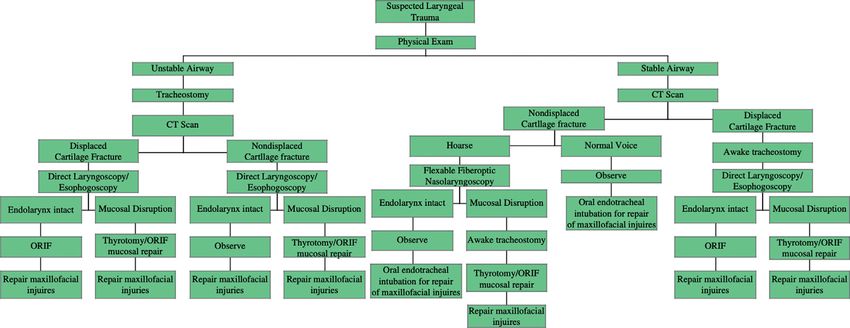
420                 Elias et al

                                                                                                             Fig. 2. Axial CT neck images of a 64-
                                                                                                             year-old male victim of a motorcycle
                                                                                                             accident with severe multisystem
                                                                                                             trauma. Injuries included (A) hyoid
                                                                                                             bone fracture, (B) a thyroid cartilage
                                                                                                             fracture, and (C) and cricoid cartilage
                                                                                                             fracture.    (D)    Three-dimensional
                                                                                                             reconstruction of the CT demon-
                                                                                                             strating the previously mentioned
                                                                                                             fractures.

             to be considered by the anesthesiologist. In the                           Nondisplaced Versus Displaced Cartilage
             presence of endolaryngeal lacerations, the pri-                            Fractures
             mary risk is that of intubating a blind submuco-
                                                                                        The CT scan answers an essential branch point in
             perichondrial pouch that produces immediate
                                                                                        our management algorithm, which is whether or
             and total airway obstruction. The stat surgical
                                                                                        not there are displaced fractures of the cartilagi-
             airway that follows not only risks the patient’s
                                                                                        nous larynx. The CT scan is especially valuable
             life but can also worsen the existing laryngotra-
                                                                                        because it can detect occult fractures missed on
             cheal injuries.
                                                                                        examination.11
                Once the airway is secured and the patient is
                                                                                           Patients without cartilage fractures or with non-
             stable, the next step in the evaluation involves
                                                                                        displaced cartilage fractures are managed in a
             physical examination, CT imaging (if not already
                                                                                        more conservative manner. Patients with nondis-
             performed), and panendoscopy. Anatomically,
                                                                                        placed cartilage fractures are carefully assessed
             the larynx is subdivided into the supraglottic lar-
                                                                                        for vocal disturbances. Signs of voice changes
             ynx, glottic larynx, and subglottic larynx, and tra-
                                                                                        should prompt a fiberoptic nasopharyngoscopy
             chea. Injury can occur at any and all of these
                                                                                        to assess for endolaryngeal injury. If the patient
             levels. Therefore, the examination is performed in
                                                                                        has already been intubated or has a tracheostomy,
             such manner where each of these areas are care-
                                                                                        a direct laryngoscopy under anesthesia is per-
             fully inspected. It is important to remember that,
                                                                                        formed instead. While under anesthesia, a bron-
             particularly with blunt trauma, disruption of the
                                                                                        choscopy and esophagoscopy are also
             laryngeal framework can occur without obvious
                                                                                        performed to assess the full extent of injury.
             external findings. For example, disruption of the
                                                                                           If no signs of endolaryngeal injury are observed,
             cricoarytenoid joint and shortening of the true
                                                                                        the patient is observed and receives supportive
             vocal cord can occur with blunt trauma and man-
                                                                                        care. If the laryngeal examination demonstrates
             ifest itself with vocal changes, without external
                                                                                        mucosal injury, consideration should be given to
             signs of injury. If left untreated, the dislocated
                                                                                        surgical repair versus serial endoscopic examina-
             arytenoid may scar in that position resulting in per-
                                                                                        tion. Mucosal lacerations are repaired primarily
             manent, more difficult to correct vocal distur-
                                                                                        through a thyrotomy (laryngofissure) approach, or
             bance. It is widely agreed on that early
                                                                                        in some cases, endoscopically. Denuded laryn-
             identification and treatment of laryngeal injuries
                                                                                        geal cartilage that is not amenable to primary
             yields superior results, ideally within the first 24
                                                                                        closure is treated with a thyrotomy approach
             to 48 hours if circumstances allow.

      Descargado para BINASSS Circulaci (binas@ns.binasss.sa.cr) en National Library of Health and Social Security de ClinicalKey.es por Elsevier en julio 15,
         2021. Para uso personal exclusivamente. No se permiten otros usos sin autorización. Copyright ©2021. Elsevier Inc. Todos los derechos reservados.
Management of Laryngeal Trauma 421
coupled with use of a laryngeal stent or laryngeal hematoma quickly results in stridor, difficulty with
keel.12 speech, and eventually dyspnea and potential
If the CT scan identifies displaced cartilage airway embarrassment. Partial or complete avul-
fractures, these are treated with open reduction sion of the epiglottis can also occur with severe
and internal fixation. Before this, many such pa- hyoid and thyroid fractures. This manifests as
tients benefit from having the airway secured dysphagia and aspiration.
with an awake tracheostomy. This avoids the
need for endotracheal intubation, which is chal- Hyoid Bone Fractures
lenging and further disrupts the displaced carti-
lage fractures. Once the airway is secured, Hyoid bone fractures are a result of high-energy
direct laryngoscopy is performed to assess for impact and, in our Legacy Emanuel series, were
endolaryngeal injury, as discussed previously. often seen in sporting injuries, such as baseball,
Consideration is given to including esophago- jet skiing, and martial arts.4 Such injuries are
scopy and/or bronchoscopy. If there are no signs also seen in suicidal hanging and in attempted
of endolaryngeal injury, the surgeon may proceed manual strangulation. Most hyoid fractures do
with open reduction and internal fixation of the not require surgical intervention. However, they
displaced cartilage segments. If there are signs are usually accompanied by temporary odyno-
of significant endolaryngeal injury, repair them phagia. Significantly displaced hyoid fractures
via laryngofissure approach followed by open are managed by either open reduction and inter-
reduction internal fixation of the cartilage nal fixation of the hyoid bone or partial hyoid
fractures. resection.
Distortion of the Glottis
ANATOMIC CONSIDERATIONS
Epiglottis Injury to the larynx can result in changes in the
anterior-posterior dimension of the vocal cords,
The epiglottis, as the superior extent of the supra-
the positioning of the vocal cords relative to each
glottic larynx, connected by ligaments to the hyoid
other, the mobility of the cords, and soft tissue
bone and thyroid cartilage, is significantly affected
injury of the cords. Any of these distortions can
by laryngeal trauma. Hyoid fractures can result in
result in voice changes. Voice changes of volume
an epiglottic hematoma (Fig. 3). An epiglottic
loss and pitch lowering indicate shortening of the
vocal cords. Voice changes of roughness indicate
asymmetry of the vocal cords, such as a unilateral
change in length or asymmetric swelling.
Displaced fractures of the thyroid cartilage
involving the anterior commissure and/or aryte-
noid dislocation can result in foreshortening of
the vocal cords. This is identified on endoscopic
examination, confirmed on CT scan, and is a
strong indication for open reduction internal fixa-
tion (Fig. 4).
Mucosal injury involving the vocal cords that re-
mains unrepaired will heal by secondary intention,
but such healing can result in synechiae between
opposing vocal cords and at the anterior commis-
sure. Careful inspection for such injuries is essen-
tial and their presence is an indication for repair.
Cricoid Injury and Cricotracheal/
Laryngotracheal Separation
Injury of the subglottic larynx can result in cricoid
Fig. 3. Illustration of an epiglottic hematoma, which
fractures and/or cricotracheal separation, both of
may result from hyoid or thyroid fracture. Note that
as the hematoma expands, the airway is at risk for crit- which result in devastating airway obstruction
ical obstruction. (From Bell RB, Verschueren DS, Dierks that is difficult to rescue in the field because these
EJ. Management of laryngeal trauma. Oral Maxillofac generally require an emergent tracheostomy. Pa-
Surg Clin North Am. 2008 Aug;20(3):415-30; with tients who are able to be stabilized are often found
permission.) to have recurrent laryngeal nerve injury.
Descargado para BINASSS Circulaci (binas@ns.binasss.sa.cr) en National Library of Health and Social Security de ClinicalKey.es por Elsevier en julio 15,
2021. Para uso personal exclusivamente. No se permiten otros usos sin autorización. Copyright ©2021. Elsevier Inc. Todos los derechos reservados.422 Elias et al
Fig. 4. Fracture of the thyroid cartilage may result in
edema, shortening, and possible avulsion of the vocal Fig. 5. Complete laryngotracheal separation with
cords resulting in significant voice changes. (From Bell tracheal retraction may result in profound airway
RB, Verschueren DS, Dierks EJ. Management of laryn- compromise and death if not treated with immediate
geal trauma. Oral Maxillofac Surg Clin North Am. airway stabilization in the form a tracheostomy. (From
2008 Aug;20(3):415-30; with permission.) Bell RB, Verschueren DS, Dierks EJ. Management of
laryngeal trauma. Oral Maxillofac Surg Clin North
Am. 2008 Aug;20(3):415-30; with permission.)
If the larynx or cricoid becomes separated from
the trachea, the trachea tends to retract inferiorly
toward the mediastinum (Fig. 5). Reconstruction This is ideally performed directly using a laryngo-
requires limited circumferential dissection of the scope, because this can address the collapse of
trachea to allow placement of bolstering sutures the potential space that occurs when a patient
for tension-free approximation of the divided is in a supine position. However, many trauma pa-
airway. tients with airway injury are at risk of cervical
spine injury and remain on cervical spine precau-
tions that limit neck extension. This difficulty is
OPERATIVE TECHNIQUE
compounded among patients with classic intuba-
Endoscopy
tion challenges, such as anterior airways and
It is beyond the scope of this article to discuss the mandibular hypoplasia. In such instances, we
nuances of performing endoscopic examination of revert back to fiberoptic examination assisted
the upper aerodigestive tract. However, we will by a videolaryngoscope. A videolaryngoscope is
discuss some pearls related to these techniques. introduced in the traditional fashion, with the tip
Flexible nasopharyngoscopy is an efficient, of the blade placed within the vallecula. This sus-
widely available (found on most hospital airway pends the base of tongue and mandible anteri-
carts), and effective way to examine the airway in orly, opening the potential space in the
an awake or sedated patient. Performing this on oropharynx, which allows the fiberoptic scope
an awake patient affords the benefit of being to be more useful.
able to visualize the larynx in function, specifically Examination of the trachea is performed with
the presence of absence of vocal cord motion. The either a flexible or rigid bronchoscope. In the trauma
effective performance of this examination in an setting, the flexible bronchoscope is the simplest to
awake trauma patient is uniquely challenging. use, does not require neck extension, and allows
Blood and swelling in the airway or difficulty with creation of video documentation of the injuries.
topical anesthesia in an agitated patient can Examination of the cervical and thoracic esoph-
make good visualization impossible. agus is performed either with a rigid or flexible
In these cases, performance of an endoscopic esophagoscopy. Rigid esophagoscopy is thought
examination in an anesthetized patient after the to be more sensitive than flexible esophagoscopy
airway has been secured is the other option. in identifying injuries.13 It also does not require
Descargado para BINASSS Circulaci (binas@ns.binasss.sa.cr) en National Library of Health and Social Security de ClinicalKey.es por Elsevier en julio 15,
2021. Para uso personal exclusivamente. No se permiten otros usos sin autorización. Copyright ©2021. Elsevier Inc. Todos los derechos reservados.Management of Laryngeal Trauma 423
insufflation to create a potential space for visuali- Surgical Exposure
zation, which runs the risk of causing soft tissue
If existing lacerations do not provide sufficient
air emphysema in the mediastinum and neck.
exposure, a limited neck exploration may be
However, as with laryngoscopy, it is difficult to
necessary. A horizontal incision is designed that
perform on patients in a cervical collar and those
is made to incorporate the incision of the tracheos-
with anatomic limitations. Also, a rigid esophago-
tomy. The incision is wide enough to explore the
scope also runs the risk of further disrupting an
lateral neck if necessitated by the nature of the
injured larynx.
injury. The skin flaps are developed in a suprafas-
cial plane over the strap musculature. The flaps are
Tracheostomy elevated superiorly to the level of the hyoid bone
and inferiorly to the clavicles.
The tracheostomy is a routine procedure that is a
If broader exposure is necessary, the sternoclei-
valuable part of the armamentarium of any sur-
domastoid muscles are skeletonized on the medial/
geon active in trauma. There are also as many
deep sides for access to create an outer tunnel.
ways to perform the procedure as there are sur-
Blunt and sharp dissection are used medial to the
geons performing it. As such, we focus this discus-
carotid sheath allowing lateral retraction to create
sion not on how to perform a tracheostomy, but
the inner tunnel. This allows wide access to the lar-
modifications for performing a tracheostomy in a
ynx, pharynx, and esophagus on either side.
patient likely to have laryngeal injury.
The median raphe is identified and dissection is
Tracheostomy in this setting is often performed
carried in this plane through the infrahyoid strap
before the patient’s cervical spine has been
muscles. The strap muscles are bluntly dissected
cleared. As such, maintaining neck immobilization
and retracted laterally until the thyroid cartilage is
is paramount. The head is typically stabilized with
visualized (Fig. 6).
tape to the bed or operating table while the ante-
Endolaryngeal repair and reconstruction is
rior portion of the cervical collar is removed for ac-
accessed by performing a laryngofissure using a
cess. Additional support to the neck is provided by
midline thyrotomy, if the exposure has not already
the anesthesiologist.
been created by the injury. A horizontal incision is
As previously mentioned, it is ideal to perform an
made through the cricothyroid membrane. Using a
awake tracheostomy. This requires a concerted
12 blade, and under the direct visualization, cut
effort between the anesthesiologist and surgeon.
from an inferior to superior direction directly be-
Although the procedure is described as “awake,”
tween the true vocal cords in the midline. This is
it is most optimally performed with some degree
important, because detachment or further disrup-
of sedation. This is typically a combination of an
tion of the vocal cord at the anterior commissure is
amnestic with a general anesthetic at a dose that
difficult to correct. An oscillating saw is used to com-
maintains spontaneous respiration. In addition to
plete the midline thyrotomy. The two sides of the thy-
oxygenation, mask ventilatory support, typically
roid cartilage can then be retracted laterally (Fig. 7).
by face mask, is essential. Good local anesthesia
is of critical importance. We typically deliver field
blocks to the cervical plexus along the anterior
border of the sternocleidomastoid muscle on
either side and infiltration from the skin down to
the trachea.
In the absence of useful overlying lacerations, a
horizontal incision is used for the tracheostomy at
a level that can be incorporated into an apron inci-
sion later for neck exploration, if needed.
Once the trachea is visualized, the tracheotomy
should ideally be placed distal to the injury, usually
between the third and fourth tracheal rings.
Although a cricothyroidotomy is the most expe-
dient in an emergency situation, it does interfere
with laryngeal reconstruction and risks limiting cri-
cothyroid mobility in the future (limiting upper Fig. 6. Retraction of the strap musculature to allow
vocal range and maximal volume). If a cricothyroi- for visualization of the cartilaginous injuries. (From
dotomy has been previously performed, the airway Bell RB, Verschueren DS, Dierks EJ. Management of
should be converted to a tracheostomy as soon as laryngeal trauma. Oral Maxillofac Surg Clin North
it is practical. Am. 2008 Aug;20(3):415-30; with permission.)
Descargado para BINASSS Circulaci (binas@ns.binasss.sa.cr) en National Library of Health and Social Security de ClinicalKey.es por Elsevier en julio 15,
2021. Para uso personal exclusivamente. No se permiten otros usos sin autorización. Copyright ©2021. Elsevier Inc. Todos los derechos reservados.424 Elias et al
If an esophageal injury has been identified, this
should be repaired before the endolaryngeal
repair.
Endolaryngeal Repair
After the laryngofissure is performed, the supra-
glottic, glottic, and subglottic larynx is directly
visualized. Mucosal lacerations, particularly if
there is exposed cartilage, are repaired primarily
with resorbable sutures, such as 4–0 and 5–0 chro-
mic gut. Do not debride lacerated mucosa,
because this makes primary closure difficult. The
arytenoids are inspected. If dislocated, they
should be reduced. The vocal cord attachments
to the thyroid cartilage are also inspected. If there
is detachment, the vocal cords are resuspended
using fine nonresorbable suture to the thyroid
cartilage at Broyles ligament (Fig. 8).
Rarely, there may be avulsion injuries of the mu-
cosa that are not amenable to primary closure.
Special consideration is given to these, particularly
if there are opposing injuries on the other side. This
may result in adhesions that inhibit vocal cord
mobility. This is managed using a laryngeal stent
or a keel. A laryngeal stent is placed with or without
a skin graft (with the underside of the graft facing
outward) (Fig. 9). The stent is then secured with
Fig. 7. Midline thyrotomy allows for lateral retrac-
tion of the thyroid and cricoid cartilages to allow
two nonresorbable sutures. The first suture is
for visualization of laryngeal and esophageal struc- passed through skin, thyroid lamina, stent, oppo-
tures. (From Bell RB, Verschueren DS, Dierks EJ. Man- site thyroid lamina, and back out through the
agement of laryngeal trauma. Oral Maxillofac Surg skin. The second suture is passed through skin,
Clin North Am. 2008 Aug;20(3):415-30; with subglottic trachea, stent, opposite wall of the tra-
permission.) chea, and back out through skin. The two sutures
are passed through an external silicone button and
tied loosely, on either side. The primary advantage
Alternatively, for surgeons with greater experi- of the stent, beyond preventing synechiae and ad-
ence, a simpler method involves starting with a hesions, is that it provides structural stabilization
midline thyroidotomy. The oscillating saw is used of the endolarynx circumferentially. By separating
to make a cut in the midline and the two halves denuded areas from each other, it allows for
of the thyroid cartilage are gently pulled to 3 to epithelial migration and healing by secondary
4 mm apart. Generally, if performed with care intention without adhesions. However, it comes
this is executed without violating the mucosa and at some risk. The stent itself may cause pressure
entering the airway. The vocal processes are visu- on the endolaryngeal mucosa, creating raw areas
ally identified as two white spots. They can be that may heal as adhesions once the stent is
divided in the midline and remain attached to their removed. This may result in stenosis later on.
respective thyroid ala. This obviates an incision in An alternative to a laryngeal stent is the use of a
the cricothyroid area and allows improved airtight keel. Similar to a stent, it is a barrier that allows for
closure. healing by secondary to intention while preventing
If the airway is inadvertently entered during thyroid adhesions. In contrast to a stent, it does not pro-
cartilage division, it will nearly always be into the vide circumferential support of the larynx. This
space between the true and false vocal cords, the avoids the risk of pressure or rubbing injury to
thinnest area of mucosa. Using an 11 blade, the vocal the endolaryngeal mucosa.
ligaments are divided. Up-angled scissors can divide The laryngofissure must be meticulously
the false cords if needed for visualization. One can repaired. Fine nonresorbable sutures are used to
then leave the inferior mucosa intact for an airtight reconstruct the anterior commissure. Special
seal after closure. attention is given to lining up these two landmarks
Descargado para BINASSS Circulaci (binas@ns.binasss.sa.cr) en National Library of Health and Social Security de ClinicalKey.es por Elsevier en julio 15,
2021. Para uso personal exclusivamente. No se permiten otros usos sin autorización. Copyright ©2021. Elsevier Inc. Todos los derechos reservados.Management of Laryngeal Trauma 425
and posterior to the trachea. This can often be
done carefully with a finger. Avoid excessive lateral
dissection, because this may compromise the
vascular supply to the skeletonized trachea. This
dissection should be carried inferiorly into the su-
perior mediastinum to allow for sufficient superior
traction.
Nonresorbable sutures, such as 2–0 Prolene, are
used to reconstruct. These sutures are passed
through the cartilage of a tracheal ring at least
one or more rings distal the injury. Superiorly, the
suture needle is then passed through the cricoid.
Fig. 8. Resuspension of Broyles ligament to the
Be sure to avoid passing through the tracheal mu-
external perichondrium. (From Bell RB, Verschueren
cosa. These sutures are placed 270 around the
DS, Dierks EJ. Management of laryngeal trauma.
Oral Maxillofac Surg Clin North Am. 2008 trachea. Once all the sutures are passed, they are
Aug;20(3):415-30; with permission.) tied down one by one, working from lateral to ante-
rior (Fig. 11). It is important to keep the patient’s
in the same horizontal plane, otherwise the vocal head out of extension, to minimize tension across
cords do not adduct evenly during function. A su- the closure. As a reminder to the patient, a Grillo
ture is passed through the thyroid cartilage and stitch, a heavy nonresorbable suture passed from
anterior commissure on one side, then through menton to sternum, is placed. This discourages
the anterior commissure and thyroid cartilage on the patient from extending his or her neck and putt-
the opposite side to line these points up correctly. ing excess tension across the repaired trachea.
The thyroid (and cricoid, if necessary) is then After the repair is complete, the patient can usu-
repaired either with suture, wire, or miniplate fixa- ally be extubated, assuming other indications for
tion (Fig. 10). When using miniplates, be sure to remaining intubated are not present. However,
choose screws that do not penetrate the endolar- keeping the patient intubated may be prudent,
yngeal mucosa. Resorbable plates and screws depending on the surgeon’s judgment and the pa-
have also been used for this indication. tient’s clinical status.
Reconstruction of Cricotracheal Separation Wound Closure
For reconstruction of a complete separation of the The skin flaps are closed in layers. Special atten-
trachea from the larynx, the trachea must be suffi- tion must be given to separation of the tracheos-
ciently mobilized. Blunt dissection is used anterior tomy from the rest of the wound. Deep sutures
are placed circumferentially around the tracheos-
tomy to seal it off. Suction drains are placed under
the skin flaps to prevent saliva or seroma accumu-
lation or a subcutaneous aerocele. Although some
authors advocate for passive drains for fear of pro-
motion of fistula formation, we have not found this
to be an issue in our experience.
POSTOPERATIVE CARE
Enteral Feeds
The patient is initially fed by a nasogastric feeding
tube because temporary dysphagia is common.
Enteral feeds are continued until the patient is
able to protect their airway. If permitted by other in-
juries, a swallow evaluation is initiated by a speech
language pathologist as early as the third to fourth
postoperative day. If aspiration is present, enteral
Fig. 9. A laryngeal stent can be secured with sutures
that extend through the skin, thyroid lamina bilater- feeds are continued until this is resolved.
ally before being tied loosely over skin buttons. If there was an esophageal injury, we typically
(From Bell RB, Verschueren DS, Dierks EJ. Manage- postpone initiation of oral feeding for 2 weeks.
ment of laryngeal trauma. Oral Maxillofac Surg Clin Before oral feeding, a modified barium swallow
North Am. 2008 Aug;20(3):415-30; with permission.) and esophogram are performed to assess for leaks.
Descargado para BINASSS Circulaci (binas@ns.binasss.sa.cr) en National Library of Health and Social Security de ClinicalKey.es por Elsevier en julio 15,
2021. Para uso personal exclusivamente. No se permiten otros usos sin autorización. Copyright ©2021. Elsevier Inc. Todos los derechos reservados.426 Elias et al
Fig. 10. A 64-year-old male victim whose CT images were presented in Fig. 4. Examination of the neck revealed
and large submental laceration and edema and ecchymosis extending down to the sternal notch, which raised
the suspicion for a laryngeal injury. (A) Clinical appearance after initial stabilization with a tracheostomy. (B) In-
traoperative exposure for repair of the laryngeal injuries. (C) Open reduction internal fixation of thyroid and
cricoid cartilages.
If leaks are present, enteral feeds are continued for or wound contracture. Circumferential injuries are
an additional 2 weeks and the study is repeated. at the greatest risk. Despite careful mucosal repair
and use of stents, stenosis can still occur. This is a
COMPLICATIONS chronic and difficult to manage problem. Even with
a patent airway, a stenotic airway can result in stri-
Several perioperative complications can occur with dor, dyspnea on mild exertion, constant shortness
laryngotracheal injuries including voice changes; of breath, and intractable fatigue. Such patients
bleeding; infection; fistula formation; and, most often undergo tracheostomy and subsequently
seriously, loss of airway. However, probably the seek tracheal “sleeve” resection.
most substantial and common long-term issue is Initial management of airway stenosis starts with
stenosis of the airway. This can occur in the pres- a careful endoscopic examination to identify the
ence of mucosal injuries that results in adhesions
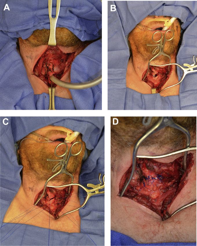
Fig. 11. Patient suffered a work-
related injury where a chain saw re-
bounded off a metal pipe resulting
in a penetrating anterior neck injury
that led to near complete cricotra-
cheal separation. An emergency med-
ical technician in the field visualized
bubbles from his neck wound and
was able to intubate his trachea
directly and secure his airway. He
was immediately taken to Legacy
Emanuel Medical Center, directly to
the operating room. (A) After hemo-
stasis was obtained and panendo-
scopy performed, the “trach” was
converted to an oral endotracheal
tube. (B) The trachea was mobilized
with blunt dissection. (C) 2–0 Prolene
sutures were used to repair the sepa-
ration. Note the sutures are passed
through a tracheal ring away from
the site of injury. (D) The patient’s
neck was placed in slight flexion and
the sutures tied down in a para-
chuting fashion.
Descargado para BINASSS Circulaci (binas@ns.binasss.sa.cr) en National Library of Health and Social Security de ClinicalKey.es por Elsevier en julio 15,
2021. Para uso personal exclusivamente. No se permiten otros usos sin autorización. Copyright ©2021. Elsevier Inc. Todos los derechos reservados.Management of Laryngeal Trauma 427
location, length, and degree of stenosis. Tracheal REFERENCES
stenosis isolated to a few rings is often amenable
to serial bronchoscopic partial laser ablations 1. Jewett BS, Shockley WW, Rutledge R. External
combined with careful dilation. In severe, refrac- laryngeal trauma analysis of 392 patients. Arch Oto-
tory tracheal stenosis below the second ring, laryngol Head Neck Surg 1999;125(8):877–80.
tracheal resection of several rings and primary 2. Schaefer SD. The acute management of external
anastomosis is performed. Stenosis involving the laryngeal trauma. A 27-year experience. Arch Oto-
cricoid is generally managed by cricoid split with laryngol Head Neck Surg 1992;118(6):598–604.
interpositional grafting. 3. Bent JP, Silver JR, Porubsky ES. Acute laryngeal
trauma: a review of 77 patients. Otolaryngol Head
SUMMARY Neck Surg 1993;109(3 Pt 1):441–9.
4. Verschueren DS, Bell RB, Bagheri SC, et al. Man-
Injuries to the larynx and trachea are rare. Howev-
agement of laryngo-tracheal injuries associated
er, a high degree of suspicion is necessary for
with craniomaxillofacial trauma. J Oral Maxillofac
trauma patients with the right mechanism and ex-
Surg 2006;64(2):203–14.
amination findings, because early identification
and treatment leads to better outcomes. Securing 5. Lynch M. Repair of the traumatized larynx. Laryngo-
the airway is the first priority, with an awake tra- scope 1951;61(1):51–65.
cheostomy being the ideal method in patients 6. Nahum AM. Immediate care of acute blunt laryngeal
with displaced laryngeal fractures with or without trauma. J Trauma 1969;9(2):112–25.
substantial endolaryngeal injury. Early open reduc- 7. Trone TH, Schaefer SD, Carder HM. Blunt and pene-
tion and internal fixation of displaced laryngeal trating laryngeal trauma: a 13-year review. Otolar-
cartilage fractures is recommended. Endolaryng- yngol Head Neck Surg (1979) 1980;88(3):257–61.
eal injuries are managed through a laryngofissure
8. Fuhrman GM, Stieg FH, Buerk CA. Blunt laryngeal
or through existing cartilage fractures. The use of
trauma: classification and management protocol.
a laryngeal stent or keel can help prevent syne-
J Trauma 1990;30(1):87–92.
chiae and laryngeal stenosis. Complete laryngo-
tracheal separation is often quickly fatal because 9. Stassen NA, Hoth JJ, Scott MJ, et al. Laryngotra-
of loss of the airway in the field. cheal injuries: does injury mechanism matter? Am
Surg 2004;70(6):522–5.
10. Shi J, Uyeda JW, Duran-Mendicuti A, et al. Multide-
CLINICS CARE POINTS
tector CT of laryngeal injuries: principles of injury
recognition. Radiographics 2019;39(3):879–92.
11. Randall DR, Rudmik L, Ball CG, et al. Airway man-
The key step in treatment of any laryngeal agement changes associated with rising radiologic
injury is the establishment of a secure airway.
incidence of external laryngotracheal injury. Can J
Early intervention (within 24–48 hours) is an Surg 2018;61(2):121–7.
important factor for improved patient out-
12. Montgomery WW, Montgomery SK. Manual for use
comes (functional speech, swallowing, and
airway patency). of Montgomery laryngeal, tracheal, and esophageal
prostheses: update 1990. Ann Otol Rhinol Laryngol
An awake tracheostomy is the airway of
1990;150(9_suppl):2–28.
choice with grade II or higher laryngeal in-
juries. 13. Schaefer SD. Management of acute blunt and pene-
trating external laryngeal trauma. Laryngoscope
2014;124(1):233–44.
Descargado para BINASSS Circulaci (binas@ns.binasss.sa.cr) en National Library of Health and Social Security de ClinicalKey.es por Elsevier en julio 15,
2021. Para uso personal exclusivamente. No se permiten otros usos sin autorización. Copyright ©2021. Elsevier Inc. Todos los derechos reservados.You can also read