Unusual Localization of Blood-Borne Loa loa Microfilariae in the Skin Depends on Microfilarial Density in the Blood: Implications for ...
←
→
Page content transcription
If your browser does not render page correctly, please read the page content below
Clinical Infectious Diseases
    Supplement Article

Unusual Localization of Blood-Borne Loa loa Microfilariae
in the Skin Depends on Microfilarial Density in the
Blood: Implications for Onchocerciasis Diagnosis in
Coendemic Areas
Yannick Niamsi-Emalio,1,a Hugues C. Nana-Djeunga,1,a Cédric B. Chesnais,2 Sébastien D. S. Pion,2 Jules B. Tchatchueng-Mbougua,3 Michel Boussinesq,2

                                                                                                                                                                                                  Downloaded from https://academic.oup.com/cid/article/72/Supplement_3/S158/6256993 by guest on 07 October 2021
María-Gloria Basáñez,4,a, and Joseph Kamgno1,5,a
1
 Centre for Research on Filariasis and other Tropical Diseases (CRFilMT), Yaoundé, Cameroon; 2Institut de Recherche pour le Développement (IRD), UMI233/INSERM U1175, Université de
Montpellier, Montpellier, France; 3Service d’Epidémiologie, Centre Pasteur du Cameroun, Membre du Réseau International des Instituts Pasteur, Yaoundé, Cameroun; 4MRC Centre for Global
Infectious Disease Analysis and London Centre for Neglected Tropical Disease Research, Department of Infectious Disease Epidemiology, School of Public Health, Imperial College London, London,
United Kingdom; and 5Faculty of Medicine and Biomedical Sciences, University of Yaoundé I, Yaoundé, Cameroon

   Background. The diagnostic gold standard for onchocerciasis relies on identification and enumeration of (skin-dwelling) Onchocerca
volvulus microfilariae (mf) using the skin snip technique (SST). In a recent study, blood-borne Loa loa mf were found by SST in individ-
uals heavily infected with L. loa, and microscopically misidentified as O. volvulus due to their superficially similar morphology. This study
investigates the relationship between L. loa microfilarial density (Loa MFD) and the probability of testing SST positive.
   Methods. A total of 1053 participants from the (onchocerciasis and loiasis coendemic) East Region in Cameroon were tested
for (1) Loa MFD in blood samples, (2) O. volvulus presence by SST, and (3) Immunoglobulin (Ig) G4 antibody positivity to Ov16 by
rapid diagnostic test (RDT). A Classification and Regression Tree (CART) model was used to perform a supervised classification of
SST status and identify a Loa MFD threshold above which it is highly likely to find L. loa mf in skin snips.
   Results. Of 1011 Ov16-negative individuals, 28 (2.8%) tested SST positive and 150 (14.8%) were L. loa positive. The range of Loa
MFD was 0–85 200 mf/mL. The CART model subdivided the sample into 2 Loa MFD classes with a discrimination threshold of 4080
(95% CI, 2180–12 240) mf/mL. The probability of being SST positive exceeded 27% when Loa MFD was >4080 mf/mL.
   Conclusions. The probability of finding L. loa mf by SST increases significantly with Loa MFD. Skin-snip polymerase chain re-
action would be useful when monitoring onchocerciasis prevalence by SST in onchocerciasis–loiasis coendemic areas.
   Keywords. Onchocerca volvulus; Loa loa; diagnosis; classification and regression tree model; skin snip technique.

Onchocerciasis is a parasitic infection caused by the filarial                                       mortality, and epilepsy [2–5]. The severity of these manifest-
nematode Onchocerca volvulus and transmitted by Simulium                                             ations depends on the burden of infection, particularly on (past
blackfly vectors. Sessile adult female worms live inside nodules                                     and/or present) O. volvulus microfilarial density (MFD) [6–9].
or worm bundles, where they mate with the (mobile) males and                                            In endemic countries, regional control and elimination ini-
periodically produce thousands of embryos or microfilariae [1].                                      tiatives have been implemented, based on vector control and/or
Microfilariae (mf) leave the nodules to populate the skin under                                      mass drug administration (MDA) with ivermectin. Elimination
the epidermis and the eyes and may live for a maximum of                                             of transmission (EOT) has been achieved in formerly endemic
2.5 years [1]. Most (99%) onchocerciasis cases occur in sub-Sa-                                      Latin American countries (Mexico, Guatemala, Colombia,
haran Africa. The infection is associated with skin pathology,                                       Ecuador) under the auspices of the Onchocerciasis Elimination
ocular lesions that progressively lead to blindness, excess                                          Program for the Americas (1993–present) [10], with the excep-
                                                                                                     tion of the Amazonian focus straddling Venezuela and Brazil
                                                                                                     [11]. In Africa, the Onchocerciasis Control Programme in
    a
    Y. N.-E., H. C. N.-D., M-G. B., and J. K. contributed equally to this work.                      West Africa (OCP; 1974–2002) and the African Programme for
   Correspondence: M. G. Basáñez, MRC Centre for Global Infectious Disease Analysis and
London Centre for Neglected Tropical Disease Research, Department of Infectious Disease
                                                                                                     Onchocerciasis Control (APOC; 1995–2015) led to substantial
Epidemiology, School of Public Health, Imperial College London, Norfolk Place, London W2 1PG,        reductions in disease burden [12] and to EOT in some foci of
UK (m.basanez@imperial.ac.uk).
                                                                                                     West and East Africa [13, 14]. Levels of precontrol endemicity
Clinical Infectious Diseases®  2021;72(S3):S158–64
© The Author(s) 2021. Published by Oxford University Press for the Infectious Diseases Society
                                                                                                     and progress towards morbidity and EOT targets have been
of America. This is an Open Access article distributed under the terms of the Creative Commons       measured via assessment of prevalence and intensity of infec-
Attribution License (http://creativecommons.org/licenses/by/4.0/), which permits unrestricted
                                                                                                     tion: in the OCP by detection and enumeration of skin mf by
reuse, distribution, and reproduction in any medium, provided the original work is properly cited.
DOI: 10.1093/cid/ciab255                                                                             the skin snip technique (SST) [15] and in APOC by nodule

S158 • cid 2021:72 (Suppl 3) • Niamsi-Emalio et al
prevalence at the beginning of the program and by SST for 3 health districts (HDs)—namely, Bertoua, Betare Oya, and
phase 1A (monitoring prevalence decline) and phase 1B (stop- Lomie—out of the 14 HDs in the East Region.
MDA) evaluations [16]. The SST consists of taking 2 or more For epidemiological monitoring, the East Region has been
bloodless skin snips (typically with a Holth-type corneoscleral organized into 5 zones or evaluation units (EUs). A cross-sec-
punch), incubating the snips in a suitable medium (eg, saline) tional survey was conducted in a community selected in each
for 24 hours, and detecting the presence (for prevalence) and of these EUs. Individuals of both sexes, aged 5 or more years,
number (for MFD) of emerged mf in the medium under a mi- and residing in the selected community were eligible for the
croscope [15]. study and invited to participate. At least 150 individuals in each
However, the microfilarial morphology of Loa loa (another selected community were sampled.
Chrysops-transmitted, filarial infection endemic in West and
Central Africa) is superficially similar to that of O. volvulus (al- Parasitological and Serological Assessment
beit L. loa mf are sheathed while those of O. volvulus are not) Loa loa MFD (Loa MFD) was quantified using calibrated thick
Downloaded from https://academic.oup.com/cid/article/72/Supplement_3/S158/6256993 by guest on 07 October 2021
when preparations are not stained, making it difficult to dis- blood smears. A sample of 50 µL of peripheral blood was col-
tinguish between these 2 species using bright-field microscopy lected by finger-prick between 10 am and 2 pm (due to diurnal
(L. loa mf measure 250‒300 μm by 6‒8 μm and those of O. vol- periodicity of L. loa mf [20]). After de-hemoglobinization and
vulus measure 220‒360 µm by 5‒9 µm). Since L. loa mf have Giemsa staining, the slides were examined using bright-field
been considered to be solely blood-dwelling, the presence of microscopy. All mf present on the slide, including L. loa and
L. loa mf in skin snips has seldom been investigated. A recent Mansonella perstans, were identified, counted, and expressed as
study demonstrated the occurrence of L. loa mf when using SST mf/mL. Here we only report Loa MFD.
and discussed its implications for potential reporting of O. vol- Onchocerciasis was assessed by 2 different methods—namely,
vulus false positives [17]. Not only would this impact individual the SST to detect mf in the skin and a rapid diagnostic test
diagnosis but also epidemiological evaluations of interven- (RDT)—to detect the presence of immunoglobulin G4 (IgG4)
tions. Therefore, this study aimed to analyze the relationship antibodies against the O. volvulus–specific antigen Ov16 [21].
between L. loa MFD in the blood (mf/mL) and the probability For SST, 2 skin biopsies (one from each posterior iliac crest)
of detecting L. loa mf in skin snips during onchocerciasis diag- were taken using a 2-mm Holth-type corneoscleral punch and
nosis in an area where infections by O. volvulus and L. loa are incubated for 24 hours in saline; the presence of emerged mf
coendemic. was assessed under an optical microscope. Antibody testing was
done using the Standard Diagnostics (SD) Bioline biplex Ov16/
METHODS Wb123 RDT as per the manufacturer’s instructions, with speci-
ficity listed at 100%. Recently published studies using the biplex
Ethical Approval RDT [22] and studies using earlier prototypes have reported a
The study received ethical clearance from the Cameroon specificity of 97.5% [23].
National Ethics Committee for Research for Human Health
(CNERSH; no. 2015/01/545/CE/CNERSH/SP). The survey Dependent Variable
was approved by and undertaken under the authority of the The dependent variable was a false-positive (+/ve) result for
Ministry of Public Health of Cameroon following the Helsinki SST, defined as the presence of mf under the microscope whilst
Declaration. Participation in this study was entirely voluntary testing negative (–/ve) for Ov16 RDT. Combining the results
and refusal to participate had no consequence for individuals. of the 2 tests produced 4 categories: (1) active O. volvulus in-
The protocol (objectives, methodology, use of collected data, fections (SST+/ve and RDT+/ve), (2) true negatives (SST–/ve
and dissemination of results) was carefully explained to all el- and RDT–/ve), (3) likely past infections (SST–/ve but RDT+/
igible individuals. Those who agreed to participate signed an ve), and (4) false positives (SST+/ve but RDT–/ve).
informed-consent form before undergoing clinical examination
and sample collection. Parents or legal guardians provided their Statistical Modeling Analysis
approval upon enrollment of minors (agedchoice of polynomial powers was based on minimizing the de- 23.1–37.1%). The SST revealed that 37 (3.5%) individuals pre-
viance information criterion (DIC) of the model. The Akaike sented with skin mf, while the Ov16 RDT revealed 42 (3.4%)
information criterion (AIC) [25, 26] was also used to select positive cases. Among the 1011 Ov16-negative individuals, 28
the best-fit model (that with the lowest AIC value). The pre- (2.8%) tested SST positive and were considered as false positives.
dicted probability of SST false positivity was estimated using Of these, 21 (75%) presented with blood L. loa mf. The range of
the function “predict” of the R software [27] using the best-fit Loa MFD among the 28 false positives was 0–85 200 mf/mL.
regression model. The characteristics of the 28 SST false-positive individuals
A Classification and Regression Tree (CART) model are shown in Supplementary Table 1. Although not statistically
(based on an iterative algorithm) was used to estimate the significant in the univariate analysis (P = .070), the number of
covariates associated with SST false positivity. CART is a false positives was approximately 3 times higher among females
nonparametric multiple regression statistical model to per- (21) than males (7). Likewise, the number of SST false positives
form supervised classifications with regard to explanatory was nearly double in individuals aged older than 40 years (18)
Downloaded from https://academic.oup.com/cid/article/72/Supplement_3/S158/6256993 by guest on 07 October 2021
variables compared with a categorical variable [28, 29]. The compared with those aged 40 years or younger (10) (P < .001).
modeling is carried out in 3 stages: (1) identification of the The median Loa MFD was 5940 (IQR, 450–13 820) mf/mL
covariates associated with the response variable, (2) classi- among the SST positives and 0 (IQR, 0–0) among the SST nega-
fication of each covariate to discriminate the response var- tives (P < .001). The proportions of SST false positives were
iable into 2 distinct groups, and (3) iterative repetition of similar between communities (P = .603) (Table 2). Multivariate
the 2 previous steps until it is no longer possible to perform analysis (Supplementary Table 2) revealed that SST false posi-
segmentation [30]. The 95% confidence interval (CI) for the tivity was significantly and negatively associated with male sex
identified threshold was obtained by bootstrapping [31], (P = .0226) and positively with Loa MFD (P < .0001).
based on the percentile method with 10 000 replicates. In
order to assess the difference in risk of being SST false posi- Predictors and Predicted Probability of Being False Positive for
Onchocerciasis
tive between different Loa MFD classes, a logistic regression
Several models were run to select the best one according to the
model was implemented to quantify, through the calculation
AIC and DIC criteria. The logistic model with fractional poly-
of odds ratios (ORs) and their 95% CIs, the association be-
nomial (for Loa MFD) was the best-fit model (lowest AIC and
tween the probability of false positivity and covariates (age,
DIC) (Supplementary Table 3). Since age and Loa MFD were
sex, community, Loa MFD).
positively and statistically significantly associated (P < .0001),
All analyses were performed with R version 3.6.1 using the
age was not included in the multivariate analysis. Males were
packages MFP (multiple fractional polynomial) and ggplot2
less likely than females to be SST false positive (P = .023). The
[27, 31]. The P value used for statistical significance was
probability of being SST false positive increased with Loa MFD
P < .05.
(P < .001).
Figure 1 illustrates the predicted probability of being SST
RESULTS
false positive as a (nonlinear) function of Loa MFD according
A total of 1053 (19.8%) individuals aged 6–85 (median, 28; IQR, to sex. For both sexes, the probability of being SST false posi-
12–48) years participated in the study (out of 5314 individuals tive increases steeply for low Loa MFD values, with the rate of
residing in the 5 study communities). The number of partici- increase slowing down for high Loa MFD values. For a given
pants per community ranged between 156 and 318, with a sex Loa MFD, females were consistently more likely to be SST false
ratio generally female biased (Table 1). positive than males. Supplementary Table 4 presents values of
The between-community prevalence of L. loa microfilaremia predicted probabilities of being SST false positive according to
varied from 5.1% (95% CI, 2.2–9.8%) to 29.7% (95% CI, Loa MFD.
Table 1. Description of the Population Tested for Loa loa and Onchocerca Volvulus in 5 Communities of the East Region, Cameroon
Commu- No. of Partici- Sex Ratio Median (IQR) No. (%) With No. (%) SST No. (%) Ov16 No. (%) Ov16 RDT Neg-
nity pants (% of Total) (F/M) Age, Years L. loa mf Positive RDT Positive ative but SST Positive
Adjela 156 (13.8) 1.7 (99/57) 26.0 (13.0–51.3) 8 (5.1) 11 (7.1) 16 (10.3) 5 (3.2)
Azemkout 318 (31.1) 1.4 (186/132) 15.0 (10.0–44.0) 45 (14.2) 11 (3.5) 4 (1.3) 10 (3.1)
Mikel 232 (22.5) 2.0 (155/77) 25.0 (11.8–42.0) 31 (13.4) 3 (1.3) 5 (2.2) 3 (1.3)
Pana 175 (16.7) 1.4 (102/73) 30.0 (15.0–50.5) 52 (29.7) 6 (3.4) 6 (3.4) 6 (3.4)
Timangolo 172 (15.9) 0.6 (65/107) 40.0 (24.0–53.3) 22 (12.8) 6 (3.5) 11 (6.4) 4 (2.3)
All 1053 (19.8) 1.4 (607/446) 28.0 (12.0–48.0) 158 (15.0) 37 (3.5) 42 (4.0) 28 (2.7)
Abbreviations: F, female; IQR, interquartile range; M, male; mf, microfilariae; Ov16 RDT, Ov16 rapid diagnostic test; SST, skin snip technique.
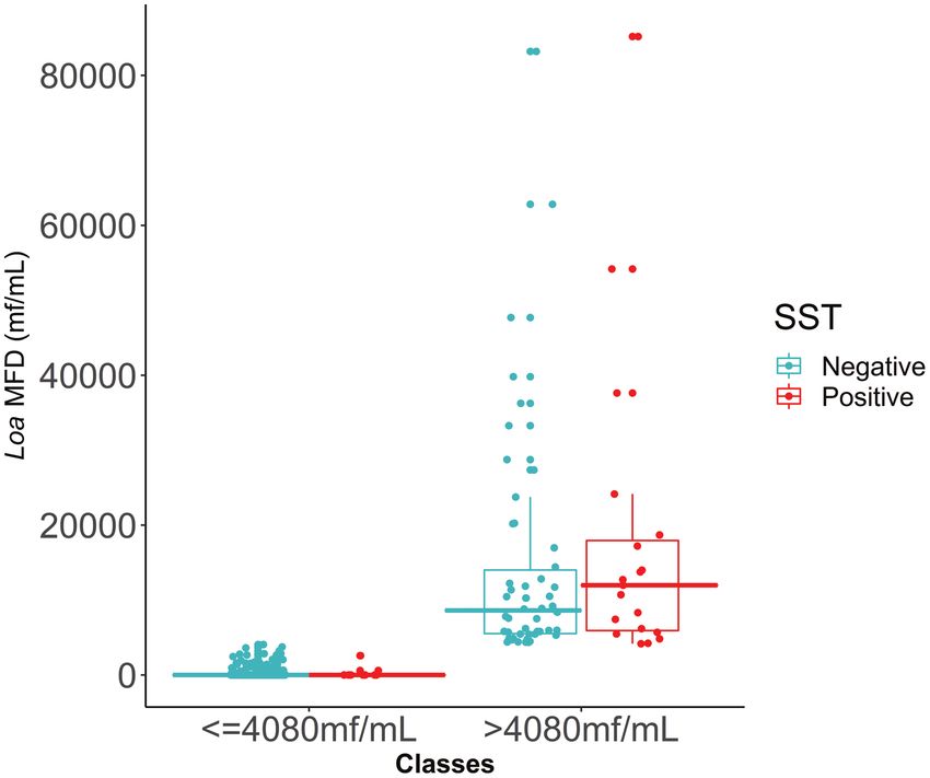
S160 • cid 2021:72 (Suppl 3) • Niamsi-Emalio et alTable 2. Univariate Association Between Skin Snip Technique False Figure 3 shows the distribution of individual Loa MFD
Positivity (for Onchocerca Volvulus) and Sex, Age, Loa loa Microfilarial
values for the 1011 Ov16-negative individuals according to the
Density, and Community of Residence Among Ov16-Negative Individuals
threshold of 4080 mf/mL and whether they tested SST negative
Variable Values P or positive. Most SST positives were found in class B and exhib-
Intercept …
ited high Loa MFD. Class B individuals were significantly more
Sex, n/N (%) .070 likely to be false SST positive than class A individuals (OR, 46.7;
Female 21/590 (3.6) P < .001). Males were less likely to be false SST positive com-
Male 7/421 (1.7) pared with females, regardless of Loa MFD category (OR, 0.3;
Loa MFD, mf/mL
P = .013) (Table 3).
Median 5940O. volvulus. Given that our overall proportion of false positives
was 2.7%, our results should be interpreted with caution.
The SST has been considered as the “gold standard” bench-
mark to detect and quantify O. volvulus microfilaridermia.
However, its sensitivity decreases during control programs
because O. volvulus MFD declines as a result of CDTI, and
although SST sensitivity can be improved by increasing the
number of snips taken [37], this may not be practical or ac-
ceptable. Besides, in areas of coendemicity with skin-dwelling
Mansonella streptocerca (endemic to Africa, mf measuring
180‒240 µm by 3‒5 µm), or blood-dwelling M. perstans (200 µm
by 4.5 µm), Mansonella ozzardi (coendemic with O. volvulus in
Downloaded from https://academic.oup.com/cid/article/72/Supplement_3/S158/6256993 by guest on 07 October 2021
the extant Amazonian focus, mf measuring 160‒205 µm by
4 µm) [38], and L. loa [17], mf of these species can occur in
skin snips and be misidentified as O. volvulus mf when assessing
microfilaridermia by SST. Consequently, there is a risk that in
Figure 3. Distribution of individual Loa MFD values according to the threshold of areas where filarial diseases are coendemic, false O. volvulus
4080 mf/mL and SST results in the 1011 Ov16-negative individuals. The 2 classes positives can be detected, with repercussions for individual di-
(≤4080 mf/mL and >4080 mf/mL) are those obtained with the CART model illus- agnosis and epidemiological evaluations. The aim of this work
trated in Figure 2. Turquoise solid circles denote SST negatives; red solid circles
represent SST positives (9 individuals with Loa MFD below the threshold and 19
was, therefore, to determine the relationship between Loa MFD
individuals with Loa MFD above the threshold). The solid horizontal lines within the and the probability of a positive diagnosis by SST while being
boxes are the medians; the lower and upper borders of the boxes are, respectively, negative by Ov16.
the first and third quartiles (IQR, Q3–Q1); the vertical bars indicate the “minimum”
Results indicate that the relationship between false positivity
and “maximum” calculated as Q1 – 1.5 × IQR and Q3 + 1.5 × IQR, respectively.
Abbreviations: CART, Classification and Regression Tree; IQR, interquartile range; to SST and Loa MFD is nonlinear and statistically significant.
Loa MFD, Loa loa microfilarial density; mf, microfilariae; Q, quartile; SST, skin snip The probability of being false positive to O. volvulus by SST in-
technique. creases sharply at low Loa MFD and less steeply at high Loa
MFD values, being consistently higher for females compared
individuals in endemic communities may be unable to mount with males—that is, for the same MFD, the probability is 2–3
an IgG4 antibody response to Ov16 [35], resulting in a false- times as high for females as for males (Supplementary Table 4).
negative outcome. In addition, there is a multiplicity of Ov16 In the Nana-Djeunga et al [17] study, O. volvulus SST positivity
assays, including various versions of the enzyme-linked immu- was significantly higher in males (who might therefore be less
nosorbent assay and the RDT used here, which have different likely to exhibit false positivity). Sex-specific exposure patterns
sensitivity and specificity characteristics, and this may vary to O. volvulus and L. loa are likely to be different [33, 39].
between laboratories or between these and field settings [34]. The discrimination of the sample through a supervised clas-
In the case of the biplex RDT used here, we accepted the value sification model yielded statistical significance for Loa MFD
of 100% specificity given by the manufacturers [34, 36] but are only. Neither age or sex nor community of residence were sig-
aware that others have reported a lower (97.5%) specificity [22, nificant predictors. The CART model produced 2 Loa MFD
23]. We assumed that when the RDT test was negative, an in- classes, the optimal discrimination value between the 2 being
dividual with an SST+/ve result would be a false positive for 4080 mf/mL, representing the threshold above which it is very
(nearly 30%) likely to find L. loa mf in skin snips. This rela-
tively low value reflected the distribution of Loa MFD in the
Table 3. Association Between Loa loa Microfilarial Density and Testing individuals examined. Half of the sample had Loa MFD less
Positive With the Skin Snip Technique for Ov16-Negative Individuals
than 6000 mf/mL, the median was 5940 mf/mL, and the 95%
Variables OR (95% CI) 95% CI (Bootstrap) P
CI for the threshold was 2180–12 240 mf/mL. Above 12 240 mf/
Intercept .013 (.00–.03) (.00–.02) …
mL, the probability of being SST false positive was greater than
Loa MFD class 35% for females and greater than 15% for males, reaching more
A: ≤4080 mf/mL 1 … than 60% for females and approximately 35% for males for Loa
B: >4080 mf/mL 46.7 (20.1–116.8) (20.6–138.0)may include that L. loa mf are in the dermal microcapillaries be further investigated and validated. Should this phenomenon
when the skin biopsy is taken. Although skin snips are sup- be confirmed, its consequences for disease manifestations and
posed to be bloodless, this may not always be achieved when onward transmission would warrant further studies.
using corneoscleral punches, sampling a large number of
Supplementary Data
individuals, and/or having a number of technicians under-
Supplementary materials are available at Clinical Infectious Diseases on-
taking the procedure who may vary in their skill to perform line. Consisting of data provided by the authors to benefit the reader,
the SST. Importantly, the association of this phenomenon with the posted materials are not copyedited and are the sole responsibility
increasing microfilaremia levels is of interest, and similar to of the authors, so questions or comments should be addressed to the
corresponding author.
what has been described with O. volvulus mf, which may be
found in blood when skin MFDs are high [40]. This phenom- Notes
enon led to the description of Microfilaria bolivarensis in heavily Authors’ contributions. H. C. N.-D., M. B., M.-G. B., and J. K. conceived
infected Amerindian populations of the Amazonian focus [41], and designed the study; Y. N.-E., C. B. C., S. D. S. P., J. B. T.-M., and M.-G.
Downloaded from https://academic.oup.com/cid/article/72/Supplement_3/S158/6256993 by guest on 07 October 2021
which was later demonstrated to be O. volvulus in blood [40]. B. analyzed the data and conducted the modeling; Y. N.-E., H. C. N.-D.,
C. B. C., S. D. S. P., and M.-G. B. wrote the paper. All authors commented on
Notwithstanding these considerations, it is increasingly rec- and revised interim and final drafts of the manuscript.
ognized that the skin may be an organ of crucial importance Acknowledgments. We thank the health authorities of the East Region of
in the transmission of (blood-dwelling) vector-borne parasites Cameroon for their support during the study, and particularly the volun-
teers from the study communities who agreed to take part in the data- and
(eg, Trypanosoma brucei gambiense [42], Leishmania donovani sample-collection phases of this study.
[43], and Plasmodium falciparum [44]) and this phenomenon Supplement sponsorship. This article appears as part of the supplement
should be rigorously investigated in loiasis. We also recommend “Sustainable control of neglected tropical diseases – surveillance and diag-
nostics”, sponsored by the NTD Modelling Consortium.
that further studies be carried out in different epidemiological
Disclaimer. The authors alone are responsible for the views, opinions,
settings and populations to assess the applicability and varia- assumptions, or any other information expressed in this manuscript, which
bility of the Loa MFD threshold identified in this study. are not necessarily the views, decisions, or policies of the institutions with
which they are affiliated.
Financial support. The data collection of this study was co-funded by
Conclusions the Bill & Melinda Gates Foundation (grant number OPP1033740; to M. B.)
The diagnosis and epidemiological evaluations of onchocerci- through the Task Force for Global Health project “Filling the Gaps: Operational
Research to Ensure the Success of the Neglected Tropical Disease Control
asis, particularly at baseline and during early phases of control
and Elimination Programs” (grant number NTD-SC/NCT 049; to J. K.); the
interventions, rely on the detection and enumeration of O. vol- African Programme for Onchocerciasis Control (APOC); and the US Agency
vulus mf by SST. In areas coendemic with loiasis, L. loa mf may for International Development (USAID) ENVISION Project. ENVISION
also be found in the skin, emerging from dermal arterioles (2011‒2019) was a global project led by RTI International and funded by the
USAID (cooperative agreement number AID-OAA-A-11-00048; to J. K.).
and venules when snips are taken with corneoscleral punches M.-G. B. and J. K. acknowledge funding from The Royal Society International
during SST, potentially confounding its results. In onchocer- Exchange Programme (grant number IES\R2\181017). M.-G. B. acknow-
ciasis–loiasis coendemic areas, a test-and-not-treat strategy ledges funding of the NTD Modelling Consortium by the Bill & Melinda
Gates Foundation (grant number OPP1184344) and joint center funding
has been developed to prevent post-ivermectin severe adverse (grant number MR/R015600/1) by the UK Medical Research Council (MRC)
events, enabling safe treatment of onchocerciasis. This strategy and the UK Department for International Development (DFID) under the
relies on LoaScope testing to measure Loa MFD and to exclude MRC/DFID Concordat agreement, which is also part of the European and
Developing Countries Clinical Trials Partnership (EDCTP2) program sup-
individuals with Loa MFD greater than 20 000 mf/mL from
ported by the European Union. J. K. was also funded by Institut Bertrand
ivermectin treatment [45]. In these areas, testing such individ- Buisson, France (subaward number 2011_TNT_01 to the Centre for Research
uals for onchocerciasis and treating them with anti-Wolbachia on Filariasis and other Tropical Diseases [CRFilMT]).
therapies (eg, doxycycline) provides a safe and appropriate al- Potential conflicts of interest. The authors: No potential conflicts of
interest. All authors have submitted the ICMJE Form for Disclosure of
ternative treatment strategy to help eliminate onchocerciasis Potential Conflicts of Interest. Conflicts that the editors consider relevant to
[46], as L. loa lacks Wolbachia endosymbionts [47]. This test- the content of the manuscript have been disclosed.
and-treat with doxycycline strategy [48] would benefit from
using methods such as real-time PCR and loop-mediated iso- References
1. Duke BOL. The population dynamics of Onchocerca volvulus in the human host.
thermal amplification (LAMP) assays for the amplification of
Trop Med Parasitol 1993; 44:61–8.
O. volvulus DNA from skin snips [34]. A next step to investigate 2. Prost A, Vaugelade J. [Excess mortality among blind persons in the West African
the impact of our results on onchocerciasis programs would be savannah zone.] Bull World Health Organ 1981; 59:773–6.
3. Kirkwood B, Smith P, Marshall T, Prost A. Relationships between mortality,
the inclusion, in a loiasis transmission dynamics model such as visual acuity and microfilarial load in the area of the Onchocerciasis Control
EPILOA [49], of the distribution of L. loa microfilarial loads to Programme. Trans R Soc Trop Med Hyg 1983; 77:862–8.
4. Pion SD, Kamgno J, Demanga-Ngangue, Boussinesq M. Excess mortality associ-
ascertain the proportion of the population who would likely test ated with blindness in the onchocerciasis focus of the Mbam Valley, Cameroon.
false-positive for O. volvulus in coendemic areas. The Loa MFD Ann Trop Med Parasitol 2002; 96:181–9.
5. Kaiser C, Pion SD, Boussinesq M. Case-control studies on the relationship be-
threshold (4080 mf/mL) above which the likelihood increases tween onchocerciasis and epilepsy: systematic review and meta-analysis. PLoS
of having an SST false-positive result for onchocerciasis should Negl Trop Dis 2013; 7:e2147.
Loa loa Microfilariae in Skin Snips • cid 2021:72 (Suppl 3) • S1636. Little MP, Breitling LP, Basáñez MG, Alley ES, Boatin BA. Association between 27. R Foundation for Statistical Computing. R: a language and environment for sta-
microfilarial load and excess mortality in onchocerciasis: an epidemiological tistical computing. Vienna, Austria: R Foundation for Statistical Computing.
study. Lancet 2004; 363:1514–21. Available at: http://www.R-project.org/. Accessed 30 October 2020.
7. Little MP, Basáñez MG, Breitling LP, Boatin BA, Alley ES. Incidence of blindness 28. Bierman AS, Brown AD, Levinton CM. Using decision trees for measuring gender
during the Onchocerciasis Control Programme in western Africa, 1971–2002. J equity in the timing of angiography in patients with acute coronary syndrome: a
Infect Dis 2004; 189:1932–41. novel approach to equity analysis. Int J Equity Health 2015; 14:155.
8. Walker M, Little MP, Wagner KS, Soumbey-Alley EW, Boatin BA, Basáñez MG. 29. Tung H-H, Chen C-Y, Line K-C, et al. Classification and regression tree analysis
Density-dependent mortality of the human host in onchocerciasis: relation- in acute coronary syndrome patients. World J Cardiovasc Dis 2012; 2:177–83.
ships between microfilarial load and excess mortality. PLoS Negl Trop Dis 2012; 30. Piarroux M, Piarroux R, Knapp J, et al.; FrancEchino Surveillance Network.
6:e1578. Populations at risk for alveolar echinococcosis, France. Emerg Infect Dis 2013;
9. Chesnais CB, Nana-Djeunga HC, Njamnshi AK, et al. The temporal relationship 19:721–8.
between onchocerciasis and epilepsy: a population-based cohort study. Lancet 31. Efron B, Tibshirani RJ. An introduction to the bootstrap. Chapman & Hall, Inc,
Infect Dis 2018; 18:1278–86. 1993.
10. Sauerbrey M, Rakers LJ, Richards FO. Progress toward elimination of onchocerci- 32. Coffeng LE, Stolk WA, Golden A, de Los Santos T, Domingo GJ, de Vlas SJ.
asis in the Americas. Int Health 2018; 10:i71–8. Predictive value of Ov16 antibody prevalence in different subpopulations for
11. Botto C, Basañez MG, Escalona M, et al. Evidence of suppression of onchocerci- elimination of African onchocerciasis. Am J Epidemiol 2019; 188:1723–32.
asis transmission in the Venezuelan Amazonian focus. Parasit Vectors 2016; 9:40. 33. Hamley JID, Walker M, Coffeng LE, et al. Structural uncertainty in onchocerci-
Downloaded from https://academic.oup.com/cid/article/72/Supplement_3/S158/6256993 by guest on 07 October 2021
12. Coffeng LE, Stolk WA, Zouré HG, et al. African Programme for Onchocerciasis asis transmission models influences the estimation of elimination thresholds and
Control 1995–2015: model-estimated health impact and cost. PLoS Negl Trop Dis selection of age groups for seromonitoring. J Infect Dis 2020; 221:510–8.
2013; 7:e2032. 34. Unnasch TR, Golden A, Cama V, Cantey PT. Diagnostics for onchocerciasis in the
13. Traore MO, Sarr MD, Badji A, et al. Proof-of-principle of onchocerciasis elimina- era of elimination. Int Health 2018; 10:i20–6.
tion with ivermectin treatment in endemic foci in Africa: final results of a study 35. Gass KM. Rethinking the serological threshold for onchocerciasis elimination.
in Mali and Senegal. PLoS Negl Trop Dis 2012; 6:e1825. PLoS Negl Trop Dis 2018; 12:e0006249.
14. Katabarwa MN, Lakwo T, Habomugisha P, et al. After 70 years of fighting an 36. PATH. Quality assurance program materials for onchocerciasis and lymphatic
age-old scourge, onchocerciasis in Uganda, the end is in sight. Int Health 2018; filariasis tests. Available at: https://www.path.org/programs/diagnostics/dx-qa/.
10:i79–88. Accessed 30 October 2020.
15. Moreau JP, Prost A, Prod’hon J. [An attempt to normalize the methodology of 37. Bottomley C, Isham V, Vivas-Martínez S, et al. Modelling neglected tropical dis-
clinico parasitologic surveys of onchocerciasis in West-Africa.] Med Trop (Mars) eases diagnostics: the sensitivity of skin snips for Onchocerca volvulus in near
1978; 38:43–51. elimination and surveillance settings. Parasit Vectors 2016; 9:343.
16. Tekle AH, Zouré HG, Noma M, et al. Progress towards onchocerciasis elimina- 38. Vivas-Martinez S, Basáñez MG, Grillet ME, et al. Onchocerciasis in the
tion in the participating countries of the African Programme for Onchocerciasis Amazonian focus of southern Venezuela: altitude and blackfly species compo-
Control: epidemiological evaluation results. Infect Dis Poverty 2016; 5:66. sition as predictors of endemicity to select communities for ivermectin control
17. Nana-Djeunga HC, Fossuo-Thotchum F, Pion SD, et al. Loa loa microfilariae in programmes. Trans R Soc Trop Med Hyg 1998; 92:613–20.
skin snips: consequences for onchocerciasis monitoring and evaluation in L. loa- 39. Whittaker C, Walker M, Pion SDS, Chesnais CB, Boussinesq M, Basáñez MG. The
endemic areas. Clin Infect Dis 2019; 69:1628–30. population biology and transmission dynamics of Loa loa. Trends Parasitol 2018;
18. Zouré HG, Wanji S, Noma M, et al. The geographic distribution of Loa loa in 34:335–50.
Africa: results of large-scale implementation of the Rapid Assessment Procedure 40. Botto C, Arango M, Yarzábal L. Onchocerciasis in Venezuela: prevalence of
for Loiasis (RAPLOA). PLoS Negl Trop Dis 2011; 5:e1210. microfilaraemia in Amerindians and morphological characteristics of the micro-
19. Cano J, Basáñez MG, O’Hanlon SJ, et al. Identifying co-endemic areas for major filariae from the Upper Orinoco Focus. Tropenmed Parasitol 1984; 35:167–73.
filarial infections in sub-Saharan Africa: seeking synergies and preventing severe 41. Godoy GA, Orihel TC, Volcan GS. Microfilaria bolivarensis: a new species of fi-
adverse events during mass drug administration campaigns. Parasit Vectors 2018; laria from man in Venezuela. Am J Trop Med Hyg 1980; 29:545–7.
11:70. 42. Capewell P, Cren-Travaillé C, Marchesi F, et al. The skin is a significant but over-
20. Hawking F. Periodicity of microfilariae of Loa loa. Trans R Soc Trop Med Hyg looked anatomical reservoir for vector-borne African trypanosomes. Elife 2016;
1955; 49:132–42. 5:e17716.
21. Golden A, Steel C, Yokobe L, et al. Extended result reading window in lateral flow 43. Doehl JSP, Bright Z, Dey S, et al. Skin parasite landscape determines host infec-
tests detecting exposure to Onchocerca volvulus: a new technology to improve tiousness in visceral leishmaniasis. Nat Commun 2017; 8:57.
epidemiological surveillance tools. PLoS One 2013; 8:e69231. 44. Meibalan E, Barry A, Gibbins MP, et al. P. falciparum gametocyte density and in-
22. Dolo H, Coulibaly YI, Sow M, et al. Serological evaluation of onchocerciasis and fectivity in peripheral blood and skin tissue of naturally infected parasite carriers
lymphatic filariasis elimination in the Bakoye and Falémé foci, Mali. Clin Infect in Burkina Faso. J Infect Dis 2019; doi:10.1093/infdis/jiz680.
Dis 2020; doi:10.1093/cid/ciaa318. 45. Kamgno J, Pion SD, Chesnais CB, et al. A test-and-not-treat strategy for oncho-
23. Steel C, Golden A, Stevens E, et al. Rapid point-of-contact tool for mapping and cerciasis in Loa loa-endemic areas. N Engl J Med 2017; 377:2044–52.
integrated surveillance of Wuchereria bancrofti and Onchocerca volvulus infec- 46. Boussinesq M, Fobi G, Kuesel AC. Alternative treatment strategies to accelerate
tion. Clin Vaccine Immunol 2015; 22:896–901.23. the elimination of onchocerciasis. Int Health 2018; 10:i40–8.
24. Royston P, Ambler G, Sauerbrei W. The use of fractional polynomials to 47. Taylor MJ, Bandi C, Hoerauf A. Wolbachia bacterial endosymbionts of filarial
model continuous risk variables in epidemiology. Int J Epidemiol 1999; nematodes. Adv Parasitol 2005; 60:245–84.
28:964–74. 48. Wanji S, Nji TM, Hamill L, et al. Implementation of test-and-treat with doxycy-
25. Kasenda B, Sauerbrei W, Royston P, et al. Multivariable fractional polynomial in- cline and temephos ground larviciding as alternative strategies for accelerating on-
teraction to investigate continuous effect modifiers in a meta-analysis on higher chocerciasis elimination in an area of loiasis co-endemicity: the COUNTDOWN
versus lower PEEP for patients with ARDS. BMJ Open 2016; 6:e011148. consortium multi-disciplinary study protocol. Parasit Vectors 2019; 12:574.
26. Pion SD, Grout L, Kamgno J, Nana-Djeunga H, Boussinesq M. Individual host 49. Whittaker C, Walker M, Pion S, Chesnais C, Boussinesq M, Basáñez MG. Evaluating
factors associated with Onchocerca volvulus microfilarial densities 15, 80 and the impact of anthelmintic-based intervention strategies for controlling Loa loa: a
180 days after a first dose of ivermectin. Acta Trop 2011; 120(Suppl 1):S91–9. mathematical modelling study using EPILOA. Am J Trop Med Hyg 2018; 99:16.
S164 • cid 2021:72 (Suppl 3) • Niamsi-Emalio et alYou can also read