Recombinant high mobility group box 1 induces cardiomyocyte hypertrophy by regulating the 14 3 3η, PI3K and nuclear factor of activated T cells ...
←
→
Page content transcription
If your browser does not render page correctly, please read the page content below
MOLECULAR MEDICINE REPORTS 23: 214, 2021

      Recombinant high‑mobility group box 1 induces cardiomyocyte
        hypertrophy by regulating the 14‑3‑3η, PI3K and nuclear
              factor of activated T cells signaling pathways
                          FEIFEI SU1,2*, MIAOQIAN SHI3*, JIAN ZHANG4*, YAN LI2 and JIANWEI TIAN1

                  1
                   Department of Cardiology, Air Force Medical Center, People's Liberation Army, Beijing 100142;
     2
       Department of Cardiology, Tangdu Hospital Affiliated to The Fourth Military Medical University, Xi'an, Shaanxi 710038;
    3
      Department of Cardiology, The Seventh Medical Centre of The People's Liberation Army General Hospital, Beijing 100700;
       4
         Department of Cardiology, Beijing Chest Hospital Heart Center, Capital Medical University, Beijing 101149, P.R. China

                                     Received September 28, 2019; Accepted September 7, 2020

                                                  DOI: 10.3892/mmr.2021.11853

Abstract. High‑mobility group box 1 (HMGB1) is released by         cells, and serves as an inflammatory cytokine. In addition,
necrotic cells and serves an important role in cardiovascular      HMGB1 serves an important role in multiple organ patholo‑
pathology. However, the effects of HMGB1 in cardiomyo‑             gies, including cerebral, liver, lung and renal injury, and
cyte hypertrophy remain unclear. Therefore, the aim of the         rheumatoid arthritis (1). It is likely that the specific properties
present study was to investigate the potential role of HMGB1       of HMGB1 exhibit an effect on cardiomyocytes. Recently,
in cardiomyocyte hypertrophy and the underlying mecha‑             it has been suggested that HMGB1 is involved in cardio‑
nisms of its action. Neonatal mouse cardiomyocytes (NMCs)          vascular diseases (2), such as cardiac fibrosis, myocardial
were co‑cultured with recombinant HMGB1 (rHMGB1).                  ischemia‑reperfusion injury, heart transplantation, aortic
Wortmannin was used to inhibit PI3K activity in cardiomyo‑         valve calcification and sepsis‑associated myocardial dysfunc‑
cytes. Subsequently, atrial natriuretic peptide (ANP), 14‑3‑3      tion (3‑7). Although these previous reports have revealed that
and phosphorylated‑Akt (p‑Akt) protein levels were detected        HMGB1 serves an essential role in the cardiovascular system,
using western blot analysis. In addition, nuclear factor of        in which our previous study (8) also reported that HMGB1
activated T cells 3 (NFAT3) protein levels were measured by        induced cardiomyocyte hypertrophy, its physiological function
western blot analysis and observed in NMCs under a confocal        in cardiomyocytes requires further investigation.
microscope. The results revealed that rHMGB1 increased                 Cardiac hypertrophy is induced by several factors,
ANP and p‑Akt, and decreased 14‑3‑3 η protein levels.              including inf lammatory factors and stresses, such as
Furthermore, wortmannin abrogated the effects of rHMGB1            hypoxia (9). This process is initially considered beneficial,
on ANP, 14‑3‑3η and p‑Akt protein levels. In addition,             but it may progress to heart failure (10,11) under prolonged
rHMGB1 induced nuclear translocation of NFAT3, which was           stress (12). It has been suggested that cardiac hypertrophy
also inhibited by wortmannin pretreatment. The results of this     may be attenuated by controlling inflammation (13). HMGB1
study suggest that rHMGB1 induces cardiac hypertrophy by           acts as an inflammatory cytokine and serves an important
regulating the 14‑3‑3η/PI3K/Akt/NFAT3 signaling pathway.           role in cardiovascular pathology (14). Therefore, increased
                                                                   levels of circulating HMGB1 may be associated with human
Introduction                                                       heart disease (15). For example, exogenous HMGB1 treat‑
                                                                   ment in acute myocardial infarction induces cardiomyocyte
High‑mobility group box 1 (HMGB1) is released by activated         survival by attenuating apoptosis and AMP‑activated protein
cells, including cardiomyocytes and damaged or necrotic            kinase‑dependent autophagy (16). Our previous study
                                                                   demonstrated that HMGB1 may induce cardiomyocyte hyper‑
                                                                   trophy (8). In addition, HMGB1 may be partially derived from
                                                                   cardiac myocytes under pressure overload and may serve a
Correspondence to: Dr Feifei Su, Department of Cardiology, Air     crucial role in cardiac dysfunction (17). Notably, maintenance
Force Medical Center, People's Liberation Army, 30 Fucheng Road,   of stable nuclear HMGB1 levels prevents heart hypertrophy
Haidian, Beijing 100142, P.R. China                                and failure by inhibiting DNA damage (18). However, to the
E‑mail: shijian@fmmu.edu.cn                                        best of our knowledge, the direct effects of exogenous HMGB1
                                                                   treatment on cardiomyocytes remain elusive.
*
    Contributed equally                                                14‑3‑3 proteins are distributed ubiquitously in all eukary‑
                                                                   otic organisms and serve a major role in stress response in
Key words: 14‑3‑3, high‑mobility group box 1, hypertrophy,
                                                                   several cells, including cardiomyocytes. The 14‑3‑3 protein
cardiomyocyte
                                                                   family includes several highly conserved acid proteins, named
                                                                   according to their different isoforms (19). The 14‑3‑3 protein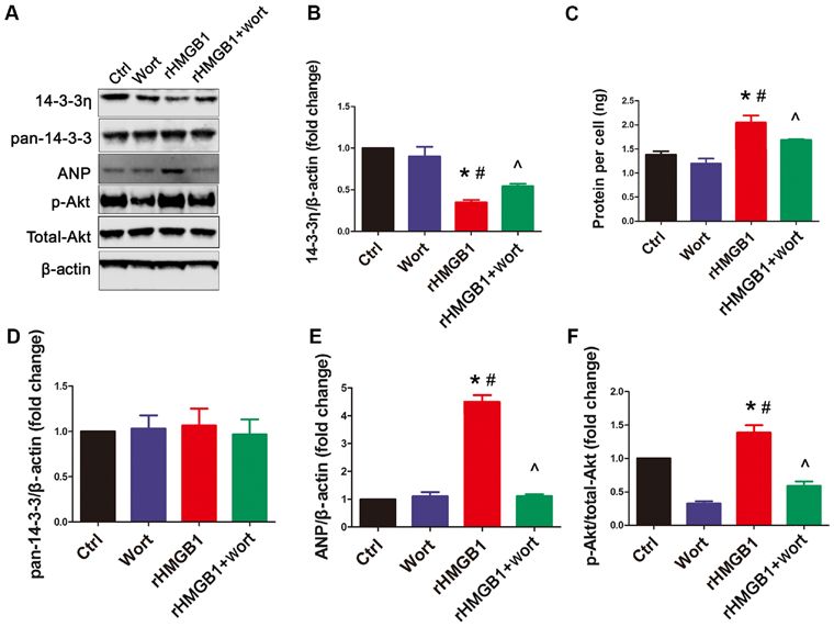
2                                  SU et al: HMGB1 INDUCES CARDIOMYOCYTE HYPERTROPHY

family includes several highly conserved acid proteins, named      wortmannin for 60 min prior to exposure to PBS or rHMGB1
according to their different isoforms (β, ε, η, γ, τ, σ and ζ)     in the Wort or rHMGB1+Wort groups, respectively.
detected in the cell cytoplasm and nucleus (20,21). 14‑3‑3η has
been reported to serve an essential role in myocardial metabo‑     NMC protein synthesis measurement. Following treatment,
lism (22,23). Additionally, depletion of 14‑3‑3η increases         NMCs were trypsinized, counted using a cell counting
cardiac hypertrophy, inflammation, fibrosis and apoptosis (24).    chamber (Beckman Coulter, Inc.) and lysed for further protein
It has previously been suggested that 14‑3‑3 proteins regulate     detection. Subsequently, protein concentration was deter‑
the nuclear translocation of the nuclear factor of activated       mined using a Bradford protein assay (Bio‑Rad Laboratories,
T cells (NFAT) (25,26), which in turn may induce pathological      Inc.). Finally, the protein synthesis per cell was calculated by
cardiac hypertrophy (27‑29). In addition, a recent study has       dividing the total amount of protein by the number of NMCs.
revealed that simvastatin upregulates 14‑3‑3 expression, which
ultimately exerts beneficial effects through cardioprotection      Reverse transcription‑quantitative PCR (RT‑qPCR).
against pressure overload (30). In addition, 14‑3‑3 proteins       Total RNA was extracted from NMCs using the TRIzol®
interact with PI3K and NFAT3‑mediated transcription                reagent (Invitrogen; Thermo Fisher Scientific, Inc.). RNA
in cardiomyocytes (19), which in turn phosphorylates its           samples were quantified spectrophotometrically at 260 nm.
downstream target, Akt, resulting in physiological cardiac         Subsequently, RT‑qPCR was performed as previously
growth (31,32). The aforementioned results indicate that           described (8). Briefly, RNA was exposed to RNAse‑free
14‑3‑3 proteins may be involved in the process of cardiac          DNase I and 5 µg total RNA was reverse transcribed into
hypertrophy. However, whether HMGB1 interacts with                 cDNA using oligo(dT) and M‑MuLV reverse transcriptase
14‑3‑3 proteins to affect cardiac hypertrophy remains unclear,     (Promega Corporation). The reverse transcription products
and, to the best of our knowledge, there are no studies on the     served as templates for PCR using gene‑specific primers
association between HMGB1 and 14‑3‑3 proteins in cardiac           (Table I) (33). qPCR was subsequently performed using a
hypertrophy.                                                       TaqMan™ Fast Advanced Master Mix (Applied Biosystems;
    Therefore, the present study hypothesized that HMGB1           Thermo Fisher Scientific, Inc.) using the 7500 Sequence
may induce cardiomyocyte hypertrophy by regulating the             Detector Real‑Time PCR system (Applied Biosystems;
14‑3‑3 and PI3K/Akt signaling pathways. To test this hypoth‑       Thermo Fisher Scientific, Inc.). The thermocycling condi‑
esis, neonatal mouse cardiomyocytes (NMCs) were isolated           tions were as follows: Initial step at 95˚C for 10 min,
and treated with HMGB1. Subsequently, NMCs were treated            followed by 40 cycles at 95˚C for 15 sec and 60˚C for 1 min.
with wortmannin, a specific PI3K inhibitor. Furthermore, to        Fluorescence signals of each gene were recorded during
assess the nuclear translocation of NFAT in NMCs, the effects      the elongation phase of each PCR cycle. The melting curve
were observed by confocal microscopy.                              analysis was used to confirm the amplification specificity,
                                                                   and the RNA abundance was expressed as ΔΔCq. GAPDH
Materials and methods                                              expression served as the internal control, and each gene was
                                                                   quantified in duplicate. Finally, the RT‑qPCR data were
Preparation of NMCs and treatment. NMCs were isolated              analyzed using the 2‑ΔΔCq method (34).
from 100 C57BL/6 mice (male; weight, 2±0.5 g; age, 1‑3 days
old), which were housed in a pathogen free facility with           Western blot analysis. NMCs were rinsed with PBS and
50% humidity at 22˚C, with a 12‑h light/dark cycle and free        lysed in buffer [Cell Lysis buffer (10X); cat. no. 9803; Cell
access to food and water. Mice were obtained from the Animal       Signaling Technology, Inc.] on ice. Subsequently, cell lysates
Center of the Fourth Military Medical University (Xi'an China)].   were centrifuged (2,000 x g; 5‑7 min; 4˚C), and the superna‑
The mice were sacrificed by decapitation and heart cell isola‑     tant was collected. Following a Bradford assay to determine
tion was performed using the Pierce Primary Cardiomyocyte          protein concentration, equal amounts of proteins (20 µg)
Isolation kit (Thermo Fisher Scientific, Inc.), according to the   were diluted in sample buffer (Thermo Fisher Scientific,
manufacturer's protocol. A monolayer of isolated NMCs was          Inc.), boiled and separated using 15% SDS‑PAGE (Thermo
plated at a density of 3x10⁶ cells/plate. NMCs were cultured       Fisher Scientific, Inc.). The separated proteins were trans‑
in DMEM (Gibco; Thermo Fisher Scientific, Inc.) containing         ferred to a nitrocellulose membrane (LI‑COR Biosciences).
1% penicillin‑streptomycin (Thermo Fisher Scientific, Inc.)        Following blocking in 5 ml 1X Odyssey ® blocking buffer
and 10% FBS (Thomas Scientific, Inc.) at 37˚C in a humidi‑         (LI‑COR Biosciences) for 1 h at room temperature, the
fied atmosphere with 5% CO2. The presence of fibroblasts           nitrocellulose membranes were incubated with primary
was minimized by the addition of cardiomyocyte growth              antibodies at 4˚C for 12 h. Subsequently, membranes were
supplement (dilution, 1:1,000; Thermo Fisher Scientific, Inc.).    washed with 1X PBS‑T (PBS with 0.1% Tween-20) and incu‑
Subsequently, NMCs were divided into the following four            bated with secondary antibodies for 1 h at room temperature.
groups: The control (Ctrl) group; the wortmannin (Wort;            Finally, immunoreactive protein bands were analyzed by
kindly provided by Dr Jun L)‑treated group; the recombinant        densitometry using the Odyssey ® infrared imaging system
HMGB1‑treated (rHMGB1) group; and the rHMGB1 plus                  (version 2.1; LI‑COR Biosciences), and the specific protein
Wort (rHMGB1+Wort) group. In addition, rHMGB1 and                  bands were visualized using an Odyssey ® scanner, then
wortmannin treatment of NMCs was performed according to            analyzed using Image Studio Lite version 5.0 (LI‑COR
our previous study (8). NMCs were treated with 200 ng/ml           Biosciences). The primary antibodies used in the present
rHMGB1 (Sigma‑Aldrich; Merck KGaA) or PBS for 24 h in              study were all diluted 1:1,000 and were: Anti‑atrial natriuretic
the rHMGB1 or Ctrl groups, respectively, and with 100 nmol/l       peptide (ANP; cat. no. ab126149; Abcam), anti‑pan14‑3‑3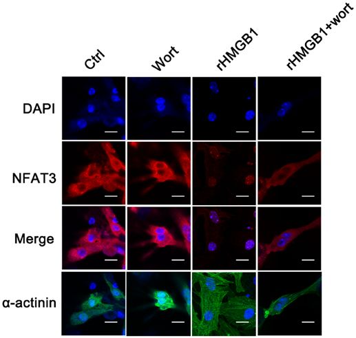
MOLECULAR MEDICINE REPORTS 23: 214, 2021                                                   3

Table I. Primers used for reverse transcription‑quantitative PCR.

Gene                                         Forward primer (5'→3')                                            Reverse primer (5'→3')

ANP                                  AGGCAGTCGATTCTGCTT                                                 CGTGATAGATGAAGGCAGGAAG
BNP                                  TAGCCAGTCTCCAGAGCAATTC                                             TTGGTCCTTCAAGAGCTGTCTC
GAPDH                                CCTTCCGTGTTCCTACCCC                                                GCCCAAGATGCCCTTCAGT

ANP, atrial natriuretic peptide; BNP, brain natriuretic peptide.

(cat. no. ab32377; Abcam), anti‑14‑3‑3η (cat. no. ab206292;                    was detected using a TUNEL assay kit (Roche Diagnostics),
Abcam), anti‑phospho‑Akt (p‑Akt) (cat. no. ab38449;                            according to the manufacturer's protocol. Briefly, following
Abcam), anti‑histone (cat. no. ab1791; Abcam), anti‑β‑tubulin                  treatment NMCs (1x10⁵ cells/well) were fixed with 4% parafor‑
(cat. no. ab6160; Abcam), anti‑NFAT3 (cat. no. orb315632;                      maldehyde at room temperature for 30 min and permeabilized
Biorbyt Ltd.) and anti‑ β ‑actin (cat. no. SC‑8432; Santa                      with 0.2% Triton X‑100 at room temperature for 30 min. The
Cruz Biotechnology, Inc.). The secondary antibodies were                       cells were subsequently incubated with TUNEL reagent for
goat anti‑mouse IRDye ® (1:10,000; cat. no. 926‑68070;                         1 h at room temperature. Thereafter, the cells were mounted
LI‑COR Biosciences), goat anti‑rabbit IRDye ® (1:10,000;                       with a mounting medium containing DAPI (1:1,000; Vector
cat. no. 926‑32211; LI‑COR Biosciences) and goat anti‑rat                      Laboratories, Inc.) at room temperature for 10 min. The
IRDye® (1:10,000; cat. no. 926‑32219; LI‑COR Biosciences).                     TUNEL‑positive and the total number of nuclei were counted
In addition, β‑tubulin and histone protein levels served as a                  in five independent experiments (3 randomly selected fields
loading control for the total protein lysate, the cytosol and                  of view) under a Nikon Eclipse 80 fluorescent microscope
nuclear extracts, respectively.                                                (magnification, x10; Nikon Corporation).

Confocal microscopy observation. NFAT3 localization in                         Statistical analysis. All data were analyzed using GraphPad
NMCs was analyzed using confocal microscopy. Briefly,                          Prism 5 (GraphPad Software, Inc.) and presented as the
NMCs were isolated and cultured on laminin pre‑coated                          mean ± SEM for continuous variables. One‑way ANOVA was
Lab‑Tek chamber slides (Thermo Fisher Scientific, Inc.).                       used to assess differences among groups followed by Tukey's
Following treatment, cells (1x10⁵ cells/well) were fixed with                  post hoc test. P
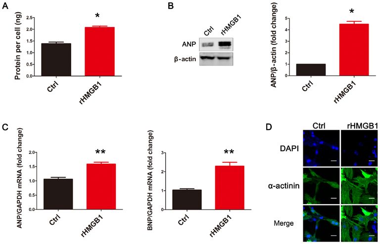
4                                          SU et al: HMGB1 INDUCES CARDIOMYOCYTE HYPERTROPHY

Figure 1. rHMGB1 induces NMC hypertrophy. rHMGB1 significantly increased (A) the protein content per cell, (B) ANP protein levels and (C) the mRNA
levels of ANP and BNP in NMCs (n=3). Data are presented as the mean ± standard error of the mean for continuous variables. One‑way ANOVA was used to
assess differences among different groups. (D) Representative confocal images from the Ctrl and rHMGB1 groups (scale bar, 20 µm). Cells were stained with
DAPI (blue signal) and α‑actinin (green signal) to identify nuclei and cardiomyocytes, respectively. rHMGB1, recombinant high‑mobility group box 1; NMC,
neonatal mouse cardiomyocytes; ANP, atrial natriuretic peptide; BNP, brain natriuretic peptide; Ctrl, control; DAPI, 2‑(4‑amidinophenyl)‑1H‑indole‑6‑car‑
boxamidine. *P
MOLECULAR MEDICINE REPORTS 23: 214, 2021                                                             5

Figure 3. rHMGB1 induces NFAT3 translocation to the nucleus.
Representative confocal images of NFAT3 localization in neonatal mouse
cardiomyocytes in different treatment groups (scale bar, 15 µm). Red
indicates NFAT3 protein, blue indicates DAPI‑stained nuclei and green       Figure 4. rHMGB1 induces NFAT3 translocation to the nucleus.
indicates α‑actinin. Each experiment was repeated four times. rHMGB1,       (A) Representative western blot results from cytosol or nuclear extracts
recombinant high‑mobility group box 1; NFAT3, nuclear factor of activated   from different treatment groups. (B) Semi‑quantitative analysis of
T cells 3; DAPI, 2‑(4‑amidinophenyl)‑1H‑indole‑6‑carboxamidine; Wort,       NFAT3 in cytosol extracts, with β ‑tubulin serving as the loading control.
wortmannin; Ctrl, control.                                                  (C) Semi‑quantitative analysis of NFAT3 in nuclear extracts, with histone
                                                                            serving as a loading control. The three independent neonatal mouse cardio‑
                                                                            myocyte cultures were used in each experiment (n=3). One‑way ANOVA was
                                                                            used to assess differences among different groups. rHMGB1, recombinant
rHMGB1‑mediated effects on 14‑3‑3η protein levels and Akt                   high‑mobility group box 1; NFAT3, nuclear factor of activated T cells 3;
phosphorylation (Fig. 2A, B and F). However, wortmannin                     Wort, wortmannin; Ctrl, control. *P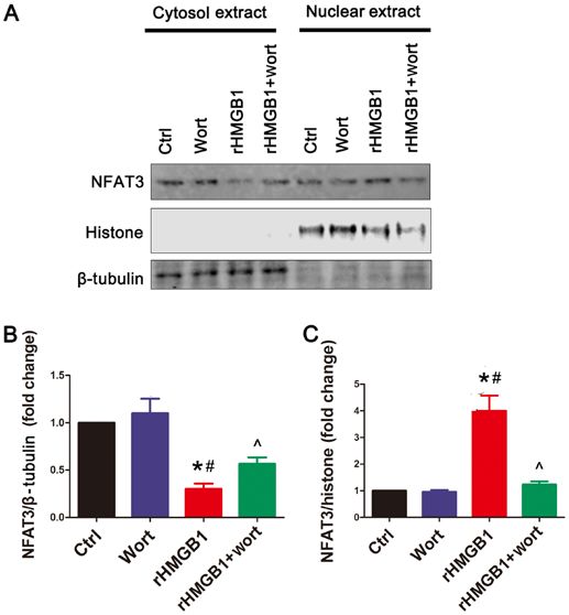
6 SU et al: HMGB1 INDUCES CARDIOMYOCYTE HYPERTROPHY
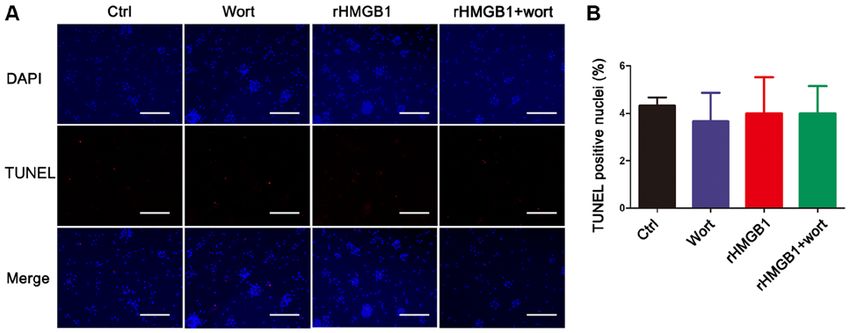
Figure 5. rHMGB1 has no effect on the apoptosis rate of neonatal mouse cardiomyocytes assessed using a TUNEL assay. (A) Fluorescence microscopic
images of TUNEL‑positive nuclei. Red indicates TUNEL‑positive nuclei and blue indicates total nuclei (scale bar, 200 µm). The experiment was repeated (five
independent experiments were performed). TUNEL‑positive nuclei were counted in four fields for each experiment under a fluorescent microscope (n=12).
(B) Data analysis of the number of TUNEL‑positive nuclei from the different groups. One‑way ANOVA was used to assess differences among the different
groups. rHMGB1, recombinant high‑mobility group box 1; TUNEL, terminal deoxynucleotidyl transferase dUTP nick‑end‑labeling; Wort, wortmannin; Ctrl,
control; DAPI, 2‑(4‑amidinophenyl)‑1H‑indole‑6‑carboxamidine.
circulation. It has been reported that endogenous HMGB1
may serve an important role in myocardium pathology (39). In
addition, patients with myocarditis display increased systemic
HMGB1 levels, suggesting its involvement in the pathogenesis
of inflammatory cardiomyopathy (2). However, the effects
of secreted HMGB1 on the cardiovascular system remain
unclear, and the effect of exogenous HMGB1 treatment on
cardiomyocytes requires further investigation. The results of
the present study revealed that rHMGB1 treatment increased
ANP protein levels, BNP and ANP mRNA synthesis, and
protein content per cell, highlighting its role in the induction
of NMC hypertrophy. In the present study, HMGB1 increased
the intracellular levels of ANP and BNP in cardiomyocytes.
However, whether HMGB1 is able to enhance the secretion
of ANP or BNP was not the main purpose of the present
study; this should be analyzed in vivo in future studies. The
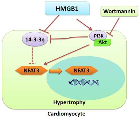
aforementioned results suggest that both endogenous and Figure 6. Proposed effects of rHMGB1 on cardiomyocyte hypertrophy.
exogenous HMGB1 serve a role in myocardial modifications, rHMGB1 may induce cardiomyocyte hypertrophy by regulating the
14‑3‑3/PI3K/Akt/NFAT3 signaling pathway. The red solid arrows and the
which is consistent with a previous study demonstrating that solid T‑shaped lines indicate the stimulatory and inhibitory effects, respec‑
exogenous HMGB1 treatment induced cardiomyocyte survival tively. The dashed line indicates the probable effect. rHMGB1, recombinant
in a murine myocardial infarction model (16). high‑mobility group box 1; NFAT3, nuclear factor of activated T cells 3.
In the present study, HMGB1 activated NFAT3 by
promoting its translocation to the nucleus, thereby upregulating
its nuclear expression. Additionally, it has been documented involved in rHMGB1‑induced hypertrophy, an indepen‑
that NFAT3 is regulated by 14‑3‑3 proteins (25,26), exhibits dent predictor of cardiovascular morbidity and mortality,
inflammatory effects and induces pathological hypertrophy in requires further investigation. The results of the present
cardiac myocytes (27‑29). study revealed that the levels of total 14‑3‑3 proteins in
14‑3‑3 proteins interact with several proteins, including NMCs did not change in the presence of rHMGB1. However,
PI3K, Akt and NFAT3 in diabetic cardiomyopathy (40). the protein levels of 14‑3‑3η were significantly decreased in
14‑3‑3 proteins are dimeric phosphoserine‑binding mole‑ rHMGB1‑treated NMCs, resulting in significant induction
cules separated into several isoforms, including the β, γ, of NMC hypertrophy. Therefore, the present results suggest
ε, ζ, η, θ and σ isoforms (19). These proteins bind to their that 14‑3‑3η, and not total 14‑3‑3, may serve a major role in
target proteins and modify their function by altering their rHMGB1‑induced NMC hypertrophy.
intracellular localization and phosphorylation status (20). NMCs were treated with a specific PI3K inhibitor prior
Several studies have concluded that 14‑3‑3 proteins, to exposure to rHMGB1. Notably, the levels of 14‑3‑3η were
particularly the 14‑3‑3η isoform, are involved in diabetic partially preserved in rHMGB1‑treated NMCs following
cardiomyopathy (40,41). However, whether 14‑3‑3 η is wortmannin pretreatment. Additionally, wortmannin partiallyMOLECULAR MEDICINE REPORTS 23: 214, 2021 7
inhibited NFAT3 nuclear translocation. The present results Competing interests
suggest that PI3K, Akt and NFAT3 may interact with 14‑3‑3η
and may influence rHMGB1‑induced hypertrophy. The The authors declare that they have no competing interests.
proposed model of rHMGB1‑mediated regulation of NMC
hypertrophy is illustrated in Fig. 6. References
Furthermore, it has been reported that cardiomyo‑
cyte hypertrophy may eventually lead to apoptosis (35). 1. Biscetti F, Flex A, Alivernini S, Tolusso B, Gremese E and
Therefore, a TUNEL assay was performed to determine Ferraccioli G: The role of high‑mobility group Box‑1 and its
crosstalk with microbiome in rheumatoid arthritis. Mediators
the rHMGB1‑induced apoptosis rate in NMCs. However, no Inflamm 2017: 5230374, 2017.
statistically significant differences were observed in the NMC 2. Bangert A, Andrassy M, Muller AM, Bockstahler M, Fischer A,
apoptosis rate between the Ctrl and the rHMGB1‑treated Volz CH, Leib C, Göser S, Korkmaz‑Icöz S, Zittrich S, et al:
Critical role of RAGE and HMGB1 in inflammatory heart
groups. The present finding is consistent with a previous disease. Proc Natl Acad Sci USA 113: E155‑E164, 2016.
study demonstrating that HMGB1 does not induce cardio‑ 3. Dong LY, Chen F, Xu M, Yao LP, Zhang YJ and Zhuang Y:
myocyte apoptosis under normal conditions (42). However, Quercetin attenuates myocardial ischemia‑reperfusion injury via
downregulation of the HMGB1‑TLR4‑NF‑kB signaling pathway.
it has been reported that endogenous HMGB1 contributes Am J Transl Res 10: 1273‑1283, 2018.
to ischemia‑reperfusion‑induced myocardial apoptosis (39). 4. Zhang W, Tao A, Lan T, Cepinskas G, Kao R, Martin CM
A potential explanation for these contrasting findings may and Rui T: Carbon monoxide releasing molecule‑3 improves
myocardial function in mice with sepsis by inhibiting NLRP3
reside in the different environments or distinct receptors on inflammasome activation in cardiac fibroblasts. Basic Res
different cells. Therefore, further investigation is required to Cardiol 112: 16, 2017.
investigate the role of exogenous HMGB1 in different cell 5. Wu RN, Yu TY, Zhou JC, Li M, Gao HK, Zhao C, Dong RQ,
Peng D, Hu ZW, Zhang XW and Wu YQ: Targeting HMGB1
types. ameliorates cardiac fibrosis through restoring TLR2‑mediated
In conclusion, the present study demonstrated that autophagy suppression in myocardial fibroblasts. Int
extracellular HMGB1 treatment induced NMC hypertrophy J Cardiol 267: 156‑162, 2018.
6. Shen W, Zhou J, Wang C, Xu G, Wu Y and Hu Z: High mobility
potentially through the 14‑3‑3η/PI3K/Akt/NFAT signaling group box 1 induces calcification of aortic valve interstitial cells
pathway. Therefore, 14‑3‑3 may be an important factor that via toll‑like receptor 4. Mol Med Rep 15: 2530‑2536, 2017.
links HMGB1, PI3K/Akt and NFAT3 in cardiomyocytes. 7. Lv Q, Li C, Mo Y and He L: The role of HMGB1 in heart trans‑
plantation. Immunol Lett 194: 1‑3, 2018.
8. Su FF, Shi MQ, Guo WG, Liu XT, Wang HT, Lu ZF and Zheng QS:
Acknowledgements High‑mobility group box 1 induces calcineurin‑mediated cell
hypertrophy in neonatal rat ventricular myocytes. Mediators
Inflamm 2012: 805149, 2012.
Not applicable. 9. Nehra S, Bhardwaj V, Kalra N, Ganju L, Bansal A, Saxena S and
Saraswat D: Nanocurcumin protects cardiomyoblasts H9c2 from
Funding hypoxia‑induced hypertrophy and apoptosis by improving oxida‑
tive balance. J Physiol Biochem 71: 239‑251, 2015.
10. Fang X, Liu Y, Lu J, Hong H, Yuan J, Zhang Y, Wang P,
The present study was supported by the National Natural Liu P and Ye J: Protocatechuic aldehyde protects against
Science Foundation of China (grant no. 81300077). isoproterenol‑induced cardiac hypertrophy via inhibition of the
JAK2/STAT3 signaling pathway. Naunyn Schmiedebergs Arch
Pharmacol 391: 1373‑1385, 2018.
Availability of data and materials 11. Wang S, Han HM, Pan ZW, Hang PZ, Sun LH, Jiang YN,
Song HX, Du ZM and Liu Y: Choline inhibits angio‑
tensin II‑induced cardiac hypertrophy by intracellular calcium
The datasets used and/or during the present study are available signal and p38 MAPK pathway. Naunyn Schmiedebergs Arch
from the corresponding author on reasonable request. Pharmacol 385: 823‑831, 2012.
12. Zwadlo C, Schmidtmann E, Szaroszyk M, Kattih B, Froese N,
Hinz H, Schmitto JD, Widder J, Batkai S, Bähre H, et al:
Authors' contributions Antiandrogenic therapy with finasteride attenuates cardiac
hypertrophy and left ventricular dysfunction. Circulation 131:
FS conceived the study. FS, MS and JZ performed the experi‑ 1071‑1081, 2015.
13. Song L, Wang L, Li F, Yukht A, Qin M, Ruther H, Yang M,
ments. YL and JT analyzed the experimental data, assessed Chaux A, Shah PK and Sharifi BG: Bone marrow‑derived
the raw data and confirm the authenticity of all the raw data. tenascin‑C attenuates cardiac hypertrophy by controlling inflam‑
FS prepared the manuscript. All authors read and approved the mation. J Am Coll Cardiol 70: 1601‑1615, 2017.
14. Gendy AM, Abdallah DM and El‑Abhar HS: The potential cura‑
final manuscript. tive effect of rebamipide in hepatic ischemia/reperfusion injury.
Naunyn Schmiedebergs Arch Pharmacol 390: 691‑700, 2017.
Ethics approval and consent to participate 15. Raucci A, Di Maggio S, Scavello F, D'Ambrosio A, Bianchi ME
and Capogrossi MC: The Janus face of HMGB1 in heart disease:
A necessary update. Cell Mol Life Sci 76: 211‑229, 2019.
The present experimental procedures were performed 16. Foglio E, Puddighinu G, Germani A, Russo MA and Limana F:
according to the ethical guidelines of the 1964 Declaration HMGB1 inhibits apoptosis following MI and induces autophagy
via mTORC1 inhibition. J Cell Physiol 232: 1135‑1143, 2017.
of Helsinki. All animal experiments were approved by the 17. Zhang L, Liu M, Jiang H, Yu Y, Yu P, Tong R, Wu J, Zhang S,
Animal Care and Welfare Ethics Committee of the Fourth Yao K, Zou Y and Ge J: Extracellular high‑mobility group box
Military Medical University (Xi'an, China). 1 mediates pressure overload‑induced cardiac hypertrophy and
heart failure. J Cell Mol Med 20: 459‑470, 2016.
18. Funayama A, Shishido T, Netsu S, Narumi T, Kadowaki S,
Patient consent for publication Takahashi H, Miyamoto T, Watanabe T, Woo CH, Abe J, et al:
Cardiac nuclear high mobility group box 1 prevents the devel‑
opment of cardiac hypertrophy and heart failure. Cardiovasc
Not applicable. Res 99: 657‑664, 2013.8 SU et al: HMGB1 INDUCES CARDIOMYOCYTE HYPERTROPHY
19. Liao W, Wang S, Han C and Zhang Y: 14‑3‑3 proteins regulate 31. DeBosch B, Treskov I, Lupu TS, Weinheimer C, Kovacs A,
glycogen synthase 3beta phosphorylation and inhibit cardiomyo‑ Courtois M and Muslin AJ: Akt1 is required for physiological
cyte hypertrophy. FEBS J 272: 1845‑1854, 2005. cardiac growth. Circulation 113: 2097‑2104, 2006.
20. Jia H, Liang Z, Zhang X, Wang J, Xu W and Qian H: 14‑3‑3 32. O'Neill BT, Kim J, Wende AR, Theobald HA, Tuinei J,
proteins: An important regulator of autophagy in diseases. Am Buchanan J, Guo A, Zaha VG, Davis DK, Schell JC, et al: A
J Transl Res 9: 4738‑4746, 2017. conserved role for phosphatidylinositol 3‑kinase but not Akt
21. Obsilova V, Kopecka M, Kosek D, Kacirova M, Kylarova S, signaling in mitochondrial adaptations that accompany physi‑
Rezabkova L and Obsil T: Mechanisms of the 14‑3‑3 protein ological cardiac hypertrophy. Cell Metab 6: 294‑306, 2007.
function: Regulation of protein function through conformational 33. Gao RR, Wu XD, Jiang HM, Zhu YJ, Zhou YL, Zhang HF,
modulation. Physiol Res 63 (Suppl 1): S155‑S164, 2014. Yao WM, Li YQ and Li XL: Traditional Chinese medicine
22. Sreedhar R, Arumugam S, Thandavarayan RA, Karuppagounder V, Qiliqiangxin attenuates phenylephrine‑induced cardiac hyper‑
Koga Y, Nakamura T, Harima M and Watanabe K: Role of trophy via upregulating PPARγ and PGC‑1α. Ann Transl Med 6:
14‑3‑3 η protein on cardiac fatty acid metabolism and macrophage 153, 2018.
polarization after high fat diet induced type 2 diabetes mellitus. 34. Schmittgen TD and Livak KJ: Analyzing real‑time PCR data by
Int J Biochem Cell Biol 88: 92‑99, 2017. the comparative C(T) method. Nat Protoc 3: 1101‑1108, 2008.
23. Sreedhar R, Arumugam S, Thandavarayan RA, Giridharan VV, 35. Lu F, Xing J, Zhang X, Dong S, Zhao Y, Wang L, Li H, Yang F,
Karuppagounder V, Pitchaimani V, Afrin R, Miyashita S, Xu C and Zhang W: Exogenous hydrogen sulfide prevents
Nomoto M, Harima M, et al: Myocardial 14‑3‑3 η protein cardiomyocyte apoptosis from cardiac hypertrophy induced by
protects against mitochondria mediated apoptosis. Cell Signal 27: isoproterenol. Mol Cell Biochem 381: 41‑50, 2013.
770‑776, 2015. 36. Hwang JH, Chu H, Ahn Y, Kim J and Kim DY: HMGB1 promotes
24. Sreedhar R, Arumugam S, Thandavarayan RA, Giridharan VV, hair growth via the modulation of prostaglandin metabolism. Sci
Karuppagounder V, Pitchaimani V, Afrin R, Harima M, Rep 9: 6660, 2019.
Nakamura M, Suzuki K, et al: Depletion of cardiac 14‑3‑3 η 37. Muller S, Ronfani L and Bianchi ME: Regulated expression and
protein adversely influences pathologic cardiac remodeling subcellular localization of HMGB1, a chromatin protein with a
during myocardial infarction after coronary artery ligation in cytokine function. J Intern Med 255: 332‑343, 2004.
mice. Int J Cardiol 202: 146‑153, 2016. 38. Tang D, Shi Y, Kang R, Li T, Xiao W, Wang H and Xiao XZ:
25. Chhabra S, Fischer P, Takeuchi K, Dubey A, Ziarek JJ, Hydrogen peroxide stimulates macrophages and monocytes to
Boeszoermenyi A, Mathieu D, Bermel W, E Davey N, Wagner G actively release HMGB1. J Leukoc Biol 81: 741‑747, 2007.
and Arthanari H: (15)N detection harnesses the slow relax‑ 39. Xu H, Yao Y, Su Z, Yang Y, Kao R, Martin CM and Rui T:
ation property of nitrogen: Delivering enhanced resolution for Endogenous HMGB1 contributes to ischemia‑reperfu‑
intrinsically disordered proteins. Proc Natl Acad Sci USA 115: sion‑induced myocardial apoptosis by potentiating the effect of
E1710‑E1719, 2018. TNF‑α/JNK. Am J Physiol Heart Circ Physiol 300: H913‑H921,
26. Faul C, Donnelly M, Merscher‑Gomez S, Chang YH, Franz S, 2011.
Delfgaauw J, Chang JM, Choi HY, Campbell KN, Kim K, et al: 40. Watanabe K, Thandavarayan RA, Gurusamy N, Zhang S,
The actin cytoskeleton of kidney podocytes is a direct target Muslin AJ, Suzuki K, Tachikawa H, Kodama M and Aizawa Y:
of the antiproteinuric effect of cyclosporine A. Nat Med 14: Role of 14‑3‑3 protein and oxidative stress in diabetic cardiomy‑
931‑938, 2008. opathy. Acta Physiol Hung 96: 277‑287, 2009.
27. Kumar S, Wang G, Liu W, Ding W, Dong M, Zheng N, Ye H 41. Thandavarayan RA, Watanabe K, Ma M, Veeraveedu PT,
and Liu J: Hypoxia‑induced mitogenic factor promotes cardiac Gurusamy N, Palaniyandi SS, Zhang S, Muslin AJ, Kodama M
hypertrophy via calcium‑dependent and hypoxia‑inducible and Aizawa Y: 14‑3‑3 protein regulates Ask1 signaling
Factor‑1α mechanisms. Hypertension 72: 331‑342 2018. and protects against diabetic cardiomyopathy. Biochem
28. Grund A, Szaroszyk M, Doppner JK, Mohammadi MM, Pharmacol 75: 1797‑1806, 2008.
Kattih B, Korf‑Klingebiel M, Gigina A, Scherr M, Kensah G, 42. Lin H, Shen L, Zhang X, Xie J, Hao H, Zhang Y, Chen Z,
Jara‑Avaca M, et al: A gene therapeutic approach to inhibit Yamamoto H, Liao W, Bin J, et al: HMGB1‑RAGE axis makes
CIB1 ameliorates maladaptive remodeling in pressure overload. no contribution to cardiac remodeling induced by pressure‑over‑
Cardiovasc Res 115: 71‑82, 2019. load. PLoS One 11: e158514, 2016.
29. Gelinas R, Mailleux F, Dontaine J, Bultot L, Demeulder B,
Ginion A, Daskalopoulos EP, Esfahani H, Dubois‑Deruy E,
Lauzier B, et al: AMPK activation counteracts cardiac hyper‑ This work is licensed under a Creative Commons
trophy by reducing O‑GlcNAcylation. Nat Commun 9: 374, 2018. Attribution-NonCommercial-NoDerivatives 4.0
30. Su F, Shi M, Zhang J, Zheng Q, Zhang D, Zhang W, Wang H and International (CC BY-NC-ND 4.0) License.
Li X: Simvastatin protects heart from pressure overload injury by
inhibiting excessive autophagy. Int J Med Sci 15: 1508‑1516, 2018.You can also read