Celiac Disease Associated With Autoimmune Myocarditis
←
→
Page content transcription
If your browser does not render page correctly, please read the page content below
Celiac Disease Associated With Autoimmune Myocarditis
       Andrea Frustaci, MD; Lucio Cuoco, MD; Cristina Chimenti, MD; Maurizio Pieroni, MD;
  Giuseppina Fioravanti, CTER; Nicola Gentiloni, MD; Attilio Maseri, MD; Giovanni Gasbarrini, MD

Background—Both celiac disease (CD) and myocarditis can be associated with systemic autoimmune disorders; however,
  the coexistence of the 2 entities has never been investigated, although its identification may have a clinical impact.
Methods and Results—We screened the serum of 187 consecutive patients with myocarditis (118 males and 69 females,
  mean age 41.7⫾14.3 years) for the presence of cardiac autoantibodies, anti–tissue transglutaminase (IgA-tTG), and
  anti-endomysial antibodies (AEAs). IgA-tTG–positive and AEA-positive patients underwent duodenal endoscopy and
  biopsy and HLA analysis. Thirteen of the 187 patients were positive for IgA-tTG, and 9 (4.4%) of them were positive
  for AEA. These 9 patients had iron-deficient anemia and exhibited duodenal endoscopic and histological evidence of
  CD. CD was observed in 1 (0.3%) of 306 normal controls (P⬍0.003). In CD patients, myocarditis was associated with
  heart failure in 5 patients and with ventricular arrhythmias (Lown class III-IVa) in 4 patients. From histological
  examination, a lymphocytic infiltrate was determined to be present in 8 patients, and giant cell myocarditis was found
  in 1 patient; circulating cardiac autoantibodies were positive and myocardial viral genomes were negative in all patients.
  HLA of the patients with CD and myocarditis was DQ2-DR3 in 8 patients and DQ2-DR5(11)/DR7 in 1 patient. The 5
  patients with myocarditis and heart failure received immunosuppression and a gluten-free diet, which elicited recovery
  of cardiac volumes and function. The 4 patients with arrhythmia, after being put on a gluten-free diet alone, showed
  improvement in the arrhythmia (Lown class I).
Conclusions—A common autoimmune process toward antigenic components of the myocardium and small bowel can be
  found in ⬎4% of the patients with myocarditis. In these patients, immunosuppression and a gluten-free diet can be
  effective therapeutic options. (Circulation. 2002;105:2611-2618.)
                                     Key Words: celiac disease 䡲 myocarditis 䡲 immune system

M      yocarditis, particularly the giant cell type,1 can be
       associated with systemic autoimmune disorders that if
unrecognized and untreated can prevent the recovery of or
                                                                            The present study reports CD prevalence in a large cohort
                                                                         of patients with biopsy-proven myocarditis and the impact of
                                                                         a gluten-free diet, alone or in combination with immunosup-
even worsen myocardial function. Celiac disease (CD) is a                pressive therapy, on cardiac arrhythmias and dysfunction.
chronic inflammatory disease of the small bowel that is
caused, in genetically susceptible individuals, by a permanent                                      Methods
intolerance to dietary wheat gliadin and related protein,                Patient Selection
resulting in small bowel mucosal inflammation, villous atro-             From January 1997 to January 2001 in our institution, 187 consec-
phy, and crypt hyperplasia. It is characterized by a classic             utive white Italian patients (118 males and 65 females, mean age
malabsorption syndrome (diarrhea, steatorrhea, and weight                41.7⫾14.3 years) had a clinical and histological diagnosis of
loss) or by minor or apparently unrelated symptoms, such as              myocarditis. Among these, 110 (75 males and 35 females [60%])
upper abdominal complaints, iron-deficiency anemia, os-                  were admitted because of heart failure and because the mean
                                                                         echocardiographic left ventricular ejection fraction (LVEF) and left
teopenic bone disease, amenorrhoea, and infertility.2– 4                 ventricular end-diastolic diameter (LVEDD) were 31.1⫾9.2% and
   Several studies have demonstrated a close association                 62.5⫾8.3 mm, respectively. The remaining 77 patients were admit-
between CD and autoimmune disorders, such as insulin-                    ted because of cardiac arrhythmias (66 patients [36 males and 30
dependent diabetes mellitus,5 thyroid disorders,6 Addison’s              females]) with preserved left ventricular function (74%; LVEF
disease,7 and connective tissue disorders.8 An increased                 55.7⫾3.7%, LVEDD 48.2⫾4.6 mm) or mildly reduced left ventric-
                                                                         ular function (26%; LVEF 42.6⫾5.4%, LVEDD 57.4⫾7.4 mm) or
prevalence of CD (5.7%) has been recently recognized in                  because of a myocarditis mimicking a myocardial infarction (11
patients with idiopathic dilated cardiomyopathy,9,10 and an              patients [7 males and 4 females]; LVEF 53.8⫾4.1%, LVEDD
immunologic associative mechanism has been suggested.                    50.1⫾3.8 mm).

  Received March 4, 2002; accepted March 22, 2002.
  From the Departments of Cardiology (A.F., C.C., M.P., A.M.), Internal Medicine (L.C., N.G., G.G.), and Transplant Surgery (F.G.), Catholic
University, Rome, Italy.
  Correspondence to Andrea Frustaci, MD, Cardiology Department, Catholic University, Largo Gemelli 8, 00168 Rome, Italy. E-mail
biocard@rm.unicatt.it
  © 2002 American Heart Association, Inc.
  Circulation is available at http://www.circulationaha.org                                        DOI: 10.1161/01.CIR.0000017880.86166.87

                                                                  2611
                                 Downloaded from http://circ.ahajournals.org/ by guest on October 10, 2015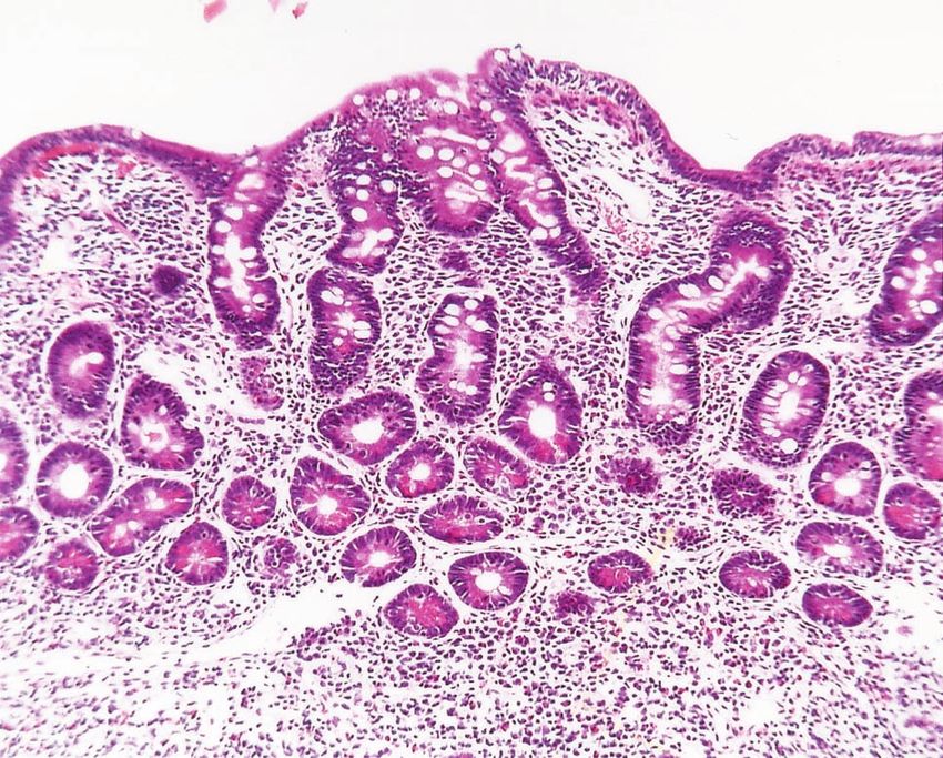
2612 Circulation June 4, 2002
No patient was diagnosed with a gastrointestinal, pancreatic, or and culture). Biopsy specimens were formalin-fixed, paraffin-
hepatic disorder, but 13 of them exhibited a sideropenic anemia with embedded, and hematoxylin-eosin–stained for histological evalua-
a negative hemoccult test refractory to oral iron administration, tion. Patients with subtotal/total villous atrophy (Marsh stage II or
suggesting the possibility of intestinal malabsorption. III) were considered to have CD, as were patients with intraepithelial
lymphocytic infiltration (Marsh stage I, intraepithelial lymphocytic
Cardiac Studies infiltration ⬎40/100 enterocytes), and they were started on a gluten-
All 187 patients underwent both noninvasive cardiac examinations free diet. After 6 to 8 months, the CD patients were reevaluated for
(ECG, Holter monitoring, exercise stress testing, and 2D echocardi- compliance to the gluten-free diet by means of clinical examination,
ography) and invasive cardiac examinations (cardiac catheterization, by evaluation of iron metabolism improvement, and by AEA and
biventricular and coronary angiography, biventricular endomyocar- tTG assessment.
dial biopsy, and electrophysiological study according to class I
indication of American College of Cardiology/American Heart HLA Analysis
Association guidelines). Additional cardiac catheterization and bi- Determination of HLA was performed in patients who were affected
opsy were obtained at 1, 3, and 5 months of follow-up in those by CD. Lymphotype plates for serological determination of HLA
patients with active myocarditis who were undergoing immunosup- phenotype were purchased from Biotest AG and One Lambda, Inc.
pressive therapy. All invasive cardiac exams were performed after
informed consent was given and were approved by the ethics Statistical Analysis
committee of our institution. Data were analyzed by using the 2 test.
Endomyocardial biopsies (3 or 4 per ventricular chamber) were
performed in the septal-apical region of both ventricles. Four to 6
endomyocardial samples obtained from each patient were processed Results
for histological and immunohistochemical studies. For histology, According to the patients’ clinical histories, no family mem-
multiple 5-m-thick sections were cut and stained with hematoxylin- ber with myocarditis or with a recent pregnancy or history of
eosin, Miller’s elastic van Gieson, Masson’s trichrome, and Ziehl- alcohol abuse was reported. By 2D echocardiogram, no
Neelsen stain. In all samples, immunohistochemical analysis for the
pericardial effusion was observed, even for those patients
characterization of inflammatory infiltrate was performed.
Histological Dallas criteria11 were used for the diagnosis of with myocarditis mimicking a myocardial infarction. Exer-
myocarditis. cise stress tests failed to induce ST-T changes suggestive of
Polymerase chain reaction (PCR) and sequencing analysis were myocardial ischemia. Ventricular arrhythmias were sup-
performed on frozen myocardial samples from all patients to detect pressed by exercise and reappeared on recovery. Coronary
RNA and DNA genomes of common cardiotropic viruses (adenovi-
rus, enterovirus, influenza A and B viruses, cytomegalovirus, hepa-
angiography was normal for all patients.
titis C virus, herpes simplex viruses, and Epstein-Barr virus) as At histological examination, diffuse or focal inflammatory
previously described.12,13 Viral types were identified when nucleo- lymphomononuclear infiltrates associated with necrosis of
tide comparisons revealed an identity of ⬎95% with known type. adjacent myocytes were observed in all patients meeting the
Dallas criteria for myocarditis. Two patients had giant cell
Serological Studies myocarditis. Different degrees of interstitial and/or replace-
Blood samples, collected at the time of endomyocardial biopsy, were
divided into aliquots and stored at ⫺80° until use. Sera of all the 187 ment fibrosis were also evident by Masson’s trichrome and
patients were screened for the presence of human IgA anti–tissue Miller’s elastic van Gieson staining. Ziehl-Neelsen staining
transglutaminase antibodies (IgA-tTGs), determined by an ELISA was negative for acid fast bacteria in all patients. Immuno-
commercial kit (Eu-tTG, IgA Umana, Eurospital) and IgA anti- phenotypical characterization of the inflammatory cells
endomysial antibodies (AEAs), detected by an indirect immunofluo-
showed the presence of activated T lymphocytes
rescence technique on commercial sections of distal monkey esoph-
agus (Antiendomysium, Eurospital). Samples were tested in (CD45RO⫹), including a moderate amount of cytotoxic
duplicate, and results were evaluated according to the manufacturer’s lymphocytes (CD8⫹). Giant cells were stained for the mac-
instructions and cut-off points. Sera containing antibody at a titer of rophage marker CD68 but not for the muscle markers actin
ⱖ1:5 were considered positive. Total serum IgA immunoglobulins and desmin, suggesting the macrophage origin of the giant
(normal value 90 to 450 mg/dL) were tested in all patients to exclude
selective IgA deficiency.
cells.15 PCR analysis showed the presence of viral genomes in
Patients’ sera were also tested to detect the presence of cardiac 86 (46%) of 187 patients. Among these, 26 had enterovirus
autoantibodies by standard indirect immunofluorescence.14 (30%), 21 had adenovirus (24%), 16 had Epstein-Barr virus
Patients positive for tissue transglutaminase (tTG) and AEAs (19%), 8 had hepatitis C virus (9.3%), 8 had cytomegalovirus
and/or cardiac autoantibodies were also screened for other autoim- (9.3%), 5 had influenza A virus (6%), and 2 had influenza B
mune disorders through the determination of organ-specific autoan-
tibodies (anti-islet cells, anti-thyroglobulin, anti–thyroid peroxidase, virus (2.4%). Sequencing analysis of enterovirus and adeno-
anti–liver-kidney microsomes, and adrenal autoantibodies) and non– virus PCR amplimers showed a high homology with coxsack-
organ-specific autoantibodies (antinuclear, anti-DNA, anticardio- ievirus B3 and B4, rhinovirus 14, and adenovirus 2 and 5.
lipin, anti sarcolemmal, antimyolemmal, and antineutrophil cyto- Comparison of influenza A virus and Epstein-Barr virus
plasm antibodies).
As a control population, we enrolled 306 healthy blood donors
showed high homology with human viral sequences. Hepati-
(212 males and 94 females, mean age 37.0 years [range 21 to 54 tis C virus–positive cases showed high homology for geno-
years]). type 1b.
Positivity for organ-specific anti-heart autoantibodies with
Gastrointestinal Studies diffuse cytoplasmic staining was evident in 53 (28%) of the
Patients positive to tTG or AEAs or to both and patients who patients.
exhibited iron-deficiency anemia underwent upper gastrointestinal
endoscopy with multiple biopsies in the descending duodenum, and No patient showed selective IgA deficiency. Thirteen of
duodenal juice samples were obtained by sterile tube for bacterio- 187 patients were positive for IgA-tTG antibodies, and all
logical and parasitological analysis (direct microscopic examination showed iron deficiency anemia refractory to oral iron replace-
Downloaded from http://circ.ahajournals.org/ by guest on October 10, 2015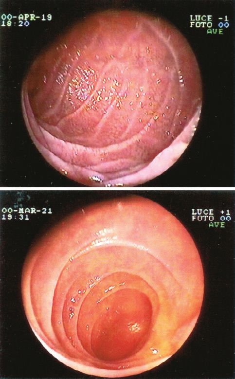
Frustaci et al Celiac Disease and Autoimmune Myocarditis 2613
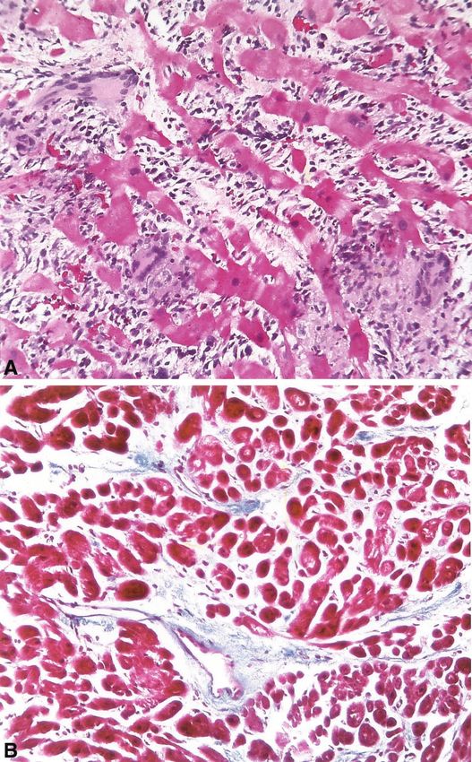
Figure 2. Duodenal biopsy from patient 2 showing total villous
atrophy with crypt hyperplasia and lymphoplasmacellular infil-
trate of lamina propria that are consistent with CD.
diagnosis was Giardia lamblia infestation, and in the remain-
ing 2 patients, duodenal diverticula with massive bacterial
overgrowth were detected by coculture of the duodenal juice.
The prevalence of CD in our myocarditis population was
4.4%. In the control population, 2 (0.6%) of 306 individuals
showed IgA-tTG positivity, but only 1 (0.3%) was AEA
positive and showed intestinal histology consistent with a
diagnosis of CD. The results were statistically significant
(P⬍0.003).
Patients With CD and Autoimmune Myocarditis
Clinical and histological data of the 9 CD patients with
Figure 1. Endoscopic appearance of descending duodenum of
patients (pts) 1 and 2 showing severe reduction of number and
autoimmune myocarditis are summarized in Table 1.
thickness of Kerckring’s folds with “scalloping” (top) and reduc- None of the CD patients had a history of recurrent
tion of number of Kerckring folds without scalloping (bottom). abdominal pain, chronic diarrhea, or weight loss, but all had
iron-deficiency anemia refractory to oral iron replacement.
PCR analysis on frozen myocardial tissue was negative, but
ment. Among tTG-positive patients, 9 showed positivity for all 9 patients were positive for anti-heart autoantibodies in the
AEA. serum. As a result of organ-specific autoantibody screening,
Endoscopy was performed in all the IgA-tTG–positive and no additional autoimmune disorder was found in the CD
AEA-positive patients. We found a normal endoscopic pat- patients. Positive antinuclear antibodies with a diffuse homo-
tern in 4 patients, a reduction of number or a loss of duodenal geneous pattern were found in 5 of 9 patients, including 1
folds (Figure 1A) in 7 patients, and an appearance as patient with giant cell myocarditis. HLA analysis showed a
“scalloped valvulae” (Figure 1B) in the 2 remaining patients. combination of DQ2-DR3 haplotypes in 8 patients and the
Histological examination of bioptic specimens was consistent presence of DQ2-DR5(11)/DR7 in 1 patient (Table 2).
with a diagnosis of CD in the 9 AEA-positive patients with No abnormalities of serum proteins or plasma electrolytes
abnormal endoscopic patterns (4.4%): 5 showed subtotal were observed in these patients.
villous atrophy and crypt hyperplasia (Marsh stage II), and In 5 patients, the clinical manifestation of cardiac disease
the other 4 patients showed total villous atrophy, crypt was progressive heart failure (Figure 3A) that failed to
hyperplasia, and lymphoplasmacellular infiltrate of the lam- improve after conventional supportive therapy administered
ina propria (Marsh stage III) (Figure 2). Among the remain- for ⬎6 months and including digitalis (0.25 mg daily),
ing 4 patients submitted to a duodenal biopsy, none showed diuretics (furosemide 25 to 50 mg daily), ACE inhibitors
histological findings of CD: 3 had eosinophilic infiltrate of (enalapril 20 mg twice daily), and carvedilol (25 to 50 mg
the lamina propria of intestinal mucosa without changes in daily); no inotropic agent was additionally provided. In the
intestinal villi and crypts and without an increase of intraepi- remaining 4 patients, myocarditis plus ventricular arrhyth-
thelial lymphocytes, and the remaining patient showed nor- mias (Lown class III-IVa) were associated with normal
mal intestinal mucosa. For 2 of these patients, the final cardiac volume and function and normal intracavity pres-
Downloaded from http://circ.ahajournals.org/ by guest on October 10, 2015
2614         Circulation             June 4, 2002

       TABLE 1.        Characteristics, Treatment, and Follow-Up of Patients With Autoimmune Myocarditis and Celiac Disease
                                                                Myocardial         Duodenal          Duodenal
       No.     Age/Sex             Clinical Presentation        Histology         Endoscopy          Histology      Treatment         Follow-Up (overall 12 months)
       1            24/F    CHF (NYHA III; LVEF 32%)⫹IDA            ALM            Scalloped            SVA/CH         GFD⫹I          Improved (NYHA I; LVEF 54%)
                                                                                    valvulae
       2            22/F    CHF (NYHA IV; LVEF 21%)⫹IDA             GCM          Reduction of           TVA/CH         GFD⫹I          Improved (NYHA I; LVEF 56%)
                                                                                duodenal folds
       3            35/F      VEB (Lown Class IVa)⫹IDA              ALM            Loss of              TVA/CH         GFD              Improved (Lown Class I)
                                                                                duodenal folds
       4            16/F         VEB (Lown Class III)⫹IDA           ALM          Reduction of           SVA/CH         GFD              Improved (Lown Class I)
                                                                                duodenal folds
       5           32/M     CHF (NYHA IV; LVEF 17%)⫹IDA             ALM            Scalloped            SVA/CH         GFD⫹I            Improved (NYHA II; LVEF
                                                                                    valvulae                                                     46%)
       6            38/F      VEB (Lown Class IVa)⫹IDA              ALM            Loss of              TVA/CH         GFD              Improved (Lown Class I)
                                                                                duodenal folds
       7            16/F    CHF (NYHA II; LVEF 36%)⫹IDA             ALM            Loss of              SVA/CH         GFD⫹I          Improved (NYHA I; LVEF 54%)
                                                                                duodenal folds
       8           36/M     CHF (NYHA III; LVEF 27%)⫹IDA            ALM            Loss of              TVA/CH         GFD⫹I          Improved (NYHA I; LVEF 48%)
                                                                                duodenal folds
       9            14/F         VEB (Lown Class III)⫹IDA           ALM          Reduction of           SVA/CH         GFD              Improved (Lown Class I)
                                                                                duodenal folds
          CHF indicates congestive heart failure; NYHA, New York Heart Association class; IDA, iron deficiency anemia; LVEF, left ventricular ejection fraction;
       VEB, ventricular ectopic beats; ALM, active lymphocytic myocarditis; GCM, giant-cell myocarditis; SVA, subtotal villous atrophy; TVA, total villous
       atrophy; CH, crypt hyperplasia; GFD, gluten-free diet; and I, immunosuppression.

sures. Ventricular arrhythmias were abolished during the                                off to 0.33 mg/kg daily for 5 months) and a gluten-free diet in
cardiac stress test. Because of the positivity of cardiac                               addition to the current supportive treatment. All patients
autoantibodies (with the absence of cardiotropic viruses at                             demonstrated an improvement in cardiac volume and func-
PCR analysis suggesting an autoimmune myocarditis) and                                  tion (Figure 3B) that was maintained at 12 months of
because of the progressive heart failure refractory to conven-                          follow-up. At control biopsy, lymphocytic and giant cell
tional therapy, the 5 patients with heart failure were treated                          myocarditis had progressed to a healed phase (Figure 4A and
with immunosuppression (azathioprine 1 mg/kg daily for 6                                4B). The 4 arrhythmic patients received a gluten-free diet
months and prednisone 1.25 mg/kg daily for 4 weeks, tapered                             alone and were followed by 2D echo and Holter monitoring

       TABLE 2.        Serological and HLA Profile of Patients With Autoimmune Myocarditis and Celiac Disease
                                                                                               Patients

                                        1                  2          3             4               5              6              7             8            9
       AEA (IgA)                       ⫹               ⫹              ⫹             ⫹              ⫹               ⫹             ⫹              ⫹            ⫹
       Anti-heart                      ⫹               ⫹              ⫹             ⫹              ⫹               ⫹             ⫹              ⫹            ⫹
       HLA pattern                  DQ2-DR3          DQ2-         DQ2-DR3       DQ2-DR3         DQ2-DR3      DQ2-DR3           DQ2-DR3       DQ2-DR3     DQ2-DR3
                                                  DR5(11)-DR7
       ANA                             ⫹               ⫹              ⫹             ⫺              ⫺               ⫹             ⫹              ⫺            ⫺
       ANCA                            ⫺               ⫺              ⫺             ⫺              ⫺               ⫺             ⫺              ⫺            ⫺
       Anti-DNA                        ⫺               ⫺              ⫺             ⫺              ⫺               ⫺             ⫺              ⫺            ⫺
       Anticardiolipin                 ⫺               ⫺              ⫺             ⫺              ⫺               ⫺             ⫺              ⫺            ⫺
       Antisarcolemmal                 ⫺               ⫺              ⫺             ⫺              ⫺               ⫺             ⫺              ⫺            ⫺
       Antimyolemmal                   ⫺               ⫺              ⫺             ⫺              ⫺               ⫺             ⫺              ⫺            ⫺
       Adrenal autoantibodies          ⫺               ⫺              ⫺             ⫺              ⫺               ⫺             ⫺              ⫺            ⫺
       Anti-islet cells                ⫺               ⫺              ⫺             ⫺              ⫺               ⫺             ⫺              ⫺            ⫺
       Anti liver-kidney               ⫺               ⫺              ⫺             ⫺              ⫺               ⫺             ⫺              ⫺            ⫺
       microsomes
       Anti-tireoglobulin,             ⫺               ⫺              ⫺             ⫺              ⫺               ⫺             ⫺              ⫺            ⫺
       anti-thyroid peroxidase
       FT3, FT4, TSH                 Normal          Normal        Normal        Normal          Normal          Normal        Normal         Normal       Normal
           AEA indicates Anti-endomysial antibodies; ANA, Antinuclear antibodies; and ANCA, Anti-neutrophils-cytoplasm antibodies.

                                      Downloaded from http://circ.ahajournals.org/ by guest on October 10, 2015
Frustaci et al Celiac Disease and Autoimmune Myocarditis 2615
Figure 3. End-diastolic (up) and end-systolic (down) frame from patient 2 before (A) and after (B) 1 month of immunosuppressive ther-
apy. Marked improvement of ejection fraction is shown (from 21% [A] to 56% [B]).
every 4 to 6 weeks. At 12 months of follow-up, cardiac infiltration of the small bowel mucosa was documented in all
arrhythmias improved from Lown class III-IVa to class I of our 9 patients. In these patients, clinical manifestation of
(Figure 5A and 5B), and the cardiac 2D echo parameters myocarditis was, in 5 cases, heart failure that markedly
remained normal. All CD patients experienced a disappear- improved after a combination of gluten-free diet and immu-
ance of the CD-specific autoantibodies 8 months after gluten nosuppressive therapy. The latter was prompted because of
withdrawal, with a normalization of iron metabolism. the severity of cardiac dysfunction (which was unresponsive
to full conventional supportive therapy), histological evi-
Discussion dence of active lymphocytic or giant cell myocarditis, nega-
A reciprocal negative interaction between the heart and small tive serology and PCR on frozen endomyocardial biopsies for
intestine is known to occur whenever either organ is severely the most common cardiotropic viruses, and, finally, the
compromised. Less recognized is the possibility of simulta- presence of circulating cardiac autoantibodies.
neous damage of the 2 organs due to a common pathogenetic It can be argued that cardiac improvement cannot be
mechanism. The present study showed the presence of an attributed with certainty to the therapeutic regimen adopted
intestinal inflammatory disease in 4.4% of a large population because a spontaneous resolution has been observed in up to
of patients with myocarditis, with a prevalence that was 14 40% of the patients with myocarditis.17 However, this kind of
times higher than that in normal control subjects. improvement is mainly attributed to patients with acute
This amount appears to be rather reliable, because for both lymphocytic myocarditis, whereas our patients had chronic
AEA and tTG antibodies, a high sensitivity (95% and 100%, heart failure and failed to respond to supportive treatment
respectively) and specificity (90% and 100%, respectively) administered for ⬎6 months. Moreover, 1 of our patients had
have been found.16 giant cell myocarditis, which is known as a progressive
The presenting symptom of intestinal malabsorption was in disease, and had a poor prognosis unless treatment with a
our series a sideropenic anemia that was refractory to oral strong immunosuppressive regimen was implemented or
iron supplementation, whereas a diagnosis of CD was ob- heart transplantation was performed.1,15
tained by both positivity of AEA and identification of the In the 4 patients with ventricular arrhythmias, only the
characteristic duodenal endoscopic and histological findings. gluten-free diet was instituted, and cardiac contractility was
In fact, a combination of villous atrophy with lymphocytic preserved, and no sustained ventricular tachycardia or synco-
Downloaded from http://circ.ahajournals.org/ by guest on October 10, 20152616 Circulation June 4, 2002
tion of many intestinal luminal antigens (such as ingested
food proteins, bacterial breakdown products, endotoxins, and
active enzymes) that can exacerbate myocardial inflamma-
tion. Indeed, some other extraintestinal findings of CD, such
as chronic unexplained hypertransaminasemia, are attributed
to the mechanism of antigenic overload.22
Furthermore, active CD is accompanied by consistent
production of IgA autoantibodies to reticulin, a common
constituent of the extracellular matrix; serum IgA antibodies
of patients with untreated CD have been reported to strongly
react against human brain– blood vessel structures,23 and this
mechanism has been hypothesized to be involved in the
abnormal nervous system manifestations frequently described
in association with CD. Recent studies have demonstrated
that anti-gliadin autoantibodies react with common epitopes
on gliadin, calreticulin, and enterocytes24 and with a nuclear
autoantigen expressed in intestinal endothelial cells and in
fibroblasts.25 On the other hand, tTG, recognized as the target
antigen of CD-specific autoantibodies, is an intracellular
enzyme that is distributed in the cells of all organs. A possible
link between tTG and cardiac damage26 and also an upregu-
lation of mRNA for tTG in rat models of cardiac failure have
been reported. These findings lead us to hypothesize that
antigenic mimicry could be actually involved in the patho-
genesis of CD-associated disorders. In our CD patients, we
were able to detect an autoimmune process against cardiac
antigens that could play a key role in the pathogenesis of
inflammatory heart damage. The evidence that improvement
of cardiac function and of ventricular arrhythmias was paral-
leled by the disappearance of AEAs and tTG in the serum
supports this hypothesis. Nevertheless, other potential mech-
anisms, such as resumed absorption of proteins with an
Figure 4. Left ventricular endomyocardial biopsy from patient 2 antioxidant or cardioprotective effect, cannot be excluded.
showing giant cell myocarditis (A) that progressed to a healed An interesting finding in the present study was the sub-
phase (B) after 1 month of immunosuppressive therapy. Stains
and magnifications were as follows: hematoxylin and eosin, clinical presentation of CD even in the presence, in some
original magnification ⫻250 (A); Masson’s trichrome, original cases, of marked histopathological lesions. It has been clearly
magnification ⫻160 (B). demonstrated that the risk of autoimmune disorders is signif-
icantly more elevated in untreated CD and that the prevalence
of autoimmune disorders in CD patients is related to the
pal event was observed. Arrhythmic patients seemed to duration of exposure to a gluten-containing diet; compared
benefit from diet, because at sequential Holter monitoring, with healthy subjects, patients with early diagnosis of CD do
the arrhythmias markedly improved (from Lown class III-IVa not show an increased prevalence of autoimmune disorders.27
to class I). This observation suggests the need for early diagnosis,
HLA analysis showed the presence of DQ2-DR3, which is
prompt instauration of a gluten-free diet, and strict compli-
commonly observed in patients with CD (DQ2 in up to 95%
ance to gluten withdrawal. Finally, malabsorption can reduce
of the cases)16 and with systemic autoimmune disorders.18,19
the availability of cardiovascular drugs and essential nutrients
Serological detection of AEA and cardiac autoantibodies,
HLA profile, negative PCR studies for cardiotropic viruses, that can, in turn, impair contractile and electrical cardiac
and responsiveness to a gluten-free diet and immunosuppres- function.
sive therapy strongly suggest the existence in our patients of
an autoimmune disorder directed toward antigenic compo- Clinical Implications
nents of both the myocardium and small bowel. Indeed, both Patients with biopsy-proven myocarditis, especially in the
myocarditis (particularly giant cell myocarditis)1 and CD20 presence of clinical findings of malabsorption, should be
are known to occur in association with systemic autoimmune screened for CD.
disorders. The observation that these entities can be combined In fact, if CD is associated with autoimmune myocarditis,
in the same patient is of clinical relevance. a gluten-free diet alone or the diet in combination with
In fact, CD is invariably associated with an increase of immunosuppressive agents can significantly improve the
intestinal permeability,21 which could lead to the transloca- clinical outcome.
Downloaded from http://circ.ahajournals.org/ by guest on October 10, 2015Frustaci et al Celiac Disease and Autoimmune Myocarditis 2617
Figure 5. Twelve-lead ECG from patient
3 showing frequent couples of ventricular
ectopic beats (A) that disappeared after
8 months on gluten-free diet (B).
Limitations of the Study 3. Maki M, Collin P. Coeliac disease. Lancet. 1997;349:1755–1759.
In our 5 patients with CD and autoimmune myocarditis 4. Schuppan D. Current concepts of celiac disease pathogenesis. Gastroen-
terology. 2000;119:234 –242.
presenting with heart failure, immunosuppressive treatment 5. Collin P, Salmi J, Hallstrom O, et al. High frequency of coeliac disease
was administered together with a gluten-free diet because of in adult patients with type-1 diabetes. Scand J Gastroenterol. 1989;24:
the severe cardiac dysfunction and hemodynamic instability 81– 84.
6. Counsell CE, Taha A, Ruddell WSJ. Coeliac disease and autoimmune
that were unresponsive to full conventional therapy. The thyroid disease. Gut. 1994;35:844 – 846.
efficacy of a gluten-free diet alone on recovery of cardiac 7. Kaukinen K, Collin P, Mykkanen AH, et al. Celiac disease and auto-
function should be tested in patients with autoimmune myo- immune endocrinologic disorders. Dig Dis Sci. 1999;44:1428 –1433.
8. Rustgi AK, Peppercorn MA. Gluten-sensitive enteropathy and systemic
carditis and CD who exhibit a myocardial compromise with a lupus erythematosus. Arch Intern Med. 1988;148:583–584.
stable hemodynamic profile and less aggressive inflammatory 9. Curione M, Barbato M, Biase L, et al. Prevalence of coeliac disease in
disease. Such an investigation could definitively clarify the idiopathic dilated cardiomyopathy. Lancet. 1999;354:222–223.
10. Chimenti C, Pieroni M, Frustaci A. Celiac disease in idiopathic dilated
relationship between the 2 entities.
cardiomyopathy. Ital Heart J. 2001;2:658 – 659.
11. Aretz H, Billingham ME, Edwards WD, et al. Myocarditis: a his-
topathologic definition and classification. Am J Cardiovasc Pathol. 1986;
Acknowledgments 1:3–14.
This study was supported by the “Myocarditis: therapeutic impact of 12. Pauschinger M, Bowles NE, Fuentes-Garcia FJ, et al. Detection of
etiological diagnosis based upon molecular and immunologic find- adenoviral genome in the myocardium of adult patients with idiopathic
ings” (MURST) project and by “Fondazione per il cuore.” left ventricular dysfunction. Circulation. 1999;99:1348 –1354.
13. Chimenti C, Calabrese F, Thiene G, et al. Inflammatory left ventricular
microaneurysms as a cause of apparently idiopathic ventricular
tachyarrhythmias. Circulation. 2001;104:168 –173.
References 14. Frustaci A, Chimenti C, Maseri A. Global bi-ventricular dysfunction in
1. Cooper LT Jr, Berry GJ, Shabetai R. Idiopathic giant-cell myocarditis patients with symptomatic coronary artery disease may be caused by
natural history and treatment: Multicenter Giant Cell Myocarditis Study myocarditis. Circulation. 1999;99:1295–1299.
Group Investigators. N Engl J Med. 1997;336:1860 –1866. 15. Frustaci A, Chimenti C, Pieroni M, et al. Giant cell myocarditis
2. Trier J. Celiac sprue. N Engl J Med. 1991;325:1709 –1719. responding to immunosuppressive therapy. Chest. 2000;117:905–907.
Downloaded from http://circ.ahajournals.org/ by guest on October 10, 20152618 Circulation June 4, 2002
16. Farrell RJ, Kelly CP. Celiac sprue. N Engl J Med. 2002;346:180 –188. 23. Pratesi R, Gandolfi L, Friedman H, et al. Serum IgA antibodies from
17. Dec GW Jr, Palacios IF, Fallon JT, et al. Active myocarditis in the patients with coeliac disease react strongly with human brain blood-vessel
spectrum of acute dilated cardiomyopathies: clinical features, histologic structures. Scand J Gastroenterol. 1998;33:817– 821.
correlates, and clinical outcome. N Engl J Med. 1985;312:885– 890. 24. Krupickova S, Tuckova L, Flegelova Z, et al. Identification of common
18. Klein J, and Sato A. The HLA system: first of two parts. N Engl J Med. epitopes on gliadin, enterocytes, and calreticulin recognised by antigliadin
2000;343:702–709. antibodies of patients with coeliac disease. Gut. 1999;44:168–173.
19. Klein J, Sato A. The HLA system: second of two parts. N Engl J Med. 25. Natter S, Granditsch G, Reichel GL, et al. IgA cross-reactivity between a
2000;343:782–786. nuclear autoantigen and wheat proteins suggests molecular mimicry as a
20. Collin P, Reunala T, Pukkala E, et al. Coeliac disease, associated dis- possible pathomechanism in celiac disease. Eur J Immunol. 2001;31:
orders and survival. Gut. 1994;35:1215–1218. 918 –928.
21. van Elburg RM, Uil JJ, Mulder CJ, et al. Intestinal permeability in 26. Iwai N, Shimoike H, Kinoshita M. Genes up-regulated in hypertrophied
patients with coeliac disease and relatives of patients with coeliac disease. ventricle. Biochem Biophys Res Commun. 1995;209:527–534.
Gut. 1993;34:354 –357. 27. Ventura A, Magazzu G, Greco L. Duration of exposure to gluten and risk
22. Bardella MT, Fraquelli M, Quatrini M, et al. Prevalence of hyper- for autoimmune disorders in patients with celiac disease: SIGEP Study
transaminasemia in adult patients an effect of gluten-free diet. Hepa- Group for Autoimmune Disorders in Celiac Disease. Gastroenterology.
tology. 1995;22:833– 836. 1999;117:297–303.
Downloaded from http://circ.ahajournals.org/ by guest on October 10, 2015Celiac Disease Associated With Autoimmune Myocarditis
Andrea Frustaci, Lucio Cuoco, Cristina Chimenti, Maurizio Pieroni, Giuseppina Fioravanti,
Nicola Gentiloni, Attilio Maseri and Giovanni Gasbarrini
Circulation. 2002;105:2611-2618; originally published online May 13, 2002;
doi: 10.1161/01.CIR.0000017880.86166.87
Circulation is published by the American Heart Association, 7272 Greenville Avenue, Dallas, TX 75231
Copyright © 2002 American Heart Association, Inc. All rights reserved.
Print ISSN: 0009-7322. Online ISSN: 1524-4539
The online version of this article, along with updated information and services, is located on the
World Wide Web at:
http://circ.ahajournals.org/content/105/22/2611
Permissions: Requests for permissions to reproduce figures, tables, or portions of articles originally published
in Circulation can be obtained via RightsLink, a service of the Copyright Clearance Center, not the Editorial
Office. Once the online version of the published article for which permission is being requested is located,
click Request Permissions in the middle column of the Web page under Services. Further information about
this process is available in the Permissions and Rights Question and Answer document.
Reprints: Information about reprints can be found online at:
http://www.lww.com/reprints
Subscriptions: Information about subscribing to Circulation is online at:
http://circ.ahajournals.org//subscriptions/
Downloaded from http://circ.ahajournals.org/ by guest on October 10, 2015You can also read