A New Disease of Strawberry, Fruit Rot, Caused by Geotrichum candidum in China
←
→
Page content transcription
If your browser does not render page correctly, please read the page content below
Vol. 54, 2018, No. 2: 92–100                                                                      Plant Protect. Sci.
https://doi.org/10.17221/76/2017-PPS

                   A New Disease of Strawberry, Fruit Rot,
                   Caused by Geotrichum candidum in China

         Wenyue Ma1, Ya Zhang 1*, Chong Wang 2, Shuangqing Liu 1 and Xiaolan Liao 1,3*
     1
  Department of Plant Protection, College of Plant Protection and 2Department of Chemistry,
 Science College, Hunan Agricultural University, Changsha, P.R. China; 3Hunan Provincial Key
Laboratory for the Biology and Control of Plant Diseases and Plant Pests, Changsha, P.R. China
                      *Corresponding authors: zhangya230@126.com; lxllxl423@163.com

Abstract

Ma W., Zhang Y., Wang Ch., Liu S., Liao X.-l. (2018): A new disease of strawberry, fruit rot, caused by Geotri-
chum candidum in China. Plant Protect. Sci., 54: 92–100.

A new disease of strawberry (Fragaria ananassa Duch.) was discovered in the Lianqiao strawberry planting base in
Shaodong County, in Hunan Province, China. In the early disease stage, leaves showed small black spots surrounded
by yellow halos, while in the late stage, a white fluffy layer of mold appeared. Fruits were covered with a white layer
of mold. The symptoms were observed using in vitro inoculation experiments. After the spray-inoculation of stabbed
leaves, small black spots surrounded by yellow halos occurred on leaves, with no clear boundary between diseased
and healthy areas. In the late stage, disease spots gradually expanded and a white fluffy layer of mold formed under
humid conditions. Unstabbed leaves had almost no disease occurrence after spray-inoculation. After the spray-
inoculation of stabbed fruits, by the late stage, a dense white layer of mold formed. According to Koch’s postulates,
the isolated strain was verified as a pathogen. The pathogenic strain, designated SDLQ16, was isolated from diseased
fruit by dilution method and tentatively identified as G. candidum based on the culture characteristics, morphologies,
physio-biochemical analysis, and phylogenetic analysis of the rDNA-ITS sequence. The fungus was able to grow on
different culture mediums, with a broad range of nutrition. The colonies on PDA medium were raised and pale white,
with a neat edge and visible hyphae. The hyphae were friable but the spores were developing. Basal hyphae rapidly
grew close to the medium to 3.2–4.2 µm in diameter, with septa and forked branches at acute angles. The solitary or
beaded spores with smooth surfaces were 3.5–7.5 µm in length and 3.5–4.5 µm in width. This strain was able to gelatin
liquefaction, proteolysis, grease, peptonised milk, urea, and so on. The pathogenicity on strawberry from strong to
weak was: fruit > leaf > stem. A BLAST algorithm was used to query SDLQ16’s rDNA-ITS sequence (cloned and de-
posited as GenBank number KU373122) against the NCBI database, and it was located in the Acinetobacter sp. branch
of a phylogenetic tree. SDLQ16 was most closely related to Geotrichum candidum ATCC34614 (GQ4580314.1), with
a sequence similarity of 99%.

Keywords: Fragaria ananassa Duch.; new pathogenic fungi; Geotrichum candidum; identification; rDNA-ITS

  Strawberry (Fragaria ananassa Duch.), an her-              favoured by consumers because of its colourful ap-
baceous perennial plant in the genus Fragaria and            pearance, sweet, sour taste, aromatic, juicy flesh,
in the family Rosaceae, is highly adaptable, widely          and rich nutrient ingredient (Wang et al. 2008a;
cultivated, and extensively distributed. Strawberry          Takahashi et al. 2009). Over the past 20 years, the
is regarded as the “queen of fruit” and is strongly          planting area and yield of strawberry have grown

Supported by Special Fund for Agro-scientific Research in the Public Interest, Grant No. 201303025, and the Sci-
entific Research Fund of Hunan Provincial Education Department, Grant No. 14B086.

92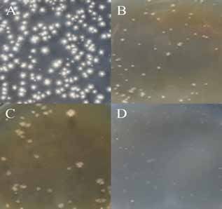
Plant Protect. Sci.                                                               Vol. 54, 2018, No. 2: 92–100
                                                                            https://doi.org/10.17221/76/2017-PPS

rapidly. The average annual planting area is 2.5 ×        culture observations, morphological characterisation,
10 5 hm 2, and the average annual yield is 4.5 × 10 8 t   a pathogenicity assay, and an internal transcribed
(FAOSTAT 2013; FAO Statistical database 2012 –            spacer (ITS) sequence analysis. This study provides
http://faostat.fao.org/site/339/default.aspx; USDA        scientific methods for identifying and diagnosing
Quick Stats 2.0. U.S 2013; Šamec et al. 2016). Europe     strawberry disease, and clarifying the pathogen type,
has the greatest strawberry production, followed by       which will aid in developing disease prevention and
the America and, in descending order: Asia, Africa,       control measures.
and Oceania. China is the most important country
for strawberry production in Asia. In recent years,
there has been a structural adjustment in the plant-               Material and Methods
ing industry in China. Planting scale of strawberry
has been expanded rapidly, because it is a cash crop        Diseased sample collection. Typical diseased fruit
with a short production cycle, high effectiveness, and    samples were collected from the Lianqiao strawberry
high efficiency (Li et al. 2004). The planting area       planting base in Shaodong County, Hunan Province,
and yield of strawberry in China, both ranking first      China in March–April 2015 on strawberry Fragaria
in the world, were 1.1 × 10 5 hm 2 and 200 t, respec-     ananassa Duch. cv. Hongyan. Fruits were gently
tively, in 2010 (Tan et al. 2003; Wang et al. 2008b;      placed into sample bags and labelled, including the
Zhao et al. 2012). However, blind large-scale plant-      date. All samples were delivered to the laboratory
ings, extensive management modes, climate change,         for identification.
farming system alterations, the disordered introduc-        Culture media, We prepared the following culture
tion of strawberry varieties, the misappropriation,       media according to the method of Fang (1998) using
and misuse of chemical pesticides, have resulted          potato dextrose agar medium (PDA; the major com-
in increasingly serious strawberry diseases and the       position: potato 200 g, glucose 20 g, agar 18 g, H 2O
continuous emergence of new diseases (Zhang et            1000 ml), beef extract–peptone agar (NA; the major
al. 2010a, b; Walker et al. 2011).                        composition: beef extract 3 g, peptone 10 g, NaCl
  The strawberry diseases previously recorded in          5 g, agar 18 g, H 2O 1000 ml, pH 7.2–7.4), Gause’s
China mainly included root rot, gray mold, pow-           No. 1 agar (GSYH; the major composition: soluble
dery mildew, anthracnose, leaf spot, and brown spot       starch 20 g, KNO 3 1 g, K 2HPO 4 0.5 g, MgSO 4·7H 2O
(Behrouz et al. 2006; Lv et al. 2010; Fang et al.         0.5 g, NaCl 0.5 g, FeSO 4·7H 2O 0.01g, agar 18 g, H 2O
2012; Sylla et al. 2013; Asad-Uz-Zaman et al.             1000 ml, pH 7.2–7.4), beef extract–nutrient broth
2015; Campos-Requena et al. 2015; Lachhab et al.          (NB; the major composition: beef extract 3 g, pep-
2015). Recently, we investigated the occurrences of       tone 10 g, NaCl 5g, H 2O 1000 ml, pH 7.2–7.4), and
strawberry diseases in 14 regions of Hunan Province,      Luria–Bertani agar (LB; the major composition:
including southern, central, western, northern, and       tryptone 10 g, yeast extract 5 g, NaCl 10 g, agar 18 g,
eastern. During this investigation, an unknown new        H 2O 1000 ml, pH 7.2–7.4).
potential disease that infected strawberry fruits was       Strawberry seedlings. Healthy, disease/pest-free
discovered in the Lianqiao strawberry planting base       seedlings with uniform growth were chosen from the
in Shaodong County. Typical diseased fruit samples        strawberry planting base and transplanted with soil
were taken to the laboratory for identification, and      into small experimental pots. The plants were placed
an isolate was grown under culture conditions. The        in a light incubator (20–23°C, 80–90% humidity) for
growth was rapid, which excluded powdery mildew           3–5 days. After reviving, the plants were used for
pathogens, the growth temperature was low, which          inoculation and experimentation.
excluded Phytophthora fruit rot, and no sclerotia or        Pathogen isolation. Strain SDLQ16 was isolated by
pycnidia were observed, which excluded southern           dilution from disease tissue. We utilised the sterile
blight (Li et al. 2005; Sylla et al. 2013; Eikemo &       knife to cut morbid and healthy at junctions from
Stensvand 2015). A thorough review of the lit-            typical sick fruits with 4–5 length and 2–3 width.
erature and historical data found no reports of this      Taken 10 tissues (about 0.5 g) into sterile tube with
disease or any similar diseases. Thus, this disease was   4.5 ml of sterile water washed repeatedly, after then
considered to be a new disease affecting strawberry.      we gained dilute spore suspension of 10 –1. Put 0.5 ml
  In present study, we systematically identified the      suspension of 10 –1 spore suspension into 10 ml tube
new disease of strawberry through pathogen isolation,     which has 4.5 ml sterile water and shaking, then we

                                                                                                             93
Vol. 54, 2018, No. 2: 92–100 Plant Protect. Sci. https://doi.org/10.17221/76/2017-PPS gained dilute spore suspension of 10–2. In turn, we got tion at 94°C for 45 s, annealing at 55°C for 45 s, and spore suspension of 10 –6. Taken 20 µl of 10 –6 spore extension at 72°C for 1 min, and a final extension suspension into PDA medium which contained strep- step at 72°C for 10 minutes. The PCR products were tomycin at a concentration of 40 µg/ml. Afterwards, held at 4°C until they were removed from the ther- uniform coated with spreader, after water being mal cycler. The target gene fragment was isolated aired, the medium were placed upside down and using 1% agarose gel electrophoresis for 20 min at incubated at 20–23°C in the dark for 24–72 hours. 150 V and 100 mA. The DNA band was recovered Utilised inoculating needle moved single colonies to and purified using a DNA gel extraction kit and another PDA medium to pure cultivate. Until gained then sequenced by Sangon (Shanghai, China). The the pure colonies which meet the requirements of sequence was deposited in GenBank to obtain an experiments (Fang 1998). accession number (KU373122). Cultural characteristics of the pathogen strain. The obtained sequence of strain SDLQ16 was Strain SDLQ16 was inoculated onto PDA, NA, GSYH, compared, using the BLAST algorithm, with other and LB and incubated in a temperature-controlled sequences in the NCBI database to identify related incubator in the dark for 24–120 hours. Colony sequences and retrieve the sequences of other species characteristics were observed, including size, shape for a phylogenetic analysis. Phylogenetic trees were (round, rhizoid, prostrate, irregular), edge (neat, constructed using the maximum likelihood method diffuse, dense, loose), surface (mouldy, powdery, in MEGA 7.0 (1000 replicates) (Gao et al. 2012). small black spotty, blossom-like, concentric annu- Pathogenicity assay. The pathogenicity of the lar), raised shape (expansion, platform, low convex, isolated pure culture was verified by Koch’s postu- convex surface), texture (fatty, filmy, sticky, brittle), lates. In the in vitro inoculation experiments, we colour, ease of picking, extent of binding to medium, collected stems, leaves, and semi-mature fruits of a production of pigments, and consistency of colour uniform size in the flowering and fruiting periods between the front and back sides of the medium. of strawberry. The samples were surface disinfected Morphological characteristics of the pathogen and washed with sterile water before use (Wei 1979; strain. Strain SDLQ16 was inoculated onto thin Moreno-Garcia & Grey 2012). PDA plates and continuously incubated at 20–23°C The in vitro inoculation experiments of stems, for 24–72 hours. First, the basic morphology of the leaves, and fruits included two treatments each. In fungus was observed, including the main and sec- Treatment 1, unstabbed samples were sprayed with ondary hyphal branches, the presence of septa, the the spores suspension, and in Treatment 2, stabbed position, production and germination of spores, and samples were sprayed with the spores suspension. the size, shape and arrangement of spores. Next, the The inoculated samples were incubated in a light formation of special structures, such as pycnidia, was incubator at 20–23°C with 85–90% humidity for examined. Attention was paid to the size, shape, and 7 days. The incidence of the disease was observed. location of these structures, which helped to identify When obvious symptoms were observed in the in- the fungus (Wei 1979). oculated tissue, re-isolation was performed using the ITS sequence analysis and phylogenetic tree diseased tissue. Additionally, an in vivo inoculation construction. Genomic DNA was extracted from the experiment was performed. Strawberry seedlings mycelia of strain SDLQ16 using the CTAB (Hexa- were transplanted into small pots and revived before decyltrimethylammonium bromide) method after the pathogenicity assay. The other procedures were 3–5 days culture. The extracted DNA was stored in performed as described in the in vitro inoculation a refrigerator at –20°C (Liu et al. 2005). experiments. The fungal rDNA gene was amplified with the uni- versal primers ITS1 (TCCGTAGGTGAACCTGCGG) and ITS4 (TCCTCCGCTTATTGATATGC). The PCR Results reaction contained 0.5 µl of template DNA, 2.5 µl of 10× Buffer (with Mg 2+), 1 µl of dNTPs (2.5 mM), Isolation and culture characteristics. A pure 0.2 µl of Taq polymerase, 0.5 µl of IS1, and 0.5 µl of culture, designated strain SDLQ16, was isolated IS4, with ddH 2O added to bring the total volume to from strawberry fruit with typical symptoms. At 25 µl. The PCR was run as follows: pre-degeneration 20–23°C, SDLQ16 was able to grow on different at 94°C for 4 min, followed by 30 cycles of degenera- media (e.g., PDA, NA, LB, and GSYH), with a wide 94
Plant Protect. Sci.                                                                    Vol. 54, 2018, No. 2: 92–100
                                                                                https://doi.org/10.17221/76/2017-PPS

 (A)                         (B)                            (A)                            (B)

                                                            (C)                             (D)
 (C)                         (D)

Figure 1. Colonial morphology of SDLQ16 on the different   Figure 2. Morphological characteristics of strain SDLQ16
medium: (A) PDA 48 h; (B) NA 48 h; (C) LB 48 h; (D)        by electron microscope: (A) hyphae mycelium, (B) branch
GSYH 96 hours                                              and branch, (C) beaded mycelium, and (D) oval spore,
                                                           round spores or cylindrical spores; 10 × 40

range of nutrition. On PDA, the colonies were raised       stage, they were fragmented to arthrospores (Figure
and pale white, with a neat edge and visible hyphae        2D). Spores, solitary or beaded (Figure 3A), 3.5–7.5 µm
at the edge; colonies were friable, with no pigments,      in length and 3.5–4.5 µm in width (Figures 3B–C), had
opaque and not shiny; sporulation was vigorous, and        smooth surfaces (Figure 3D). Single or multiple short
the colony surface was white and powdery. After            hyphae were rarely observed at the septum on the top
6 months of culture, white paste-like secretions ap-       of the hyphae. However, when they matured, the top
peared on the PDA, but the sexual stage of spores was      hyphae easily produced septa and formed beaded hy-
not observed. On NA, the colonies were raised, with        phae. The reproduction mode of strain SDLQ 16 was
an unapparent colour, neat edge, developed mycelia,        fession. The colony centre of this strain existed concen-
weak sporulation, and no pigments, being opaque            tric circles. This strain was able to gelatin liquefaction,
and not shiny. Additionally, SDLQ16 could grow on          proteolysis, grease, peptonised milk, urea, and so on.
LB and showed similar characteristics as observed on       These characteristics were basically consistent with
the NA medium. On GSYH, the colonies were small,           the pattern strain ATCC 34614 (Table 2).
with developed mycelia, weak sporulation, and no             Pathogenicity of the pathogen. Stabbed and un-
pigments, and were opaque and not shiny (Figure 1).        stabbed stems, leaves, and semi-mature fruits of
The growth rate analysis showed that PDA sup-
                                                           Table 1. Colony growth rate of strain SDLQ16 cultured for
ported the fastest growth (growth rate 1.49 mm/day),
                                                           5 days in different culture medium
followed by NA (growth rate 1.29 mm/day) and LB
(growth rate 1.17 mm/day); GSYH supported the              Culture Colony diameter
                                                                                   Average value            Growth rate
slowest growth (Table 1). These results indicate that      medium       (mm)
                                                                                      (mm)                   (mm/day)
strain SDLQ16 has a wide range of nutrition.               type     I     II  III
  Morphological features. Strain SDLQ16 did not            PDA        7.30 7.50 7.60      7.47 ± 0.15a      1.49 ± 0.03a
form aerial hyphae. Basal hyphae rapidly grew close to     NA         6.50 6.40 6.40      6.43 ± 0.06b      1.29 ± 0.01b
the medium, 3.2–4.2 µm in diameter, with septa and         LB         5.80 5.80 6.00      5.87 ± 0.12   c
                                                                                                            1.17 ± 0.02c
forked branches at acute angles (Figure 2A). Single or     GSYH       2.10 2.30 1.90      2.10 ± 0.20d      0.42 ± 0.04d
multiple short hyphae often adhered to the septa at the
base of the hyphae (Figure 2B). However, these hyphae      Values ± SD followed by different letters indicate significantly
were usually short and easily formed septa in a beaded     different scores in the same phase, according to Duncan’s
pattern (Figure 2C). When hyphae matured in the late       multiple range tests at the P = 0.05 level (SPSS 11.50)

                                                                                                                      95
Vol. 54, 2018, No. 2: 92–100 Plant Protect. Sci.
https://doi.org/10.17221/76/2017-PPS
Table 2. Characteristic of strain SDLQ16 morphology and observed on uninjured stems (Figure 4B). Injured
physiological biochemistry in PDA medium leaves had a more rapid occurrence, but it was still
1–2 days slower than on fruits. Small black spots
Strain
Morphology idex surrounded by yellow halos occurred on the leaf
SDLQ16 ATCC 34614§
surface, with an unapparent boundary between dis-
Colony colour milky white milky white eased and healthy areas (Figures 4C1–C2). Uninjured
Spore shape short rod rod leaves underwent slow or no disease occurrence
Spore size 3.5–7.5 μm 3.0–7.6 μm (Figures 4D1–D2). Injured fruits exhibited a rapid
Diaphragm exist exist occurrence. The fruit colour turned dark red and the
Colony shape flat flat fruit surface wilted rapidly; however, fruits did not
Branch two fork two fork
(D2)
Mycelium width 3.2–4.2 μm 2.6–7.0 μm
(A) (B)
Mycelium shape blanket-like powdery
Reproduction mode fissiparity fissiparity
Colony concentric circles exist exist
Gelatin liquefaction liquefaction liquefaction
Proteolysis hydrolysis hydrolysis
Grease degradation degradation
Peptonised milk peptonisation peptonisation
Urea assimilation assimilation
§
standard strain
(C1) (C1) (D1)
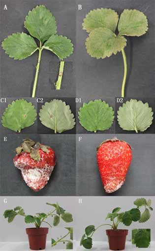
strawberry were spray-inoculated and cultured for
3–5 days at 85–90% relative humidity. Injured stems
underwent a rapid disease occurrence and turned
black, with dark spots forming in the late stage (Fig- (E) (F)
ure 4A). However, the disease occurrence was the
slowest on the stems. Almost no occurrence was
(A) (B)
(G) (H)
(C) (D)
Figure 4. Symptoms of strain SDLQ16 infecting stra-
wberry: (A) The inoculated stems with injury in vitro;
(B) The inoculated stems without injury in vitro; (C1)
Positive of the inoculated leaves with injury in vitro; (C2)
Opposite of the inoculated leaves with injury in vitro; (D1)
Positive of the inoculated leaves without injury in vitro;
(D2) Opposite of the inoculated leaves without injury in
vitro; (E) The inoculated stems with injury in vitro; (F)
Figure 3. Morphological characteristics of strain SDLQ16 The inoculated stems without injury in vitro; (G) The
by scanning electron microscope: (A) 1000, (B) 5000, (C) inoculated leaves with injury in vivo; (H) The inoculated
5000, and (D) 10 000× stems with injury in vivo
96Plant Protect. Sci. Vol. 54, 2018, No. 2: 92–100
https://doi.org/10.17221/76/2017-PPS
crack. On the fruit surface, white hyphae appeared ITS sequence analysis and phylogenetic tree con-
in the early stage, and a white layer of mold formed struction. The genomic DNA of strain SDLQ16 was
in the late stage. Uninjured fruits underwent a slow PCR amplified. The agarose gel electrophoresis of
occurrence, generally 3–4 days later than injured the PCR product revealed a clear 345-bp band. Fur-
fruits. The disease signs were not uniform, and the ther sequencing analysis verified that the rDNA-ITS
fruit colour turned dark slowly. Fruits were wilted sequence of this strain was 350 bp, in agreement with
but did not crack. Under humid conditions, the the electrophoresis results. The obtained rDNA-ITS
pathogen invaded slowly in the early stage, forming sequence was deposited in GenBank under accession
a small amount of hyphae; however, a white layer of number KU373122.
mold also formed in the late stage (Figures 4E–F). We compared the rDNA-ITS sequence of ‘SDLQ16’,
Spores or hyphae were picked from strawberry stems, using the BLAST algorithm, with other sequences in
leaves, and fruits to re-isolate and purify the patho- the NCBI database and found this strain in the Geo-
gen. The same colony characteristics were obtained trichum sp. branch of a phylogenetic tree. ‘SDLQ16’
as for the originally isolated pathogen. The results was most closely related to Geotrichum candidum
of the in vivo inoculation experiment were gener- ATCC 34614 (GQ4580314.1), with a sequence simi-
ally consistent with those of the in vitro inoculation larity of 99% (Figure 5). ‘SDLQ16’ was tentatively
experiments (Figures 4G–H). identified as G. candidum based on the phylogenetic
Figure 5. Bootstrap consensus tree constructed using the maximum likelihood method from 345 bp of ribosomal
internal transcribed spacer DNA obtained from the SDLQ16 and Geotrichum population. The tree was rooted to
Aspergillus helicothrix. The isolate SDLQ16 are indicated in bold font
97Vol. 54, 2018, No. 2: 92–100 Plant Protect. Sci.
https://doi.org/10.17221/76/2017-PPS
analysis of the rDNA-ITS sequence, in combination we only found the asexual stage of the pathogenic
with morphological and culture characteristics and fungus, and no sexual stage was observed during
a physico-biochemical analysis. culturing. Further studies are needed to investigate
whether the sexual stage of this strain is the same as
previously reported. This will include determining
Discussion how it overwinters and when and under what con-
ditions it germinates, as well as revealing primary
Our group has discovered a new disease affecting infections, the infection conditions, and the mode
strawberry. The isolate’s identity was determined of transmission in the field. G. candidum is strongly
using cultured characteristics, morphological ob- virulent towards carrots (Horita & Hatta 2016).
servations, and molecular biology techniques, and Bourret et al. (2013) indicated a weak pathogenic-
it was verified to be a pathogen, meeting Koch’s ity of G. candidum to tomatoes that the wounded
postulates. The pathogen strain was tentatively tomatoes could be infected with G. candidum and
identified as G. candidum. It belongs to fungus of gotten rot disease at a given tempreature of 20°C.
deuteromycotina, hyphomycetes, hyphomycetales, Yaghmour et al. (2012) suggested that injury in-
and moniliaceae (Barnett & Hunter 1998). A creases the susceptibility of peaches and nectarines
review of the literature and historical data showed to G. candidum, and the rot disease appeared after
no research on this pathogen affecting strawberry. 5 days under a constant 22–26°C. A number of studies
Thus, this is the first report of a strawberry disease have demonstrated that G. candidum is the major
caused by G. candidum. pathogen of citrus (Eckert 1978; Talibi et al. 2012).
The traditional classification and identification of The present study shows that the pathogenicity of
plant pathogens are usually based on culture char- G. candidum is not only on different host plants but
acteristics, colony characteristics, morphological also has regional differences, which may be related
features, and a pathogenicity index. The identifica- to the pathogenicity genes of G. candidum that are
tion of Geotrichum fungi mainly involves colony present in different areas, resulting in differences
morphology, the presence of septa in hyphae, hyphal in pathogenic performance. However, the causes of
branching, and the angle between main and secondary the different pathogenicity levels requires further
hyphae, as well as sporulation, and the germination, study. In the present research, G. candidum could
position, and size of spores. Geotrichum are widely cause strawberry to rot in Shaodong County, Hunan
distributed, and these fungi show great variation Province, China. This similarity could result from
under different geographical and environmental the high water content observed in both peaches
conditions (Liu et al. 2011). In recent years, the and strawberries which belong to different genera
extensive use of DNA identification techniques has of the family Rosaceae, or from regional differences,
effectively compensated for the shortcomings of indicating that G. candidum is pathogenic to some
traditional classification methods (Sun et al. 2015). plants. Thus, for biological product development, or
rDNA-ITS is the target most widely used to classify product degradation or preservation, G. candidum
and identify plant pathogenic fungi. The combination should be used with caution, and not arbitrarily
of traditional classification methods with DNA-based discarded, to prevent unnecessary losses.
techniques has substantially improved the accuracy In addition, under the conditions of low tempera-
of the identification of plant pathogenic fungi. In ture, high humidity, and insufficiency light, the fun-
particular, for closely related or sibling species of gus showed a high virulence toward its host, leading
Geotrichum that share a close phylogenetic relation- to a large disease incidence. Different organs of
ship (Blair et al. 2008). plants have different sensitivities to the fungi, and
G. candidum is a member of the genus Geotrichum. the fruit is more sensitive to this fungi than stems
The sexual stage (named Galactomyces geotrichum) and leaves. Thus, the disease rate was high when the
belongs to the family Candida, order Saccharomy- pathogen infected wounded tissue, indicating it was
cetales, and class Hemiascomycetes; the asexual stage a weak pathogen. The lower incidence of disease
(named G. candidum) belongs to the genus Oospora, development on the stem may be the result of the
family Moniliaceae, and phylum Deuteromycotina. lower stem water content compared with that of
A study indicated that G. candidum has more than the fruit and blade. The cortex thickness, stomatal
10 synonyms (Ma et al. 2007). In the present study, density, pore size, fruit and leaf ages may also af-
98Plant Protect. Sci. Vol. 54, 2018, No. 2: 92–100
https://doi.org/10.17221/76/2017-PPS
fect the disease rate. Here, G. candidum is reported FAOSTAT (2013): Searchable online statistical database
as a new pathogenic fungi of strawberry in China; from Food and Agriculture Division of the United Nations.
however, at the present, there is limited research on Available at http://faostat.fao. org/site/567/DesktopDe-
its treatment or pathogenic mechanisms, such as fault.aspx?PageID=567#ancor (accessed June 24, 2014).
the biological characteristics, epidemic laws, host Gao B.D., Huang W., Xia H. (2012): A new rice disease, black
range, the mechanisms of infection, fungicides and sheath spot, caused by Curvularia fallax in China. Plant
antagonism-based screenings are seldom used. To Disease, 8: 1224–1224.
increase our understanding of strawberry rot disease, Horita H., Hatta Y. (2016): Sour rot of carrot caused by
the above aspects require further studies as we work Geotrichum candidum in Japan. Journal of General Plant
toward disease prevention and control. Pathology 1: 65–68
Lachhab N., Sanzani S.M., Bahouaoui M.A., Boselli M.,
References Ippolito A. (2015): Effect of some protein hydrolysates
against gray mould of table and wine grapes. European
Asad-Uz-Zaman M., Bhuiyan M.R., Khan M.A.I., Bhui- Journal of Plant Pathology, 9: 1–10.
yan M.A., Latif M.A. (2015): Integrated options for the Li T.H., Wang L. (2004): The situation of production and
management of black root rot of strawberry caused by trade and strategy analysis of the sustainable development
Rhizoctonia solani Kuhn. Comptes Rendus Biologies, 2: of chinese strawberry industry. Chinese Agricultural Sci-
112–120. ence Bulletin, 6: 372–375.
Barnett H.L., Hunter B.B. (1998): Illustrated Genera of Li Y., Jin W., Huang J.W. (2005): Identification and biological
Imperfect Fungi. 4th Ed. Minnesota, Burgess Publishing characteristics of Sclerotium rolfsii Sacc causing southern
Co.: 34–35. blight on strawberry. Journal of Huazhong Agricultural
Behrouz E.M., Marie T.C., Odile C. (2006): Superoxide University, 3: 250–253
dismutase responses of strawberry cultivars to infection Liu S.H., Lu J.P., Zhu R.L., Dai F.M. (2005): A rapid and
by Mycosphaerella fragariae. Journal of Plant Physiology, simple extraction method for plant pathogenic fungi.
2: 147–153. Acta Phytopathologica Sinica, 4: 362–365.
Blair J.E., Coffey M.D., Park S.Y., Geiser D.M., Kang S. Liu T.M., Wang H.H., Wang J.J. (2011): Relationship analysis
(2008): A multi-locus phylogeny for phytophthora utiliz- between genetic diversity of Geotrichum candidum and
ing markers derived from complete genome sequences. geographical regions. Biotechnology Bulletin, 5: 121–125.
Fungal Genetics Biology, 45: 266–277. Lv R.L., Xie J.T., Fu Y.P., Jiang D.H. (2010): Identification
Bourret T.B., Kramer E.K., Rogers J.D., Glawe D.A. (2013): of pathogen that causes strawberry brown blotch disease
Isolation of Geotrichum candidum pathogenic to tomato and observation of biological characteristics. Journal of
(Solanum lycopersicum) in Washington State. North Huazhong Agricultural University, 4: 427–430.
American Fungi, 14: 1–7. Ma K., Liu G.Q., Li J.X., Yao S., Cheng C. (2007): The phy-
Campos-Requena V.H., Rivas B.L., Pérez M.A., Figueroa logenetic analysis of 15 Geotrichum strains based on 26S
C.R. Sanfuentes E.A. (2015): The synergistic antimi- rRNA gene D1/D2 region sequencing. Acta Microbio-
crobial effect of carvacrol and thymol in clay/polymer logica Sinica, 2: 359–362.
nanocomposite films over strawberry gray mold. LWT- Moreno-Garcia S., Grey O. (eds) (2012): Fungi. Vancouver,
Food Science and Technology, 1: 390–396. Innsmouth Free Press.
Eckert J.W. (1978): Pathological diseases of fresh fruits and Šamec D., Maretić M., Lugarić I., Mešic A., Salopek-Sondi
vegetables. Journal of Food Biochemistry 3: 243–250. B., Duralija B. (2016): Assessment of the differences in the
Eikemo H., Stensvand A. (2015): Resistance of strawberry physical, chemical and phytochemical properties of four
genotypes to leather rot and crown rot caused by Phy- strawberry cultivars using principal component analysis.
tophthora cactorum. European Journal of Plant Pathology, Food Chemistry, 194: 828–834.
2: 407–413. Sun D.W., Cao J.F., Pei W.H., Yin G.F., Ma J.Q., Pan K.H.,
Fang Z.D. (1998): The Research Methods of Plant Pathology. Wu K., Zhao Q., Duan Z.Q., Yang M.Y., Wang L., Zhao
3rd Ed. Beijing, China Agriculture Press: 45–50. Z.J. (2015): Identification of a new phytophthora blight
Fang X.P., Chen W.Y., Xin Y., Zhang H., Yan C., Yu H., Liu disease on konjac in Yunnan. Acta Phytopathologica
H., Xiao W.F., Wang S.Z., Zheng G.Z., Liu H.B., Jin L., Ma Sinica, 1: 84–87.
H.S., Ruan S.L. (2012): Proteomic analysis of strawberry Sylla J., Alsanius B.W., Krüger E., Becker D., Wohanka W.
leaves infected with Colletotrichum fragariae. Journal of (2013): In vitro compatibility of microbial agents for
Proteomics, 13: 4074–4090. simultaneous application to control strawberry powdery
99Vol. 54, 2018, No. 2: 92–100 Plant Protect. Sci.
https://doi.org/10.17221/76/2017-PPS
mildew (Podosphaera aphanis). Crop Protection, 51: in French vineyards in sympatry with Botrytis cinerea.
40–47. Phytopathology, 12: 1433–1445.
Talibi I., Askarne L., Boubaker H., Boudyach E.H., Msanda Wei J.C. (1979): Determinative Manual of Common Fungi
F., Saadi B., Ait Ben Aoumar A. (2012): Antifungal activ- System. Shanghai, Science Press: 15–20. (in Chinese)
ity of Moroccan medicinal plants against citrus sour rot Yaghmour M.A., Bostock R.M., Morgan D.P., Michailides
agent Geotrichum candidum. Letters in Applied Micro- T.J. (2012): Biology and sources of inoculum of Geotri-
biology, 2: 155–161. chum candidum causing sour rot of peach and nectarine
Takahashi H., Yamasaki A., Shoji K., Kawagishi K., Taguchi fruit in California. Plant Disease, 2: 204–210.
T., Yoshida Y., Morishita M. (2009): Present status and Zhang J., Zhang L., Li G.Q., Yang L., Jiang D.H., Zhuang
prospects of strawberry breeding and cropping type in W.Y., Huang H.C. (2010a): Botrytis sinoallii: a new species
Northern Japan. Acta Horticulturae, 842: 475–478. of the grey mould pathogen on Allium crops in China.
Tan C.H., Dai H.P., Lei J.J. (2003): Development trend of Mycoscience, 6: 421–431.
strawberry production and trade in the world. World Zhang J., Wu M.D., Li G.Q., Yang L., Yu L., Jiang D.H.
Agriculture, 5: 10–12. (2010b): Botrytis fabiopsis, a new species causing choco-
Wang G., Zhang Y.T., Dong J., Zhang L.X. (2008a): Retro- late spot of broad bean in central China. Mycologia, 5:
spection and prospect of strawberry breeding in China. 1114–1126.
Journal of Plant Genetic Resources, 2: 272–276. Zhao M.Z., Wang J., Wang Z.W., Qian Y.M., Wu W.M.
Wang F., Ma Y., Gao X.Y., Zhang Z.H. (2008b): Study on (2012): Countermeasures of strawberry sustainable de-
the identification techniques for determining strawberry velopment in Sanghai Zhejiang Jiangsu province and
cultivar’ s resistance to anthracnose. Journal of Fruit Sci- development status of strawberry industry in the world.
ence, 4: 542–547. Jiangsu Academy of Agricultural Sciences, 2: 1–3.
Walker A.S., Gautier A., Confais J., Martinho D., Viaud
Received: 2017–06–11
M., Pêcheur L., Dupont J., Fournier E. (2011): Botrytis
Accepted after corrections: 2017–08–30
pseudocinerea, a new cryptic species causing gray mold
Published online: 2017–10–03
100You can also read