Recurrence of Functional Versus Organic Mitral Regurgitation After Transcatheter Mitral Valve Repair: Implications from Three-Dimensional ...
←
→
Page content transcription
If your browser does not render page correctly, please read the page content below
Recurrence of Functional Versus Organic
                         Mitral Regurgitation After Transcatheter
                       Mitral Valve Repair: Implications from Three-
                       Dimensional Echocardiographic Analysis of
                       Mitral Valve Geometry and Left Ventricular
                              Dilation for a Point of No Return
                     Thomas Buck, MD, PhD, FACC, FESC, Nora Eiswirth, MD, Ahmed Farah, MD, Heike Kahlert, MD,
                     Polykarpos C. Patsalis, MD, Philipp Kahlert, MD, PhD, FACC, FESC, FAHA, and Bj€
                                                                                                   orn Plicht, MD,
                                               FESC, Dortmund, Essen, and Bochum, Germany

               Background: MitraClip implantation has become the standard transcatheter mitral valve repair (TMVR) tech-
               nique for severe mitral regurgitation (MR). However, approximately one third of patients have poor outcomes,
               with MR recurrence at follow-up. The aim of this study was to investigate whether quantitative analysis of mitral
               valve (MV) geometry on three-dimensional (3D) echocardiography can identify geometric parameters associ-
               ated with the recurrence of severe functional MR (FMR) versus organic MR (OMR) at 6-month follow-up after
               TMVR using the MitraClip.

               Methods: Sixty-one patients with severe FMR (n = 45) or OMR (n = 16) who underwent transesophageal 3D
               echocardiography before and 6 months after TMVR were retrospectively analyzed. MV geometry was quan-
               tified using 3D echocardiography software. Vena contracta area (VCA) at 6-month follow-up was used to
               define two outcome groups: patients with good results with VCA < 0.6 cm2 (MR < 0.6) and those with MR recur-
               rence with VCA $ 0.6 cm2 (MR $ 0.6).

               Results: MR recurrence was found in 34% of all study patients (21 of 61). In patients with FMR, signif-
               icant differences between MR < 0.6 and MR $ 0.6 were found at baseline for tenting index (1.13 vs
               1.23, P = .004), tenting volume (2.8 vs 4.0 ml, P = .04), indexed left ventricular (LV) end-diastolic vol-
               ume (68.0 vs 99.9 ml/m 2, P = .001), and VCA (0.71 vs 1.00 cm2, P = .003); no significant parameters of
               MR recurrence were found in patients with OMR. Multivariate analysis identified indexed LV end-
               diastolic volume as the strongest independent determinant of MR recurrence. Receiver operating
               characteristic analysis identified a tenting index of 1.185 (area under the curve 0.79) and indexed
               LV end-diastolic volume of 88 ml/m2 (area under the curve 0.76) to best discriminate between
               MR < 0.6 and MR $ 0.6.

               Conclusions: MR recurrence after TMVR in patients with FMR is associated with advanced LV dilation and MV
               tenting before TMVR, which provides clinical implications for a point of no return beyond which progressive LV
               dilation with MV geometry dilation and tethering cannot be effectively prevented by TMVR. In contrast, no sig-
               nificant determinants of MR recurrence and progressive MV annular dilation could be identified in patients with
               OMR. (J Am Soc Echocardiogr 2021;-:---.)

               Keywords: Mitral valve insufficiency, Functional mitral regurgitation, Organic mitral regurgitation, Transcath-
               eter mitral valve repair, Real-time 3D echocardiography, Treatment outcome

From the Department of Cardiology, Klinikum Westfalen, Heart Center Westfalen,          Reprint requests: Thomas Buck, MD, FESC, FACC, Heart Center Westfalen,
Dortmund, Germany (T.B., A.F., B.P.); the Department of Cardiology and                  Department of Cardiology, Klinikum Westfalen, Am Knappschaftskrankenhaus 1,
Vascular Medicine, University Clinic Essen, West-German Heart and Vascular              Dortmund 44309, Germany (E-mail: thomas.buck@klinikum-westfalen.de).
Center, Essen, Germany (T.B., N.E., H.K., P.C.P., P.K., B.P.); and the                  0894-7317/$36.00
Department of Cardiology and Angiology, University Clinic Bergmannsheil, Ruhr
                                                                                        Copyright 2021 by the American Society of Echocardiography.
University, Bochum, Germany (P.C.P.).
                                                                                        https://doi.org/10.1016/j.echo.2021.02.017
Conflicts of Interest: The authors declare that there are no conflicts of interest or
funding for this article.
                                                                                                                                                                 1
2 Buck et al                                                                                      Journal of the American Society of Echocardiography
                                                                                                                                             - 2021

           Abbreviations
                                       Mitral regurgitation (MR), which      METHODS
                                       is the most common valve dis-
  3D = Three-dimensional               ease (with 2% to 3% of the gen-       Study Design and Objectives
  ALPM = Anterolateral-to-             eral population having at least          Of 133 consecutive patients who underwent TMVR using the
  posteromedial                        moderate to severe MR),1 ulti-        MitraClip (Abbott Laboratories, Abbott Park, IL) for the treatment
                                       mately results in volume over-        of FMR and OMR between March 2009 and February 2014, 61 pa-
  AML = Anterior mitral leaflet        loading of the heart and              tients (45 with FMR, 16 with OMR) who had complete 3D echocar-
  AP = Anterior-posterior              progressive heart failure.2 Mitral    diographic data sets acquired within 7 days before TMVR and at 6-
                                       valve (MV) surgery restores           month follow-up were retrospectively analyzed. The percentages of
  APML = Anterior-to-posterior
                                       leaflet       closure,3      while    patients with FMR (73.8%) and OMR (26.2%) in our study group
  mitral leaflet angle
                                       transcatheter-based       edge-to-    were similar to the ratio in the total group of 133 consecutive patients
  AUC = Area under the curve           edge MV repair has become an          (72.3% vs 27.7%) and similar to recent real-world data.20 Although all
  FMR = Functional mitral              accepted alternative in higher-       patients were scheduled for follow-up transesophageal echocardiog-
  regurgitation                        risk patients.4 However, approxi-     raphy at the university outpatient department, we experienced a rela-
                                       mately one-third of transcatheter     tively high no-show rate because patients were not included in a
  LV = Left ventricular                MV repair (TMVR) patients have        prospective follow-up study, or patients refused to undergo transeso-
  LVEDVi = Indexed left                poor outcomes (e.g., dyspnea          phageal echocardiographic reexamination. Of the 133 consecutive
  ventricular end-diastolic            New York Heart Association            patients, six patients were excluded from our analysis because intra-
  volume                               functional class III or IV, recur-    procedural MR reduction by TMVR was less than one grade or MR
                                       rent severe MR at 1-year              grade was >2. All patients were receiving guideline-directed medical
  ML = Mitral leaflet
                                       follow-up).5,6 Although the           therapy at the time of the heart team evaluation as well as after
  MR = Mitral regurgitation            mechanism of MR recurrence af-        TMVR.21 The diagnoses of the 16 patients with OMR comprised
  MV = Mitral valve                    ter TMVR is not completely un-        the following: one with A2 flail, one with P1 flail, five with P2 flail,
                                       derstood, it is likely to be          two with P3 flail, two with posterior mitral leaflet (PML) prolapse,
  MVQ = Mitral valve                   different from surgical failure7      two with anterior mitral leaflet (AML) prolapse, one with bileaflet
  quantification                       because of the fundamentally          prolapse, and two with degenerated MVs without prolapse or flail.
  NPV = Negative predictive            different repair approaches:          On the basis of MR severity at follow-up, patients were divided into
  value                                rather than annular downsizing,3      two groups: those with good TMVR results with VCA <
                                       TMVR bridges the leaflets.4           0.6 cm2 (MR < 0.6) and those with poor results with MR recurrence
  OMR = Organic mitral
                                          Exercise capacity and MR           with VCA $ 0.6 cm2 (MR $ 0.6). This VCA cutoff value correlated
  regurgitation
                                       severity are MV repair out-           well with a biplane vena contracta width (VCW) $ 0.8 cm,22 as sug-
  PML = Posterior mitral leaflet       comes,8,9 but understanding           gested in current guidelines.8,9 Intraprocedural MR was graded
  PPV = Positive predictive            MR        recurrence      requires    (1 = mild, 2 = moderate, 3 = severe) according to current guide-
  value                                measuring   MV   apparatus geome-     lines.8,9,23
                                       try, which is possible using trans-      We hypothesized that MV geometric parameters are associated
  TMVR = Transcatheter mitral          esophageal three-dimensional          with TMVR outcome. Thus, the following were assessed for FMR
  valve repair                         (3D) echocardiography with            and OMR, respectively: (1) identification of baseline differences of
  VCA = Vena contracta area            advanced MV quantification            MV geometry between the MR < 0.6 and MR $ 0.6 groups associ-
                                       (MVQ) software.10,11 MVQ              ated with 6-month outcome, (2) observation of follow-up differences
  VCW = Vena contracta width
                                       quantifies predictors of surgical     in MV geometry between the MR < 0.6 and MR $ 0.6 groups asso-
                                       repair complexity12,13; although      ciated with differences in 6-month outcome, (3) identification of dif-
there are only limited data for TMVR patients, it has been shown             ferences in MV geometry remodeling between the MR < 0.6 and
that larger preprocedural mitral annular area predicts #50% 24-              MR $ 0.6 groups, and (4) investigation of the impact of left-heart
hour vena contracta area (VCA) reduction14 and a predominant                 chamber sizes and function on MV geometry and TMVR outcome.
reduction in annular anterior-posterior (AP) diameter in patients               TMVR was performed under general anesthesia according to estab-
with functional MR (FMR).15-19 Only limited follow-up data exist af-         lished procedural standards.4 Implantation of more than one
ter TMVR, but it could be hypothesized that acute AP diameter                MitraClip was performed, if necessary, to achieve an intraprocedural
reduction cannot durably resist annular dilation without stabilization       MR reduction of at least one grade or to grade < 2, which is the
by annuloplasty.                                                             accepted definition of procedural success,4 while keeping the mean
   No study has yet investigated the association of MV apparatus ge-         transmitral pressure gradient #5 mm Hg on the basis of intraproce-
ometry on left atrial and left ventricular (LV) dilation and LV function     dural echocardiographic monitoring.24 The study was approved by
in patients with FMR versus organic MR (OMR) before and at 6-                the institutional committee for human research.
month follow-up after TMVR to understand MR recurrence in
FMR and OMR as two fundamentally different MV diseases.
                                                                             Three-Dimensional Echocardiography
   We therefore performed comprehensive 3D echocardiographic
MVQ analyses before and 6 months after TMVR, hypothesizing                     Transesophageal 3D echocardiography at baseline and follow-up
that differences in baseline MV geometry are associated with MR              was performed under light sedation (2–4 mg midazolam to avoid
recurrence and can identify a ‘‘point of no return’’ of advanced MV          impaired hemodynamics) using a standard scanner (iE33; Philips
dilation beyond which further progressive annular dilation and MR            Medical Systems, Andover, MA) with a 3D probe (X7-t2). The 3D
cannot be effectively prevented or reversed by TMVR.                         echocardiographic MV data sets were acquired using live 3D zoom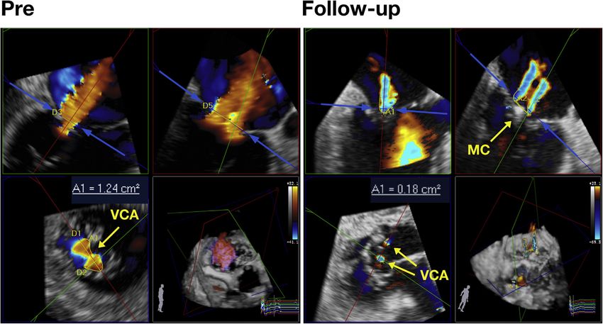
Journal of the American Society of Echocardiography                                                                            Buck et al 3
Volume - Number -

                                                                       MV Quantification
  HIGHLIGHTS                                                             MV geometry was assessed using MVQ software (Philips
                                                                       Healthcare), which provides quantitative 3D parameters10,11 that
   Recurrence of FMR is determined by LV dilation before Mitra-       are not obtainable by two-dimensional echocardiography (Figure 2).
    Clip implantation.                                                 Geometric parameters were grouped into the following aspects of
   In patients with FMR, a point of no return of MV tethering ex-     the MV apparatus: (1) Annular size was measured by
    ists.                                                              annulus(ALPM) as anterolateral-to-posteromedial annular diameter,
   Remodeling after MitraClip placement is markedly different         annulus(AP) as anterior-to-posterior annular diameter, annulus(area)
    between FMR and OMR.                                               as the minimum area spanning the saddle-shaped mitral annulus, an-
                                                                       nulus(circ) as nonplanar annular circumference, annulus(height) as
   Recurrence of OMR after MitraClip implantation could not be
                                                                       the height of the saddle-shaped annulus, and annulus(ALPM/AP)
    predicted.
                                                                       as the ratio of annulus(ALPM) to annulus(AP) (the mitral annular
   MV remodeling after MitraClip placement can be comprehen-          sphericity index). (2) Leaflet size was measured by ML(area) as
    sively analyzed by 3D echo.                                        exposed total mitral leaflet (ML) area, AML(area) as total AML
                                                                       area including coapting leaflet area, PML(area) as total PML area,
                                                                       PML/AML(area) as the ratio of PML(area) to AML(area), PML/
mode and narrowed color Doppler full volume with four to six sub-
                                                                       ML(area) as the ratio of PML(area) to ML(area), and AML/
volumes to obtain a color Doppler frame rate of $14 volumes/sec.
                                                                       ML(area) as the ratio of AML(area) to ML(area). (3) Degree of tenting
Patients with atrial fibrillation were not excluded.
                                                                       was determined by tenting(vol) as ML tenting volume, tenting index
                                                                       as the ratio of total ML area (ML(area)) to mitral annular area (annu-
                                                                       lus(area))—this index being similarly used in earlier studies27—and
MR Quantification                                                      angle(APML) as the anterior to posterior ML angle. (4) Degree of pro-
  MR severity, as quantified by VCA measurement using color            lapse was measured by prolapse(vol) as the volume of ML prolapse.
Doppler 3D echocardiography, is equally applicable to single, multi-
ple, and asymmetric VCAs.14,22,25 Baseline VCW was measured in
four- and two-chamber view, and their biplane mean was used.22,26      Two-Dimensional Echocardiography
All VCA measurements using 3D software (QLAB version 9.2;                Left-heart chamber size and function were assessed using transtho-
Philips Healthcare, Best, The Netherlands; Figure 1, Supplemental      racic two-dimensional echocardiography: LV end-diastolic volume by
Videos 1–6 available at www.onlinejase.com, Supplemental               the biplane summation-of-disks method indexed to body surface area
Figures 1–3) were performed by a single experienced clinician          (LVEDVi), LV ejection fraction (LVEF), left atrial volume by the
(T.B.). VCAs of multiple jets were summed as previously described.14   biplane summation-of-disks method indexed to body surface area
A VCA cutoff of $0.6 cm2, which correlates well with biplane VCW       (LAVi), and LV dP/dt estimated using the time interval between 1
$ 0.8 mm, was defined as severe MR.22                                  and 3 m/sec on the MR velocity continuous-wave Doppler spectrum.

Figure 1 Example of planimetry of VCA. Planimetry of asymmetric VCA (1.24 cm2) before MitraClip (MC) and at follow-up in the same
patient with two VCAs (total of 0.18 cm2) separated by the MitraClip (MC).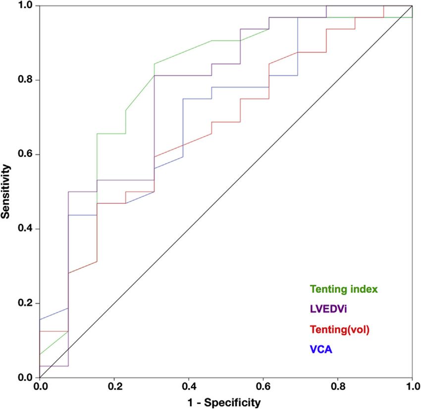
4 Buck et al                                                                              Journal of the American Society of Echocardiography
                                                                                                                                     - 2021

Figure 2 Three-dimensional parameters of MV geometry. Illustration of 3D parameters of MV geometry derived from MVQ analysis.
Parameters are indicated by green lines and grids (see green arrows). A, Anterior; AL, anterolateral; Ao, aorta; P, posterior; PM, post-
eromedial.
Journal of the American Society of Echocardiography Buck et al 5
Volume - Number -
Table 1 Patient baseline characteristics
All FMR OMR
(n = 61) (n = 45) (n = 16) P
Age, y 73.6 6 10.7 71.7 6 11.1 79.0 6 6.7 .016
Gender, male/female 43/18 34/11 9/7 .15
VCA, cm2 0.77 6 0.40 0.80 6 0.41 0.71 6 0.39 .48
LVEDVi, mL/m2 73.1 6 29.7 77.2 6 30.9 60.7 6 22.2 .06
LVEF, % 39.6 6 14.8 36.4 6 13.9 49.1 6 13.6 .003
LAVi, mL/m2 62.4 6 20.8 64.1 6 20.4 57.5 6 22.0 .29
Number of clips 1.11 1.07 1.20 .17
Intraprocedural MR grading (0–3) 2.8 6 0.3 2.7 6 0.4 2.9 6 0.2 .013
before MitraClip repair
Intraprocedural MR grading (0–3) 1.5 6 0.7 1.5 6 0.7 1.5 6 0.6 .96
after MitraClip repair
Data are expressed as mean 6 SD or as numbers.
LAVi, Left atrial volume; LVEF, LV ejection fraction.
Statistical Analysis
Measurements are presented as mean 6 SD, and t tests were used
to assess differences between groups. Paired t tests were applied to
comparisons within study groups between baseline and follow-up.
Unpaired t tests were applied to comparisons between the
MR < 0.6 and MR $ 0.6 groups, which were of different sizes. P
values < .05 were considered to indicate statistical significance.
Intraobserver and interobserver variability were determined on the
basis of actual differences between repeated measurements for MV
geometric parameters and VCA, and relative variability was calcu-
lated by dividing the absolute difference by the mean of the measure-
ment pair and showed as a percentage. Different linear regression
models were fitted to estimate the effects and 95% CIs of the
different tenting and LV dilation parameters on study outcome as
defined by VCA at 6-month follow-up. Here, VCA at 6-month
follow-up was included as a continuous variable. Stepwise model se-
lection was used to identify the most relevant predictor. Receiver
operating characteristic and cross-table analysis were performed to
determine cutoff values for MR < 0.6 versus MR $ 0.6 and to calcu-
late area under the curve (AUC), sensitivity, specificity, positive pre-
dictive value (PPV), and negative predictive value (NPV).
Calculations were performed using SPSS version 21 (IBM, Armonk,
NY).
Figure 3 Study design. Overview of study design and charac-
teristics of the two study groups (MR < 0.6 and MR $ 0.6).
RESULTS
Intraprocedural MR grading of color Doppler jet size before and after
TMVR showed satisfactory MR reduction in all 61 patients (grade
2.75 vs 1.53, with MR reduction to grade < 2 in 38 patients and was significantly smaller compared with baseline (0.54 6 0.30 cm2,
from grade 3 to grade 2 in 23 patients) independent of MR mecha- P < .0001) with 40 patients (66%) having VCAs < 0.6 cm2 (mean,
nism (FMR, 2.69 vs 1.52; OMR, 2.93 vs 1.53; Table 1). 0.36 6 0.13 cm2) in the MR < 0.6 group compared with 21 (34%)
with VCAs $ 0.6 cm2 (mean, 0.88 6 0.21 cm2) in the MR $ 0.6
group (Figure 3). Among all patients with FMR, VCA remained signif-
MR Quantification icantly smaller at follow-up compared with baseline (P < .001;
VCA at baseline was 0.77 6 0.40 cm2 in the whole study group, with Table 2); however, 13 of 45 patients (28.9%) had poor results with
a narrow VCWof 0.63 6 0.23 cm, a broad VCWof 1.61 6 0.50 cm, a MR recurrence (i.e., MR $ 0.6), whereas among patients with
biplane VCW of 1.12 6 0.31 cm, and a ratio of broad VCW to narrow OMR, VCA was not significantly reduced at follow-up compared
VCW of 2.72 6 1.0, indicating strong asymmetry. At follow-up, VCA with baseline (P = .36; Table 2) with eight of 16 patients (50%) having6 Buck et al Journal of the American Society of Echocardiography
- 2021
Table 2 Comparison of measurements in patients with FMR and OMR at baseline versus 6-month follow-up
FMR OMR
Baseline Follow-up P Baseline Follow-up P
Mitral annulus
Annulus(ALPM), mm 38.6 6 4.8 38.5 6 4.7 .70 38.6 ± 3.1 40.2 ± 4.0 .03
Annulus(AP), mm 32.3 6 4.1 32.2 6 4.5 .82 31.3 6 3.9 32.3 6 4.1 .28
Annulus(ALPM/AP) 1.20 6 0.11 1.20 6 0.12 .89 1.25 6 0.14 1.26 6 0.11 .78
Annulus(area), mm2 1,094 6 244 1,098 6 245 .80 1,073 6 194 1,139 6 215 .10
Annulus(circ), mm 121.0 6 14.1 121.3 6 13.6 .76 120.5 6 10.5 122.7 6 11.3 .15
Annulus(height), mm 5.79 6 1.58 5.56 6 1.59 .32 5.67 6 1.55 5.64 6 1.43 .91
MLs
ML(area), mm2 1,264 6 295 1,251 6 306 .55 1,207 6 230 1,261 6 282 .14
AML(area), mm 2
845 ± 206 796 ± 216 .003 696 6 130 704 6 176 .78
PML(area), mm2 516 6 136 542 6 131 .15 573 ± 170 635 ± 196 .04
PML/AML(area) 0.62 ± 0.14 0.71 ± 0.17 .003 0.83 6 0.25 0.93 6 0.29 .07
PML/ML(area) 0.41 ± 0.06 0.44 ± 0.07 .006 0.47 6 0.08 0.50 6 0.08 .07
AML/ML(area) 0.67 ± 0.06 0.63 ± 0.06 .001 0.58 6 0.08 0.56 6 0.09 .12
Tenting
Tenting(vol), mL 3.2 6 1.9 3.1 6 1.9 .41
Tenting index 1.16 ± 0.10 1.14 ± 0.08 .002
Angle(APML) 117.5 ± 18.8 120.9 ± 16.8 .047
Prolapse(vol), mL 1.2 6 1.0 1.1 6 1.1 .38
VCA, cm2 0.80 ± 0.41 0.52 ± 0.29Journal of the American Society of Echocardiography Buck et al 7
Volume - Number -
Table 3 Comparison of measurements between MR < 0.6 and MR $ 0.6 subgroups at baseline and 6-month follow-up
MR < 0.6, BL MR $ 0.6, BL P MR < 0.6, FU MR $ 0.6, FU P
FMR
Mitral annulus
Annulus(ALPM), mm 38.7 6 5.3 38.6 6 3.5 .95 38.2 6 4.8 39.2 6 4.3 .50
Annulus(AP), mm 32.3 6 4.3 32.4 6 3.6 .90 31.6 6 4.7 33.8 6 3.5 .14
Annulus(ALPM/AP) 1.20 6 0.10 1.20 6 0.11 .85 1.22 6 0.13 1.16 6 0.10 .17
Annulus(area), mm2 1,104 6 265 1,069 6 191 .68 1,074 6 250 1,159 6 228 .29
Annulus(circ), mm 121.5 6 15.4 120.6 6 10.8 .85 119.9 6 13.7 124.7 6 13.3 .30
Annulus(height), mm 5.85 6 1.37 5.64 6 2.06 .69 5.62 6 1.66 5.42 6 1.46 .70
MLs
ML(area), mm2 1,245 6 306 1,313 6 272 .49 1,190 ± 287 1,400 ± 309 .04
AML(area), mm2 830 6 221 880 6 165 .47 759 6 216 888 6 194 .07
PML(area), mm2 521 6 135 505 6 143 .72 525 6 123 585 6 145 .16
PML/AML(area) 0.65 6 0.15 0.57 6 0.12 .13 0.72 6 0.18 0.67 6 0.14 .30
PML/ML(area) 0.42 ± 0.07 0.38 ± 0.05 .047 0.45 6 0.07 0.42 6 0.06 .18
AML/ML(area) 0.67 6 0.06 0.67 6 0.05 .66 0.63 6 0.06 0.64 6 0.05 .82
Tenting
Tenting(vol), mL 2.8 ± 1.9 4.0 ± 1.9 .04 2.6 ± 1.7 4.4 ± 2.1 .003
Tenting index 1.13 ± 0.09 1.23 ± 0.10 .004 1.11 ± 0.06 1.20 ± 0.098 Buck et al Journal of the American Society of Echocardiography
- 2021
Table 4 Comparison of differences of measurements between baseline and 6-month follow-up in MR < 0.6 versus MR $ 0.6
subgroups
MR < 0.6, DBL-FU P MR $ 0.6, DBL-FU P P, DMR < 0.6 vs DMR $ 0.6
FMR
Mitral annulus
Annulus(ALPM), mm 0.50 6 2.96 .34 0.65 6 2.83 .42 .24
Annulus(AP), mm 0.72 6 2.77 .18 1.34 6 2.79 .11 .029
Annulus(ALPM/AP) 0.02 6 0.12 .46 0.03 6 0.12 .35 .24
Annulus(area), mm2 29.9 6 103.8 .11 89.8 ± 123.6 .02 .002
Annulus(circ), mm 1.55 6 5.49 .11 4.86 6 6.48 .052 .002
Annulus(height), mm 0.23 6 1.34 .35 0.22 6 1.93 .68 1.00
MLs
ML(area), mm2 55.0 ± 130.0 .02 87.0 6 176.1 .10 .005
AML(area), mm 2
71.7 ± 103.5Journal of the American Society of Echocardiography Buck et al 9
Volume - Number -
and PML(area) to be significantly larger in the MR $ 0.6 versus (2.4 6 1.6%) and 30.4 6 58.4 mm2 (5.0 6 4.0%); annulus(circ),
MR < 0.6 group, indicating that the increased MV size predominantly 1.3 6 2.4 mm (1.8 6 1.1%) and 1.1 6 3.6 mm (2.4 6 2.1%); annu-
affected the PML region. lus(height), 0.4 6 0.8 mm (12.8 6 12.1%) and 0.5 6 1.0 mm
Analysis of MV remodeling from baseline to follow-up revealed de- (18.6 6 16.3%); ML(area), 9.8 6 44.1 mm2 (2.8 6 1.6%) and
creases in the tenting parameters of tenting index and tenting(vol) in 27.7 6 61.0 mm2 (4.1 6 3.4%); AML(area), 3.2 6 34.7 mm2
MR < 0.6 patients with FMR, indicating a significant decrease in (3.4 6 2.9%) and 18.5 6 59.0 mm2 (6.4 6 4.0%); PML(area),
leaflet tethering (Table 4). In the MR $ 0.6 patients with FMR, only 2.8 6 48.8 mm2 (7.0 6 5.7%) and 18.7 6 64.3 mm2
annulus(area) increased from baseline to follow-up, whereas in pa- (9.4 6 6.1%); tenting(vol), 0.1 6 0.5 ml (14.2 6 16.1%) and
tients with OMR, nearly all parameters of MV annular dilation 0.4 6 0.5 ml (17.6 6 18.5%); angle(APML), 5.5 6 8.3
increased significantly (Table 4). Comparing differences of MV re- (5.5 6 6.1%) and 2.9 6 13.1 (9.9 6 4.5%); and prolapse(vol),
modeling between MR < 0.6 and MR $ 0.6, we found significant dif- 0.00 6 0.2 ml (29.6 6 20.9%) and 0.1 6 0.2 ml (32.9 6 19.2%).
ferences in patients with FMR with decreases in MV annular size and
tenting in the MR < 0.6 group compared with increases in
annulus(AP), annulus(circ), annulus(area), ML(area), and tenting(vol) Cutoff Values for a ‘‘Point of No Return’’ of Progressive MV
in the MR $ 0.6 group. In patients with OMR, differences in MV re- Tethering in Patients with FMR
modeling between MR < 0.6 and MR $ 0.6 were more strongly
As significantly larger LVEDVi, larger VCA, and larger tenting param-
related to the increase of MV annular diameters annulus(ALPM)
eters (i.e., tenting index and tenting(vol)) at baseline were associated
and annulus(AP) and the increases in annulus(area) and ML(area).
with MR recurrence (MR $ 0.6) in patients with FMR (Table 3), the
Analysis of AML and PML leaflet sizes from baseline to follow-up re-
receiver operating characteristic analysis was performed only in pa-
vealed a decrease in AML(area) in the MR < 0.6 FMR group, while
tients with FMR to determine a point of no return of progressive
PML(area) remained similar. In contrast, PML(area) and PML/
MV tethering. Receiver operating characteristic analysis identified
ML(area) increased from baseline to follow-up in the MR $ 0.6
the following as optimal cutoff values to determine progressive LV
group, while AML(area) remained similar. However, only an increase
dilation and MR recurrence: 88 ml/m2 for LVEDVi (sensitivity
in PML(area) was found in the MR $ 0.6 OMR group.
81.3%, specificity 69.2%, AUC 0.76, PPV 86.7%, NPV 60.0%;
Figure 4), 0.85 cm2 for VCA (sensitivity 75.0%, specificity 61.5%,
Quantification of LV and Left Atrial Size and Function AUC 0.70, PPV 82.8%, NPV 50.0%), 1.185 for tenting index (sensi-
tivity 84.4%, specificity 69.2%, AUC 0.79, PPV 87.1%, NPV 64.3%),
Analyzing the FMR and OMR patient groups revealed no significant
and 3.75 ml for tenting(vol) (sensitivity 68.8%, specificity 53.9%,
changes in LVEDVi, LAVi, and LVEF between baseline and follow-up
AUC 0.67, PPV 78.6%, NPV 41.2%), whereby the cutoff values for
(Table 2). Only in patients with FMR was LV dP/dt significantly
tenting index and LVEDVi were the most sensitive to predict MR
reduced at follow-up. Comparing MR $ 0.6 with MR < 0.6 at base-
recurrence. Of note, 12 of 13 patients with FMR with MR recurrence
line, LVEDVi was significantly larger for MR $ 0.6 versus MR < 0.6
in our study had LVEDVi $ 88 ml/m2.
among patients with FMR (99.9 6 36.1 vs 68.0 6 23.5 ml/m2,
Separate linear regression models revealed bivariate associations
P = .001), and LV dP/dt was significantly reduced (648 6 146 vs
between all parameters included and VCA at 6-month follow-up
942 6 422 mm Hg/sec, P = .04), which may potentially be associated
(LVEDVi: b = 0.005 [95% CI, 0.002 to 0.007; P < .001]; tenting(vol):
with poor TMVR outcome (Table 3). In patients with OMR, only
b = 0.06 [95% CI, 0.01 to 0.10; P = .015]; tenting index: b = 1.32
LVEF was significantly larger for MR $ 0.6 versus MR < 0.6
[95% CI, 0.54 to 2.1; P = .001]; VCA: b = 0.30 [95% CI, 0.10 to
(Table 3). At follow-up among patients with FMR, not only was
0.50; P = .004]). The effect of baseline LVEDVi on MR recurrence
LVEDVi found to be significantly larger in the MR $ 0.6 group
did not change after including all variables into one multivariate
compared with the MR < 0.6 group (Table 3), but there was also a
model (b = 0.005; 95% CI, 0.001 to 0.009; P = .02), while all other
larger increase from baseline to follow-up (99.9 6 36.1 vs
effects decreased (tenting[vol]: b = 0.07 [95% CI, 0.14 to 0.01;
112.2 6 51.2 ml/m2); furthermore, the difference between the
P = .10]; tenting index: b = 0.08 [95% CI, 0.45 to 2.15; P = .19];
LVEDVi decrease in the MR < 0.6 group and the increase in the
and VCA: b = 0.19 [95% CI, 0.01 to 0.39; P = .07]) compared
MR $ 0.6 group was significant (D6.1 6 12.1 ml/m2 vs
with the previous models fitted separately for each parameter.
D 12.3 6 26.6 ml/m2, P = .002; Table 4). Compared with this, we
Using stepwise model selection, only LVEDVi remained as the most
found a significant decrease in LVEDVi in the MR $ 0.6 patients
relevant determinant. Thus, LVEDVi seems to have the strongest inde-
with OMR.
pendent influence on outcome. R2 for LVEDVi was 0.27, meaning
that 27% of the variance of VCA was explained by LVEDVi.
Observer Variability
Intra- and interobserver variability was determined in 20 study pa-
tients, including 15 patients with FMR and five with OMR, and 17 DISCUSSION
data sets acquired before TMVR and three at follow-up. Repeated
3D measurements were performed on the same volume of the car- To the best of our knowledge, this is the first study to comprehensively
diac cycle. Intraobserver variability of VCA measurements was analyze the association of 3D echocardiographic MV geometry in
0.04 6 0.08 cm2 (9.2 6 10.9%), whereas interobserver variability concert with LV size and function on MR recurrence and MV remod-
was 0.03 6 0.06 cm2 (6.0 6 7.0%). Intra- and interobserver variability eling after TMVR and the differences between patients with FMR and
values, respectively, for MV geometric parameters were as follows: those with OMR. In all, 34% of patients (21 of 61) had MR recurrence
annulus(ALPM), 0.3 6 0.9 mm (1.5 6 2.2%) and 0.8 6 1.1 mm at follow-up, which is in line with the reported 34% of patients (26 of
(2.4 6 2.9%); annulus(AP), 0.3 6 0.9 mm (1.6 6 2.4%) and 76) with MR > 2+ at 1 year after TMVR in the EVEREST study,4 but
0.3 6 1.5 mm (3.0 6 3.6%); annulus(area), 8.8 6 33.7 mm2 the rates were higher compared with the ACCESS-EU (A Two-Phase10 Buck et al Journal of the American Society of Echocardiography
- 2021
rence after surgical MV repair,29 but this information for TMVR
outcome is rare in 1-year outcome studies.4-6 Compared with our
results for LVEDVi (MR < 0.6 = 68.0 ml/m2 vs
MR $ 0.6 = 99.9 ml/m2), Altiok et al. and Mantegazza et al.
described no preprocedural difference in LVEDV and LVEDVi in
the two outcome groups (205.2 vs 205.0 ml and 89 vs 93 ml/m2).
Remodeling of MV Geometry at 6-Month Follow-up after
TMVR
We found significant differences in MV geometry remodeling be-
tween the MR < 0.6 and MR $ 0.6 groups and between patients
with FMR and those with OMR at follow-up. In patients with FMR,
we found an improvement of MV geometry with decreases in
LVEDVi and the MV tenting parameters of tenting index and tenting(-
vol) in the MR < 0.6 group but no reduction of annular size and a
moderate increase in MV annular dilation and tenting with an in-
crease of LVEDVi in the MR $ 0.6 group. Thus, good results in the
MR < 0.6 FMR group were less related to reverse remodeling of
MV annular dilation but rather to reduced leaflet tethering that re-
sulted from reverse LV remodeling, as indicated by decreased
Figure 4 Receiver operating characteristic analysis of parame- LVEDVi, tenting index, and tenting(vol) (Table 4, Figure 5).
ters associated with MR recurrence. Receiver operating charac- Contrary to recent reports of reverse LV remodeling after successful
teristic analysis revealed the largest AUCs for tenting index TMVR,14,17,19,30 we did not find a significant improvement of MV ge-
(0.79; green line) and LVEDVi (0.76; violet line) as the strength ometry in the MR < 0.6 OMR group. Moreover, we did not observe a
for identifying a cutoff value for the development of MR recur- significant reduction in the MV annular dimensions of annulus(AP)
rence. Tenting(vol) (AUC 0.67; red line) and VCA (AUC 0.70; and annulus(ALPM) at 6-month follow-up in patients with
blue line) showed less strength. MR < 0.6 in either the FMR or OMR group, which is in contrast to
other studies showing reductions in annulus(AP) immediately after
TMVR,15-17,31 at 6-month follow-up,31 and after 1 year.32 MV remod-
Observational Study of the MitraClip System in Europe) study (21.1% eling at 6-month follow-up in the MR $ 0.6 OMR group was charac-
[69 of 327]) and the recent COAPT (Cardiovascular Outcomes terized by increases in MV annular diameters annulus(ALPM) and
Assessment of the MitraClip Percutaneous Therapy for Heart annulus(AP) and increases in annulus(area) and ML(area), most likely
Failure Patients With Functional Mitral Regurgitation) study (22.8% as a result of marked worsening of MR in this group, in which prolap-
[51 of 222]).6,28 se(vol) was much larger already at baseline and also at follow-up
In our assessment of the results to understand MR recurrence after compared with the MR < 0.6 OMR group. Thus, a large prolapse(vol)
TMVR, we found (1) that significantly increased MV tethering at base- is a potential determinant of difficult or unsuccessful TMVR, but it did
line in patients with FMR with MR recurrence, who also had signifi- not achieve statistical significance, potentially because of the limited
cantly larger values of tenting index, tenting(vol), LVEDVi, and study group size. We considered the decrease in LVEDVi observed
VCA, was associated with poor 6-month outcome, and (2) that there in patients with OMR with MR $ 0.6 to be caused by increased trans-
were significant differences in MV geometric remodeling between pa- mitral LV ejection toward the low-pressure system under the condi-
tients with MR < 0.6 and MR $ 0.6 as well as between patients with tion of lowered LV afterload as a result of worsened MR, this
FMR and those with OMR. pathomechanism being characteristic of acute MR in presence of pre-
served LV function.33
The markedly different characteristics of MV remodeling after
Parameters Associated with 6-Month Outcome after TMVR in the two mechanisms of FMR and OMR demonstrated in
TMVR our study support the understanding of FMR and OMR as two funda-
According to our hypothesis of 3D MV geometric parameters being mentally different MV diseases, where FMR is the consequence of
associated with 6-month outcome before TMVR, we found in pa- ventricular or atrial dysfunction and OMR is caused by progressive
tients with FMR that stronger MV tethering with larger tenting index primary valve disease.34
and tenting(vol) in the presence of larger LVEDVi and VCA (Table 3)
was associated with recurrent MR at 6-month follow-up, with the
multivariate analysis identifying LVEDVi to be the strongest indepen- Clinical Implications
dent determinant of MR recurrence. Although Altiok et al.14 Our findings support the concept of a ‘‘point of no return’’ in patients
described larger preprocedural annulus(area) in patients in whom with FMR with advanced LV dilation and severe MV tethering,
TMVR resulted in a VCA reduction of only #50% as another poten- beyond which TMVR cannot successfully prevent progression of
tial predictor, and Mantegazza et al.19 described a larger preprocedural MV dilation and tethering, whereas in patients with less dilated left
annulus(AP) ($4.44 cm) to be predictive of MR reduction of less ventricles this process has not yet been triggered or can be successfully
than two grades at 6-month follow-up, neither could be observed reversed. This may explain the differences in outcomes in the recent
in our study. Furthermore, moderate to severe preprocedural LV dila- MITRA-FR (Multicentre Study of Percutaneous Mitral Valve Repair
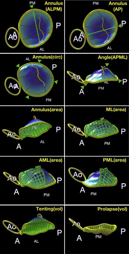
tion was reported to predict progressive LV dilation and MR recur- MitraClip Device in Patients With Severe Secondary MitralJournal of the American Society of Echocardiography Buck et al 11 Volume - Number - Figure 5 Principle of MV remodeling after MitraClip implantation in patients with FMR. Illustration of MV remodeling after MitraClip im- plantation in MR < 0.6 versus MR $ 0.6 patients with FMR. MR < 0.6 patients showed reductions of LVEDVi and ML tethering at follow-up (FU; bottom left) compared with preprocedural state. Decreases in AML(area), ML(area), and tenting index are indicated by downward arrows. Patients with MR $ 0.6 presenting with larger LVEDVi and MV tethering at baseline (top right) compared with MR < 0.6 patients showed increased dimensions of LVEDVi, PML(area), ML(area), annulus(area), and annulus(AP) at follow-up, indicated by upward arrows (bottom right). A, Anterior; Ao, aorta; MC, MitraClip; P, posterior; PM, posteromedial.
12 Buck et al                                                                                       Journal of the American Society of Echocardiography
                                                                                                                                               - 2021

Regurgitation) and COAPT studies (e.g., MITRA-FR patients who had           ACKNOWLEDGMENTS
worse clinical outcomes had more advanced LV dilation with larger
LVEDVi compared with COAPT study patients).28,35 Evidence of                We wish to acknowledge Mirjam Frank, MD, and B€      orge Schmidt,
progressive MV geometric dilation and tethering due to worsening            MD, PhD, Institute of Medical Informatics, Biometry and
of LV impairment by LV dilation and LV dP/dt lowering in patients           Epidemiology of University Essen, Germany, for their support in sta-
with FMR has also been reported to be associated with MR recur-             tistical analyses.
rence in prior studies on surgical MV repair.36,37 As our study results
demonstrate that remodeling of MV geometry after TMVR beyond
this point of no return is determined not only by leaflet clipping but      SUPPLEMENTARY DATA
also by forces external to the mitral apparatus (i.e., LV dilation and
consecutive MV dilation and tethering), further studies should inves-       Supplementary data to this article can be found online at https://doi.
tigate whether outcomes in patients with LV dilation and MV teth-           org/10.1016/j.echo.2021.02.017.
ering beyond this point of no return can be improved by combining
TMVR with annuloplasty techniques. Furthermore, our study results
imply that progressive MV tethering promoted by advanced LV dila-
tion is the cause of MR recurrence and not the consequence; thus, it is     REFERENCES
important to better understand the reasons of progressive LV failure
with MV tethering in patients with FMR beyond this point of no re-           1. Nkomo VT, Gardin JM, Skelton TN, Gottdiener JS, Scott CG, Enriquez-
turn. Ongoing LV failure potentially fostered by abnormal intraven-             Sarano M. Burden of valvular heart diseases: a population-based study.
tricular fluid dynamics in dilated left ventricles with poor ejection           Lancet 2006;368:1005-11.
                                                                             2. Zile MR. Chronic aortic and mitral regurgitation. Clin Cardiol 1991;9:
fractions has recently been reported to be aggravated after TMVR,
                                                                                239-53.
potentially promoting long-term adverse LV remodeling.38
                                                                             3. Carpentier A, Chauvaud S, Fabiani JN, Deloche A, Relland J, Lessana A,
                                                                                et al. Reconstructive surgery of mitral valve incompetence: ten-year
Limitations                                                                     appraisal. J Thorac Cardiovasc Surg 1980;79:338-48.
Limitations of our study include its single-center design and the small      4. Feldman T, Kar S, Rinaldi M, Fail P, Hermiller J, Smalling R, et al. Percuta-
                                                                                neous mitral repair with the MitraClip system: safety and midterm dura-
patient numbers (61 of 133); for example, the small number of pa-
                                                                                bility in the initial EVEREST (Endovascular Valve Edge-to-Edge Repair
tients with OMR may prevent the identification of anatomic determi-             Study) cohort. J Am Coll Cardiol 2009;54:686-94.
nants of recurrent MR in patients with OMR. As another limitation,           5. Puls M, Lubos E, Boekstegers P, von Bardeleben RS, Ouarrak T, Butter C,
the sensitivity and specificity of the LVEDVi cutoff value were not             et al. One-year outcomes and predictors of mortality after MitraClip ther-
derived from an independent validation population but from the                  apy in contemporary clinical practice: results from the German transcath-
study population from which this value was optimized; thus the accu-            eter mitral valve interventions registry. Eur Heart J 2016;37:70
racies presented represent a ‘‘best case’’ and should be tested in an in-       3-12.
dependent population. However, we reported the AUCs from the                 6. Maisano F, Franzen O, Baldus S, Sch€afer U, Hausleiter J, Butter C, et al.
study cohort data as being independent of this limitation. All proced-          Percutaneous mitral valve interventions in the real world: early and 1-
ures were performed with the first-generation MitraClip system.                 year results from the ACCESS-EU, a prospective, multicenter, non-
However, the new MitraClip NTR device has the same size as the                  randomized post-approval study of the MitraClip therapy in Europe. J
                                                                                Am Coll Cardiol 2013;62:1052-61.
first-generation MitraClip system, and therefore effects on MV geom-
                                                                             7. Kongsaerepong V, Shiota M, Gillinov AM, Song JM, Fukuda S,
etry should be similar. With the recently introduced larger MitraClip           McCarthy PM, et al. Echocardiographic predictors of successful versus un-
XTR device, improved intraprocedural coaptation in severe FMR                   successful mitral valve repair in ischemic mitral regurgitation. Am J Cardiol
cases should be obtainable. Another aspect potentially limiting the             2006;98:504-8.
applicability of our study results to current TMVR therapy is the use        8. Lancellotti P, Moura L, Pierard LA, Agricola E, Popescu BA, Tribouilloy C,
of 1.1 clips compared with a mean of 1.5 or 1.7 clips per procedure             et al. European Association of Echocardiography recommendations for
reported by others,28,39 thus reflecting a learning curve in the field          the assessment of valvular regurgitation. Part 2: mitral and tricuspid regur-
of TMVR therapy (i.e., larger numbers of implanted clips and less               gitation (native valve disease). Eur J Echocardiogr 2010;11:307-32.
restrictive selection of patients according to the EVEREST criteria).        9. Baumgartner H, Falk V, Bax JJ, De Bonis M, Hamm C, Holm PJ, et al. 2017
                                                                                ESC/EACTS guidelines for the management of valvular heart disease. Rev
                                                                                Esp Cardiol (Engl Ed) 2018;71:110.
                                                                            10. O’Gara P, Sugeng L, Lang R, Sarano M, Hung J, Raman S, et al. The role of
CONCLUSION                                                                      imaging in chronic degenerative mitral regurgitation. JACC Cardiovasc
                                                                                Imaging 2008;1:221-37.
Comprehensive 3D echocardiographic analysis of MV geometry and              11. Salcedo EE, Quaife RA, Seres T, Carroll JD. A framework for systematic
two-dimensional echocardiographic LV size and function revealed                 characterization of the mitral valve by real-time three-dimensional transe-
MR recurrence in patients with FMR to be strongly associated with               sophageal echocardiography. J Am Soc Echocardiogr 2009;22:1087-10
                                                                                99.
advanced LV dilation and MV tethering at baseline, with LVEDVi be-
                                                                            12. Chandra S, Salgo IS, Sugeng L, Weinert L, Tsang W, Takeuchi M, et al.
ing the strongest independent determinant. In patients with OMR,                Characterization of degenerative mitral valve disease using morphologic
however, no significant determinants of MR recurrence could be                  analysis of real-time three-dimensional echocardiographic images: objec-
identified despite associations with progressive MV annular dilation,           tive insight into complexity and planning of mitral valve repair. Circ Car-
leaflet enlargement, and prolapse size. Finally, the study results pro-         diovasc Imaging 2011;4:24-32.
vide strong clinical evidence for a point of no return in patients          13. Chikwe J, Adams DH, Su KN, Anyanwu AC, Lin HM, Goldstone AB, et al.
with FMR beyond which progressive LV dilation and MV tethering                  Can three-dimensional echocardiography accurately predict complexity
cannot be effectively reversed by TMVR.                                         of mitral valve repair? Eur J Cardiothorac Surg 2012;41:518-24.
Journal of the American Society of Echocardiography Buck et al 13
Volume - Number -
14. Altiok E, Hamada S, Brehmer K, Kuhr K, Reith S, Becker M, et al. Analysis mitral regurgitation using 3D TEE. JACC Cardiovasc Imaging 2012;5:
of procedural effects of percutaneous edge-to-edge mitral valve repair by 669-76.
2D and 3D echocardiography. Circ Cardiovasc Imaging 2012;5:748-55. 26. Grayburn PA, Weissman NJ, Zamorano JL. Quantitation of mitral regurgi-
15. Schmidt FP, von Bardeleben RS, Nikolai P, Jabs A, Wunderlich N, tation. Circulation 2012;126:2005-17.
M€ unzel T, et al. Immediate effect of the MitraClip procedure on mitral 27. Ryan LP, Jackson BM, Parish LM, Sakamoto H, Plappert TJ, St J-S M, et al.
ring geometry in primary and secondary mitral regurgitation. Eur Heart J Mitral valve tenting index for assessment of subvalvular remodeling. Ann
Cardiovasc Imaging 2013;14:851-7. Thorac Surg 2007;84:1243-9.
16. Herbrand T, Eschenhagen S, Zeus T, Kehmeier E, Hellhammer K, 28. Stone GW, Lindenfeld J, Abraham WT, Kar S, Lim DS, Mishell JM, et al.
Veulemans V, et al. Acute reverse annular remodeling during MitraClip. Transcatheter mitral-valve repair in patients with heart failure. N Engl J
Eur J Med Res 2017;22:33. Med 2018;379:2307-18.
17. Schueler R, Momcilovic D, Weber M, Welz A, Werner N, Mueller C, et al. 29. Acker MA, Parides MK, Perrault LP, Moskowitz AJ, Gelijns AC, Voisine P,
Acute changes of mitral valve geometry during interventional edge-to- et al. Mitral-valve repair versus replacement for severe ischemic mitral
edge repair with the MitraClip system are associated with midterm out- regurgitation. N Engl J Med 2014;370:23-32.
comes in patients with functional valve disease: preliminary results from 30. Scandura S, Ussia GP, Capranzano P, Caggegi A, Sarkar K, Cammalleri V,
a prospective single-center study. Circ Cardiovasc Interv 2014;7:390-9. et al. Left cardiac chambers reverse remodeling after percutaneous mitral
18. Al Amri I, Debonnaire P, van der Kley F, Schalij MJ, Bax JJ, Marsan NA, valve repair with the MitraClip system. J Am Soc Echocardiogr 2012;25:
et al. Acute effect of MitraClip implantation on mitral valve geometry in 1099-105.
patients with functional mitral regurgitation: insights from three- 31. Patzelt J, Zhang Y, Magunia H, Ulrich M, Jorbenadze R, Droppa M, et al.
dimensional transoesophageal echocardiography. EuroIntervention Improved mitral valve coaptation and reduced mitral valve annular size af-
2016;11:1554-61. ter percutaneous mitral valve repair (PMVR) using the MitraClip system.
19. Mantegazza V, Pasquini A, Agati L, Fusini L, Muratori M, Gripari P, et al. Eur Heart J Cardiovasc Imaging 2017;19:785-91.
32. Schueler R, Kaplan S, Melzer C, Ozt€ € urk C, Weber M, Sinning JM, et al.
Comprehensive assessment of mitral valve geometry and cardiac remod-
eling with 3-dimensional echocardiography after percutaneous mitral Impact of interventional edge-to-edge repair on mitral valve geometry.
valve repair. Am J Cardiol 2018;122:1195-203. Int J Cardiol 2017;230:468-75.
20. Schillinger W, H€ unlich M, Baldus S, Ouarrak T, Boekstegers P, Hink U, 33. Gaasch WH, Meyer TE. Left ventricular response to mitral regurgitation:
et al. Acute outcomes after MitraClip therapy in highly aged patients: re- implications for management. Circulation 2008;118:2298-303.
sults from the German Transcatheter Mitral Valve Interventions (TRAMI) 34. Boudoulas KD, Boudoulas H. Floppy mitral valve (FMV)/mitral valve pro-
registry. EuroIntervention 2013;9:84-90. lapse (MVP) and the FMV/MVP syndrome: pathophysiologic mecha-
21. Nishimura RA, Otto CM, Bonow RO, Carabello BA, Erwin JP, Guyton RA, nisms and pathogenesis of symptoms. Cardiology 2013;126:69-80.
et al. 2014 AHA/ACC guideline for the management of patients with 35. Obadia JF, Messika-Zeitoun D, Leurent G, Iung B, Bonnet G, Piriou N,
valvular heart disease: executive summary: a report of the American Col- et al. Percutaneous repair or medical treatment for secondary mitral regur-
lege of Cardiology/American Heart Association Task Force on Practice gitation. N Engl J Med 2018;379:2297-306.
Guidelines. J Am Coll Cardiol 2014;63:2438-88. 36. Braun J, Bax JJ, Versteegh MI, Voigt PG, Holman ER, Klautz RJ, et al. Pre-
22. Kahlert P, Plicht B, Schenk IM, Janosi RA, Erbel R, Buck T. Direct assess- operative left ventricular dimensions predict reverse remodeling following
ment of size and shape of noncircular vena contracta area in functional restrictive mitral annuloplasty in ischemic mitral regurgitation. Eur J Cardi-
versus organic mitral regurgitation using real-time three-dimensional echo- othorac Surg 2005;27:847-53.
cardiography. J Am Soc Echocardiogr 2008;21:912-21. 37. Digiammarco G, Liberi R, Giancane M, Canosa C, Gallina S, Di
23. Zoghbi WA, Adams D, Bonow RO, Enriquez-Sarano M, Foster E, Francesco A, et al. Recurrence of functional mitral regurgitation in
Grayburn PA, et al. Recommendations for noninvasive evaluation of patients with dilated cardiomyopathy undergoing mitral valve
native valvular regurgitation: a report from the American Society of Echo- repair: how to predict it. Interact Cardiovasc Thorac Surg 2007;6:
cardiography developed in collaboration with the Society for Cardiovas- 340-4.
cular Magnetic Resonance. J Am Soc Echocardiogr 2017;30:303-71. 38. Filomena D, Cimino S, Maestrini V, Cantisani D, Petronilli V, Mancone M,
24. Neuss M, Schau T, Isotani A, Pilz M, Sch€ opp M, Butter C. Elevated mitral et al. Changes in intraventricular flow patterns after MitraClip implant in
valve pressure gradient after mitraclip implantation deteriorates long-term patients with functional severe mitral regurgitation. J Am Soc Echocardiogr
outcome in patients with severe mitral regurgitation and severe heart fail- 2019;32:1250-3.
ure. JACC Cardiovasc Interv 2017;10:931-9. 39. Rahhab Z, Kortlandt FA, Velu JF, Schurer RAJ, Delgado V, Tonino P, et al.
25. Hyodo E, Iwata S, Tugcu A, Arai K, Shimada K, Muro T, et al. Direct mea- Current MitraClip experience, safety and feasibility in The Netherlands.
surement of multiple vena contracta areas for assessing the severity of Neth Heart J 2017;25:394-400.You can also read