Clinical Significance of Timing of Intubation in Critically Ill Patients with COVID-19: A Multi-Center Retrospective Study - MDPI
←
→
Page content transcription
If your browser does not render page correctly, please read the page content below
Journal of
           Clinical Medicine

Article
Clinical Significance of Timing of Intubation in
Critically Ill Patients with COVID-19: A Multi-Center
Retrospective Study
Yong Hoon Lee 1 , Keum-Ju Choi 2 , Sun Ha Choi 1 , Shin Yup Lee 1 , Kyung Chan Kim 3 ,
Eun Jin Kim 3, * and Jaehee Lee 1, *
 1   Division of Pulmonary and Critical Care Medicine, Department of Internal Medicine, School of Medicine,
     Kyungpook National University, Daegu 41944, Korea; yhlee2020@knu.ac.kr (Y.H.L.);
     sunha20@knu.ac.kr (S.H.C.); shinyup@knu.ac.kr (S.Y.L.)
 2   Department of Internal Medicine, Daegu Veterans Hospital, Daegu 42835, Korea; tvbogo@naver.com
 3   Department of Internal Medicine, Daegu Catholic University School of Medicine, Daegu 42472, Korea;
     solar903@chol.com
 *   Correspondence: ejkim77@cu.ac.kr (E.J.K.); jaelee@knu.ac.kr (J.L.); Tel.: +82-53-650-4274 (E.J.K.);
     +82-53-420-5536 (J.L.)

 Received: 31 July 2020; Accepted: 31 August 2020; Published: 2 September 2020

 Abstract: The effect of intubation timing on the prognosis of critically ill patients with coronavirus 2019
 (COVID-19) is not yet well understood. We investigated whether early intubation is associated with
 the survival of COVID-19 patients with acute respiratory distress syndrome (ARDS). This multicenter,
 retrospective, observational study was done on 47 adult COVID-19 patients with ARDS who were
 admitted to the intensive care unit (ICU) in Daegu, Korea between February 17 and April 23, 2020.
 Clinical characteristics and in-hospital mortality were compared between the early intubation and
 initially non-intubated groups, and between the early and late intubation groups, respectively. Of the
 47 patients studied, 23 (48.9%) were intubated on the day of meeting ARDS criteria (early intubation),
 while 24 (51.1%) were not initially intubated. Eight patients were never intubated during the
 in-hospital course. Median follow-up duration was 46 days, and 21 patients (44.7%) died in the
 hospital. No significant difference in in-hospital mortality rate was noted between the early group and
 initially non-intubated groups (56.5% vs. 33.3%, p = 0.110). Furthermore, the risk of in-hospital death
 in the early intubation group was not significantly different compared to the initially non-intubated
 group on multivariate adjusted analysis (p = 0.385). Results were similar between early and late
 intubation in the subgroup analysis of 39 patients treated with mechanical ventilation. In conclusion,
 in this study of critically ill COVID-19 patients with ARDS, early intubation was not associated with
 improved survival. This result may help in the efficient allocation of limited medical resources, such
 as ventilators.

 Keywords: COVID-19; acute respiratory distress syndrome; intubation; respiratory failure; mortality;
 intensive care units

1. Introduction
     Coronavirus disease 2019 (COVID-19), an infectious disease caused by severe acute respiratory
syndrome coronavirus 2 (SARS-CoV-2), was declared a pandemic by the World Health Organization
(WHO, Geneva, Switzerland) in March 2020 [1]. Since then, the global spread has continued, and as of
July 2020, the cumulative number of confirmed patients has exceeded 16 million, and the death toll has
reached nearly 660,000 [2]. The clinical spectrum of COVID-19 is variable, ranging from asymptomatic
infection to acute respiratory distress syndrome (ARDS) and even death [3,4]. The prevalence of

J. Clin. Med. 2020, 9, 2847; doi:10.3390/jcm9092847                                www.mdpi.com/journal/jcm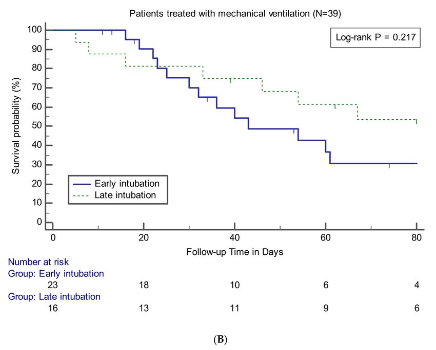
J. Clin. Med. 2020, 9, 2847                                                                          2 of 13

hypoxic respiratory failure in COVID-19 is approximately 20% [5], and recent reports of inpatients
with COVID-19 showed that approximately 25% were admitted to an intensive care unit (ICU) [4,6,7].
Although it was reported that dexamethasone reduced the mortality in COVID-19 patients receiving
invasive mechanical ventilation (MV) [8], due to lack of a proven effective antiviral agent to date, the
timely application of MV and lung protective strategies also play an important role as a life-saving
intervention in critically ill patients with COVID-19 [9]. In terms of public health, it is an important
task to secure a sufficient supply of ICU beds and ventilators for potential surges in demand, especially
in areas in the early phase of an outbreak [10].
      In ARDS, the timing of intubation may be related to clinical outcomes. A previous study reported
that ARDS patients undergoing late intubation had markedly higher mortality rates compared to those
who were intubated early in the course of the illness [11]. Similarly, current treatment guidelines
recommend early intubation in a controlled setting in case of worsening of respiratory status in
COVID-19 patients with hypoxia [9]. However, early intubation in COVID-19 is not always beneficial.
Performing unnecessary intubation in patients who otherwise would have improved without invasive
MV can interfere with life-saving treatment for other, more severe patients in medical resource-limited
settings [12]. In addition, since endotracheal intubation itself could be associated with an increased risk
of aerosolization and transmission of the virus [13], reducing the frequency of unnecessary intubation
is beneficial for healthcare worker protection. To our knowledge, no study exists on the prognostic
effect of intubation timing in a subgroup of critically ill COVID-19 patients with ARDS. Therefore, we
determined how the clinical characteristics and outcomes differ according to the timing of intubation
in COVID-19 patients admitted to the ICU with ARDS, and we investigated whether early intubation
has a survival benefit in such patients.

2. Materials and Methods

2.1. Study Design and Participants
      Data were collected from consecutive hospitalized adults (≥18 years old) with laboratory-confirmed
SARS-CoV-2 infection who subsequently were admitted to ICUs at the three tertiary referral
hospitals in Daegu, Korea between 17 February and 23 April 2020. According to WHO
guidelines [14], laboratory confirmation for SARS-CoV-2 was defined as a positive result on real-time
reverse transcription-polymerase chain reaction assay of nasal and pharyngeal swabs. During the study
period, all critically ill patients with COVID-19 who had ARDS during the clinical course were eligible
for inclusion. Patients with a “do not intubate” order were excluded. ARDS was defined according to
the Berlin definition [15]. Patients were included independent of the requirement for positive-pressure
ventilation, considering that the purpose of our study was to investigate the relationship between
intubation timing and prognosis, and that the natural course of ARDS does not start immediately
upon intubation. Therefore, all patients with a history of acute respiratory failure within 1 week of a
known clinical insult, with hypoxemia (PaO2/FiO2 ≤ 300 mmHg) and bilateral pulmonary infiltrates
on chest radiograph not fully explained by heart failure or volume overload, were considered to have
ARDS. The decision for ICU admission, oxygen therapy, respiratory support, and intubation was at
the discretion of the attending physician. This study was approved by the institutional review board
of each institution. Given the retrospective nature of our study, requirements for informed written
consent were waived.

2.2. Data Collection and Definitions
     Demographic and baseline characteristics, including age, sex, body mass index, presenting
symptoms, vital signs, comorbid conditions, and initial laboratory findings, were obtained from
the electronic medical records. Illness severity was evaluated using the Acute Physiological and
Chronic Health Evaluation (APACHE) II, and Sequential Organ Failure Assessment scores. Septic
shock was defined according to the third international consensus definitions for sepsis and septic
J. Clin. Med. 2020, 9, 2847                                                                          3 of 13

shock (Sepsis-3) [16]. Acute cardiac injury was diagnosed if serum concentrations of cardiac troponin I
(TNI) were above the upper limit of the reference range (>0.04 ng/mL). Acute kidney injury (AKI) was
identified according to the definition of the Acute Kidney Injury Network [17] as an increase in serum
creatinine level to ≥0.3 mg/dL, an increase in baseline serum creatinine level to ≥150%, or initiation of
dialysis without a history of chronic kidney disease. Data on treatment and medical events in the ICU
also were reviewed. Data on serial ventilatory parameters were not available. The number of patients
who had died, been discharged, and remained admitted in the hospital as of 2 July 2020 were recorded.

2.3. Classification by Intubation Timing and Status
      Patients were classified into two groups based on the previous study by
Kangelaris et al. [11]: (1) Early intubation: intubated/mechanically ventilated and meeting
ARDS criteria on the same day (within 24 h), and (2) initially non-intubated: not intubated on the
day of meeting ARDS criteria. The initially non-intubated group was divided further into two
subgroups: (A) never intubated: not requiring intubation throughout the entire hospital stay and (B)
late intubation: not intubated on the day of ARDS diagnosis, but intubated on a subsequent study day.

2.4. Outcomes
      The primary outcome was in-hospital mortality, and the main causes of death also were identified.
Other outcome variables included ventilator-free days (VFDs), defined as the number of days alive and
free of MV to hospital discharge or death, and the total number of days of ICU stay and MV application
in survivors to hospital discharge.

2.5. Statistical Analysis
     Data were expressed as medians (interquartile ranges, IQRs) for continuous variables and numbers
and percentages for categorical variables. For bivariate analysis, the Mann–Whitney U test or t-test was
used for continuous variables, and the χ2 or Fisher’s exact test for categorical variables. Survival curves
were developed using the Kaplan–Meier method with log-rank test. A bivariate Cox proportional
hazard model was used to adjust the effect of potential confounders on the association between
intubation status and in-hospital mortality. The multivariate analysis model incorporated variables that
varied according to intubation status with a p value < 0.05 or that were considered clinically important.
Variables from laboratory tests with missing values were excluded. All statistical procedures were
performed using SPSS software (version 24.0, SPSS Inc., Chicago, IL, USA) and MedCalc software
(version 19.2.1, Ostend, Belgium). p < 0.05 was considered statistically significant when a two-tailed
test was performed.

3. Results

3.1. Patient Characteristics
     Of the 47 patients studied (mean follow-up 46 days; IQR, 24–86 days), 23 (48.9%) were intubated
on the day of ARDS diagnosis (early intubation) and 16 (34%) were not initially intubated, but
subsequently required intubation during follow-up (late intubation). The median time interval from
ARDS diagnosis to intubation in the late intubation group was 3 days (IQR, 1–7 days). Eight patients
(17%) were never intubated during the follow-up period. All patients in the initially nonintubated
group received oxygen via high-flow nasal cannula (HFNC) either before intubation or throughout the
treatment period.
     Overall, median age of the 47 patients was 70 years (IQR, 63–77 years) and 28 (59.6%) were
male. Demographics and baseline characteristics according to intubation status are summarized
in Table 1. All patients were divided into early intubation and initially non-intubated groups for
comparative analysis. In addition, a subgroup of patients treated with MV was divided into the
early and late intubation groups. Age, sex, comorbid conditions, and presenting symptoms did not
J. Clin. Med. 2020, 9, 2847 4 of 13
show significant differences between the groups. However, among the initial vital signs, respiratory
rate was significantly higher in the early intubation than in the initially non-intubated groups
(median, 28 breaths per minute (bpm); IQR, 22–34 vs. 21 bpm; IQR, 20–26, p = 0.007).
Table 1. Demographics and baseline characteristics of critically ill COVID–19 patients with ARDS.
Early Initially
Late Intubation
Variables Intubation Nonintubated p Value a p Value b
(n = 16)
(n = 23) (n = 24)
Age 72 (64–76) 69 (60–78) 0.655 66 (59–77) 0.475
Male 14 (60.9) 14 (58.3) 0.859 10 (62.5) 0.918
Body mass
22.8 (21.0–26.7) 25.6 (22.5–27.1) 0.167 25.1 (22.7–27.3) 0.241
index, kg/m2
Comorbidities
Any
16 (69.6) 19 (79.2) 0.450 13 (81.2) 0.480
comorbidities
Hypertension 10 (43.5) 11 (45.8) 0.871 8 (50) 0.688
Diabetes 10 (43.5) 8 (33.3) 0.474 7 (43.8) 0.987
Chronic kidney
1 (4.3) 2 (8.3) >0.999 2 (12.5) 0.557
disease
Dementia 2 (8.7) 3 (12.5) >0.999 1 (6.2) >0.999
Cerebrovascular
0 (0) 2 (8.3) 0.489 1 (6.2) 0.410
disease
Malignancy 2 (8.7) 4 (16.7) 0.666 4 (25) 0.205
Cardiovascular
4 (17.4) 4 (16.7) >0.999 2 (12.5) >0.999
disease
Chronic lung
3 (13.0) 1 (4.2) 0.348 1 (6.2) 0.631
disease
Chronic liver
1 (4.3) 2 (8.3) >0.999 0 (0) >0.999
disease
Duration of
symptoms
before 7 (5–11) 5 (4–12) 0.280 5 (3–10) 0.143
admission,
days
Presenting
symptoms
Fever 18 (78.3) 16 (66.7) 0.374 9 (56.2) 0.174
Dyspnea 19 (82.6) 17 (70.8) 0.341 12 (75) 0.694
Cough 12 (52.2) 14 (58.3) 0.671 8 (50) 0.894
Sputum 10 (43.5) 10 (41.7) 0.900 6 (37.5) 0.709
Myalgia 5 (21.7) 5 (20.8) >0.999 4 (25) >0.999
Fatigue 3 (13.0) 7 (29.2) 0.286 6 (37.5) 0.123
Diarrhea 2 (8.7) 5 (20.8) 0.416 2 (12.5) >0.999
Vital signs at
the time of ICU
admission
Mean arterial
pressure, 93 (90–107) 93 (86–102) 0.539 93 (86–97) 0.388
mmHg
Heart rate,
85 (76–124) 88 (80–100) 0.915 92 (74–100) 0.808
beats/min
Respiratory
rate, 28 (22–34) 21 (20–26) 0.007 21 (20–29) 0.057
breaths/min
Body
temperature, 37.3 (36.4–37.8) 36.8 (36.5–37.4) 0.781 36.9 (36.6–37.4) 0.863
°C
Data are presented as median (interquartile range) or n (%). a Comparison between the early intubation and initially
non-intubated groups. b Comparison between the early and late intubation groups. Abbreviation: ARDS, acute
respiratory distress syndrome; ICU, intensive care unit.J. Clin. Med. 2020, 9, 2847 5 of 13
3.2. Laboratory Indices, Severity of Illness, and Clinical Course
Laboratory findings on hospital admission are shown in Table 2. Of all patients, creatine kinase-MB
(CK-MB) was significantly higher in the early intubation than in the initially non-intubated groups
(median, 1.5 U/L; IQR, 1–4.3 vs. 1.1 U/L; IQR, 0.8–1.8, p = 0.025). This difference also was observed
between the early and late intubation groups (median, 1.5 U/L; IQR, 1.0–4.3 vs. 1.0 U/L; IQR, 0.8–1.4,
p = 0.019). Laboratory tests other than CK-MB did not show a significant difference between the groups.
Table 2. Initial laboratory findings of critically ill COVID–19 patients with ARDS.
Early Initially Late
Variables Intubation Nonintubated p Value a Intubation p Value b
(n = 23) (n = 24) (n = 16)
White blood
6.89 (4.73–9.82) 7.16 (5.21–9.55) 0.907 7.16 (5.9–9.96) 0.679
cells, 103 /L
Hemoglobin,
13.0 (11.2–14.4) 13.2 (11.5–14.4) 0.935 13.1 (10.9–14.1) 0.828
g/dL
Hematocrit, % 37.6 (33.1–42) 39.3 (33.2–41.2) 0.849 38.7 (31.8–40.6) 0.706
Platelets, 103 /L 197 (146–296) 211 (156–295) 0.702 220 (156–295) 0.396
C–reactive 9.98 10.27
0.983 11 (7.39–17.43) 0.668
protein, mg/dL (5.66–15.59) (6.46–13.83)
Procalcitonin,
0.27 (0.12–0.74) 0.11 (0.1–0.21) 0.044 0.13 (0.1–0.21) 0.082
mmol/L
Lactate,
1.8 (1.3–2.5) 1.6 (1.3–2.3) 0.557 1.8 (1.3–2.4) 0.947
mmol/L
Albumin, g/dL 3.3 (3–3.5) 3.4 (3.3–3.5) 0.298 3.4 (3.3–3.7) 0.329
AST, U/L 48 (35–81) 50 (35–91) 0.717 50 (33–65) 0.875
ALT, U/L 27 (19–34) 20 (13–57) 0.302 22 (13–53) 0.346
Total bilirubin,
0.62 (0.4–0.85) 0.54 (0.32–0.83) 0.537 0.6 (0.3–0.83) 0.607
mg/dL
BUN, mg/dL 15.7 (12.5–24.7) 15.6 (8.9–22.6) 0.609 16.8 (11.8–35.3) 0.842
Creatinine,
0.9 (0.7–1.2) 0.9 (0.67–1.45) 0.741 0.95 (0.7–1.9) 0.484
mg/dL
Sodium,
134 (132–139) 137 (133–138) 0.407 137 (132–139) 0.330
mmol/L
Potassium,
3.9 (3.3–4.3) 4 (3.3–4.7) 0.327 3.9 (3.3–4.6) 0.427
mmol/L
Glucose, mg/dL 156 (117–197) 139 (111–168) 0.312 161 (122–172) 0.966
LDH, U/L 486 (410–559) 442 (370–632) 0.606 468 (344–698) 0.818
D-dimer,
2.0 (0.77–3.87) 2.5 (1.23–6.4) 0.447 2.03 (1.07–3.74) 0.642
ug/mL
Prothrombin
1.13 (1.02–1.27) 1.08 (1.02–1.26) 0.910 1.08 (1.02–1.27) 0.908
time, INR
NT–proBNP,
826 (243–1376) 570 (316–1395) 0.648 540 (372–2026) 0.917
pg/mL
Troponin I,
0.03 (0.02–0.18) 0.02 (0.01–0.02) 0.073 0.02 (0.01–0.02) 0.114
ng/mL
CK–MB, U/L 1.5 (1–4.3) 1.1 (0.8–1.8) 0.025 1 (0.8–1.4) 0.019
a
Data are presented as median (interquartile range). Comparison between the early intubation and initially
non-intubated groups. b Comparison between the early and late intubation groups. Abbreviation: ARDS,
acute respiratory distress syndrome; AST, aspartate aminotransferase; ALT, alanine aminotransferase; BUN,
blood urea nitrogen; LDH, lactate dehydrogenase; NT–proBNP, n–terminal probrain natriuretic peptide; CK–MB,
creatine kinase–MB.
The APACHE II score was significantly higher in the early intubation than in the initially
non-intubated groups (median, 15; IQR, 10–17 vs. 11; IQR, 8–14; p = 0.042; Table 3). On arterial blood
gas testing at the time of ARDS diagnosis, the early intubation group had a significantly lower pH
(median, 7.34; IQR, 7.31–7.44 vs. 7.45; IQR, 7.42–7.50; p = 0.001) and PaO2/FiO2 ratio (median, 86;
IQR, 69–123 vs. 144; IQR, 70–206; p = 0.028), and higher PaCO2 (median, 37.6 mmHg; IQR, 33.3–50.2J. Clin. Med. 2020, 9, 2847 6 of 13
vs. 32.2 mmHg; IQR, 26.8–36.5; p = 0.001) than the initially non-intubated group. In the subgroup
analysis with patients treated with MV, pH and PaCO2 in the early intubation group showed significant
differences compared to values in the late intubation group (median, 7.34; IQR, 7.31–7.44 vs. 7.43; IQR,
7.40–7.49; p = 0.013 for pH and 37.6 mmHg; IQR, 33.3–50.2 vs. 32.3 mmHg; IQR, 23.8–37.1; p = 0.002
for PaCO2).
Table 3. Severity of illness and clinical course of critically ill COVID–19 patients with ARDS.
Early Initially Late
Variables Intubation Nonintubated p Value a Intubation p Value b
(n = 23) (n = 24) (n = 16)
Severity of
illness on ICU
admission
Septic shock 4 (17.4) 2 (8.3) 0.416 1 (6.2) 0.631
Acute kidney
7 (30.4) 5 (20.8) 0.450 4 (25) >0.999
injury
Acute cardiac
8 (34.8) 3 (12.5) 0.071 2 (12.5) 0.152
injury
SOFA score 3 (2–7) 2 (2–4) 0.134 3 (2–4) 0.336
APACHE II
15 (10–17) 11 (8–14) 0.042 14 (8–15) 0.252
score
ABGA at the
time of
diagnosis of
ARDS
pH 7.34 (7.31–7.44) 7.45 (7.42–7.5) 0.001 7.43 (7.4–7.49) 0.013
PaCO2 , mmHg 37.6 (33.3–50.2) 32.2 (26.8–36.5) 0.001 32.3 (23.8–37.1) 0.002
PaO2 , mmHg 77.3 (55.3–85) 67.8 (55–82.3) 0.389 67.8 (55.7–79.7) 0.339
HCO3 , mmol/L 22.6 (21.1–25.4) 22.4 (18.8–25.5) 0.672 20.8 (17.4–26.3) 0.259
PF ratio 86 (69–123) 144 (70–206) 0.028 120 (62–188) 0.204
ICU
management
HFNC 13 (56.5) 24 (100) 0.999 5 (31.2) 0.711
Tracheostomy 9 (39.1) 7 (29.2) 0.471 7 (43.8) 0.773
ECMO 3 (13.0) 4 (16.7) >0.999 4 (25) 0.415
Medical
treatment
Antiviral
agents
Lopinavir-ritonavir 20 (87.0) 16 (66.7) 0.101 11 (68.8) 0.235
Darunavir–cobicistat 3 (13.0) 7 (29.2) 0.286 5 (31.2) 0.235
Antibiotics 23 (100) 24 (100) 16 (100)
Hydroxychloroquine 20 (87.0) 22 (91.7) 0.666 14 (87.5) >0.999
Glucocorticoid 18 (78.3) 19 (79.2) >0.999 15 (93.8) 0.370J. Clin. Med. 2020, 9, 2847 7 of 13
Table 3. Cont.
Early Initially Late
Variables Intubation Nonintubated p Value a Intubation p Value b
(n = 23) (n = 24) (n = 16)
Medical event
during ICU
care
Septic shock 20 (87.0) 15 (62.5) 0.055 14 (87.5) >0.999
Acute kidney
10 (43.5) 7 (29.2) 0.307 7 (43.8) 0.987
injury
Acute cardiac
10 (43.5) 5 (20.8) 0.096 4 (25) 0.237
injury
VAP or HAP 7 (30.4) 1 (4.2) 0.023 1 (6.2) 0.109
CRBSI 4 (17.4) 3 (12.5) 0.701 3 (18.8) >0.999
Bleeding 3 (13.0) 3 (12.5) >0.999 3 (18.8) 0.674
CPCR 1 (4.3) 3 (12.5) 0.609 2 (12.5) 0.557
Data are presented as median (interquartile range) or n (%). a Comparison between the early intubation and
initially non-intubated groups. b Comparison between the early and late intubation groups. Abbreviation:
ARDS, acute respiratory distress syndrome; ICU, intensive care unit; SOFA, Sepsis–related Organ Failure
Assessment; APACHE II, Acute Physiology and Chronic Health Evaluation; ABGA, arterial blood gas analysis;
PF ratio, arterial partial pressure of oxygen (PaO2)/fraction of inspired oxygen (FiO2) ratio; MV, mechanical
ventilation; PEEP, positive end expiratory pressure; HFNC, high–flow nasal cannula; NM blockade, neuromuscular
blockade; CRRT, continuous renal replacement therapy; ECMO, extracorporeal membrane oxygenation; VAP,
ventilator–associated pneumonia; HAP, hospital–acquired pneumonia; CRBSI, catheter–related bloodstream
infection; CPCR, cardiopulmonary–cerebral resuscitation.
Among the treatment modalities, the frequency of HFNC use was significantly lower in the early
intubation compared to the initially non-intubated (56.5%; n = 13 vs. 100%; n = 24; p < 0.001) or late
intubation groups (56.5%; n = 13 vs. 100%; n = 16; p = 0.002). Among the initial ventilator parameters,
plateau pressure of the early intubation group was significantly lower than that of the late intubation
group (median, 27 mmHg; IQR, 22–29 vs. 29 mmHg; IQR, 26–32; p = 0.014). During intensive treatment
in the ICU, the incidence of ventilator-associated (VAP) or hospital-acquired (HAP) pneumonia was
significantly higher in the early intubation than the initially non-intubated groups (30.4%; n = 7 vs.
4.2%; n = 1; p = 0.023). In patients treated with MV, VAP incidence tended to be higher in the early
than in the late intubation groups, but there was no statistical significance (30.4%; n = 7 vs. 6.2%; n = 1;
p = 0.109).
3.3. Clinical Outcomes
At the end of the study period, four patients (8.5%) remained hospitalized, 21 (44.7%) had died in
the hospital, and 22 (46.8%) had been discharged. COVID-19–related ARDS was the most common
cause of death (52.4%, n = 11), followed by VAP (19%, n = 4), catheter-related blood stream infection
(9.5%, n = 2), acute myocardial infarction (9.5%, n = 2), and AKI (4.8%, n = 1). One died of unknown
cause, who suffered from sudden cardiac arrest while receiving intensive care without intubation,
and died after cardiopulmonary resuscitation. Among the survivors (n = 26), 38.5% (n = 10), 34.6%
(n = 9), and 26.9% (n = 7) were in the early intubation, late intubation, and never intubated groups,
respectively (Figure 1). Of the 19 survivors treated with MV, 94.7% (n = 18) were weaned from the
ventilator successfully.
Data on clinical outcomes between the groups are presented in Table 4. The relevant variables
were compared between the early intubation group and the other groups for all patients or patients
who underwent intubation and MV. There was no statistically significant difference in in-hospital
mortality between the early intubation and initially non-intubated groups (56.5% vs. 33.3%, p = 0.110)
and between the early and late intubation groups (56.5% vs. 43.8%, p = 0.433). Survival curve analysis
also showed that the early intubation group had no significant difference compared to the other
groups (Figure 2). The findings of no significant difference in mortality rate according to the timing of
intubation were observed consistently at the three institutions participating in this study (data notJ. Clin. Med. 2020, 9, 2847 8 of 13
shown). No significant differences between the groups were noted in terms of causes of death. VFDs in
the early intubation group were significantly lower than those in the initially non-intubated (median,
9 days; IQR, 0–18 vs. 28 days; IQR, 9–45; p = 0.008) or late intubation (median, 9 days; IQR, 0–18 vs.
25 days; IQR, 7–45; p = 0.033) groups. Among the survivors, there were no significant differences
between the groups in terms of number of days of ICU stay or MV use.
Figure 1. Proportion of intubations implemented among 26 patients who survived after intensive care
for COVID-19 with ARDS.
Table 4. Clinical outcomes of critically ill COVID–19 patients with ARDS.
Early Initially Late
Variables Intubation Nonintubated p Value a Intubation p Value b
(n = 23) (n = 24) (n = 16)
In-hospital
13 (56.5) 8 (33.3) 0.110 7 (43.8) 0.433
mortality
Main cause of
death
COVID-19
7/13 (53.8) 4/8 (50) >0.999 4/7 (57.1) >0.999
related ARDS
VAP 3/13 (23.1) 1/8 (12.5) >0.999 1/7 (14.3) >0.999
CRBSI 2/13 (15.4) 0/8 (0) 0.505 0/7 (0) 0.521
Acute kidney
0/13 (0) 1/8 (12.5) 0.381 1/7 (14.3) 0.350
injury
Myocardial
1/13 (7.7) 1/8 (12.5) 0.999 1/7 (14.3) >0.999
infarction
Unknown c 0/13 (0) 1/8 (12.5) 0.381 0/12 (0)
Ventilator-free
9 (0–18) 28 (9–45) 0.008 25 (7–45) 0.033
days
ICU days d 13 (7–33) 14 (7–48) 0.691 47 (13–74) 0.101
Days of MV d 10 (4–24) 6 (0–25) 0.325 20 (9–57) 0.177
Data are presented as median (interquartile range) or n (%). Median duration of follow–up was 46 days (IQR,
24–86 days). Abbreviation: ARDS, acute respiratory distress syndrome; VAP, ventilator–associated pneumonia;
CRBSI, catheter–related blood stream infection; ICU, intensive care unit; MV, mechanical ventilation. a Comparison
between the early intubation and initially non-intubated groups. b Comparison between the early and late intubation
groups. c A patient in the never-intubated group died suddenly due to unknown cause. d Among survivors to
hospital discharge.J. Clin. Med. 2020, 9, 2847 9 of 13
Figure 2. Cont.J. Clin. Med. 2020, 9, 2847 10 of 13
Figure 2. Kaplan–Meier curves showing survival probability during follow-up in patients (A) or in
patients treated with MV (B).
3.4. Effects of Early Intubation on Mortality
A Cox proportional hazards model was used to analyze whether early intubation independently
affected survival outcome. Variables included in the model were respiratory rate, arterial pH, PaCO2,
PaO2/FiO2, use of HFNC, plateau pressure, VAP during ICU stay, and APACHE II score. In adjusted
analysis, early intubation showed no significant effect on in-hospital mortality compared to the initially
non-intubated group (adjusted hazard ratio (aHR) = 2.278, 95% confidence interval (CI) = 0.356–14.585;
p = 0.385). The same analysis was conducted on patients treated with MV and adjusted based on
variables that differed between the early and late intubation groups: respiratory rate, PaCO2, use of
HFNC, plateau pressure, and APACHE II score. Likewise, early intubation had no significant survival
benefit compared to late intubation (aHR = 1.964, 95% CI = 0.351–11.004; p = 0.442).
4. Discussion
Our study was conducted to examine how clinical features and outcomes differed depending
on the timing of intubation and to verify whether early intubation is associated with the survival of
critically ill COVID-19 patients with ARDS. Almost half of the patients had tracheal intubation on the
day of meeting ARDS criteria. The disease severity of this patient subset (early intubation) tended to
be higher than that of other groups. After adjusting for potential confounding variables, including
severity of illness, early intubation had no survival benefit.
According to a recent systematic review of the mortality rates of patients with COVID-19 in
ICUs [18], the reported overall mortality rate was 25.7%, which is lower than our findings. However,
the actual mortality rate may be higher, as mortality rates from previous studies varied from 8% to
66.7% and more than half of the patients were still in the ICU at publication. Rather, considering that
only 8.5% of patients were hospitalized at the time of data collection and whose definite outcome were
unknown, the in-hospital mortality rate of 44.7% in our study also appeared to be a similar finding.J. Clin. Med. 2020, 9, 2847 11 of 13
The main finding of our study is somewhat in conflict with the study of
Kangelaris et al. [11], who showed that late intubation in critically ill non–COVID-19 patients
with ARDS had a significantly higher risk of death compared to early intubation. First of all, it seems
necessary to consider that age, comorbidities, and the baseline severity of illness of the study population
are different. Our study population’s age was far higher than that of Kangelaris et al. (70 vs. 55 years).
Additionally, the average APACHE II score in the early intubation group reported by Kangelaris et al.
was 31, but in our study, the median value was 14. Such differences in study population may reflect
the tendency to be admitted preemptively to the ICU for close monitoring and use of the ventilator
when necessary, even if the general condition of the COVID-19 patients is slightly deteriorated because
the natural clinical course has not been understood clearly to date. This suggests that our patients
were intubated when in a less severe condition or earlier in the course of the disease than those of
Kangelaris et al. [11]. Nevertheless, because the definition of early intubation was the same as in that
study, our findings suggested that the ARDS due to COVID-19 may differ from other common causes
in terms of the effect of intubation timing on prognosis.
In our study, the frequency of VAP was higher in the early intubation group than in the other
groups, which may be partly related to the relatively low VFDs in the same group. Although the etiology
of VAP varies, the duration of MV is known to be an important determinant for VAP development, and
the risk tends to be higher, especially in the early stages of ventilation support [19]. In addition, VAP
was the most common cause of death except COVID-19 itself in our study, and although there was
no statistical significance, the frequency of VAP as the cause of death tended to be higher in the early
intubation group than in the other groups. This supported the possibility that early intubation itself
contributed to VAP risk in our patients with COVID-19, and consequently had some negative impact
on clinical outcomes. On the other hand, the mortality rate of the late intubation group did not show a
significant difference compared to that in the early intubation group, and all of the never-intubated
patients survived using HFNC except one case of sudden death. Our findings can help to allocate
and reserve ventilators more efficiently in clinical settings where COVID-19 confirmed cases are
rapidly increasing.
Beneficial effects of HFNC in critically ill patients with hypoxemic respiratory failure have been
suggested in several studies [20–22]. A recent report also showed the potential for HFNC to be
successful as a first-line treatment in ARDS [23]. In our study, more than half of the survivors initially
were non-intubated, and a quarter survived without endotracheal intubation, all of whom used HFNC
as the initial oxygen supply. These results suggested that HFNC also can be useful in COVID-19
patients with ARDS. In addition, one report showed that the risk of air or contact surface contamination
by HFNC was not higher compared to a conventional oxygen mask, and patients with HFNC tend to
be more comfortable [24,25]. Therefore, in terms of managing an ICU that cares for COVID-19 patients,
securing a sufficient number of HFNCs seems as important as the procurement of ventilators.
Because of the need for adherence to airborne precautions and personal protective
equipment [26], medical staff involved in the management of patients with COVID-19 find cases difficult
to deal with quickly in the event of a sudden deterioration. Moreover, emergency intubation may
increase the risk of nosocomial infection of healthcare providers, so treatment guidelines recommend
early intubation in a controlled setting if respiratory status worsens [9]. Therefore, our results should
be applied carefully in clinical practice, and a predictive model that can identify critically ill COVID-29
patients at risk for respiratory deterioration that requires intubation is needed. Among the patients
who initially were non-intubated in our study, the initial vital signs and severity scoring systems, which
were readily available, were not significantly different between the never-intubated and late intubation
groups (Supplemental Tables S1–S3). Although there were differences in some laboratory tests, it seems
difficult to attach meaning due to the small sample size. Thus, further research with a large number of
patients on this subject is necessary to properly screen for patients requiring proactive intubation
Our study has several limitations. First, this is a retrospective study that included only 47 critically
ill patients. Patients in the early intubation group had higher disease severity, which was estimated toJ. Clin. Med. 2020, 9, 2847 12 of 13
have had a significant effect on the relatively higher mortality rate of those patients, and the difference
in mortality rates seems not to be statistically significant due to the small sample size. Although we
attempted to adjust variables including the severity of illness by multivariate analysis, there may be
a statistical limitation related to sample size, which should be taken into account when interpreting
our findings. Second, no serial ventilator data were available. If there were intergroup differences in
adherence to lung protective ventilation strategies, which could affect treatment outcomes, it could
have influenced our findings as a confounder. Third, some laboratory tests had missing values and
were excluded from multivariate analysis. Fourth, data on long-term outcomes, such as pulmonary
function or quality of life after discharge, were not available. Despite these limitations, our findings
can be an important reference for COVID-19 critical care, if validated in future large-scale studies.
In summary, in this study of critically ill patients with COVID-19 and ARDS, more than half of the
survivors were not intubated on the day of meeting ARDS criteria, and some were never intubated.
There were no significant differences in in-hospital mortality between the early intubation group and
the other groups. Furthermore, after adjustment for possible confounding factors, early intubation
was not associated with improved survival. Our results may help in the efficient allocation of limited
medical resources, such as ventilators.
Supplementary Materials: The following are available online at http://www.mdpi.com/2077-0383/9/9/2847/
s1, Table S1: Demographics and baseline characteristics of initially non-intubated patients, Table S2: Initial
laboratory findings of initially non-intubated patients, Table S3: Severity of illness and clinical course of initially
non-intubated patients.
Author Contributions: Conceptualization, Y.H.L.; methodology, S.H.C. and E.J.K.; validation, S.Y.L., E.J.K. and
J.L.; formal analysis, Y.H.L.; investigation, K.-J.C., S.Y.L., K.C.K. and J.L.; data curation, Y.H.L., S.H.C. and E.J.K.;
writing—original draft preparation, Y.H.L. and K.-J.C.; writing—review and editing, K.C.K. and J.L.; supervision,
E.J.K. and J.L. All authors have read and agreed to the published version of the manuscript.
Funding: This research received no external funding.
Acknowledgments: We would like to thank all medical staff for their effort in COVID-19 patient care.
Conflicts of Interest: The authors declare no conflict of interest.
References
1. World Health Organization. General’s Opening Remarks at the Media Briefing on COVID-19.
Available online: https://www.who.int/dg/speeches/detail/who-director-general-s-opening-remarks-at-the-
media-briefing-on-covid-19---11-march-2020 (accessed on 31 July 2020).
2. World Health Organization. Coronavirus Disease 2019 (COVID-19): Situation Report-192, 30 July 2020.
Available online: https://www.who.int/docs/default-source/coronaviruse/situation-reports/20200730-covid-
19-sitrep-192.pdf?sfvrsn=5e52901f_4 (accessed on 31 July 2020).
3. Chen, N.; Zhou, M.; Dong, X.; Qu, J.; Gong, F.; Han, Y.; Qiu, Y.; Wang, J.; Liu, Y.; Wei, Y. Epidemiological and
clinical characteristics of 99 cases of 2019 novel coronavirus pneumonia in Wuhan, China: A descriptive
study. Lancet 2020, 395, 507–513. [CrossRef]
4. Wang, D.; Hu, B.; Hu, C.; Zhu, F.; Liu, X.; Zhang, J.; Wang, B.; Xiang, H.; Cheng, Z.; Xiong, Y. Clinical
characteristics of 138 hospitalized patients with 2019 novel coronavirus–infected pneumonia in Wuhan,
China. JAMA 2020, 323, 1061–1069. [CrossRef]
5. Wu, Z.; McGoogan, J.M. Characteristics of and important lessons from the coronavirus disease 2019
(COVID-19) outbreak in China: Summary of a report of 72 314 cases from the Chinese Center for Disease
Control and Prevention. JAMA 2020, 323, 1239–1242. [CrossRef] [PubMed]
6. Zhou, F.; Yu, T.; Du, R.; Fan, G.; Liu, Y.; Liu, Z.; Xiang, J.; Wang, Y.; Song, B.; Gu, X. Clinical course and risk
factors for mortality of adult inpatients with COVID-19 in Wuhan, China: A retrospective cohort study.
Lancet 2020, 395, 1054–1062. [CrossRef]
7. Wu, C.; Chen, X.; Cai, Y.; Zhou, X.; Xu, S.; Huang, H.; Zhang, L.; Zhou, X.; Du, C.; Zhang, Y. Risk factors
associated with acute respiratory distress syndrome and death in patients with coronavirus disease 2019
pneumonia in Wuhan, China. JAMA Intern. Med. 2020, 180, 934–943. [CrossRef] [PubMed]J. Clin. Med. 2020, 9, 2847 13 of 13
8. The RECOVERY Collaborative Group. Dexamethasone in hospitalized patients with Covid-19—Preliminary
report. N. Engl. J. Med. 2020. [CrossRef]
9. Alhazzani, W.; Møller, M.H.; Arabi, Y.M.; Loeb, M.; Gong, M.N.; Fan, E.; Oczkowski, S.; Levy, M.M.;
Derde, L.; Dzierba, A. Surviving Sepsis Campaign: Guidelines on the management of critically ill adults
with Coronavirus Disease 2019 (COVID-19). Intensive Care Med. 2020, 65, 854–887. [CrossRef] [PubMed]
10. Wells, C.R.; Fitzpatrick, M.C.; Sah, P.; Shoukat, A.; Pandey, A.; El-Sayed, A.M.; Singer, B.H.; Moghadas, S.M.;
Galvani, A.P. Projecting the demand for ventilators at the peak of the COVID-19 outbreak in the USA.
Lancet Infect. Dis. 2020. [CrossRef]
11. Kangelaris, K.N.; Ware, L.B.; Wang, C.Y.; Janz, D.R.; Hanjing, Z.; Matthay, M.A.; Calfee, C.S. Timing of
Intubation and Clinical outcomes in Adults with ARDS. Crit. Care Med. 2016, 44, 120–129. [CrossRef]
12. Arulkumaran, N.; Brealey, D.; Howell, D.; Singer, M. Use of non-invasive ventilation for patients with
COVID-19: A cause for concern? Lancet Respir. Med. 2020. [CrossRef]
13. Fowler, R.A.; Guest, C.B.; Lapinsky, S.E.; Sibbald, W.J.; Louie, M.; Tang, P.; Simor, A.E.; Stewart, T.E.
Transmission of severe acute respiratory syndrome during intubation and mechanical ventilation.
Am. J. Respir. Crit. Care Med. 2004, 169, 1198–1202. [CrossRef] [PubMed]
14. World Health Organization. Clinical Management of Severe Acute Respiratory Infection When Novel
Coronavirus (2019-ncov) Infection Is Suspected: Interim Guidance. 2020, p. 21. Available online: https:
//apps.who.int/iris/handle/10665/330893 (accessed on 2 July 2020).
15. Force, A.D.T.; Ranieri, V.; Rubenfeld, G.; Thompson, B.; Ferguson, N.; Caldwell, E. Acute respiratory distress
syndrome. JAMA 2012, 307, 2526–2533.
16. Singer, M.; Deutschman, C.S.; Seymour, C.W.; Shankar-Hari, M.; Annane, D.; Bauer, M.; Bellomo, R.;
Bernard, G.R.; Chiche, J.-D.; Coopersmith, C.M. The third international consensus definitions for sepsis and
septic shock (Sepsis-3). JAMA 2016, 315, 801–810. [CrossRef] [PubMed]
17. Mehta, R.L.; Kellum, J.A.; Shah, S.V.; Molitoris, B.A.; Ronco, C.; Warnock, D.G.; Levin, A. Acute Kidney
Injury Network: Report of an initiative to improve outcomes in acute kidney injury. Crit. Care 2007, 11, R31.
[CrossRef] [PubMed]
18. Quah, P.; Li, A.; Phua, J. Mortality rates of patients with COVID-19 in the intensive care unit: A systematic
review of the emerging literature. Crit. Care 2020, 24, 1–4. [CrossRef] [PubMed]
19. Weinstein, R.A.; Bonten, M.J.; Kollef, M.H.; Hall, J.B. Risk factors for ventilator-associated pneumonia:
From epidemiology to patient management. Clin. Infect. Dis. 2004, 38, 1141–1149.
20. Sztrymf, B.; Messika, J.; Bertrand, F.; Hurel, D.; Leon, R.; Dreyfuss, D.; Ricard, J.-D. Beneficial effects of
humidified high flow nasal oxygen in critical care patients: A prospective pilot study. Intensive Care Med.
2011, 37, 1780. [CrossRef]
21. Sztrymf, B.; Messika, J.; Mayot, T.; Lenglet, H.; Dreyfuss, D.; Ricard, J.-D. Impact of high-flow nasal cannula
oxygen therapy on intensive care unit patients with acute respiratory failure: A prospective observational
study. J. Crit. Care 2012, 27, 324.e9–324.e13. [CrossRef]
22. Parke, R.L.; McGuinness, S.P.; Eccleston, M.L. A preliminary randomized controlled trial to assess effectiveness
of nasal high-flow oxygen in intensive care patients. Respir. Care 2011, 56, 265–270. [CrossRef]
23. Messika, J.; Ahmed, K.B.; Gaudry, S.; Miguel-Montanes, R.; Rafat, C.; Sztrymf, B.; Dreyfuss, D.; Ricard, J.-D.
Use of high-flow nasal cannula oxygen therapy in subjects with ARDS: A 1-year observational study.
Respir. Care 2015, 60, 162–169. [CrossRef]
24. Leung, C.; Joynt, G.; Gomersall, C.; Wong, W.; Lee, A.; Ling, L.; Chan, P.; Lui, P.; Tsoi, P.; Ling, C. Comparison
of high-flow nasal cannula versus oxygen face mask for environmental bacterial contamination in critically
ill pneumonia patients: A randomized controlled crossover trial. J. Hosp. Infect. 2019, 101, 84–87. [CrossRef]
[PubMed]
25. Roca, O.; Riera, J.; Torres, F.; Masclans, J.R. High-flow oxygen therapy in acute respiratory failure.
Respir. Care 2010, 55, 408–413. [PubMed]
26. Cheung, J.C.-H.; Ho, L.T.; Cheng, J.V.; Cham, E.Y.K.; Lam, K.N. Staff safety during emergency airway
management for COVID-19 in Hong Kong. Lancet Respir. Med. 2020, 8, e19. [CrossRef]
© 2020 by the authors. Licensee MDPI, Basel, Switzerland. This article is an open access
article distributed under the terms and conditions of the Creative Commons Attribution
(CC BY) license (http://creativecommons.org/licenses/by/4.0/).You can also read