Lab recharge 2019 Life science research solutions for academia - Isolation and Cell culture
←
→
Page content transcription
If your browser does not render page correctly, please read the page content below

Lab recharge 2019
Life science research solutions for academia

Isolation and   Cell culture   Sample prep   Purification   Analysis
   cloning

                                                                  EUR
Table of contents
    Tools to support your science ....................................................................3

    Isolation and cloning....................................................................................4
    Next-generation sequencing: Challenges (and solutions!) in sample preparation...................... 4
    Featured products...................................................................................................................................................... 5

    Cell culture.....................................................................................................6
    Glutamine stability in cell culture media......................................................................................................... 6
    Featured products...................................................................................................................................................... 7

    Sample prep..................................................................................................8
    Improve lab efficiency through better filtration........................................................................................... 8
    Featured products...................................................................................................................................................... 9
    New lab start-up programme ........................................................................................................................... 10

    Purification..................................................................................................11
    ÄKTA™ start.................................................................................................................................................................11
    Sample preparation with the Protein Prep syringe filter for ÄKTA systems.................................11
    Use of affinity chromatography for antibody purification.................................................................... 12
    Featured products................................................................................................................................................... 13

    Analysis ...................................................................................................... 14
    Use of Western blotting to verify protein identity and correct molecular weight.....................14
    Featured products................................................................................................................................................... 15

2
Tools to support your science

 Handbooks                                           Apps
 Click here to request principles and                Click to download our online tools
 methodology handbooks                               to help you save time in the lab

 Web                                                 Samples and
 eu.fishersci.com                                    custom quotations
 gelifesciences.com                                  Click here to connect with your Fisher
                                                     representative and request product
                                                     information and samples

For more information please visit eu.fishersci.com
                                                                                              3
Isolation and cloning

    Next-generation sequencing: Challenges
    (and solutions!) in sample preparation
    Sample prep can have a big impact
    on next-generation sequencing
    (NGS) outcomes. Here are some
    simple things you can do to get
    your NGS off to a good start.
    By GE Healthcare.

    How to address common
    challenges in next-generation
    sequencing sample preparation
    Getting reliable data in next-generation
    sequencing (NGS) is all about the DNA
    (or RNA) you put in. How can you make
                                                  Your fragmentation method can also          modify blocked 3’ ends or fix DNA nicks.
    sure your input DNA gives you the
                                                  affect your final DNA yield. Physical       These simple repairs help make more
    quality sequencing results you need?
                                                  fragmentation can result in unexpectedly    fragments suitable for sequencing.
    DNA for sequencing might come from a          small DNA fragments which can be lost,
    variety of sources, including fresh tissue,   reducing the amount of DNA available        Challenge 3: Impurities
    formalin-fixed paraffin-embedded              for sequencing. If you have the option,     Okay, so you have enough DNA, and it’s
    (FFPE) tissue, cultured cells, and liquid     enzymatic fragmentation can provide         in pretty good condition. What else do
    biopsies. Each source comes with its          better predictability and control over      you have in that tube?
    own challenges for maximizing the three       fragmentation.
    key aspects:                                                                              Producing reliable results in sequencing
    • Quantity                                    Challenge 2: Integrity                      requires samples free of proteins, organic
    • Integrity                                   Having enough DNA won’t make for            solvents and surfactants. You might also
    • Purity                                      accurate sequencing if your DNA is          have tissue-specific contaminants to
                                                  degraded. Degradation can affect all        consider.
    Challenge 1: Yield                            kinds of samples, but long-term storage     Researchers often measure DNA
    Different workflows and kits vary             and exposure to fixatives, as you might     purity by looking at the 260:280 nm
    significantly in the amount of starting       find in FFPE samples, can exacerbate        absorbance ratio. A high-purity sample
    material required. Your workflow might        the damage.                                 should have a 260:280 ratio of 1.8 to
    require you to use a specific type of kit,                                                2.0. Nucleic acids have an absorbance
                                                  A DNA integrity number (DIN)
    and therefore starting DNA, or vice versa.
                                                  measurement can indicate the level of       maximum at 260 nm and finding a ratio
    It’s important to understand which            DNA damage. Although not a perfect          below 1.8 can indicate contamination.
    workflows and kits suit your application      predictor of usability, or parameters       As a secondary check, measure the
    and the typical amounts of starting           such as library complexity, DIN             260:230 ratio, which will detect the
    material they need. If the two don’t match    measurement is an easy method to            presence of commonly used solvents
    up, can you try another approach?             check DNA integrity.                        and surfactants, such as phenol and
    If your sample is insufficient, what can      Extracting a little more DNA can            EDTA. Values between 2.0 and 2.2
    we learn from those studying at the           compensate for low quality to some          indicate high purity.
    single-cell level? Commercially available     extent. However, there’s not much
    whole genome amplification (WGA)                                                          • Remove hemoglobin by preferential
                                                  you can do about previous storage
    kits provide the opportunity to expand                                                      lysis of red blood cells early in your
                                                  conditions, unless you can choose
    your starting material from nanograms                                                       workflow.
                                                  newer samples or those that haven’t
    to micrograms in a matter of hours.                                                       • Remove heparin by washing.
                                                  gone through such harsh processing.
    This technique provides improved                                                          • Do a phenol–chloroform extraction to
    coverage compared to PCR-based                If you can’t acquire better samples, DNA      reduce protein contamination.
    amplification and is associated with          repair might improve your outcomes.         • Use a phenol-free extraction kit to
    fewer amplification errors.                   Several commercial kits can, for example,     remove phenol contamination.

    Read full article and blogs here.
4
Isolation and cloning

Featured products
illustra™ NAP columns
These disposable columns are prepacked with Sephadex™ G-25 DNA grade and
require only gravity to run. They allow rapid and efficient purification of DNA and
oligonucleotides in less than 15 min.
• Useful for small-scale purification, desalting and buffer exchange
• Available in three sizes depending on sample volume: 0.5 mL, 1 mL or 2.5 mL

illustra GenomiPhi™ V2 DNA amplification kit
Offers highly efficient and representative whole-genome amplification with 4–7 μg
yield from nanogram amounts of DNA sample.
•   Representative isothermal amplification of the whole genome
•   No template: independent, background amplification product
•   Outperforms PCR-based whole genome amplification techniques
•   Quick and simple automation-friendly protocol; no thermal cycler required
•   Yields high-quality DNA (e.g., for high-throughput genotyping, hybridization,
    and DNA archival)

illustra single cell GenomiPhi DNA amplification kit
This kit combines the novel capabilities of Phi29 DNA polymerase with an optimized
formulation to amplify genomic DNA from 1–1000 cells without background interference.
• Amplifications can be completed in less than 3 hours with less than 15 min
  hands-on time
• Whole genome amplification of genomic DNA from as little as a single cell

                   Try illustra Ready-To-Go™ PCR† beads.
                   For robust and reproducible performance in standard PCR amplifications. Learn more here.

Ordering information
Chemistry                 Format    Description                                            Volume         Pack size   Item       Price* (€)
Sephadex G-25             Column    illustra NAP-5 columns                                 0.5 mL         50 rxn      17085302      343,76
Sephadex G-25             Column    illustra NAP-10 columns                                1 mL           50 rxn      17085402      368,94
Sephadex G-25             Column    illustra NAP-25 columns                                2.5 mL         50 rxn      17085202      469,00
Sephadex G-25             Column    illustra MicroSpin™ G-25 columns                       10 to 100 μL   50 rxn      27532501      201,00
Silica based              Column    illustra GFX™ PCR DNA and gel band purification kit    10 to 50 μL    100 rxn     28903470      191,34
Silica based              Column    illustra GFX PCR DNA and gel band purification kit     10 to 50 μL    250 rxn     28903471      449,00
Phi29 DNA polymerase      Kit       illustra single cell GenomiPhi DNA amplification kit   1–1000 cells   25 rxn      29108107      392,00
Phi29 DNA polymerase      Kit       illustra single cell GenomiPhi DNA amplification kit   1–1000 cells   100 rxn     29108039    1 410,00
Phi29 DNA polymerase      Kit       illustra GenomiPhi V2 DNA amplification kit            10 ng gDNA     25 rxn      25660030      171,00
Phi29 DNA polymerase      Kit       illustra GenomiPhi V2 DNA amplification kit            10 ng gDNA     100 rxn     25660031      470,00

For more information please visit eu.fishersci.com
                                                                                                                                              5
Cell culture

    Glutamine stability in cell culture media
    Glutamine is an essential amino acid for most cell lines. When stored as a dry powder or frozen solution,
    glutamine is relatively stable, but decomposes in aqueous solutions. The rate of decomposition depends
    strongly on the solution temperature. This study describes the stability of glutamine in cell culture media
    at different storage temperatures, and the effects of glutamine decomposition on cell growth.

    Introduction                                      taken and analyzed using a BioProfile™             Results
                                                      chemical analyzer (Nova Biomedical) for
    Since it was first isolated in 1932, glutamine                                                       Stability study
                                                      glutamine and ammonium levels.
    has been the key to many aspects of                                                                  Seven common cell lines were grown over
    mammalian cell culture. Not only does this        Cell growth was compared in fresh medium           four passages in DMEM supplemented
    amino acid stimulate cell growth and              and medium stored 30 days at 2°C–8°C,              with 10% Dialyzed FBS.
    antibody production, it is also a major           22°C, and 37°C using seven cell lines. One
    energy source in cell culture. The                bottle, at each of the three conditions,           Growth studies
    consumption of glutamine by cells in              was supplemented with 10% Dialyzed                 Initial readings of glutamine and ammonium
    culture produces ammonia and pyrroli-             FBS and analyzed to determine a base               levels were performed on day 30. The
    done carboxylic acid. As the ammonia              line for glutamine and ammonium levels.            growth studies clearly illustrate the effects
    from glutamine metabolism accumulates,                                                               of medium storage temperatures on cell
    it inhibits cellular metabolism. Ammonia          Growth studies                                     growth. Cells grew relatively well in medium
    also appears in medium during storage             Two types of growth studies were                   stored at 2°C–8°C, marginally well
    due to the spontaneous decomposition              performed using the seven cell lines in            in medium stored at 22°C, and
    of glutamine. Thus, the proper storage of         control medium and medium that had                 poorly in medium stored at 37°C. The
    cell culture medium is critical.                  been stored at the three experimental              additional growth studies show that re-
                                                      conditions for 30 days:                            supplementation of glutamine enhances
    This study examines the decomposition
                                                                                                         cell growth in medium stored 30 days at
    of glutamine over time at different               1. Multiple passage studies were                   37°C. However, growth was only about
    temperatures and the resulting effects on            conducted by averaging cell yields              50% or less of the control in some cell
    cell culture. Daily samples were analyzed            from three to four day cultures.                lines. This study also indicates that some
    for glutamine and ammonia levels. Growth
                                                      2. Growth curve studies were                       cell lines may be able to grow well in
    studies were conducted using the following
                                                         conducted by measuring cell counts              moderate ammonia levels.
    cell lines: SP20, AIF, NSO, CHO, VERO,
    BHK-21, and MRC-5. Results clearly                   and medium profiles daily.                      Ammonia growth study
    demonstrate the instability of glutamine          Additional growth studies were performed           The data from the ammonia growth study
    in media at different storage temperatures                                                           indicate that ammonia has a significant
                                                      to differentiate effects caused by
    and the effects of the resulting ammonia                                                             effect on cell growth. All four studied cell
                                                      accumulation of the ammonium ion
    on cell growth. Proper storage and                                                                   lines show significantly decreased growth
                                                      from effects caused by the depletion
    monitoring of glutamine containing media,                                                            when ammonium concentrations are
                                                      of L-glutamine. Medium stored at 37°C
    or the option of purchasing media without                                                            4 mmol/L or greater.
                                                      for 30 days was supplemented with
    glutamine, are also emphasized.
                                                      L-glutamine back to the initial 4 mmol/L.

    Material and methods                                                                                 Conclusions
                                                      Ammonia growth study
                                                                                                         Glutamine decomposition in cell culture
    Stability study                                   To further investigate the effects of              medium is properly of concern to many
    The stability study to examine the                ammonium accumulation and L-glutamine              researchers. The breakdown of this key
    degradation of glutamine in cell culture          depletion, an additional growth study was          amino acid has negative effects on cell
    medium was performed at common                    performed. Fresh bottles of DMEM/High              culture. This study clearly demonstrates
    laboratory temperatures. HyClone™                 Glucose without L-glutamine were                   the relative stability of glutamine in
    DMEM/High Glucose without L-glutamine             supplemented with 10% Dialyzed FBS and             medium stored at different temperatures
    was supplemented by adding L-glutamine            4 mmol/L L-glutamine. The medium was               and the effects of ammonia buildup on
    solution to a concentration of approximately      divided into six 250 mL samples. The               the growth of some commonly used
    4 mmol/L. Two bottles were placed at              samples were spiked with an ammonium               cell lines.
    each of the three temperature conditions:         chloride solution to create six conditions:        We recommend storing liquid medium
    2°C–8°C (laboratory cooler), 22°C                 0, 2, 4, 6, 8, and 10 mmol/L ammonium.             containing glutamine at 2°C–8°C, or
    (laboratory bench), and 37°C (cell culture        AIF, SP20, BHK21, and CHO cell lines were          purchasing medium without glutamine
    incubator). Samples were periodically             grown over four passages.                          and adding it at the time of use.

    Request a copy of data file 29184840 AA–Glutamine stability in cell culture media–to review full results.
6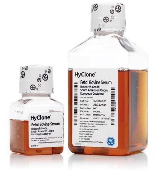
Cell culture

Featured products
HyClone South-American FBS
Research Grade Fetal Bovine Serum (FBS), South American origin is a cost-effective
solution for many cell culture research applications. Typically sourced from Brazil and
Uruguay, South American FBS complies with EU regulations, with strict manufacturing
and quality control procedures to ensure a consistent and high-quality serum.

Research classical media small volume
Cell culture media include inorganic salts, amino acids, carbohydrates, vitamins, and other
nutrients capable of sustaining cell growth. HyClone quality media provide consistent cell
culture performance in basic research and biopharmaceutical manufacturing.

Dissociation and cryopreservation
Trypsin cell detachment solution: the HyClone portfolio includes various concentrations
of trypsin protease.
• Derived from porcine pancreas
• Gamma irradiated prior to hydration and filling
• Formulated without calcium and magnesium

HyCryo-STEM maintains differentiation potential and minimizes spontaneous differentiation of stem cells.
• Designed to freeze cells sensitive to the cryopreservation process
• Helps maintain cell stemness, providing healthy and stable stocks of stem cells for downstream applications
• Provided at a 2X concentration for addition to cells suspended in their own conditioned growth medium to minimize osmotic
  shock during cryopreservation
• Chemically defined and serum-free to ensure lot-to-lot consistency
HyClone HyCryo-STEM medium is a serum-free product intended for cryopreservation and storage in biomanufacturing of stem
cell lines. The medium is designed for use with neural progenitor or stem cells, embryonic stem cells (ESCs), and induced pluripotent
stem (iPS) cells. This chemically defined formulation contains dimethyl sulfoxide (DMSO) and protein components to protect cells
during the cryopreservation process.

                     Try Whatman™ syringe filters to prepare your sample.
                     Click here for further information.

Ordering information
Product type         Format   Description                                                           Volume   Pack size    Item            Price* (€)
Fetal bovine serum   Bottle   HyClone Fetal Bovine Serum, South American Origin                     500 mL   1 × 500 mL   SV30160.03         150,00
Fetal bovine serum   Bottle   HyClone Fetal Bovine Serum, South American origin, Heat Inactivated   500 mL   1 × 500 mL   SV30160.03HI       170,00
Fetal bovine serum   Bottle   HyClone Fetal Bovine Serum, South American origin, Irradiated         500 mL   1 × 500 mL   SV30160.03 IR      170,00
Classical media      Bottle   HyClone DMEM/high glucose with L-glutamine, w/o sodium pyruvate       500 mL   1 × 500 mL   SH30022.01          18,13
Classical media      Bottle   HyClone MEM with EBSS, L-glutamine                                    500 mL   1 × 500 mL   SH30024.01           17,77
Classical media      Bottle   HyClone RPMI 1640 media, with L-glutamine                             500 mL   1 × 500 mL   SH30027.01          18,13
Cell detachment      Bottle   HyClone Trypsin                                                       100 mL   1 × 100 mL   SV30031.01          16,30
Cryopreservation     Bottle   HyClone HyCryo                                                        100 mL   1 × 100 mL   SR300001.02        146,29
Cryopreservation     Bottle   HyClone HyCryo-STEM                                                   100 mL   1 × 100 mL   SR300002.02        140,19

For more information please visit eu.fishersci.com
                                                                                                                                                       7
Sample prep

    Improve lab efficiency through better filtration
    Do you consider particle retention, loading capacity, and liquid flow rate when choosing a filter or
    device? Perhaps there is a better filter out there for your application. Or perhaps your analysis
    might be easier, quicker, or produce results that are more consistent if you switched your filter to
    a different grade.
    Here are three key characteristics to consider when identifying the right filter.

    1. Particle retention                      This can happen either through              factors that will often be specific to
    For cellulose and glass microfibre         chemical interaction with the sample        the solid/liquid being filtered. But, for
    papers, it is expressed as a “nominal      or by increasing the time to results due    comparison purposes, a typical water
    retention rating”, and quoted at 98%       to a slower flow rate than that of an       flow rate is measured and provided
    efficiency to allow for secondary          untreated filter. By knowing the weight     for each grade under gravity and
    filtration effects. For membrane           of filtrate that you want to retain on      normalized to a certain diameter.
    filters with defined pore sizes, it is     the filter, you can choose a filter that
                                                                                           Try our Whatman Filter Selector App
    an absolute retention rating.              will safely accommodate your needs          to find out if you are using the most
                                               without the downsides of a filter that      appropriate filtration solution for
    2. Loading capacity                        is more complex than is needed.             your samples.
    Filters with the highest loading
    capacities are chemically treated          3. Liquid flow rate
    and are more expensive than their          The flow rate describes the speed at
    untreated counterparts. Treatment          which a liquid flows through the filter.
    might also interfere with analysis.        In practice, this is dependent on several

    For more information please visit eu.fishersci.com
8
Sample prep

Featured products
                                                                                               NEW
Whatman Uniflo™ syringe filters
Disposable filter units designed to provide clean filtrate from small volumes up to 100 mL.
Available in a variety of membrane choices with a polypropylene overmold housing.
Whatman Uniflo syringe filters are available with:
•   13 or 25 mm diameters
•   0.22 µm or 0.45 µm pore sizes
•   Sterile or non-sterile options
•   Individual printing on the filter for easy identification
•   Bench-top space saving packaging
•   Bulk pack sizes available

Whatman GD/X™ syringe filters
These filters are specifically designed for filtration of viscous or otherwise hard-to-filter
samples with high solids content.
• High loading capacity for samples with high solids content
• Three layer glass fibre prefiltration stack for filtering larger sample volumes with less
  back pressure build-up
• Process three to seven times more sample volume than filters without prefilter

Mini-UniPrep™ syringeless HPLC filters
Whatman Mini-UniPrep syringeless filters integrate an autosampler vial, filtration
membrane, plunger, and cap/septa into one consumable product. They are built for fast
and easy HPLC/UHPLC sample preparation.
• Compatible with most major autosamplers for high throughput analysis
• All-in-one filtration device for quick and cost-effective sample processing

                     Learn more about how you can add more security to your ÄKTA chromatography
                     system runs by using our new:
                     Protein Prep syringe filter for ÄKTA systems – see page 11

Ordering information
Membrane              Format              Description                       Format/pore size    Pack size   Item         Price* (€)
PVDF                  Non sterile         Whatman Uniflo syringe filter     25 mm 0.45 µm       500/pk      9909-2504       617,94
Nylon                 Non sterile         Whatman Uniflo syringe filter     13 mm 0.45 µm       500/pk      9910-1304       660,00
PTFE                  Non sterile         Whatman Uniflo syringe filter     25 mm 0.22 µm       500/pk      9911-2502       621,00
PES                   Non sterile         Whatman Uniflo syringe filter     25 mm 0.45 µm       500/pk      9912-2504       621,00
PVDF                  Sterile             Whatman Uniflo syringe filter     25 mm 0.45 µm       45/pk       9913-2504       117,87
PES                   Sterile             Whatman Uniflo syringe filter     25 mm 0.45 µm       200/pk      9915-2504       525,00
RC                    Non sterile         Whatman GD/X 25 syringe filter    25 mm 0.2 µm        150/pk      6887-2502       459,88
RC                    Non sterile         Whatman GD/X 25 syringe filter    25 mm 0.45 µm       150/pk      6882-2504       511,89
RC                    Non sterile         Mini-UniPrep syringeless filter   0.2 µm              100/pk      UN203NPERC      208,02
RC                    Non sterile         Mini-UniPrep syringeless filter   0.45 µm             100/pk      UN203NPURC      208,02

For more information please visit eu.fishersci.com
                                                                                                                                      9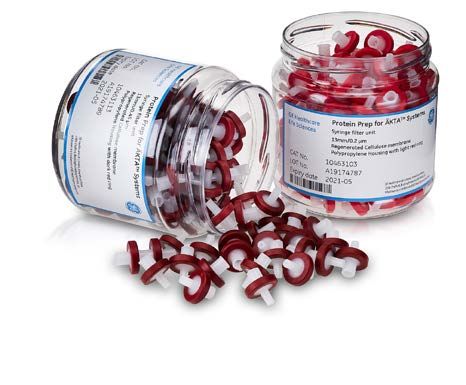
New lab start-up programme
     Visit the following link to find out more about the GE product offering
     in the Fisher new lab start-up programme:

     eu.fishersci.com/go/nlsu

               For more information please visit eu.fishersci.com

10
Purification
REQUEST
ÄKTA start DEMO
An easy-to-learn and easy-to-use system to remove the hassles
of manual protein purification
Purify tagged proteins and antibodies easily. Gain insight from real-time
monitoring. Evaluate and share your results.
User friendly—Easy-to-use touchscreen display allows you to start the run
at the touch of a button
Convenient—Easy transition from manual to automatic purification
Gain deeper insights—Gain valuable insights from real-time monitoring
and control software
Simplify your workflow—Purify tagged proteins and antibodies easily
using prepacked column
Request demo here.
NEW
Sample preparation with the
Protein Prep syringe filter for ÄKTA systems
Protein Prep syringe filters are ready-to-use with polycarbonate housing
and a regenerated cellulose membrane that is low protein binding and
broadly compatible with common solvents. Syringe filtration has been
shown to reduce debris residue in the column that could otherwise impact
performance and column life. In addition, the Protein Prep syringe filter
is lot certified for low levels of extractable particles that might otherwise
interfere with chromatograms.
Protein Prep syringe filter for ÄKTA systems
• 13 mm or 30 mm diameter
• 0.2 µm or 0.45 µm pore size
Tips for choosing the right filter
• Use 13 mm diameter filter for sample volumes < 10 mL
Protein Prep syringe filter for ÄKTA systems
• Use 0.2 μm pore size filter if the particle size of the chromatography
resin is < 30 μm
Request your sample here.
For more information please visit eu.fishersci.com
11Purification
Use of affinity chromatography for
antibody purification
How does antibody purification work? Protein L
Fv Heavy chain Light chain
ligand
Antibodies are members of a family of molecules, the immunoglobulins.
Polyclonal antibodies, monoclonal antibodies (mAb), and antibody gion
re Fa
le
fragments are usually purified by affinity chromatography. Resins r iab b
Va ion
eg
containing an immobilized ligand (e.g., protein A, protein G, or protein L) an
tr
t
ns
are used to capture antibodies and antibody fragments (right). Co Protein A ligand
Protein G ligand
Fc
IgG, which is by far the most common immunoglobulin, is commonly Kappa and
lambda
purified with protein G and protein A, both of which have a strong ligands
affinity to the Fc region of IgG. Protein L has a strong affinity to the
variable region of kappa light chains.
IgG
What do antibody purification schemes look like?
Antibody purification protocols typically are challenged by two factors. The first is specifically capturing as many antibodies as
possible in the first step as well as controlling the degradation of the sample. The second is removing the remaining impurities
and minimizing the aggregate content. Below you will find suitable protocols to choose from.
The 2-step protocol is the recommended best choice for research use. The 3-step protocol considers upscaling or process
development needs. SEC is not used as a final step to remove aggregates, fragment or other impurities due to the limitations
of sample volume. Instead, a combination of IEX steps is used.
Antibody in starting material
1-step protocol 2-step protocol 3-step protocol
Manual use or system System use System use
(ÄKTA start/ÄKTA pure) (ÄKTA start/ÄKTA pure) (ÄKTA pure/ÄKTA avant)
AC AC AC
Isolation of antibodies from initial sample Isolation of antibodies from initial sample Isolation of antibodies from initial sample
(serum, cell culture..) (serum, cell culture..) (serum, cell culture..)
B2
CIEX (Bind/Elute mode)
Further removal of host cell proteins (HCP),
leached protein A ligand and MAb aggregates,
fragments and other isoforms from the
elution in the first step
B2
SEC AIEX (Flow through mode)
B1 Neutralization of low pH elution buffer and Final removal of remaining impurities of HCP,
removing aggregates and/or fragments DNA and viruses. MAb flows through
C C C
Moderate antibody purity (> 80%) High antibody purity (95-99%) Very high antibody purity (> 99%)
High antibody yield Good purity/yield balance Moderate antibody yield
C Optional concentration B1 Buffer exchange to neutralize low pH elution buffer B2 Optional buffer exchange to prepare for IEX
Combining techniques for antibody purification with regards to yield and purity. Steps in circles are optional and may only be applied on
an as required basis. AC = affinity chromatography, CIEX = cation ion exchange chromatography, AIEX = anion exchange chromatography,
SEC = size exclusion chromatography.
12Purification
Featured products
MabSelect™ PrismA
These HiTrap™ columns are prepacked with MabSelect PrismA protein A
chromatography resin. This affinity resin in the packed column has been
improved with an optimized high-flow agarose base matrix and a genetically
engineered protein ligand, allowing efficient cleaning between monoclonal
antibody purification runs. This allows future demands in monoclonal antibody
purification to be met, including processing of many bispecific antibodies.
• Enhanced dynamic binding capacity compared with other protein A resins
• Excellent alkaline stability enables efficient cleaning and sanitization using 120
0.5 to 1.0 M NaOH 100
Remaining relative DBC (%)
Convenient HiTrap format for easy connection to a syringe, peristaltic pump 80
or chromatography systems such as an ÄKTA system for convenient process
60
optimization.
40
HiTrap MabSelect PrismA
Free wall poster! 20 HiTrap rProtein A FF
HiTrap Protein A HP
0
So how do you best combine chromatography techniques to obtain the right 0 5 10 15 20 25 30
purity and yield of your protein? Whether you want to purify a tagged, antibody Cycle
or native protein our free wall poster helps you effectively combine the main DBC of MabSelect PrismA, Protein A Sepharose™
chromatography techniques to obtain a powerful purification protocol. High Performance, and rProtein A Sepharose
Fast Flow for a polyclonal human OgG after multiple
Request your poster here! cycles with 1 M NaOH included in each cycle.
Try our new Benchkote™ sheets for ÄKTA avant, pure or start and protect top
buffer tray from buffer spillages and salt deposits.
Click here for more information.
.00
Ordering information
Resin Format Description Volume Pack size Item Price* (€)
– – ÄKTA start system – 1 29022094 6 627,00
– – Frac 30 collector – 1 29023051 1 299,00
– – UNICORN™ start 1.1 (DVD + activation code) – 1 DVD 29276964 1 121,00
MabSelect PrismA Pre-packed columns HiTrap MabSelect PrismA 5 mL/column 5 columns 17549854 4 212,00
MabSelect PrismA Pre-packed columns HiTrap MabSelect PrismA 1 mL/column 5 columns 17549852 1 072,00
Sephacryl™ S-200 HR Pre-packed columns HiPrep™ 16/60 Sephacryl S-200 HR 120 mL 1 column 17116601 639,00
Sephacryl S-300 HR Pre-packed columns HiPrep 16/60 Sephacryl S-300 HR 120 mL 1 column 17116701 639,00
Superdex™ 200 Increase Pre-packed columns Superdex 200 Increase 10/300 GL 24 mL/column 1 column 28990944 2 140,00
Sephadex Pre-packed columns HiTrap desalting column 5 mL/column 5 columns 17140801 229,00
RC membrane Non sterile Protein Prep syringe filter for ÄKTA systems 13 mm 0.45 µm 150/pk 10463113 332,00
RC membrane Non sterile Protein Prep syringe filter for ÄKTA systems 13 mm 0.2 µm 150/pk 10463103 332,00
RC membrane Non sterile Protein Prep syringe filter for ÄKTA systems 30 mm 0.45 µm 150/pk 10463033 365,00
RC membrane Non sterile Protein Prep syringe filter for ÄKTA systems 30 mm 0.2 µm 150/pk 10463043 365,00
For more information please visit eu.fishersci.com
13Analysis
Use of Western blotting to verify protein
identity and correct molecular weight
Western blotting, also known as immunoblotting, is a well-established and widely used technique for
the detection and analysis of proteins. The method is based on building an antibody:protein complex
via specific binding of antibodies to proteins immobilized on a membrane and detecting the bound
antibody with one of several detection methods. The Western blotting method is one of the most
commonly used methods in life science research. Western blotting has long been used for qualitative
protein analysis to confirm protein presence and to approximately estimate protein amount. The
development of highly sensitive detection reagents, however, together with advanced imaging
techniques has made Western blotting a potential tool for quantitative protein analysis.
Chemiluminescence Fluorescence
In most contemporary ECL™ systems a Fluorescence detection is a direct method reproducibility, fluorescence detection
luminol peroxide detection reagent is where the secondary antibody is is the preferred method for quantitative
added to the membrane and reacts with conjugated to a fluorophore, thus avoiding Western blotting applications. In addition,
the horseradish peroxidase enzyme the need for ancillary detection reagents. if selected fluorescent dyes are spectrally
(HRP) conjugated to the secondary resolvable (i.e., emit light of different
Fluorescence occurs when molecules
antibody. HRP catalyzes the oxidation wavelengths), they can be used as labels
called fluorophores absorb light. In their
of luminol in a multistep reaction and to allow multiplexing – the simultaneous
ground state, fluorophores do not emit
is accompanied by the emission of low- detection of more than one target in a
light, but when subjected to light (excitation)
intensity light at 428 nm, which can be single sample.
their energy levels are raised to a brief
measured with light-sensitive X-ray film
but unstable excited state. As fluorophores Fluorescence detection is recommended
or with a CCD imager.
return to their ground state, they release for quantitation. This is because the signal
light at a lower energy, higher wavelength stability and multiplexing capabilities result
(emission) than that of the excitation light. in reproducible data and normalization
Due to the stable signal, resulting in high of target proteins in just one step.
ECL
nt
eage Excitation Emission Excitation Emission
ECL r
Detection reagent reacts with HRP
and generates light emission
HRP
Cy™5 Cy3
Secondary antibody
conjugated with HRP
recognizes the
primary antibody
CyDye™ conjugated
Primary antibody binds specifically
secondary antibodies
to the target protein
Primary antibodies Proteins on
Proteins on membrane membrane
after transfer from gel
Protein 1 Protein 2
For more information please visit eu.fishersci.com
14Analysis
Featured products
Western blotting detection
Amersham™ ECL detection reagents
ECL based on horseradish peroxidase (HRP)-conjugated secondary antibodies has
become the most commonly used detection method for Western blotting. It is a
sensitive detection method, where the light emission is proportional to protein quantity.
Minute quantities of proteins can be detected and quantitated.
• Longer shelf life: up to 18 month shelf life on ECL Select™ and Prime products
• Stability: ECL Select and ECL Prime products are stable and stored at room
temperature
Imaging
Amersham Hyperfilm™ ECL detection film
This is a sensitive film for the detection of chemiluminescent signals in Western
blotting assays.
• Clear background for excellent contrast and band visibility
• Publication-quality images
• Learn more here: gelifesciences.com/wbfaq
Amersham Western blotting membranes
GE Healthcare Life Sciences offers a broad selection of nitrocellulose (NC) and
polyvinylidene difluoride (PVDF) Western blotting membranes, with pore size ranges
to suit your application requirements.
• Optimized for chemiluminescent and fluorescent detection
• Excellent protein binding capacity over a wide size range
• New larger pack sizes reduce your price per blot by up to 30%
Amersham ECL Rainbow™ molecular weight markers.
Accurate size determination of your protein on gels and blots. Download a brochure here.
Ordering information
Chemistry Format Description Volume/size Pack size Item Price* (€)
Chemiluminescent Reagent ECL Western blotting detection reagent For 2000 cm² membrane 1 pack RPN2209 241,74
Chemiluminescent Reagent ECL Select WB detection reagent For 1000 cm² membrane 1 pack RPN2235 312,12
Chemiluminescent Reagent ECL Prime Western blotting detection reagent For 3000 cm² membrane 1 pack RPN2236 527,34
Chemiluminescent Kit Amersham QuickStain kit 1 µg/mL to 20 mg/mL 1 pack RPN4000 122,13
Chemiluminescent Kit Full range Rainbow molecular weight marker 250 µL 1 pack RPN800E 221,34
Chemiluminescent Sheets Amersham Hyperfilm ECL 5 × 7 inches 1 pack 28906835 183,00
Chemiluminescent Roll Amersham Hybond™ PVDF 0.2 µm 260 mm × 4 m 1 roll 10600021 341,33
Chemiluminescent Roll Amersham Protran™ NC 300 mm × 4m 0.45 µm 1 roll 10600016 323,34
– Sheets GB003 blotting paper 460 × 570 mm 50/pk 10427826 514,55
For more information please visit eu.fishersci.com
15GE, the GE monogram, ÄKTA, Amersham, Benchkote, CyDye, ECL, ECL Select, GenomiPhi, GFX, HiPrep, HiTrap, Hybond, HyClone, Hyperfilm, illustra, MabSelect, MicroSpin, Mini-UniPrep, Protran, Rainbow, Ready-To-Go, Sephacryl, Sephadex, Sepharose, Superdex, UNICORN, Uniflo, Whatman, and Whatman GD/X are trademarks of General Electric Company. BioProfile is a trademark of Nova Biomedical. © 2019 General Electric Company. All offers in this issue are valid until 31 December 2019. * GE recommended list prices. † The Polymerase Chain Reaction (PCR) is covered by patents owned by Roche Molecular Systems and F Hoffmann-La Roche Ltd. A license to use the PCR process for certain research and development activities accompanies the purchase of certain reagents from licensed suppliers such as GE Healthcare and affiliates when used in conjunction with an authorized thermal cycler. Phi 29 DNA polymerase and its use for DNA synthesis is covered by US patent numbers 5,854,033, and 5,576,204. Ready to Go RT-PCR Beads: Use of this product is covered by one or more of the following US patents and corresponding patent claims outside the US: 5,789,224, 5,618,711, and 6,127,155. The purchase of this product includes a limited, non-transferable immunity from suit under the foregoing patent claims for using only this amount of product for the purchaser’s own internal research. No right under any other patent claim, no right to perform any patented method, and no right to perform commercial services of any kind, including without limitation reporting the results of purchaser’s activities for a fee or other commercial consideration, is conveyed expressly, by implication, or by estoppel. This product is for research use only. Diagnostic uses under Roche patents require a separate license from Roche. Further information on purchasing licenses may be obtained by contacting the Director of Licensing, Applied Biosystems, 850 Lincoln Centre Drive, Foster City, California 94404, USA. All goods and services are sold subject to the terms and conditions of sale of the company within GE Healthcare which supplies them. A copy of these terms and conditions is available on request. Contact your local GE Healthcare representative for the most current information. Every effort will be made to give reasonable notice of price changes but we reserve the right to change prices without further notice. All prices exclude value added or sales tax and may be subject to change without notice. These offers cannot be used in conjunction with any previously arranged purchase agreement. KA6831190319BR-EUR © 2018 Thermo Fisher Scientific Inc. All rights reserved. Trademarks used are owned as indicated at fishersci.com/trademarks.
You can also read