Statement of Concern Regarding the Accuracy and Integrity of Clinical and Preclinical Data Supporting the Ongoing Clinical Evaluation of Compound ...
←
→
Page content transcription
If your browser does not render page correctly, please read the page content below
Statement of Concern Regarding the Accuracy
  and Integrity of Clinical and Preclinical Data
 Supporting the Ongoing Clinical Evaluation of
Compound PTI-125, Also Known As Simufilam
                  August 18, 2021

                                    Jordan A. Thomas
                                    Labaton Sucharow LLP
                                    140 Broadway
                                    New York, New York 10005
                                    (212) 907-0700 (main)
                                    907-0836 (direct)
                                    jthomas@labaton.com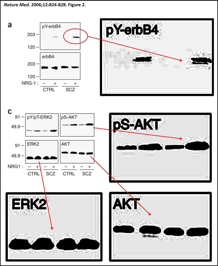
TABLE OF CONTENTS

A.   Executive Summary ............................................................................................................ 1

B.   Background ......................................................................................................................... 3

C.   Major Concerns ................................................................................................................... 4

     C.1. Concern #1: Integrity of Clinical Biomarker Data ................................................... 4

     C.2. Concern #2: Integrity of Western Blot Data ............................................................ 6

                C.2.1. Example #1: Manipulated Western Blot; Neuroscience
                       2005,135:247-261 – Figure 5a. ................................................................... 8

                C.2.2. Example #2. Falsified Western Blot; Biol Psych 2010,67:522 –
                       Figure 1a. .................................................................................................... 8

                C.2.3. Example #3: Reused/Misrepresented Western Blot; PLoS ONE
                       2008;3:e1554 – Figure 7a. ........................................................................ 10

                C.2.3. Example #4: Band Insertion Into Western Blots. Numerous
                       publications. .............................................................................................. 11

     C.3. Concern #3: Integrity of Analyses Involving Human Brain Tissue ...................... 14

                C.3.1. Implausibility of Reported Pharmacology in Postmortem Human
                       Brain Tissue. ............................................................................................. 14

                C.3.2. Evidence of Manipulation in Data from Human Tissue ........................... 16

D.   Implications and Recommendations ................................................................................. 18

E.   Appendix ........................................................................................................................... 21

     E.1.     Six Additional Areas of Concern ........................................................................... 21

              Suspicious Claim #1: Remarkably High Affinity Binding Between PTI-125
              and Filamin A ......................................................................................................... 21

              Suspicious Claim #2: Remarkably High Affinity Binding Between
              Naloxone and Filamin A ........................................................................................ 22

              Suspicious Claim #3: Isoelectric Focusing Experiments in Multiple Papers
              Indicate 100% of Filamin in Altered Conformation in Alzheimer’s Disease
              and largely Restored to Correct Conformation by PTI-125 ................................... 23

              Suspicious Claim #4: Novel Blood Diagnostic SavaDx Represents Plasma
              Filamin A Level...................................................................................................... 24

                                                                  i
Suspicious Claim #5: PTI-125/Simufilam Improves Memory in a Mouse
       Model of Alzheimer’s Disease ............................................................................... 25

       Suspicious Claim #6: PTI-125/Simufilam Blocks the Interaction Between
       β-amyloid and α7- Nicotinic Acetylcholine Receptors. ......................................... 27

E.2.   Additional Suspicious Western Blots: .................................................................... 30

       Additional examples of probable band duplication in J Neurosci
       2012;32:9773-9784. ............................................................................................... 32

                                                      ii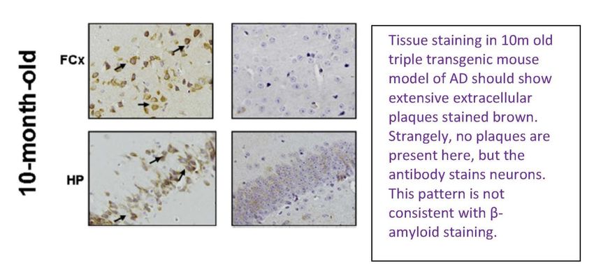
A.     Executive Summary

       For over 15 years, Cassava Sciences (previously Pain Therapeutics, Inc, PTI) has funded

the lab of Dr. Hoau-Yan Wang at City University of New York (CUNY). Together with Dr.

Lindsay Burns at Cassava, Dr. Wang has published nearly a dozen papers connecting Filamin A

protein with pain and Alzheimer’s disease (AD).

       Cassava Sciences created a drug candidate called simufilam (previously PTI-125) that

they claim binds Filamin A and has beneficial effects in biochemical and animal models of AD.

The studies from Drs. Wang and Burns discussed in this dossier were used by Cassava Sciences

to garner NIH grants and to open an investigational new drug (IND) application to study

simufilam in AD patients. They form the basic science foundation for two completed clinical

trials (phase IIa and IIb) which exposed over 70 patients to simufilam. Cassava Sciences is

currently recruiting 200 additional patients for a follow-up open-label trial.

       This report raises concerns about the quality and integrity of the laboratory-based studies

surrounding this drug candidate. To preface the analysis that follows, no other labs have

confirmed this research connecting Filamin A to pain or AD. No other labs have confirmed that

simufilam binds or modifies Filamin A or has effects in AD models.

       In this document, three primary concerns are raised:

       •   The validity of clinical biomarker data: Biomarker analysis from patients treated with

           simufilam in Cassava’s double-blind study forms a primary basis of Cassava’s claim

           that simufilam engages its target in the central nervous system, but there are concerns

           about the integrity of this data. The CSF samples in this study were analyzed by an

           outside lab, which found that simufilam was ineffective in improving the primary

           biomarker end point and showed high variability in other biomarkers. However,

           Cassava Science had these samples bioanalyzed again and the data were finalized in
an academic lab, which apparently refers to Dr. Wang. This re-analysis showed that

           simufilam rapidly and robustly improved a wide array of CSF biomarkers. Whereas

           Cassava has not fully published this reanalysis, Cassava’s 26 July 2021 poster

           presumably describing aspects of that work shows signs of data manipulation.

       •   The integrity of western blot analyses: Western blotting was extensively used by Drs.

           Wang and Burns over the past 15 years to support their foundational scientific claims

           and underscores their SavaDx clinical plasma biomarker. Detailed analysis of the

           western blots in the published journal articles from Drs. Wang and Burns shows a

           series of anomalies. The extent of these anomalies forms a 15-year pattern that

           strongly suggests systematic data manipulation and misrepresentation.

       •   The integrity of analyses involving human brain tissue: Simufilam is reported to bind

           to its target and modify a range of downstream molecules in experiments conducted on

           post-mortem human brain tissue from subjects with Alzheimer’s disease and

           neurological controls. The same human brain specimens are used across the studies

           from 2008-2017, so the results are premised on human neurons remaining viable up to

           13 hours after death, then being successfully reanimated after nearly 10 years in frozen

           archival without any advanced cryopreservative techniques. The complex, multi-step

           cellular processes the authors claim to observe in tissue that has been dead for a

           decade are contrary to a basic understanding of neurobiology. As with the western

           blot data, there are anomalies in the presentation of the data which again strongly

           suggest manipulation.

       In the appendix, six additional areas of concern are raised. These frequent errors and

anomalies occur in a pattern which is frequently favorable to the authors’ hypotheses and is of

                                                 2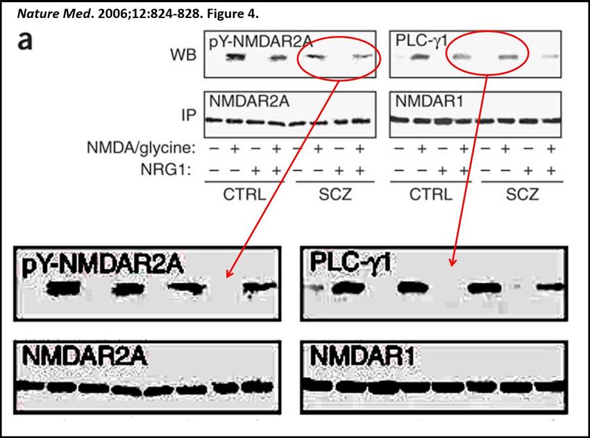
sufficient magnitude to strongly suggest scientific misconduct. This scientific work is

foundational to the link between simufilam and its supposed target Filamin A in AD.

Consequently, urgent action is advisable to limit patient exposure to this drug, until an

appropriate investigation is completed.

       Finally, we make six specific recommendations:

       •   The NIH and CUNY should audit the publications and lab of Dr. Wang to determine

           the existence and extent of data manipulation and possible fraud in all papers and

           grant applications from Drs. Wang and Burns.

       •   The FDA should audit both these publications and the IND application for simufilam’s

           use in AD.

       •   The FDA should audit all clinical biomarker studies of simufilam in AD.

       •   The FDA should oversee 3rd party reanalysis of all clinical biomarker studies of

           simufilam in AD.

       •   The FDA should pause ongoing trials with simufilam pending these investigations.

       •   The academic journals which published the studies discussed herein should review

           and retract them to correct the public record, if the concerns remain after adequate

           investigation.

B.     Background

       This letter details a long-standing pattern of seemingly intentional data manipulation and

misrepresentation in scientific papers and corporate disclosures authored primarily by Drs. Hoau-

Yan Wang, Associate Medical Professor, City University of New York, and Lindsay A Burns,

Sr. Vice President of Neuroscience at Cassava Sciences. All the information detailed herein was

obtained from public, non-proprietary sources. These apparent falsifications have helped garner

                                                 3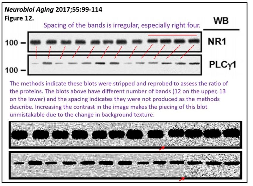
>$5,000,000 in NIH grants for preclinical/clinical studies, attract >$250,000,000 in public

fundraising by Cassava Sciences and misdirect therapeutic studies for patients suffering from

Alzheimer’s Disease (AD). In the interest of the safety of patients with Alzheimer’s disease

enrolled in Cassava Sciences’ ongoing clinical trials, as well as the NIH and other stakeholders,

the biomedical and financial communities must be made aware of these apparent falsehoods. The

laboratory of Dr. Wang and Cassava Sciences warrant an audit to comprehensively evaluate the

integrity of the scientific data.

        For >15 years, Dr. Wang has collaborated with Cassava Sciences, formerly known as Pain

Therapeutics Incorporated (PTI). Cassava Sciences is developing simufilam, a drug which was

initially designated PTI-125, as a disease modifying treatment for Alzheimer’s disease.

Simufilam is claimed to bind to bind a cytoskeleton-associated protein called Filamin A and

thereby benefit a range of Alzheimer’s disease related neuropathologies. This line of research is

unique to Dr. Wang and Cassava Sciences.

        In reviewing this work, several results were encountered that are most unexpected and are

probably unique to Drs. Wang, Burns and Cassava Sciences. Consequently, we investigated the

published journal articles and other public sources of data underlying the development of

simufilam in greater detail. This initial analysis suggests a pattern of clear errors and anomalies

that are consistent with data manipulation and misrepresentation. These findings undercut the

foundational science on which simufilam therapy is based.

C.      Major Concerns

        C.1.    Concern #1: Integrity of Clinical Biomarker Data

        NIH STTR grants (AG057329 & AG060878) funded Cassava Sciences' double-blind

placebo-controlled phase II trial of PTI-125 (50, 100 mg QD) in 64 AD patients (NCT04079803).

The primary end points reported were changes from baseline (day 1 to day 28) for a series of CSF

                                                  4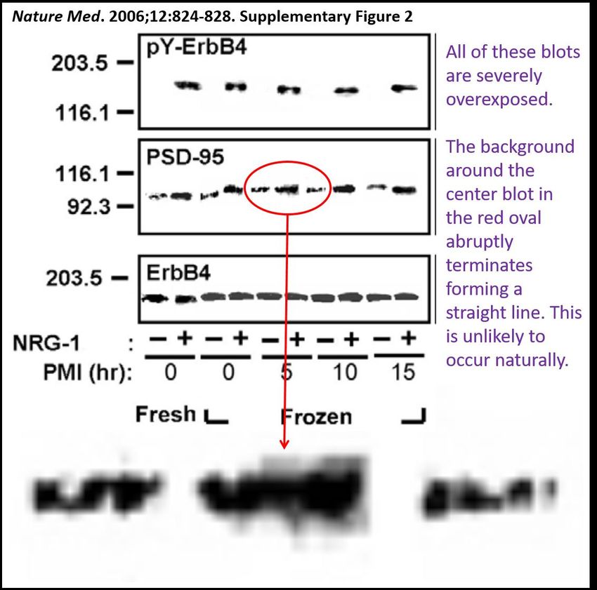
biomarkers including Abeta42, total tau, P-tau181, neurogranin, neurofilament light chain, and

YKL-40. On 15 May 2020, Cassava Sciences reported that this study missed its primary end

points. However, on 14 September 2020, Cassava Sciences reported that bioassays done by an

external group were in error, and that when patient samples were retested and finalized in what

we believe to be Dr. Wang’s lab, PTI-125/simufilam was claimed to robustly improve all

biomarkers.

       On 26 July 2021, Cassava Sciences presented a poster at the Alzheimer’s Association

International Conference entitled “SavaDx, a Novel Plasma Biomarker to Detect…” regarding

their clinical biomarkers. This poster, featuring Dr. Wang as first author, can be found on their

corporate website (https://www.cassavasciences.com/company-presentations | "SavaDx, a Novel

Plasma Biomarker to Detect Alzheimer’s Disease, Confirms Mechanism of Action of

Simufilam"). Figures 4 and 5 of this poster describe effects of 28-day treatment with simufilam

(PTI-125) on plasma P-Tau181. Figure 4 shows the percent change from baseline (CFB) and

figure 5 shows the absolute biomarker values for individuals before and after treatment.

However, Figures 4 and 5 cannot be from the same data set. In Figure 5, one patient in the 100

                                                 5
mg group (at the arrow) had a P-Tau181 level which increased from ~1.5 to 5 pg/ml during the

28-day treatment period, ~235% change from baseline. However, in figure 4 there is no data

point in the 100 mg treatment groups showing a CFB >40%. If the correct data point (+235%)

were averaged in with the other points in figure 4, any beneficial effect of 100 mg simufilam

would likely have been negated.

       As a side-note, CSF analysis was also performed on the 13 patients in the phase 2a study

and was published by Drs. Wang and Burns in early 2020 in the Journal of Prevention of

Alzheimer’s Disease 7;256-264. Remarkably, this manuscript was accepted for publication Nov.

6, 2020 seven days after submission October 31, 2020. If those dates are correct, it seems highly

unlikely to have been subjected to rigorous peer review.

       These clinical biomarker data present two significant problems. First, it seems that the

primary biomarker data set we have with simufilam in Alzheimer’s disease that was entirely

produced and finalized by an external lab found that the drug had no effect on clinical biomarkers.

Cassava replaced this with a reanalysis that was finalized by an academic lab (presumably Dr.

Wang) and showed that simufilam showed remarkable benefit. Second, plasma biomarker data

from these same patients, which were just presented by Cassava Sciences, contains evidence of

manipulation. If there’s no biomarker signal, and there is apparent misrepresentation of clinical

data the continuation of the ongoing Cassava trials may put patients at risk without the

claimed evidence of biomarker benefit. All the clinical biomarker results should be audited

and replicated by an independent third party.

       C.2.    Concern #2: Integrity of Western Blot Data

       Many experiments in the work by Drs. Wang and Burns involve western blotting. Using

this technique, proteins from tissue samples are separated on “gels” in a series of vertical lanes;

the proteins are then transferred to a paper-like membrane, and antibodies are used to detect

                                                  6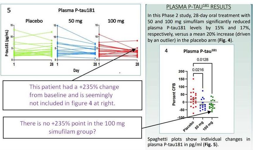
specific proteins on the membrane, producing an image of specific proteins or “bands”.

        Each band generally has a slightly different shape. As noted in an article posted on

Retraction Watch about data manipulation and focused on Western blots

(https://retractionwatch.com/2016/04/19/one-in-25-papers-contains-inappropriately-duplicated-

images-screen-finds/), “In Western blots, every band has their own characteristics, they’re like

faces.” That article further noted the significant number of cases of inappropriately duplicated or

manipulated Western Blots: “… in no way suggest that Western blotting is a flawed method.

Indeed, it suggests that Western blots are harder to fake in an undetectable way than other

experimental data.” The western blot data presented by Wang and Burns are almost always

overexposed and highly processed, which has been repeatedly seen in previously reported

examples of image manipulation. In the following sections, we present a series of examples with

strong evidence of image manipulation. In the appendix, we include additional examples which

raise red flags.

                                                 7
C.2.1. Example #1: Manipulated Western Blot; Neuroscience 2005,135:247-
261 – Figure 5a.
In figure 5a of their 2005 paper Neuroscience 135;247–261, the authors appear to have
“spliced together” gels from different experiments. Telltale signs that the Gα bands in Figure 5a
likely come from different gels are circled in red below. The cropped borders of an adjacent
protein band are present indicating the bands were taken from another blot.
C.2.2. Example #2. Falsified Western Blot; Biol Psych 2010,67:522 –
Figure 1a.
The western blot in Figure 1a (below right) of Dr. Wang’s 2010 paper in Biological
Psychiatry 67:522 contains four bands that closely resemble an image published in Figure 12a
(left) of the Wang and Burns 2005 Neuroscience 135: 247 paper mentioned in C.2.1. These eight
boxed bands come from different experimental conditions that were allegedly conducted many
years apart, using different samples. The authors appear to have vertically compressed the bands
8in the 2010 paper, but expanding them here shows they are strikingly similar to those in the 2005
paper. As the sample passes through the gel, it creates a small amount of streaking which causes a
distinctive irregular shape in the upper portion of each band; the pattern of this streaking is identical
in the two images. This degree of congruence could not have occurred by chance or error; it
suggests a complex cross-publication dimension to Cassava Science’s band duplication behavior
and, in this case, it is hard to imagine that the duplication was not intentional. It is recommended
that the original full-length images with appropriate molecular weight markers are obtained
to validate band migration from both the 2005 and 2010 papers for independent review.
Because of the seriousness of this duplication, if the original materials are not available, both of
these papers must be retracted.
As a side-note, this western blot was produced on x-ray film, not as a digital image.
9C.2.3. Example #3: Reused/Misrepresented Western Blot; PLoS ONE
2008;3:e1554 – Figure 7a.
In their 2008 paper PLoS ONE 3:e1554, Drs. Wang and Burns again present a series of
overexposed and selectively cropped gels that appear to show spliced experiments (i.e., two
separate experiments combined as if they were done simultaneously). Suggestive signs include
the sharp upper and right border for the band in the Gαo lane (lane 2 from the left in both panels;
light blue dashed boxes). Further, Figure 7a of that paper appears to show two IDENTICAL
panels (red arrows) for what are reported as different experiments. The similarity in these
images could not have occurred by chance. All original full-length gel images with appropriate
molecule weight markers to validate band migration, from this paper should be requested and
analyzed. If they are not available, this paper should be retracted.
10C.2.3. Example #4: Band Insertion Into Western Blots. Numerous
publications.
The foundational paper from Drs. Wang and Burns that links Filamin A and PTI-
125 to Alzheimer’s disease is The Journal of Neuroscience, 2012 32:9773–9784. This paper
appears to contain a collection of questionable western blots. Most of the paper comprises
western blots that are of low quality, over exposed and selectively cropped. In this paper, the
authors appear to have duplicated and transposed bands. There are dozens of questionable image
features in this paper, only a small sampling is presented here. Numerous additional examples of
this pattern of behavior in other manuscripts are included in the appendix.
In Figure 1a, the four Filamin A bands in the top set are more similar to each than can be
expected by chance and appear to be duplicates. The images at right are magnified, showing that
the pixels containing the bands are essentially identical. Additionally, the blots are not aligned
and the spacing is irregular. Because FLNA is a large protein (~290kDa), it does not migrate in
the gel very far; therefore, this degree of misalignment is suspicious. Moreover, the thin white
halos surrounding each band are concerning. There are optical reasons why a halo (or ringing
artifact) could occur, but this artifact is most common when components from multiple images are
combined using photo editing software. This halo artifact is more prominent in the questionable
blots, and extends in some cases into the frame around the blot which is hard to explain as an
optical phenomenon.
11Figure 6b: The four rightmost bands appear to be identical to each other. This degree of
similarity is unlikely to occur by chance.
Figure 9a: The five rightmost actin bands have a distinctive shape, but are nevertheless
identical to each other. That these bands all have apparently identical “dipper” shapes cannot
occur by chance. As above, the thin white border surrounding each band is prominently seen
again.
Figure 11a: The five leftmost tau bands appear to be identical to each other, AND the 3
rightmost tau bands appear to be identical to each other. These degrees of similarity are unlikely
occur by chance.
There are many other examples that strongly suggest data manipulation in this Journal of
12Neuroscience paper. Individually, each of these examples is concerning, but together they form
a pattern that strongly calls into question the integrity of this publication (and the other
publications from these authors with similar patterns of band insertion). The work in question
here serves as THE foundational research linking PTI-125 (Simufilam) to Alzheimer’s disease.
Unless the authors can produce full length unaltered gels with appropriate molecule weight
markers to validate band migration, for all experiments in this paper, it should be retracted.
Importantly, data in this paper were part of the package used to garner NIH grant
AG060878 and open an FDA investigational new drug application to study PTI-125
(Simufilam) in Alzheimer’s disease patients.
13C.3. Concern #3: Integrity of Analyses Involving Human Brain Tissue
C.3.1. Implausibility of Reported Pharmacology in Postmortem Human
Brain Tissue.
PTI-125/Simufilam is reported to bind to Filamin and alter its conformation. In so doing,
it allegedly blocks the interaction between β-amyloid and the α7-nicotinic acetylcholine receptor.
This supposedly modifies a range of downstream molecules and signaling pathways including
NMDA signaling, Toll-like receptor signaling (causing an anti-inflammatory effect) and
decreasing tau phosphorylation.
This is a complex mechanism. In one key line of experiments, the authors report that this
entire mechanism can be observed in post-mortem human brain tissue from subjects with
Alzheimer’s disease and neurological controls. This data is contained in Neurobiology of Aging
2017;55:99-114. This builds on similar experiments in The Journal of Neuroscience
2009;29:10961-10973 and The Journal of Neuroscience 2012;32:9773-9784.
In these experiments, post-mortem human brain tissue is warmed from -80°C to -20°C
and chopped into 200micron x 200micron x 3mm blocks with a McIlwain chopper (as a side
note, a McIlwain chopper doesn’t effectively cut frozen tissue). The resulting chopped tissue is
treated with β-amyloid and the experimental drug for 1 hour. They then report a massive
increase in tau phosphorylation (modification of the tau protein by enzymatic addition of a
phosphate group to the protein; up to 10 fold) from β-amyloid treatment in untreated samples;
and that tau phosphorylation was blocked by addition of PTI-125. It is unlikely that the enzyme
responsible for phosphorylation would survive the initial -80°C freezing step. Moreover, the
phosphorylation experiments are reported to have been performed at 4°C, but it is unlikely that
the enzyme responsible for phosphorylation would be active at 4°C (enzymes generally work
best at body temperature—37°C).
14In a similar experiment, NMDA-receptor signaling was evaluated after incubating minced
human brain from patients with AD and neurological controls with NMDA/glycine along with β-
amyloid and the experimental drug for 1 hour. NMDA signaling was reported blocked by β-
amyloid and in AD and rescued in both cases by the experimental drug. For similar reasons, these
reported results are unlikely.
The methodology for the post-mortem human brain experiments among the three
studies are virtually word-for-word identical. The age and post-mortem interval for the
groups of subjects are the same (down to the decimal points) in each of the three papers. It
is therefore reasonable to assume the same human brain specimens were used across the
studies from 2008-2017, so the results are premised on the enzymes in the human brain
extracts remaining active up to 13 hours post-mortem before freezing, remaining active
after nearly 10 years in frozen archival without any advanced cryopreservative techniques,
and being active at 4°C.
Importantly, the authors report that there was a marked, rapid increase in the Arc protein
observed as evidence of NMDA receptor activity with this approach. The suggestion is that
post-mortem human brain tissue, frozen for a decade, thawed and chopped, (1) has intact NMDA
receptor signaling, (2) is able to transmit that signal to the cell body through an intact dendrite,
(3) has the functioning cellular apparatus to rapidly produce the Arc protein and (4) enough
intact neurons are present to mediate a >4 fold rise in Arc levels in this tissue. In reality, neurons
in the human brain do not survive extended post-mortem intervals and long-term freezing.
The complex, multi-step cellular processes the authors claim to observe in tissue that has
been dead for a decade are contrary to a basic understanding of neurobiology. Claims of this
magnitude require extensive, detailed verification, but the authors provide no evidence of tissue
15viability. We are not aware of any other research group which has effectively used this
technique. As with the western blot data, there are anomalies in the presentation of the data from
this human tissue, which again strongly suggest manipulation.
C.3.2. Evidence of Manipulation in Data from Human Tissue
Figure 12 of Neurobiology of Aging 2017;55:99-114 uses Western blotting to support their
conclusion that PTI-125 improves NMDAR (NR1) function. Their analysis includes a
normalization step. In figure 12a (top portion), the NR1 blot that that is used for normalization
contains 12 bands whereas all the other blots in this figure contain 13 bands.
Also, the NR1 bands show different spacing than do bands in the PLCγ1 blot, which
strongly suggests that the NR1 and PLCγ1 Western blots could not have derived from the
same gel. This directly conflicts with the author’s claim in the method section of this paper that,
“Proteins were transferred to nitrocellulose membrane and the levels of PSD-95, and signaling
proteins were measured using Western blotting with specific antibodies for PSD-95, nNOS,
phospholipase C-γ1, protein kinase C, pY402PyK2, and pY416Src. Blots were stripped and
reprobed with anti-NR1 to assess loading.” The italicized sentence indicates that the gel
membrane was analyzed for PLCγ1, and the same membrane was re-analyzed for NR1. This
process does not introduce or remove band lanes.
16Another major problem with the 12-band blot is that the spacing of the bands is irregular.
This is particularly obvious on the right half (lanes 7-12). This asymmetry in band spacing is
incompatible with the regular shape of the combs used for gel loading. Therefore, the 12-band
blot was almost certainly pasted together from different sources. Further evidence that the bands
likely derive from different sources is apparent when the contrast of the image is adjusted. As
shown in the magnified panels in the figure below, in the NR1 (top row) there is a sharp contrast
between the background for the leftmost 8 bands and the background for the rightmost 4 bands,
marked with a red arrow. In the magnified panel for PLCγ1 (bottom row), there is also evidence
of splicing. Again, the red arrow denotes a sharp background contrast between the leftmost 9
bands and the rightmost 3 bands.
For these reasons, the primary data for this paper should be audited. If the primary data
17do not support the authors’ highly unlikely claims, the paper should be retracted. These
questionable experiments used donated cadaveric human tissue, which, if the experimental data
are shown to be manipulated, is a particularly egregious ethical transgression.
D. Implications and Recommendations
In summary, it appears that Drs. Wang and Burns in published PubMed indexed
manuscripts and through disclosures with Cassava Sciences have misrepresented preclinical and
clinical research results for more than 15 years. This initial examination of their published
western blots identified many dozens of examples of protein bands that appear to have been
duplicated and/or misrepresented, a Western blot that was used twice to represent different
experimental conditions, and a normalization blot that appears to have been manually
constructed. Some bands appear to have been “reused” in papers concerning different research
topics that were published five years apart.
The volume of problematic material uncovered in publicly available sources indicates a
thorough audit would likely unveil significant additional scientific misconduct and data
manipulation. It is essential that the scientific team behind Cassava Sciences’ Simufilam provide
the original blots with molecular weight markers to validate these published papers and clinical
biomarker data, which include SavaDx.
It is worth repeating, the preclinical and clinical foundations linking Filamin A to
Alzheimer’s disease derive only from the publications of Drs. Wang and Burns. As shown
above, ALL of these papers have evidence of apparent intentional scientific misrepresentation.
Cassava Sciences’ Alzheimer’s disease clinical biomarker data with PTI-125/simufilam showed
no evidence of efficacy when tested by an outside lab, and only showed apparent efficacy when
re-analyzed in an academic lab—likely Dr. Wang’s lab as he is listed as the first author on the
18poster (26 July 2021) describing the re-analyzed data. Now, Cassava Science’s 26 July 2021
analysis of clinical biomarker results with PTI-125/simufilam also shows evidence of data
manipulation.
Finally, the methodology allegedly used to evaluate the function of simufilam in
postmortem human brain tissue defies logic and the data presented again have clear hallmarks of
manipulation.
In the interests of the NIH, Main Street investors, and most importantly Alzheimer’s
disease patients, especially those currently taking simufilam in Cassava Sciences clinical
trials, the issues noted above should be investigated with expediency.
Again, we make six specific recommendations:
• NIH and CUNY should audit the publications and lab of Dr. Wang to determine the
existence and extent of data manipulation and fraud in all papers and grant applications
from Drs. Wang and Burns.
• The FDA should audit both these publications and the IND application for simufilam’s
use in AD.
• The FDA should audit all clinical biomarker studies of simufilam in AD.
• The FDA should oversee 3rd party reanalysis of all clinical biomarker studies of simufilam
in AD.
• The FDA should pause ongoing clinical trials with simufilam immediately pending these
investigations.
• The academic journals which published the studies discussed herein should review the
manuscripts and retract them to correcdt the public record, if the concerns remain after
adequate investigations.
19In particular, there are six papers that require close scrutiny:
• Wang et al. J Prev Alzheimers Dis. 2020;7(4):256-264
• Wang et al. Neurobiol Aging. 2017 Jul;55:99-114
• Wang et al. J Neurosci. 2012 Jul 18;32(29):9773-84
• Wang et al. Biol Psychiatry 2010;67: 522
• Wang, Frankfurt and Burns PLoS One. 2008 Feb 6;3(2):e1554
• Wang et al. Neuroscience. 2005;135(1):247-61
Additionally, the following corporate presentation should be examined:
• (https://www.cassavasciences.com/company-presentations | "SavaDx, a Novel Plasma
Biomarker to Detect Alzheimer’s Disease, Confirms Mechanism of Action of
Simufilam").
20E. Appendix
E.1. Six Additional Areas of Concern
Six further aspects of the research by Drs. Wang and Burns are incompatible with
scientific norms, and these claims raise further suspicions. These issues are enumerated below.
In addition to the many examples of apparent Western blot manipulation and clinical data
misreporting noted above, a number of additional western blots are included at the end of this
appendix which raise additional red flags.
Suspicious Claim #1: Remarkably High Affinity Binding Between PTI-
125 and Filamin A
Figure 1B (below) in the Neurobiology of Aging 2017;55:99-114 paper claims that PTI-
125 has femtomolar binding affinity for filamin A in Alzheimer’s disease brain. There is scant
precedent for a small molecule to bind so potently to a cytoskeletal protein. The claimed affinity
seems higher than that of any other small molecule binding to any cytoskeletal protein. Figure
1b in this paper also shows that PTI-125 displacement occurs over 7 orders of magnitude. This
“shallow” displacement is highly unusual/unprecedented. An experienced pharmacologist could
advise that this is suspicious / implausible. The authors should be asked for the raw data.
21Suspicious Claim #2: Remarkably High Affinity Binding Between
Naloxone and Filamin A
Naloxone is an old and intensively studied drug that binds with nanomolar affinity to
opiate receptors. Figure 3 (below) of the PLoS ONE 2008;3:e1554 paper claims that Naloxone
[3H]NLX binds with low picomolar affinity to Filamin A. As Filamin A is present in brain, it is
puzzling why previous studies have not reported picomolar binding affinity for naloxone in
brain. Also unusual is the “shallow” displacement curve in figure 3 that spans 4-5 orders of
magnitude. An experienced opiate receptor pharmacologist could advise that this figure is
suspicious / implausible. The authors should be asked for the raw data.
22Suspicious Claim #3: Isoelectric Focusing Experiments in Multiple
Papers Indicate 100% of Filamin in Altered Conformation in
Alzheimer’s Disease and largely Restored to Correct Conformation by
PTI-125
In Figure 2 (below) of the 2017 Neurobiology of Aging 2017 55:99-114 paper, the
authors present a gel showing that Filamin A isoelectric point shifts from 5.9 in control to 5.3 in
Alzheimer’s disease (purple arrows for lanes 1 and 4). This is suspicious for two reasons. First,
Alzheimer’s disease affects only a small subset of neurons in a diseased brain, so it is
scientifically unclear how 100% of Filamin A could shift. Second, isoelectric focusing gels do
not typically “look” like the image below. Especially for a 290 kD protein like Filamin A, one
would not expect such crisp bands in isoelectric focusing. An experienced biochemist could
advise that this figure is suspicious / implausible. This is especially suspect considering the
apparent pattern of band manipulation by Drs. Wang and Burns on Western blots. Similar
experiments are shown in other publications. The authors should be asked for the raw data.
23Suspicious Claim #4: Novel Blood Diagnostic SavaDx Represents
Plasma Filamin A Level
Figure 2 (below) in the Cassava Sciences July 26, 2021 poster presentation at AAIC is a
collection of Western blots showing that treatment of Alzheimer’s disease patients with
simufilam lowers their plasma levels of “SavaDx”, which the poster defines as “i.e. altered
Filamin A levels”. Owing to how large (290 kD) proteins run on gels, an experienced
biochemist would advise that the blots in figure 2 likely do not represent the 290 kD protein
Filamin A. The poster oddly labels the bands as “SAVA Dx” even though they define them as
“i.e. altered Filamin A levels”.
Considering all of the apparently manipulated western blots in papers from Drs. Wang
and Burns, this is particularly suspect. The original blots for this figure should be audited for
authenticity.
24Suspicious Claim #5: PTI-125/Simufilam Improves Memory in a
Mouse Model of Alzheimer’s Disease
In Neurobiol Aging 2017;55:99-114, figure 9 shows a pre-clinical study of simufilam in a
mouse model of AD and misinterprets the data as showing “improvements in memory.” It is
dubious that any legitimate experiment approximating the methodology described could yield the
reported result.
For instance, the third panel (shown below) shows data from a Y-maze which is used to
assess memory in mice. Animals are placed in an apparatus made of three tubes which interlock
in the middle, like a Mercedes Benz emblem. The test is based on two observations about mouse
behavior – (1) when they are put in a new environment, they will explore it and (2) they prefer to
explore a new area rather than areas recently explored. After a mouse explores one arm of the y-
maze and returns to the center, they must decide which of the other two tubes to enter next. A
normal mouse will generally avoid the tube that was most recently explored resulting in a pattern
where they spontaneously alternate between each of the tubes. Normal mice would be expected
to follow this pattern 70-80% of the time as a rough estimate. If a mouse has memory
impairment, the selection of which tube to enter will be random, and the alternation rate should
be about 50%. Remarkably, wild type mice and transgenic mice in Wang’s study spontaneously
alternated less than 20% of the time, which is an atypical result. Drug treatment in 6 month old
transgenic mice, increased the rate of alternation to over 30%. This raises a number of issues:
(1) this pattern of results is unlikely to occur and suggests, at the least, the experiment was
conducted incorrectly, and (2) if the result were legitimate, the drug treatment changing the
mice’s behavior to closer to 50% spontaneous alternation (i.e., closer to random) would be more
accurately interpreted as evidence of worse memory performance.
A mouse neurobehavioral specialist would likely advise that there are significant
25problems with all of the behavioral and memory data presented in the paper. Importantly, this is
the only pre-clinical cognitive/memory data that has been published supporting simufilam’s
efficacy as a cognitive enhancer. This data should be audited.
26Suspicious Claim #6: PTI-125/Simufilam Blocks the Interaction
Between β-amyloid and α7- Nicotinic Acetylcholine Receptors.
Most of the western blots in these papers take advantage of a process known as co-
immunoprecipitation. In this technique, tissue is ground up until it is liquefied and an antibody is
used to catch a protein of interest. When the antibody and the protein it binds are isolated, any
other proteins that bind to the target protein will also be isolated. This approach enables
scientists to evaluate if two proteins interact with each other.
As a standard laboratory practice, the first step in evaluating a co-immunoprecipitation
sample is to perform a western blot to confirm that the target protein was captured. It obviously
makes little sense to proceed to analyze other proteins, if the target protein was not captured.
Drs. Wang and Burns consistently follow this convention. Examples are shown below.
However, there is one exception. The control blot demonstrating efficient capture of the
target protein is omitted every time co-immunoprecipitation of β-amyloid is presented. A series
of these co-immunoprecipitation experiments is shown below, each omitting this necessary blot.
There are numerous other examples throughout the publications. The authors used this technique
to build the case that β-amyloid interacts with α7-nicotinic acetylcholine receptors. The fact that
27they deviated from a standard of practice they strictly follow in other settings is suspicious. It is
also noteworthy that a significant fraction of the western blots shown elsewhere in the document
to have been manipulated are associated with β-amyloid co-immunoprecipitation experiments
(the center and right example in the figure following also contain two of the more-egregious
examples of western blot falsification).
The authors appear to have used the same β-amyloid antibody to perform tissue staining
in a transgenic mouse model of AD. Despite the authors’ claims, this staining does not show any
extracellular β-amyloid plaques (see following figure). It is clear that this antibody is
malfunctioning in the tissue staining. Consequently, it is reasonable to be concerned that it is
non-functional in the co-immunoprecipitation as well.
28These observations strongly call into question the assertion that PTI-125/simufilam alters
the interaction between β-amyloid and any of its supposed targets. The authors should show
clear validation of effective immunoprecipitation of β-amyloid in every one of these instances.
29E.2. Additional Suspicious Western Blots:
In the 2005 Wang and Burns paper Neuroscience 135 247–261, one can see bands with
unique features that appear spliced into multiple gels. This suggests that experiments were not
conducted as described. One example of this is Figure 5B (below).
In this Western blot, the Gα bands in the s/olf lanes have peculiar “double decker” shapes.
Close inspection reveals that three of these double decker bands (green) are more similar to each
other than would be expected AND another two of these double deckers (blue) are also more
similar to each other than would be expected.
The congruence of these oddly shaped bands are unlikely to have occurred by chance and
raises the possibility of band duplication and data manipulation.
Another striking example of probable band duplication occurs in Figure 12a of this paper.
Here, the actin band from the striatum brain region treated with “Vehicle” is indistinguishable
30from the actin band from the spinal cord region treated with Morphine. The uncanny
resemblance of these “battleship” shaped bands and the precise alignment of the dot artifacts
suggest that one or both were intentionally inserted, perhaps with the intention of
misrepresenting the results.
The seemingly identical battleship shape of
these protein bands from different
experiments are unlikely to be due to
It is recommended that the original full-length images with appropriate molecule
weight markers to validate band migration from this paper be requested and analyzed. If they
are not available, this paper should be retracted.
31Additional examples of probable band duplication in J Neurosci
2012;32:9773-9784.
One can see that the four Filamin A bands in the bottom set of Figure 1A appear to be
identical to each other. This degree of similarity is unlikely to occur by chance, and the thin
white borders surrounding each band could be due to merging multiple images in a photo editing
software.
Thin white halos surround each band
Another important consideration is that the Wang and Burns 2012 Journal of
Neuroscience paper uses human specimens from Alzheimer’s disease patients. Any intentional
misuse of such material violates the World Medical Association Declaration of Helsinki
regarding ethical use of donated human tissue.
32Figure 12A (below) of the Journal of Neuroscience paper, used human Alzheimer’s
disease tissue to establish the SavaDx biomarker and effects of PTI-125/simufilam. The ten
filamin A (FLNA) bands appear identical in size and shape. As protein bands on Western blots
typically have unique features, ten consecutive indistinguishable bands are exceedingly unlikely
to occur by chance and were probably manually duplicated.
33A subsequent paper alleging to connect PTI-125 with Alzheimer’s disease is 2017
Neurobiol Aging 55: 99-114. Again, this paper largely comprises a series of overexposed, and
apparently manipulated and cropped Western blots. Band duplication appears to occur
throughout this paper.
As just one of many examples, Figure 8B contains Western blots from mice treated with
PTI-125. The top blot displays a western blot using an antibody for IRβ (see label on the right).
The similarity in size and shape of the bands in the purple boxes seemingly could not have
occurred by chance. This and many other blots in this paper appear to have been manipulated.
These five indistinguishable bands are all
exactly 12 pixels high and 20 pixels wide.
34The following example of a manipulated western blot occurred earlier than the examples
referenced in the primary document. Dr. Wang was the first author of this 2002 paper in the
Journal of Biological Chemistry 278:P31547-32553 and it is one of the few examples presented
in this document without Dr. Burns as a co-author. The apparent manipulation applied to this
blot is similar to that shown in C2.2.1. The marks highlighted at the red arrow do not form
naturally and are likely produced by clipping multiple blots together. These blots are also
severely overexposed. This study purports to establish that β-amyloid binding to the alpha7
nicotinic acetylcholine receptor induced tau phosphorylation, which is one of the pathways
simufilam is supposed to interrupt.
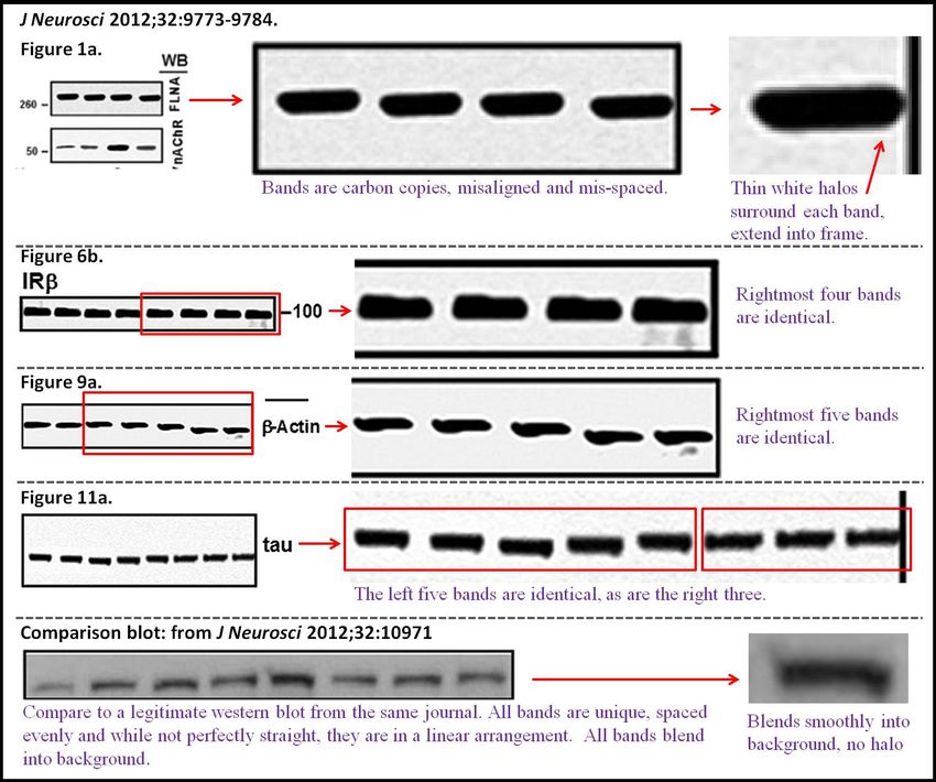
35Because of the contemporaneous examples of western blot manipulation, we undertook
an evaluation the author’s highest profile publication, a 2006 publication in Nature Medicine
12:824-828. Dr. Wang is the co-first author of this work. There are numerous suspicious
appearing blots in this publication, as well. Again, blots are suspiciously over-exposed. In the
supplementary material accompanying that published manuscript, we encounter the blot shown
below. The background has more-or-less been obliterated, except for a small area circled in the
red oval. Linear termination of the background signal is suspicious for the original blot having
been cut and reassembled. Because of the low quality of this image, we evaluated the images in
the main manuscript (which are higher quality), to assess for evidence of tampering.
Importantly, this manuscript purports to establish the validity of the functional
characterization of NMDA receptor signaling in post-mortem, frozen human brain material
which is called into question in section C.3.1. Evidence of tampering with this evidence further
calls into question the validity of this unusual technique.
3637
The images in the main text are of higher quality, enabling clearer evaluation. Increasing

                                        38
the contrast in the images published as Figure 4 (below) clearly reveals evidence of linear cuts in
the blots. Importantly, there is clearly a smooth background between the two darker bands and a
textured background only behind the dark bands. This was not likely done for cosmetic reasons,
it strongly suggests a manufactured/fraudulent result. There is no legitimate explanation for this
pattern of findings. This high-profile manuscript should be reviewed by the publisher and
retracted. All subsequent manuscripts built on this technique should likewise be reviewed.
39You can also read