The Silencing of Casein Kinase I Attenuated Neuromuscular Impairment in a Preclinical Mouse Model of Amyotrophic Lateral Sclerosis
←
→
Page content transcription
If your browser does not render page correctly, please read the page content below
https://www.scientificarchives.com/journal/journal-of-experimental-pathology

    Journal of Experimental Pathology                                                                        Research Article

The Silencing of Casein Kinase I Attenuated Neuromuscular
Impairment in a Preclinical Mouse Model of Amyotrophic
Lateral Sclerosis

Sergio Gonzalez-Gonzalez1*, Bastien Caumes2, Chantal Cazevieille3
1
    In vivex. www.invivex.com 177b avenue Louis Lumière. 34400 Lunel. France
2
    Eurofins Amatsigroup. 17 Rue des Vautes, 34980 Saint-Gély-du-Fesc. France
3
    Institute for Neurosciences of Montpellier. COMET. 80, rue Augustin Fliche. 34091 Montpellier. France
*
    Correspondence should be addressed to Sergio Gonzalez-Gonzalez; sergiogonzalez@invivex.com
Received date: November 05, 2020, Accepted date: March 19, 2021
Copyright: © 2021 Gonzalez-Gonzalez S, et al. This is an open-access article distributed under the terms of the Creative Commons
Attribution License, which permits unrestricted use, distribution, and reproduction in any medium, provided the original author
and source are credited.

Abstract
Amyotrophic Lateral Sclerosis (ALS) is a fatal progressive neurodegenerative disease characterized by the destruction of the
motor neurons. It usually affects people between 40 and 60-year-old and the average survival from onset to death is 3–4 years.
Despite the severity of the disease and the high health care and social costs, no cure or viable long-term effective treatment has
been identified. Moreover, the failure to translate positive preclinical results from the SOD1 mouse model into clinical efficacy has
raised questions about the translational suitability of this model. For this reason, the TDP-43 transgenic mouse model was created
by overexpressing the mutant human TDP-43 gene, mutation directly related with ALS. In this study we characterized this mouse
strain TDP-43, recognized as a preclinical mouse model for ALS disorder. We observed neuromuscular disorders, peripheral nerve
electrophysiological impairment and histological anomalies at 3 months old. We also demonstrated that intrathecal injection of AAV1
expressing shRNA for casein kinase-1δ (CK1δ) attenuated the peripheral degenerative phenotype in this ALS model. Our data confirm
that TDP-43 mouse strain is a robust and reproducible model to analyze the neuropathy disorders of ALS and that gene therapy
silencing CK1δ is a promising therapy for human ALS disorder, and can be used as a positive reference control for additional new
drugs efficacy studies targeting ALS.

Introduction                                                        showing accumulations in the cytoplasm of cortical and
                                                                    motor neurons [3]. Mutations in this protein have been
 Amyotrophic Lateral Sclerosis (ALS) is a fatal progressive         associated with cases of ALS, but also have been associated
neurodegenerative disease, which results in the destruction         with tau protein-independent cases of frontotemporal
of upper and/or lower motor neurons in the brain and spinal         dementia (FTD), generating the idea of a pathological
cord. It usually affects people between 40 and 60-year-old          spectrum between ALS and FTD based on alterations
and the average survival from onset to death is 3–4 years           in TDP-43 and other related proteins [4]. Today, it is
[1]. Despite the severity of the disease and the high health        recognized that TDP-43 proteinopathy, characterized by
care and social costs, no cure or viable long-term effective        hyperphosphorylation, truncation, ubiquitination, and/
treatment has been identified, with only two therapeutic            or nuclear depletion in neurons, is the prominent and
agents (riluzole and edaravone) already approved by Food            common pathological feature of sporadic and familiar
and Drug Administration but having limited efficacy or              ALS [1]. Furthermore, it is present in rare diseases such
serving only for specific groups of patients [2].                   as Perry syndrome or Alexander disease, but also in the
                                                                    prevalent Alzheimer’s disease (AD) and recently in the
 In 2006, the trans-activating response region DNA                  limbic-predominant age-related TDP-43 encephalopathy
binding protein of 43 kDa, known as TDP-43, was identified          [2,3]. In addition, TDP-43 pathology is a secondary
as one of the main hallmarks of sporadic and familial ALS,          feature of several other neurodegenerative disorders,

J Exp Pathol. 2021
Volume 2, Issue 2                                                                                                                          53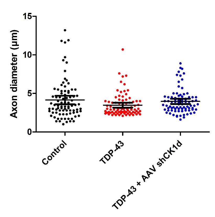
Gonzalez-Gonzalez S, Caumes B, Cazevieille C. The Silencing of Casein Kinase I Attenuated Neuromuscular
Impairment in a Preclinical Mouse Model of Amyotrophic Lateral Sclerosis. J Exp Pathol. 2021;2(2):53-62.

including Parkinson’s disease, and Huntington’s disease,           PBS. Then, cells were lysed using dry ice/ethanol bath and
where its presence may aggravate the primary existing              centrifuged 15 min at 5000 rpm to discard cell debris. The
proteinopathy [4]. Increasing number of publications               cleared supernatant and the cleared medium were pooled
demonstrated that in ALS, TDP-43 concentration is                  and filtrated using a 0.22 μm filter. The viral solution was
increased in cytoplasm leading to cytoplasmic inclusion            filtrated through a cation-exchange membrane Mustang
formation [5,6]. Mitochondrial localization of TDP-43              S acrodisc (Pall Corporation) to deplete empty particles
depends on internal motifs M1 (aa 35–41), M3 (aa 146–              and later, filtrated through an anion-exchange membrane
150), and M5 (aa 294–300), which consists of continuous            Mustang Q acrodisc (Pall corporation) to retain AAV viral
stretch of hydrophobic amino acids [7]. Owing to its               particles. Then, viruses were eluted and concentrated
poor in vitro solubility and high aggregation propensity,          using centrifugal concentrators Amicon tube. The viral
the complete structure of TDP-43 has remained elusive              titer used was 1×10¹² vg/mL.
thus far. Several groups, however, have determined high
resolution structures of some of its domains and its holistic       Cell culture and transfection
structure is now evolving.
                                                                     Human embryonic kidney (HEK)-293T cells were
 CK-1 was the first kinase identified to phosphorylate             grown in Dulbecco’s modified Eagle’s medium (DMEM)
TDP-43 in vivo in more than 29 different sites, being 18           supplemented with 2 mM L-glutamine, 100 U/ml
of them located in the C-terminal glycine-rich region [8].         penicillin/streptomycin, and 5% (v/v) heat inactivated
Moreover, different stress signaling cause CK-1-dependent          Foetal Bovine Serum (FBS) (all from Invitrogen). Cells
phosphorylation of TDP-43 triggering its cytosolic                 were maintained at 37°C in an atmosphere of 5% CO2, and
mislocalization and accumulation [9,10]. Furthermore,              were passaged every 3 or 4 days when they were 80-90%
TDP-43 binds directly to and regulates the expression of           confluent. HEK-293T cells growing in 6-well dishes were
CK-1ε mRNA [11].                                                   transiently transfected with 1 µg of cDNA of mouse CK-1δ-
                                                                   flag using Lipofectamine method (Invitrogen). 48 hours
 Importantly, chemical inhibition of CK-1, mainly the δ and        after transfection, cells were infected with increasing
ε isoforms, has been proposed as potential treatment for           concentration of viral particles. 24 hours after infection,
different neurodegenerative diseases including ALS, FTD            cells were lysed and the expression level of CK-1δ was
and Alzheimer’s disease [12]. In this way, and as showed in        determined by western blot.
the Figure 1a, we developed an AAV1 expressing the shRNA
for mouse CK-1δ in order to reduce the expression of CK-            Western blot
1δ in motor neurons. We demonstrated that the silencing
of CK-1δ by intrathecal injection significantly increased            Cell lysed samples were denaturalized and separated
neuromuscular performances and reduced motor neuron                in 10% SDS-polyacrylamide gel and transferred onto
degeneration attenuating ALS neuropathy. These changes             nitrocellulose membranes. Membranes were blocked for
were probably induced by the reduction of the TDP-43               60 min with 5 mL of Licor Blocking Buffer. The following
phosphorylation and the accumulation and mislocalization           primary antibodies were incubated overnight at 4°C in the
of this protein. Our data confirm that the silencing of CK-        same blocking buffer: rabbit anti-lamin B1 (1:100, Sigma)
1δ can be used as a positive reference control for additional      and mouse anti-flag (1:100, Sigma). The following day the
new drugs efficacy studies targeting ALS disorder.                 membranes were washed 3 times for 10 minutes in TBS
                                                                   Tween-20 (0.1% V/V) and the incubated for 1 hour at room
Materials and Methods                                              temperature with the secondary fluorescence antibodies :
                                                                   donkey anti-rabbit IRDye 800 (1:10.000, Licor) and Goat
 Cloning and virus production                                      anti-mouse IRDye 680 (1:10.000, Licor). After secondary
                                                                   antibody incubation, the membranes were washed three
  The      mouse        CK-specific       shRNA       sequence     times for 10 min each with TBS Tween-20 (0.1% V/V).
TGATAAGTCGTATTGAGTA and shRNA scramble                             Visualization of the bands was performed using Licor
(control) sequences AGTTCCAGTACGGCTCCAA were                       scanning devise.
separately cloned into a pAAV vector using XhoI and EcoRV
restriction sites. Each cloning was validated by sequencing.        Animal housing
Then, to produce high-titer adeno-associated virus (AAV1),
three 15 cm dishes of 70-80% confluent HEK293T cells                Male Prp-TDP43A315T mice and wild type control were
were transfected with 71 μg of pAAV expression vector, 20          kept in the A1 animal house facility. Mice were housed
μg of pAAV1 capsid and 40 μg of pHelper (Cell Biolabs,             in ventilated and clear plastic boxes and subjected to
Inc.). 48 h later transfection, the medium was collected,          standard light cycles (12 hours in 90-lux light, 12 hours
pooled and centrifuged 15 min at 2000 rpm to spin down             in the dark). Baseline experiments started at 1 month old
floating cells. In parallel, cells were scraped and collected in   and ALS phenotype was characterized at 2 and 3 months

J Exp Pathol. 2021
Volume 2, Issue 2                                                                                                        54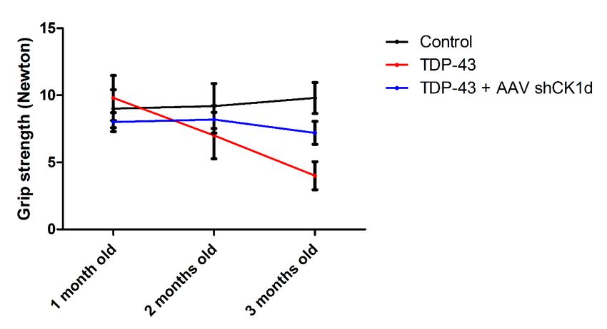
Gonzalez-Gonzalez S, Caumes B, Cazevieille C. The Silencing of Casein Kinase I Attenuated Neuromuscular
Impairment in a Preclinical Mouse Model of Amyotrophic Lateral Sclerosis. J Exp Pathol. 2021;2(2):53-62.

old. All animal experiments were approved by the Comité        anesthetized with ketamine/xylazine mixture. A pair
d’Ethique en Expérimentation Animale du Languedoc-             of steel needle electrodes (AD Instruments. MLA1302)
Roussillon, Montpellier, France, and the Ministère de          was placed subcutaneously along the nerve at the sciatic
l’Enseignement Supérieur et de la Recherche, Paris,            notch (proximal stimulation). A second pair of electrodes
France.                                                        was placed along the tibial nerve above the ankle (distal
                                                               stimulation). Supramaximal square-wave pulses, lasting
 Intrathecal injection                                         10 ms at 1 mA were delivered using a PowerLab 26T (AD
                                                               Instruments). CMAP was recorded from the intrinsic
 In ketamine/xylazine anesthetized animals, a 10               foot muscles using steel electrodes. Both amplitudes and
µl Hamilton needle was gently and vertically (or tilt          latencies of CMAP were determined. The distance between
slightly 70-80°) inserted in the intersection of the L5-       the 2 sites of stimulation was measured alongside the skin
L6 intervertebral space. The angle was reduced to              surface with fully extended legs, and NCVs were calculated
approximately 30° slowly when it connected the bone, then      automatically from sciatic nerve latency measurements.
the needle was slipped into the intervertebral space. 8 μL     The left sciatic nerves were analyzed in this study.
of the AAV1-shRNA CK1d solution was injected at a speed
of 1 μL/4 seconds. The needle was retained approximately       Sciatic nerve histology by Transmission Electron
1 min after finishing AAV1 delivery.                           Microscopy (TEM)

 Balance beam test                                              The sciatic nerves of all mice were fixed for 20 minutes
                                                               in situ with 4 % PFA and 2.5% glutaraldehyde in 0.1 M
 Balance beam test is a narrow “walking bridge” that           phosphate buffer (pH 7.3). Then, nerves were removed and
rodents can cross to test balance and neurosensory             post-fixed overnight in the same buffer. After washing for
coordination. The beam (thickness 6 cm) was elevated with      30 minutes in 0.2 M PBS buffer, the nerves were incubated
the help of two feet with platforms to hold mice. The time     with 2% osmic acid in 0.1 M phosphate buffer for 90
required to cross the beam from side to side was quantified    minutes at room temperature. Then, samples were washed
for each animal. Each animal underwent 3 trials/day at 5       in 0.2 M PBS buffer, dehydrated using ethanol gradient
minutes intervals. For each day, values from the 3 trials      solutions, and embedded in epoxy resin. Ultrathin (70-nm)
were averaged for each animal, normalized according to         cross sections were cut and stained with 1% uranyl acetate
animal weight, and then averaged for each treated group.       solution and lead citrate and analyzed using a HITACHI
                                                               H7100 electron microscope at the COMET platform. The
 Rotarod                                                       left sciatic nerves were analyzed in this study.
 A rotating rod apparatus was used to measure                   Statistics
neuromuscular coordination and balance. Mice were first
given a 2-day pretraining trial to familiarize them with the    Data are represented as mean ± SEM. Statistical
rotating rod. Latency to fall was measured at a successively   significance was determined using a 2-tailed Student’s t
increased speed from 4 to 40 rpm over a 300-second time        test or 1-way ANOVA, followed by a Dunnett’s multiple
period. Each animal underwent 3 trials a day. For each         comparison post-hoc test. A P value of less than 0.05 was
day, values from the 3 trials are averaged for each animal,    considered significant. n= 5 animals/group.
normalized according to animal weight, and then averaged
for each group.                                                Results
 Grip test                                                      In vitro validation of shRNA for CK-1δ

 Neuromuscular strength of mice was assessed in                 In vitro HEK293T cells were transfected with 1 µg of cDNA
standardized grip strength tests for hind limbs. Limbs grip    mouse CK-1δ-flag. 24 hours after transfection, cells were
strength was measured by supporting each rodent on a           infected with increasing concentrations of AAV1-shRNA
metal grid and pulling the animal’s tail toward a horizontal   CK-1δ or shRNA scramble (negative control). 48 hours
grid connected to a gauge. The maximum force (measured         after infection, cells were lysed and the concentration of
in newtons) exerted on the grid before the animal              mouse CK-1δ was determined by western blot using mouse
lost its grip was recorded, and the mean of 3 repeated         anti-flag antibody. Lamin B1 was used as protein control
measurements was calculated. All data were normalized          loading.
according to animal weight.
                                                                Our data confirmed that the plasmid shRNA mouse CK-
 Sciatic nerve electrophysiology                               1δ and the viral particles AAV1 expressing shRNA for CK-
                                                               1δ were able to decrease the expression of mouse CK-1δ in
 Standard electromyography was performed on mice               vitro (Figure 1b).

J Exp Pathol. 2021
Volume 2, Issue 2                                                                                                   55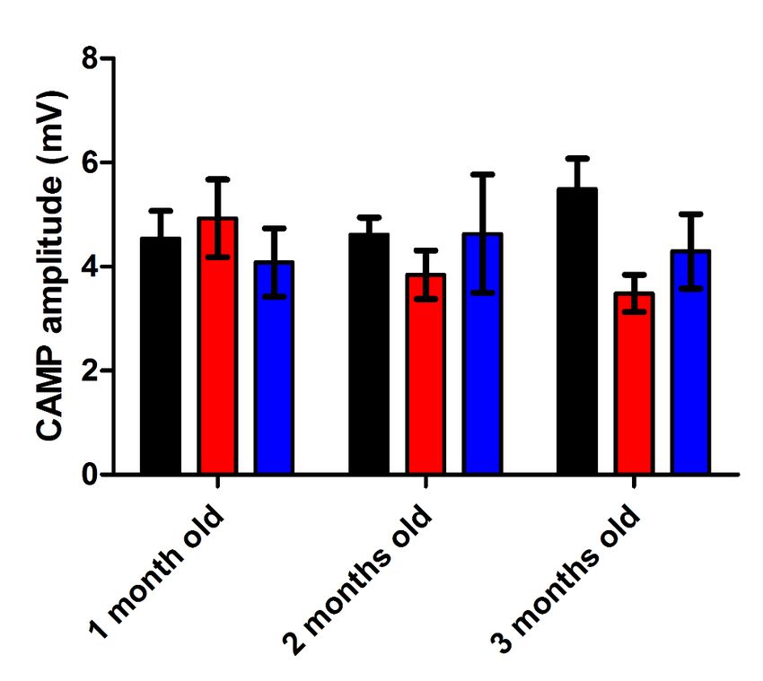
Gonzalez-Gonzalez S, Caumes B, Cazevieille C. The Silencing of Casein Kinase I Attenuated Neuromuscular
Impairment in a Preclinical Mouse Model of Amyotrophic Lateral Sclerosis. J Exp Pathol. 2021;2(2):53-62.

 Figure 1

  a                                                                 b                       Transfection Mouse CK1δ (1µg/mL)

      AAV1-shRNA CK1δ
                 Sense        Loop       Antisense Terminator

                                                                                                                   AAV1-shRNA CK1δ

                                                                                                             1E8   1E7 1E6 1E5
                                                                               80kDa
                                                                    Lamin B1
                                                                                60kDa

                                                                      CK1δ
                                                                               40kDa

 Figure 1: a. Schematic representation of the intrathecal viral particles injection. b. Western blot image of the in vitro validation
 of the plasmid and viral particles expressing shRNA – CK1δ.

Decrease of neuromotor impairment in ALS                            TDP-43 and TDP-43 + shCK1δ groups at 3 months old
mouse model induced by CK-1δ silencing                              suggesting a partial protective effect of the viral particle
                                                                    injection in the walking impairment (Figure 2b).
 No mortality and no pathological clinical signs were
observed during the study. As expected, significant                   Then, the neuromuscular strength was analyzed using
decrease of body weight was observed in the TDP-43                  grip test. Similar grip strengths were observed in control,
groups compared to the control group at 3 months old.               TDP-43 and TDP-43 + shCK1δ groups at 1 month old (9.0
Moreover, a slight but significant difference of body weight        ± 1.4 N; 9.8 ± 1.7 N; 8.0 ± 0.7 N respectively). At 2 months
was also observed between TDP-43 and TDP-43 + AAV                   old, the grip strength was decreased at 7.0 ± 1.7N in the
shCK1d at 3 months old. No significant difference was               TDP-43 group whereas the grip strength of control group
observed in the TDP-43 + AAV shCK1d group compared                  remained at 9.2 ± 1.7N. However, no statistical difference
to the control group (Figure 2a) suggesting an effect of the        was observed between TDP-43 and control groups at this
intrathecal AAV injection in the body weight loss induced           time point. Moreover, no differences in the grip strength
by ALS phenotype.                                                   were observed in the TDP-43 + shCK1δ at 2 months old
                                                                    compared to the baseline (Figure 2c). At 3 months old,
 Walking performances were analyzed using balance                   the grip strength of the TDP-43 group was significantly
beam test. Similar beam cross times were observed in                decreased at 4.0 ± 1.0 N whereas the control animals
control, TDP-43 and TDP-43 + shCK1δ groups at 1 month               presented strength of 9.8 ± 1.2N. Moreover, even if the
old (5.4 ± 0.3 s; 5.6 ± 0.4 s; 6.0 ± 0.3 s respectively). At 2      TDP-43 + shCK1δ group presented a slight decrease of
months old, the beam cross time was increased at 8.1 ± 0.9          the grip strength compared to the baseline, no statistical
seconds in the TDP-43 group mice whereas the cross time             differences were observed between TDP-43 + shCK1δ
of control animals remained at 6.2 ± 0.6 s. A slight increase       versus control and TDP-43 animals at 3 months old (Figure
of the beam cross time was also observed in the TDP-43              2c). Taken together these data also suggested a protective
+ shCK1δ compared to control. However, no statistical               effect of the viral particle injection in the neuromuscular
differences were observed between all groups at this time           strength.
point (Figure 2b). At 3 months old, the beam cross time
of the TDP-43 group was significantly increased at 10.8 ±            Finally, the coordination and balance were analyzed
0.6 seconds whereas the control animals presented a beam            using rotarod. Similar rotarod latencies were observed in
cross time of 6.5 ± 0.4 s. Moreover, the TDP-43 + shCK1δ            control, TDP-43 and TDP-43 + shCK1δ groups at 1 month
group also presented a slight but significant increase of the       old (73.2 ± 8.1 s; 70.0 ± 6.5 s; 71.6 ± 6.2 s respectively).
cross time compared to the control animals at 3 months              At 2 months old, the rotarod latency was decreased at
old. However, a statistical difference was observed between         55.0 ± 6.4s in the TDP-43 group whereas the latency of

J Exp Pathol. 2021
Volume 2, Issue 2                                                                                                                    56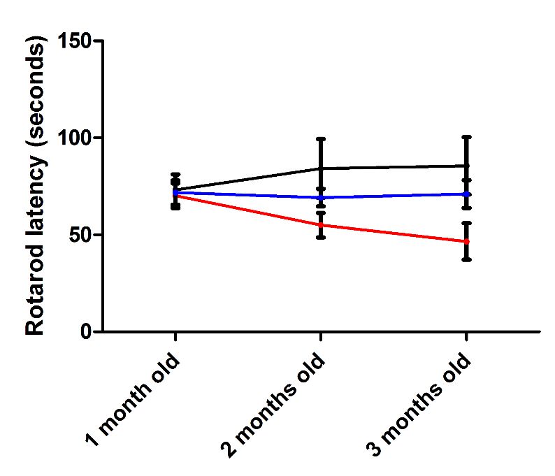
Gonzalez-Gonzalez S, Caumes B, Cazevieille C. The Silencing of Casein Kinase I Attenuated Neuromuscular
       Impairment in a Preclinical Mouse Model of Amyotrophic Lateral Sclerosis. J Exp Pathol. 2021;2(2):53-62.

re 2
         a                                                         b
                                                    ns

                                                    #        ***
                                                                                                           #

                                                                                                               ***
                                                                                                           *

         c                                                         d
                                                                                                          ns
                                                        ns
                                                                                                               *
                                                              *
                                                        ns                                                ns

       Figure 2: Analysis of the neuromuscular performance. a. Animal body weight of control, b. Balance beam cross time, c. Rotarod
       latency, and d. grip strength of control, TDP-43 and TDP-43 + AAV shRNA CK1δ groups at 1, 2 and 3 months old. Values are mean.
       Error bars indicate SEM. Statistical tests are repeated measures 1-way ANOVA test comparing groups to control (*) or TDP-43 (#)
       values. #* p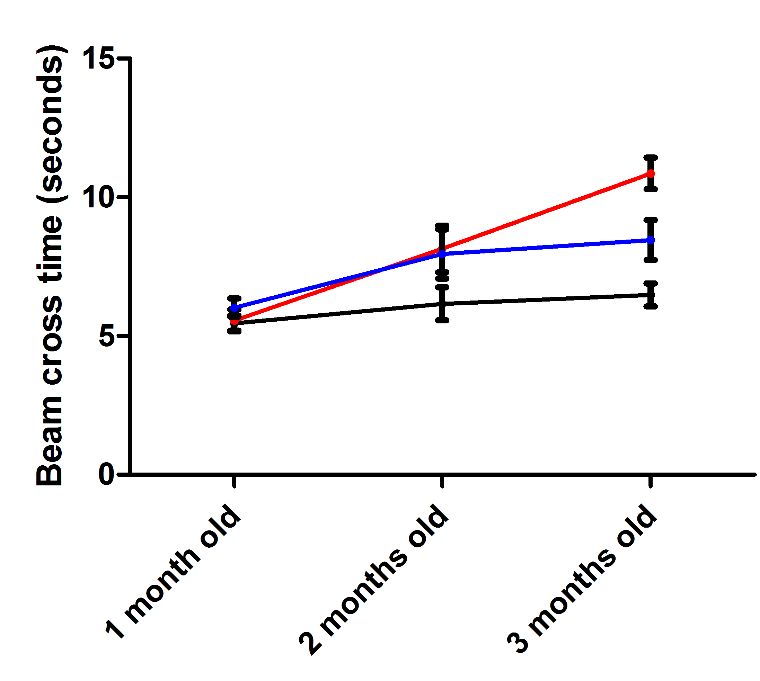
Gonzalez-Gonzalez S, Caumes B, Cazevieille C. The Silencing of Casein Kinase I Attenuated Neuromuscular
Impairment in a Preclinical Mouse Model of Amyotrophic Lateral Sclerosis. J Exp Pathol. 2021;2(2):53-62.

Figure 3

  a                                                    b
                             ns          ns                                      ns          ns
                                                                                  ns         ns

                                                                                 *          **

 Figure 3: Analysis of the sciatic nerve conduction. a. compound action muscle potential (CAMP) analysis represented in mV and
 b. nerve conduction velocity represented in m/s of control, TDP-43 and TDP-43 + AAV shRNA CK1δ groups at 1, 2 and 3 months
 old. Values are mean. Error bars indicate SEM. Statistical tests are repeated measures 1-way ANOVA test comparing groups to
 control or TDP-43 values. * p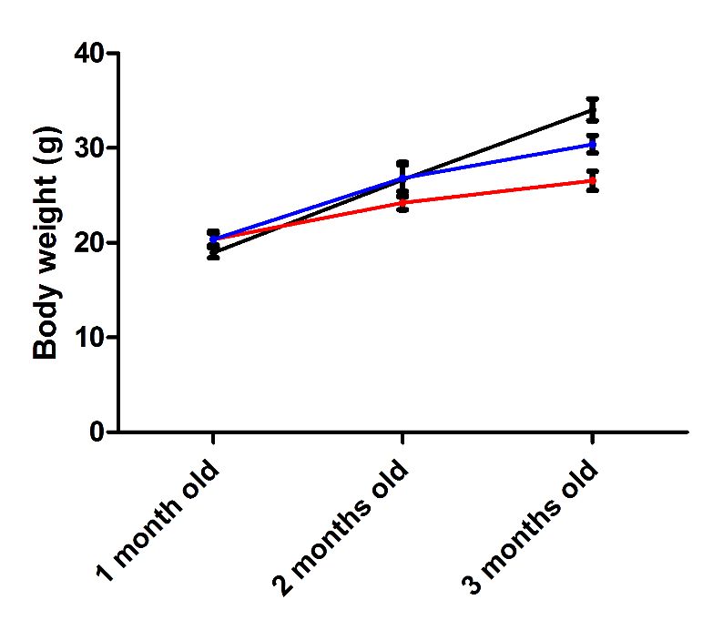
Gonzalez-Gonzalez S, Caumes B, Cazevieille C. The Silencing of Casein Kinase I Attenuated Neuromuscular
Impairment in a Preclinical Mouse Model of Amyotrophic Lateral Sclerosis. J Exp Pathol. 2021;2(2):53-62.
e4 a b *
ns
#
Control
TDP-43
c **
#
TDP-43 +
AAV shCK1δ
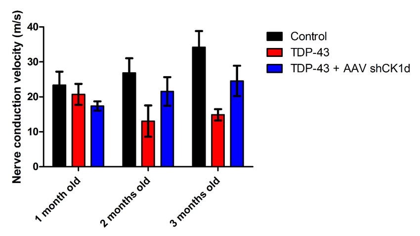
Figure 4: Histological analysis of sciatic nerve. a. Representative image of transmission electron microscopy of sciatic nerve of
control, TDP-43 and TDP-43 + AAV shRNA CK1δ mice at 3 months old. Scale bar: 10 µm. b. Analysis of the axon diameter and, b.
quantification of the number of axons par 100 µm2 of control, TDP-43 and TDP-43 + AAV shRNA CK1δ groups at 3 months old.
Values are mean. Error bars indicate SEM. Statistical tests are repeated measures 1-way ANOVA test comparing groups to control
or TDP-43 values. # * p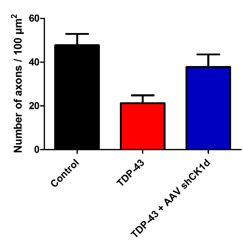
Gonzalez-Gonzalez S, Caumes B, Cazevieille C. The Silencing of Casein Kinase I Attenuated Neuromuscular
Impairment in a Preclinical Mouse Model of Amyotrophic Lateral Sclerosis. J Exp Pathol. 2021;2(2):53-62.

strategy for the severe ALS. They demonstrated that the           number 1. It comprises of an N-terminal region (aa 1–102)
treatment of the ALS preclinical model TDP-43 with a              with a nuclear localization signal (NLS, aa 82–98), two
CK-1δ inhibitor decreased of TDP-43 phosphorylation,              RNA recognition motifs: RRM1 (aa 104–176) and RRM2
inducing motor neuron survival and decreasing astroglial          (aa 192–262), a nuclear export signal (NES, aa 239–250),
and microglial reactivity [19]. This publication served as a      a C-terminal region (aa 274–414) which encompasses a
solid proof of concept for the potential therapy of ALS with      prion-like glutamine/asparagine-rich (Q/N) domain (aa
CK-1δ or dual CK-1δ/ε inhibitors showing a promising              345–366) and a glycine-rich region (aa 366–414) [26,27].
neuroprotective effect [19,20]. For this reason, in this study    TDP-43 is predominantly localized in the nucleus but also
we developed an AAV1 expressing the shRNA for mouse               shuttles to the cytoplasm for some of its functions [28].
CK-1δ in order to reduce the expression of CK-1δ in motor         In ALS, there is an increase in the cytoplasmic TDP-43
neurons looking to reproduce Martinez-Gonzalez results            concentration leading to cytoplasmic inclusion formation
from a gene therapy point of view. We demonstrated that           [29,30].
the silencing of CK-1δ by intrathecal injection reduced the
motor neuron degeneration attenuating ALS neuropathy              Conclusion
and corroborating Martinez-Gonzalez previous results.
Our data demonstrated that the knockdown of CK-1δ                  In this study we described the neuromotor disorders,
using in vivo shRNA was able to partially reduce, but not         peripheral     nervous     system     electrophysiological
to stop, the ALS phenotype in the TDP-43 mouse model.             impairment and histological anomalies observed in the
For this reason, we concluded that the silencing of CK-1δ         preclinical ALS mouse model TDP-43 at 3 months old.
is not perfect solution to ALS but a good attempt fighting        Moreover, we demonstrated that intrathecal injection
against ALS in mouse models.                                      of AAV1 expressing shRNA for CK-1δ attenuated the
                                                                  peripheral nerve degeneration of ALS model. Our data
  The study of the post-translational regulation of TDP-43        confirm that TDP-43 mouse strain is a robust and
has placed the phosphorylation of this protein at specific        reproducible model to analyze the neuropathy disorders of
residues, which is dependent on certain protein kinases, as       ALS and that gene therapy silencing CK1δ is a promising
a key event for regulating its cellular activities and also for   therapy for human ALS disorder. This silencing strategy
its dysregulation associated with pathological conditions         can be used as a positive reference control for additional
[21,22]. In fact, phospho-TDP-43 is distributed in brain
                                                                  new drugs efficacy studies targeting ALS.
areas of FTD and ALS patients [21]. Different kinases
have been recently involved in TDP-43 phosphorylation.
                                                                  References
Among these, protein casein kinase-1 [22], tau tubulin
kinase 1 [23] and cell division cycle kinase 7 [22] are the        1. Hergesheimer RC, Chami AA, de Assis DR, Vourc’h
best characterized.                                               P, Andres CR, Corcia P, et al. The debated toxic role of
                                                                  aggregated TDP-43 in amyotrophic lateral sclerosis: a
 As published by Gou and collaborators in 2020 [24],
                                                                  resolution in sight?. Brain. 2019 May 1;142(5):1176-94.
aberrant phosphorylation of various ALS-related proteins
by kinases could affect the localization and function of           2. Palomo V, Tosat-Bitrian C, Nozal V, Nagaraj S, Martin-
these proteins. However, the exact role these alterations         Requero A, Martinez A. TDP-43: a key therapeutic target
play in motor neuron degeneration remains elusive.                beyond amyotrophic lateral sclerosis. ACS Chemical
Confirming our hypothesis, multiple studies showed that           Neuroscience. 2019 Feb 20;10(3):1183-96.
mutations in genes encoding different kinases can cause or
confer susceptibility to ALS, suggesting that alterations in       3. Nelson PT, Dickson DW, Trojanowski JQ, Jack CR,
the function of specific kinases and/or their downstream          Boyle PA, Arfanakis K, et al. Limbic-predominant age-
targets are vital to motor neuron survival. Taken together,       related TDP-43 encephalopathy (LATE): consensus
these data suggest a potential key role of kinases in ALS         working group report. Brain. 2019 Jun 1;142(6):1503-27.
genetics and pathophysiology [24].
                                                                   4. Davis SA, Gan KA, Dowell JA, Cairns NJ, Gitcho MA.
 In motor neurons, TDP-43 has also been shown in humans           TDP-43 expression influences amyloidβ plaque deposition
to be a low molecular weight neurofilament mRNA-                  and tau aggregation. Neurobiology of Disease. 2017 Jul
binding protein [25]. It has also shown to be a neuronal          1;103:154-62.
activity response factor in the dendrites of hippocampal
neurons suggesting possible roles in regulating mRNA               5. Neumann M, Sampathu DM, Kwong LK, Truax AC,
stability, transport and local translation in neurons [26].       Micsenyi MC, Chou TT, et al. Ubiquitinated TDP-43 in
The TDP-43 protein contains 414 amino acids and the               frontotemporal lobar degeneration and amyotrophic
encoding gene TARDBP is located on the chromosome                 lateral sclerosis. Science. 2006 Oct 6;314(5796):130-3.

J Exp Pathol. 2021
Volume 2, Issue 2                                                                                                      60
Gonzalez-Gonzalez S, Caumes B, Cazevieille C. The Silencing of Casein Kinase I Attenuated Neuromuscular
Impairment in a Preclinical Mouse Model of Amyotrophic Lateral Sclerosis. J Exp Pathol. 2021;2(2):53-62.
6. Winton MJ, Igaz LM, Wong MM, Kwong LK, 16. Wegorzewska I, Bell S, Cairns NJ, Miller TM, Baloh
Trojanowski JQ, Lee VM. Disturbance of nuclear and RH. TDP-43 mutant transgenic mice develop features of
cytoplasmic TAR DNA-binding protein (TDP-43) induces ALS and frontotemporal lobar degeneration. Proceedings
disease-like redistribution, sequestration, and aggregate of the National Academy of Sciences. 2009 Nov
formation. Journal of Biological Chemistry. 2008 May 3;106(44):18809-14.
9;283(19):13302-9.
17. Cozza G, Pinna LA. Casein kinases as potential
7. Wang W, Wang L, Lu J, Siedlak SL, Fujioka H, Liang J, therapeutic targets. Expert opinion on therapeutic targets.
et al. The inhibition of TDP-43 mitochondrial localization 2016 Mar 3;20(3):319-40.
blocks its neuronal toxicity. Nature Medicine. 2016
Aug;22(8):869-78. 18. Gu J, Hu W, Tan X, Qu S, Chu D, Gong CX, et al.
Elevation of casein kinase 1ε associated with TDP-43 and
8. Liachko NF, McMillan PJ, Guthrie CR, Bird TD, Leverenz tau pathologies in Alzheimer’s disease. Brain Pathology.
JB, Kraemer BC. CDC7 inhibition blocks pathological 2020 Mar;30(2):283-97.
TDP-43 phosphorylation and neurodegeneration. Annals
of Neurology. 2013 Jul;74(1):39-52. 19. Martínez-González L, Rodríguez-Cueto C, Cabezudo
D, Bartolomé F, Andrés-Benito P, Ferrer I, et al. Motor
9. Nonaka T, Suzuki G, Tanaka Y, Kametani F, Hirai neuron preservation and decrease of in vivo TDP-43
S, Okado H, Miyashita T, Saitoe M, Akiyama H, Masai phosphorylation by protein CK-1δ kinase inhibitor
H, Hasegawa M. Phosphorylation of TAR DNA-binding treatment. Scientific Reports. 2020 Mar 10;10(1):1-2.
protein of 43 kDa (TDP-43) by truncated casein kinase
1δ triggers mislocalization and accumulation of TDP-43. 20. Salado IG, Redondo M, Bello ML, Perez C, Liachko
Journal of Biological Chemistry. 2016 Mar 11;291(11):5473- NF, Kraemer BC, et al. Protein kinase CK-1 inhibitors as
83. new potential drugs for amyotrophic lateral sclerosis.
Journal of Medicinal Chemistry. 2014 Mar 27;57(6):2755-
10. Hicks DA, Cross LL, Williamson R, Rattray M. 72.
Endoplasmic reticulum stress signalling induces casein
kinase 1-dependent formation of cytosolic TDP-43 21. Guedes ÁC, Santin R, Costa AS, Reiter KC, Hilbig
inclusions in motor neuron-like cells. Neurochemical A, Fernandez LL. Distinct Phospho-TDP-43 brain
Research. 2019 Jul 6:1-1. distribution in two cases of FTD, one associated with ALS.
Dementia & Neuropsychologia. 2017 Sep;11(3):249-54.
11. Krach F, Batra R, Wheeler EC, Vu AQ, Wang R,
Hutt K, Rabin SJ, Baughn MW, Libby RT, Diaz-Garcia S, 22. Liachko NF, McMillan PJ, Guthrie CR, Bird TD, Leverenz
Stauffer J. Transcriptome–pathology correlation identifies JB, Kraemer BC. CDC7 inhibition blocks pathological
interplay between TDP-43 and the expression of its kinase TDP-43 phosphorylation and neurodegeneration. Annals
CK1E in sporadic ALS. Acta Neuropathologica. 2018 of Neurology. 2013 Jul;74(1):39-52.
Sep;136(3):405-23.
23. Liachko NF, McMillan PJ, Strovas TJ, Loomis E,
12. Gu J, Hu W, Tan X, Qu S, Chu D, Gong CX, Iqbal K, Greenup L, Murrell JR, et al. The tau tubulin kinases
Liu F. Elevation of casein kinase 1ε associated with TDP- TTBK1/2 promote accumulation of pathological TDP-43.
43 and tau pathologies in Alzheimer’s disease. Brain PLoS Genetics. 2014 Dec 4;10(12):e1004803.
Pathology. 2020 Mar;30(2):283-97.
24. Guo W, Vandoorne T, Steyaert J, Staats KA, Van Den
13. Rosen DR, Siddique T, Patterson D, Figlewicz DA, Bosch L. The multifaceted role of kinases in amyotrophic
Sapp P, Hentati A, et al. Mutations in Cu/Zn superoxide lateral sclerosis: genetic, pathological and therapeutic
dismutase gene are associated with familial amyotrophic implications. Brain. 2020 Jun 1;143(6):1651-73.
lateral sclerosis. Nature. 1993 Mar;362(6415):59-62.
25. Wang IF, Wu LS, Chang HY, Shen CK. TDP-43,
14. Gurney ME, Cutting FB, Zhai P, Andrus PK, Hall ED. the signature protein of FTLD-U, is a neuronal activity-
Pathogenic mechanisms in familial amyotrophic lateral responsive factor. Journal of Neurochemistry. 2008
sclerosis due to mutation of Cu, Zn superoxide dismutase. May;105(3):797-806.
Pathologie-biologie. 1996 Jan 1;44(1):51-6.
26. Cohen TJ, Lee VM, Trojanowski JQ. TDP-43
15. Gordon PH, Meininger V. How can we improve clinical functions and pathogenic mechanisms implicated in TDP-
trials in amyotrophic lateral sclerosis?. Nature Reviews 43 proteinopathies. Trends in Molecular Medicine. 2011
Neurology. 2011 Nov;7(11):650-4. Nov 1;17(11):659-67.
J Exp Pathol. 2021
Volume 2, Issue 2 61Gonzalez-Gonzalez S, Caumes B, Cazevieille C. The Silencing of Casein Kinase I Attenuated Neuromuscular
Impairment in a Preclinical Mouse Model of Amyotrophic Lateral Sclerosis. J Exp Pathol. 2021;2(2):53-62.
27. Jiang LL, Zhao J, Yin XF, He WT, Yang H, Che 29. Neumann M, Sampathu DM, Kwong LK, Truax AC,
MX, et al. Two mutations G335D and Q343R within Micsenyi MC, Chou TT, et al. Ubiquitinated TDP-43 in
the amyloidogenic core region of TDP-43 influence its frontotemporal lobar degeneration and amyotrophic
aggregation and inclusion formation. Scientific Reports. lateral sclerosis. Science. 2006 Oct 6;314(5796):130-133.
2016 Mar 31;6(1):1-1.
30. Winton MJ, Igaz LM, Wong MM, Kwong LK,
28. Ayala YM, Zago P, D’Ambrogio A, Xu YF, Petrucelli Trojanowski JQ, Lee VM. Disturbance of nuclear and
L, Buratti E, et al. Structural determinants of the cellular cytoplasmic TAR DNA-binding protein (TDP-43) induces
localization and shuttling of TDP-43. Journal of Cell disease-like redistribution, sequestration, and aggregate
Science. 2008 Nov 15;121(22):3778-85. formation. Journal of Biological Chemistry. 2008 May
9;283(19):13302-9.
J Exp Pathol. 2021
Volume 2, Issue 2 62You can also read