Kappa opioid receptors in the central amygdala modulate spinal nociceptive processing through an action on amygdala CRF neurons
←
→
Page content transcription
If your browser does not render page correctly, please read the page content below
Ji and Neugebauer Molecular Brain      (2020) 13:128
https://doi.org/10.1186/s13041-020-00669-3

 SHORT REPORT                                                                                                                                       Open Access

Kappa opioid receptors in the central
amygdala modulate spinal nociceptive
processing through an action on amygdala
CRF neurons
Guangchen Ji1,2 and Volker Neugebauer1,2,3*

  Abstract
  The amygdala plays an important role in the emotional-affective aspects of behaviors and pain, but can also
  modulate sensory aspect of pain (“nociception”), likely through coupling to descending modulatory systems. Here
  we explored the functional coupling of the amygdala to spinal nociception. We found that pharmacological
  activation of neurons in the central nucleus of the amygdala (CeA) increased the activity of spinal dorsal horn
  neurons; and this effect was blocked by optogenetic silencing of corticotropin releasing factor (CRF) positive CeA
  neurons. A kappa opioid receptor (KOR) agonist (U-69,593) was administered into the CeA by microdialysis. KOR
  was targeted because of their role in averse-affective behaviors through actions in limbic brain regions. Extracellular
  single-unit recordings were made of CeA neurons or spinal dorsal horn neurons in anesthetized transgenic Crh-Cre
  rats. Neurons responded more strongly to noxious than innocuous stimuli. U-69,593 increased the responses of CeA
  and spinal neurons to innocuous and noxious mechanical stimulation of peripheral tissues. The facilitatory effect of
  the agonist was blocked by optical silencing of CRF-CeA neurons though light activation of halorhodopsin
  expressed in these neurons by viral-vector. The CRF system in the amygdala has been implicated in aversiveness
  and pain modulation. The results suggest that the amygdala can modulate spinal nociceptive processing in a
  positive direction through CRF-CeA neurons and that KOR activation in the amygdala (CeA) has pro-nociceptive
  effects.
  Keywords: Amygdala, Kappa opioid receptor, Spinal dorsal horn, Nociception, Optogenetics

Introduction                                                                           as from thalamic and cortical regions through the baso-
The amygdala has emerged as an important node of the                                   lateral amygdala network [2]. Importantly, synaptic plas-
emotional-affective aspects of pain and pain modulation                                ticity in the CeA in different pain models has been
[1–6]. The central nucleus (CeA) serves major amygdala                                 linked to emotional responses such as vocalizations,
output functions and receives pain-related information                                 aversive behaviors in conditioned place preference/aver-
through the spino-parabrachio-amygdala pathway as well                                 sion assays, and anxio-depressive behaviors [7–16]. The
                                                                                       contribution of the amygdala to sensory aspects of pain
* Correspondence: volker.neugebauer@ttuhsc.edu
1
                                                                                       such as hypersensitivity in pain models is less clear. Ma-
 Department of Pharmacology and Neuroscience, Texas Tech University
                                                                                       nipulations of amygdala activity provided evidence for
Health Sciences Center, School of Medicine, 3601 4th St, Lubbock, TX
79430-6592, USA                                                                        dual pro- and anti-nociceptive effects [4, 17–26] but
2
 Center of Excellence for Translational Neuroscience and Therapeutics, Texas           others had little, if any effect on hypersensitivity [15, 16,
Tech University Health Sciences Center, Lubbock, TX, USA
                                                                                       27–32]. The current view is that distinct amygdala
Full list of author information is available at the end of the article

                                        © The Author(s). 2020 Open Access This article is licensed under a Creative Commons Attribution 4.0 International License,
                                        which permits use, sharing, adaptation, distribution and reproduction in any medium or format, as long as you give
                                        appropriate credit to the original author(s) and the source, provide a link to the Creative Commons licence, and indicate if
                                        changes were made. The images or other third party material in this article are included in the article's Creative Commons
                                        licence, unless indicated otherwise in a credit line to the material. If material is not included in the article's Creative Commons
                                        licence and your intended use is not permitted by statutory regulation or exceeds the permitted use, you will need to obtain
                                        permission directly from the copyright holder. To view a copy of this licence, visit http://creativecommons.org/licenses/by/4.0/.
                                        The Creative Commons Public Domain Dedication waiver (http://creativecommons.org/publicdomain/zero/1.0/) applies to the
                                        data made available in this article, unless otherwise stated in a credit line to the data.
Ji and Neugebauer Molecular Brain (2020) 13:128 Page 2 of 10
circuits and cell types serve different functions related to another set of experiments, single-unit recordings of
different aspects of pain. spinal dorsal horn neurons were made in anesthetized
Nociceptive plasticity in the spinal dorsal horn plays a naïve rats before and during administration of U-69,593
critical role in pain-related hypersensitivity [33, 34]. How- into the CeA by microdialysis (15 min) and during op-
ever, there is little information about the modulation of tical silencing of CRF-CeA neurons while U-69,593 ad-
dorsal horn activity by the amygdala, although amygdala ministration continued for another 15 min. For the
neurons project to and can modulate descending pain optogenetic experiments, an AAV vector expressing
control centers such as periaqueductal gray PAG [2, 35, halorhodopsin was injected into the CeA 4–5 weeks be-
36]. A recent study showed that morphine injection into fore. In these experiments, a microdialysis fiber and an
the CeA, but not anterior cingulate cortex (ACC), reduced optical fiber were inserted into the CeA.
the responses to spinal dorsal horn neurons to noxious
mechanical stimulation in a neuropathic pain model [37]. Systems electrophysiology
And block of kappa opioid receptors (KOR) in the CeA re- Amygdala
stored the loss of diffuse noxious inhibitory control Extracellular single-unit recordings were made from
(DNIC) in a neuropathic pain model, implicating amyg- neurons in the lateral-capsular division of the CeA in
dala KOR in descending pain modulation. the right hemisphere as described previously [14, 32, 42,
Here we used a selective KOR agonist (U-69,593) to 43]. Rats were anesthetized with isoflurane (3–4% induc-
manipulate CeA neuronal activity and measure the con- tion, 2% maintenance; precision vaporizer, Harvard Ap-
sequences on spinal dorsal horn neurons. Interactions paratus, Holliston, MA). Core body temperature was
between the KOR and CRF systems have been linked to maintained at 37 °C with a homeothermic blanket sys-
aversiveness, anxiety and stress responses [38], and tem. Using a stereotaxic frame (David Kopf Instruments,
amygdala CRF-CeA neurons project to brain areas in- Tujunga, CA), a craniotomy was performed at the sutura
volved in pain modulation such as PAG [39]. Therefore, frontoparietalis level for the insertion of the recording
we tested the contribution of CRF-CeA neurons to the electrode (glass-insulated carbon filament electrode, 4–6
spinal effects of KOR activation in the amygdala, using MΩ) and a microdialysis probe for drug or vehicle ad-
optogenetic silencing of CRF-CeA neurons. ministration (see “Intra-amygdala drug application by
microdialysis”), or an optical fiber for delivering yellow
Materials and methods (590 nm) light pulses (see “Optogenetics”). A recording
Animals electrode was inserted stereotaxically into the CeA with
Male, hemizygous transgenic and wildtype Crh-Cre rats a microdrive (David Kopf Instruments) using the follow-
on Wistar background [39–41] (initial breeding pairs ing coordinates: 2.3–2.8 mm caudal to bregma, 3.8–4.2
kindly provided by Dr. Robert Messing, UT Austin), mm lateral to midline, 7–8 mm deep. The recorded sig-
250–350 g at time of testing, were housed on a 12-h nals (action potentials/spikes) were amplified, band-pass
light-dark cycle with unrestricted access to food and filtered (300 Hz to 3 kHz), and processed by an interface
water. On the day of the experiment, animals were accli- (1401 Plus; Cambridge Electronics Design, CED, Cam-
mated to the laboratory for at least 1 h. All procedures bridge, UK). Spike2 software (version 4; CED) was used
were approved by the Institutional Animal Care and Use for spike sorting and data analysis. Spike size and config-
Committee (IACUC) at the Texas Tech University uration were monitored continuously. For each neuron,
Health Sciences Center (TTUHSC) and conformed to a spike template was created during a 5 min baseline re-
the policies and recommendations of the National Insti- cording period, capturing the waveform within set limits
tutes of Health (NIH) Guide for the Care and Use of La- of variability for amplitude, duration, and rise time. Only
boratory Animals. those neurons were included in the study that showed a
spike configuration that matched the preset template
Experimental protocol and could be clearly discriminated from activity in the
Single-unit recordings of CeA neurons were made in background throughout the experiment. Neurons were
anesthetized naïve rats before, during and after adminis- identified by monitoring background activity and re-
tration of a KOR agonist (U-69,593) into the CeA by mi- sponses to search stimuli, i.e., compression of the
crodialysis (15 min). In some experiments, an AAV contralateral hind paw at innocuous (100 g/6 mm2) and
vector expressing halorhodopsin was injected into the noxious (500 g/6 mm2) intensities with a calibrated for-
CeA 4–5 weeks before the electrophysiology recordings ceps. Neurons were selected that had a receptive field in
to determine the effect of optical silencing of CRF-CeA the hindpaw and were activated more strongly by nox-
neurons on the activity of CRF-CeA neurons. In these ious than innocuous mechanical stimuli. Neuronal activ-
experiments, a recording electrode and a microdialysis ity was measured as spikes/s for 10 min (background
or optical fiber were inserted into the CeA region. In activity in the absence of intentional stimulation) andJi and Neugebauer Molecular Brain (2020) 13:128 Page 3 of 10
then during mechanical test stimulation (compression of Optogenetics
the paw for 15 s). The interval between the innocuous For optical silencing of CRF-CeA neurons, a viral vector
stimulus and the noxious stimulus was 15 s, and mea- (rAAV5/EF1a-DIO-eNpHR3.0-eYFP, 1 μl, 1012 units/
surements were repeated about every 5 min. For net 100 μl) packaged by the vector core facility at the Uni-
evoked responses, background activity (spikes/15 s) pre- versity of North Carolina, Chapel Hill, NC was injected
ceding the stimulus was subtracted from the total num- into the CeA using a 5 μl Hamilton syringe (33 gauge) to
ber of spikes during stimulation (15 s). express halorhodopsin in CRF neurons of the transgenic
rats. Coordinates were as follows: 1.8–2.3 mm caudal to
Spinal cord bregma, 4.0–4.5 mm lateral to midline, and 7.5 mm deep.
Extracellular single-unit recordings were made from wide 4–5 weeks were allowed for vector expression before the
dynamic range (WDR) neurons in the left spinal dorsal electrophysiology experiments. Light sensitive eNpHR3.0
horn as described previously [44, 45]. WDR neurons re- (halorhodopsin) channels were activated with yellow
spond more strongly to stimuli of noxious than innocuous (590 nm) laser light pulses (20 Hz, 1–5 mW, 3–15 min;
intensities. Rats were anesthetized with isoflurane (3–4% Opto Engine LLC, Midvale, UT) through an optical fiber
induction, 2% maintenance). Body temperature was main- (200 μm diameter) inserted into the CeA. Pulsed optical
tained at 37 °C with a homeothermic blanket system. A activation of halorhodopsin in CRF-CeA neurons has
small laminectomy at vertebral levels T13-L2 exposed the been shown to silence these neurons [40].
lumbar spinal segments. Then the animal was mounted in
a stereotaxic frame (David Kopf Instruments), the dura Histological verification of recording, drug administration
mater was opened, and a small pool was formed with agar and optical stimulation sites
to cover the exposed spinal cord with mineral oil. A glass At the end of each experiment, the recording site in the
insulated carbon filament electrode (4–6 MΩ) was CeA or spinal dorsal horn was marked with an electro-
inserted perpendicularly to the spinal cord surface using a lytic lesion by injecting DC (250 μA for 3 min) through
microdrive (David Kopf Instruments) to record the activity the recording electrode. The brain or spinal lumbar en-
of WDR neurons in the deep dorsal horn (300–1000 μm) largement was removed and submerged in 10% formalin
of the lumbar enlargement of the spinal cord (L5/6). Sig- and potassium ferrocyanide. Tissues were stored in 30%
nals (background activity and evoked responses) were re- sucrose before they were frozen-sectioned at 50 μm and
corded and analyzed as described for the amygdala. stained with hematoxylin and eosin. Lesion/recording
sites and locations of the tips of the microdialysis and
Intra-amygdala drug application by microdialysis optical fibers were identified under bright-field micros-
A KOR agonist (U-69,593, Tocris Bioscience, R&D Sys- copy and plotted on standard diagrams (Fig. 1).
tems, Minneapolis, MN) was administered stereotaxic-
ally into the CeA by microdialysis while neurons in the Statistical analysis
CeA or spinal dorsal horn were recorded. At least 1 h All averaged values are given as the mean ± SE. Statistical
before recordings started, a microdialysis probe (CMA/ significance was accepted at the level P < 0.05. GraphPad
Microdialysis 11, 240 μm diameter, 6 kDa; Solna, Prism 7.0 software was used for all statistical analyses.
Sweden) was inserted stereotaxically into the CeA (1.8– Statistical analysis was performed on the raw data. Paired
2.3 mm caudal to bregma, 4.0–4.5 mm lateral to midline, t-tests were used where appropriate. For multiple compar-
8.0 mm deep) at a 50 angle to allow simultaneous posi- isons, ANOVA was used with Bonferroni posthoc tests.
tioning of the recording electrode or optical fiber (see
Optogenetics). The probe was connected to an infusion Results
pump (Harvard Apparatus) with polyethylene tubing. Experiments were designed to test the hypothesis
Artificial cerebrospinal fluid (ACSF; in mM: 117 NaCl, that KOR activation in the amygdala under normal
4.7 KCl, 1.2 NaH2PO4, 2.5 CaCl2, 1.2 MgCl2, 25 conditions increases activity of spinal dorsal horn
NaHCO3, and 11 glucose) was continuously perfused neurons through the activation of CRF neurons in
through the fiber at 5 μl/min. ACSF served as control the amygdala (CeA).
before and after administration of U-69,593. U-69,953
stock solution was diluted in ACSF to the final concen- Facilitatory effects of KOR activation on CeA neurons
tration (100 μM), which is 100-fold higher than the Extracellular single-unit recordings of 19 CeA neurons
intended target concentration in the tissue due to the were made in 9 animals (1–2 neurons were studied in
concentration gradient across the dialysis membrane each animal). Recording sites in the lateral and capsular
and diffusion in the brain tissue [15, 45–48]. U-69,953 CeA are shown in Fig. 1d. Neurons were selected that
was administered into the CeA at a rate of 5 μl/min for responded more strongly to noxious than innocuous stim-
at least 15 min to establish equilibrium in the tissue. uli as in our previous studies [14, 49, 50]. In 8 CeAJi and Neugebauer Molecular Brain (2020) 13:128 Page 4 of 10 Fig. 1 Recording, drug administration and optical stimulation sites. a-d Coronal brain slices. Numbers next to diagrams indicate distance from bregma. a Location of tips of microdialysis probes in the CeA for drug application (n = 12 sites). b Location of tips of optical fibers for optogenetic stimulation of CRF-CeA neurons (n = 8 sites). c Confocal image of eYFP fluorescence in CRF neurons in the CeA following viral vector (rAAV5/EF1a-DIO-eNpHR3.0-eYFP) injection to express halorhodopsin (see Materials and Methods, Optogenetics). d Site of electrolytic lesions indicating recording sites in CeA. e Spinal cord slice showing recoding sites in the dorsal horn of lumbar segment L4. a-e Scale bars, 500 μm neurons the effect of a KOR agonist (U-69,593, 100 μM in injection of viral vector (rAAV5/EF1a-DIO- microdialysis probe, 15 min) administered into the CeA by eNpHR3.0-eYFP) into CeA (see Fig. 1b; for details microdialysis was tested (Fig. 2). U-69,593 increased back- see Materials and Methods, “Optogenetics”). Optical ground activity and evoked responses to innocuous and silencing with yellow light pulses (590 nm, 20 Hz, 1– noxious mechanical stimuli (compression of the hindpaw 5 mW, 3–5 min) significantly decreased the back- with a calibrated forceps; for details see Materials ground activity of CeA neurons (P < 0.001 compared Methods, “Systems Electrophysiology”). Facilitatory effects to baseline, paired t-test, n = 11; Fig. 2c and f). La- were significant (P < 0.05 and P < 0.001, compared to pre- belled CRF neurons in the CeA following AAV injec- drug ACSF, paired t-test, n = 8; Fig. 2a, b, d, e). Drug ap- tion are shown in Fig. 1c. The data suggest that plication sites in the CeA are shown in Fig. 1a. KOR activation has facilitatory effects on CeA neu- In another 11 CeA neurons the effect of optical si- rons whereas silencing of CRF neurons has inhibi- lencing of CRF neurons was tested 4–5 weeks after tory effects.
Ji and Neugebauer Molecular Brain        (2020) 13:128                                                                                     Page 5 of 10

 Fig. 2 Facilitatory effects of KOR activation and inhibitory effects of CRF-CeA silencing on CeA neurons. Extracellular single-unit recordings of 19
 neurons in the lateral and capsular CeA in anesthetized rats. a-b Peristimulus time histograms show action potentials (spikes) per second in an
 individual CeA neuron before (pre-drug control in ACSF) and during administration of U-69,593 (100 μM in microdialysis probe, 15 min) into the CeA.
 Background activity and evoked responses (see Materials Methods) increased. c Peristimulus time histogram shows effects of optical silencing of
 halorhodopsin expressing CRF-CeA neurons by yellow light pulses (590 nm, 20 Hz, 1–5 mW, 3 min) on action potential firing (see Materials and
 Methods, Optogenetics). d Time course of effects of U-69,593 in the same CeA neuron. e Summary of effects of U-69,593 in CeA neurons (n = 8).* P <
 0.05, *** P < 0.001, compared to pre-drug ACSF, paired t-test. f Summary of effects of optical silencing of CRF-CeA neurons (n = 11). *** P < 0.001,
 compared to baseline before optical stimulation, paired t-test. e, f Bar histograms show mean ± SE for the sample of neurons

Facilitatory effects of KOR activation in CeA on spinal                       Extracellular single-unit recordings of 10 spinal dorsal
dorsal horn neurons were blocked by silencing CRF                             horn WDR neurons were made in 7 animals (Fig. 3). Re-
neurons in the CeA                                                            cording sites in the deep dorsal horn of the left side of
Using this information, we studied the effects of amyg-                       the lumbar cord are shown in Fig. 1e. WDR neurons
dala KOR activation on spinal dorsal horn neurons and                         responded more strongly to noxious than to innocuous
examined the contribution of CRF neurons in the CeA.                          stimuli applied to the hind paw (see Materials and
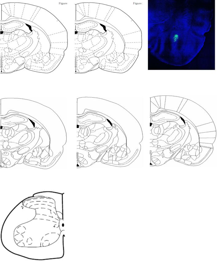
Ji and Neugebauer Molecular Brain         (2020) 13:128                                                                                     Page 6 of 10

 Fig. 3 Facilitatory effects of KOR activation in CeA on spinal dorsal horn neurons were blocked by silencing CRF neurons in the CeA. Extracellular
 single-unit recordings of 10 spinal dorsal horn WDR neurons in anesthetized rats. a-c Peristimulus time histograms show action potentials (spikes) per
 second in an individual spinal WDR neuron before (pre-drug control in ACSF; A) and during administration of U-69,593 (100 μM in microdialysis probe,
 15 min; B) into the CeA and during continued administration of U-69,593 while CRF-CeA neurons were silenced optogenetically (590 nm, 20 Hz, 1–5
 mW, 3 min; C). Optical silencing of CRF-CeA neurons inhibited the facilitatory effects of U-69,593. d Time course of effects of U-69,593 in the CeA and
 optical silencing of CRF-CeA neurons on the same WDR neuron. e Summary of effects of U-69,593 in the CeA and optical silencing of CRF-CeA
 neurons on WDR neurons (n = 10). * P < 0.05, ** P < 0.01, compared to pre-drug ACSF; + P < 0.05, ++ P < 0.01, compared to U-69,593 alone; ANOVA with
 Bonferroni posthoc tests. See text for results of ANOVA. Bar histograms show mean ± SE for the sample of neurons

Methods, “Systems Electrophysiology”). Administration of                      responses to innocuous (F2,23 = 7.43, P < 0.01) and noxious
U-69,593 (100 μM in microdialysis probe, 15 min) into the                     stimuli (F2,23 = 6.103, P < 0.05, compared to pre-drug
right CeA by microdialysis significantly increased                            ACSF, ANOVA with Bonferroni posthoc tests, n = 10;
Ji and Neugebauer Molecular Brain   (2020) 13:128                                                                  Page 7 of 10

Fig. 3a, b, d, e). In 6 of these neurons, the effect of optical   diffuse noxious inhibitory control (DNIC), a measure of
silencing (590 nm, 20 Hz, 1–5 mW, 15 min) of CRF-CeA              descending pain control [56, 57].
neurons was tested during continued administration of U-             Evidence for functional links between amygdala KOR
69,593 (100 μM in microdialysis probe) into the CeA for           and CRF systems [38] let us to explore the contribution
another 15 min (Fig. 3c, d, e). Optical silencing of CRF-         of CRF neurons to the descending modulation of noci-
CeA neurons significantly inhibited the facilitatory effects      ceptive processing. CRF neurons in the CeA project to
of U-69,593 on responses to innocuous (P < 0.01, F2,23 =          extra-amygdalar targets to promote averse-affective be-
7.43) and noxious stimuli (P < 0.05, F2,23 = 6.103; com-          haviors [39, 41, 67–69]. Amygdala CRF functions are
pared to U-69,593 alone; ANOVA with Bonferroni post-              under tonic inhibitory control of KOR as shown with a
hoc tests, n = 6). Pulsed activation of halorhodopsin was         KOR antagonist that enhanced the synaptic effects of
used (see Methods, Optogenetics) that was shown before            CRF on medial CeA neurons [70]. We used optogenetics
to silence CRF-CeA neurons [40]. Effects of optical silen-        to silence CRF neurons in transgenic Crh-Cre rats and
cing were reversible. Effects of U-69,593 and optical silen-      found inhibitory effects on CeA neurons as well as in-
cing on background activity did not reach the level of            hibition of the facilitatory effects of U-69,593 on spinal
statistical significance (F2,23 = 0.9726, ANOVA).                 neurons. The CeA cell type recorded in this in vivo
                                                                  study is not known but it is possible that they were CRF
                                                                  neurons that were inhibited directly with optical silen-
Discussion                                                        cing. This scenario is supported by the fact that CRF-
The data presented here show that pharmacological acti-           CeA neurons project to brainstem areas involved in de-
vation of KOR in the CeA with U-69,593 increases activ-           scending pain modulation such as the PAG [39]. Silen-
ity of neurons in the CeA and in the spinal dorsal horn,          cing of CRF neurons could also have effects on other
suggesting a functional connection and positive correl-           types of CeA neurons that form long range projections
ation between neuronal activity in these regions. This            including those containing somatostatin [71]. CRF neu-
connectivity was disrupted when CRF neurons in the                rons can excite [72] whereas somatostatin neurons in-
CeA were silenced optogenetically, which suggests an              hibit [71] PAG neurons. In addition to coupling to
important contribution of CRF neurons to amygdala                 brainstem centers involved in descending pain modula-
output coupled to descending modulation of spinal noci-           tion, the amygdala CeA could influence spinal nocicep-
ceptive processing.                                               tive processing through indirect influences on cortical
   We focused on KOR to explore the amygdala-spinal               regions such as ACC [73, 74] via cholinergic neurons in
cord connection because KOR in the amygdala have                  substantia innominata and nucleus basalis of Meynert
been linked to aversiveness, anxiety and stress responses         [75]. CeA neurons do not directly project to cortical re-
[38, 51–54] as well as to averse-affective behaviors in           gions; main amygdala input to ACC arises from the
stress- or injury-induced pain conditions [16, 55–57].            basolateral amygdala [76]. Therefore, the amygdala can
KOR is expressed in the CeA at particularly high levels           exert facilitatory influences on spinal nociceptive pro-
[58]. We used a selective KOR agonist (U-69,593) [59] at          cessing either through ascending or descending output
a concentration that was based on data in the literature          to pain control systems [73, 77, 78] by activating facilita-
from electrophysiological studies in brain slices (see [60,       tory or inhibiting inhibitory modulation. This remains to
61]. U-69,593 has been shown to decrease synaptic in-             be determined.
hibition of medial CeA neurons through a presynaptic                 On a technical note, prolonged application of U-69,
action [60, 61]. U-69,593 also produced an outward                593 does not produce desensitization of CeA neurons
current in medial CeA neurons, indicative of a postsyn-           [60]. This is important because our experimental proto-
aptic inhibitory effect [62, 63]. Increased activity of           col tested optical silencing of CRF-CeA neurons on the
lateral-capsular CeA neurons by U-69,593 in the present           facilitatory effect of prolonged administration of U-69,
study is consistent with disinhibition observed in the            593 on spinal nociceptive processing. Therefore, reversal
brain slice studies [60, 61]. The facilitatory effects of         of the effects of U-69,593 by optical silencing of CRF-
KOR activation in the CeA on spinal dorsal horn re-               CeA neurons was not due to desensitization. Finally,
sponses suggest a positive correlation and functional             pulsed optical activation of halorhodopsin has been re-
connection between amygdala activity and spinal noci-             ported to evoke rebound spiking after hyperpolarization
ceptive processing.                                               in certain cell types and brain regions [79, 80], which
   We studied KOR function in the right CeA because of            may not result in the desired silencing of neuronal activ-
evidence for right-hemispheric lateralization of CeA              ity. However, pulsed optical activation of halorhodopsin
function [49, 64, 65] and KOR function in the CeA re-             silenced CRF-CeA neurons without rebound spiking
lated to pain modulation [16, 56, 57, 66]. For example,           [40], which could be due to cell type, region and possibly
blockade of KOR in the right, but not left, CeA restored          species specific differences.
Ji and Neugebauer Molecular Brain (2020) 13:128 Page 8 of 10
Conclusion 5. Veinante P, Yalcin I, Barrot M. The amygdala between sensation and affect: a
This short communication provides important novel in- role in pain. J Mol Psychiatry. 2013;1:9.
6. Thompson JM, Neugebauer V. Amygdala plasticity and pain. Pain Res
formation about amygdalo-spinal interactions and the Manag. 2017;2017:8296501.
contribution of amygdala KOR and CRF systems. The 7. Han JS, Neugebauer V. mGluR1 and mGluR5 antagonists in the amygdala
data support the concept of a positive correlation and inhibit different components of audible and ultrasonic vocalizations in a
model of arthritic pain. Pain. 2005;113:211–22.
functional link between amygdala (CeA) and spinal noci- 8. Ji G, Li Z, Neugebauer V. Reactive oxygen species mediate visceral pain-
ceptive processing. Details of the neural circuitry and related amygdala plasticity and behaviors. Pain. 2015;156:825–36.
cell-types remain to be determined. 9. Minami M. Neuronal mechanisms for pain-induced aversion behavioral
studies using a conditioned place aversion test. Int Rev Neurobiol. 2009;85:
Abbreviations 135–44.
ACC: Anterior cingulate cortex; BLA: Basolateral nucleus of the amygdala; 10. Bourbia N, Ansah OB, Pertovaara A. Corticotropin-releasing factor in the
CeA: Central nucleus of the amygdala; CRF: Corticotropin releasing factor; rat amygdala differentially influences sensory-discriminative and
KOR: Kappa opioid receptor; PAG: Periaqueductal grey emotional-like pain response in peripheral neuropathy. J Pain. 2010;11:
1461–71.
Acknowledgments 11. Ansah OB, Bourbia N, Goncalves L, Almeida A, Pertovaara A. Influence of
We thank Dr. Robert Messing, University of Texas at Austin, for kindly amygdaloid glutamatergic receptors on sensory and emotional pain-related
providing the initial Crh-Cre rat breeding pairs. behavior in the neuropathic rat. Behav Brain Res. 2010;209:174–8.
12. Sagalajev B, Bourbia N, Beloushko E, Wei H, Pertovaara A. Bidirectional
Authors’ contributions amygdaloid control of neuropathic hypersensitivity mediated by
JG and VN conceived the study, designed the experiments and wrote the descending serotonergic pathways acting on spinal 5-HT3 and 5-HT1A
manuscript. JG carried out the experiments. Both authors participated in the receptors. Behav Brain Res. 2015;282:14–24.
data analysis and interpretation of results and read and approved the final 13. Ji G, Fu Y, Ruppert KA, Neugebauer V. Pain-related anxiety-like behavior
manuscript. requires CRF1 receptors in the amygdala. Mol Pain. 2007;3:13–7.
14. Ji G, Zhang W, Mahimainathan L, Narasimhan M, Kiritoshi T, Fan X, Wang J,
Funding Green TA, Neugebauer V. 5-HT2C receptor knockdown in the amygdala
Support for the authors’ work was provided by National Institutes of Health inhibits neuropathic-pain-related plasticity and behaviors. J Neurosci. 2017;
(NIH) grants NS038261, NS106902 and NS109255. 37:1378–93.
15. Thompson JM, Yakhnitsa V, Ji G, Neugebauer V. Small conductance calcium
Availability of data and materials activated potassium (SK) channel dependent and independent effects of
All data generated or analyzed during this study are included in this riluzole on neuropathic pain-related amygdala activity and behaviors in rats.
published article. Data files. Neuropharmacology. 2018;138:219–31.
used for this manuscript are available via a direct and reasonable request to 16. Navratilova E, Ji G, Phelps C, Qu C, Hein M, Yakhnitsa V, Neugebauer V,
the corresponding. Porreca F. Kappa opioid signaling in the central nucleus of the amygdala
author and approval from Texas Tech University Health Sciences Center promotes disinhibition and aversiveness of chronic neuropathic pain. Pain.
(TTUHSC). 2019;160:824–32.
17. Carrasquillo Y, Gereau RW. Activation of the extracellular signal-regulated
Ethics approval and consent to participate kinase in the amygdala modulates pain perception. J Neurosci. 2007;27:
All animal experiments were approved by the Institutional Animal Care and 1543–51.
Use Committee (IACUC) at Texas Tech University Health Sciences Center, 18. Rea K, Olango WM, Harhen B, Kerr DM, Galligan R, Fitzgerald S, Moore M,
Lubbock, TX (#14006, approved through 06/21/2021). Roche M, Finn DP. Evidence for a role of GABAergic and glutamatergic
signalling in the basolateral amygdala in endocannabinoid-mediated fear-
Consent for publication conditioned analgesia in rats. Pain. 2013;154:576–85.
Not applicable. 19. Rea K, Roche M, Finn DP. Modulation of conditioned fear, fear-conditioned
analgesia, and brain regional c-Fos expression following administration of
Competing interests muscimol into the rat basolateral amygdala. J Pain. 2011;12:712–21.
The authors declare that they have no competing interests. 20. Butler RK, Ehling S, Barbar M, Thomas J, Hughes MA, Smith CE, Pogorelov
VM, Aryal DK, Wetsel WC, Lascelles BDX. Distinct neuronal populations in
Author details the basolateral and central amygdala are activated with acute pain,
1
Department of Pharmacology and Neuroscience, Texas Tech University conditioned fear, and fear-conditioned analgesia. Neurosci Lett. 2017;661:
Health Sciences Center, School of Medicine, 3601 4th St, Lubbock, TX 11–7.
79430-6592, USA. 2Center of Excellence for Translational Neuroscience and 21. McGaraughty S, Heinricher MM. Microinjection of morphine into various
Therapeutics, Texas Tech University Health Sciences Center, Lubbock, TX, amygdaloid nuclei differentially affects nociceptive responsiveness and RVM
USA. 3Garrison Institute on Aging, Texas Tech University Health Sciences neuronal activity. Pain. 2002;96:153–62.
Center, Lubbock, TX, USA. 22. Ortiz JP, Heinricher MM, Selden NR. Noradrenergic agonist administration
into the central nucleus of the amygdala increases the tail-flick latency in
Received: 21 July 2020 Accepted: 9 September 2020 lightly anesthetized rats. Neuroscience. 2007;148:737–43.
23. Min MY, Yang HW, Yen CT, Chen CC, Chen CC, Cheng SJ. ERK, synaptic
plasticity and acid-induced muscle pain. Commun Integr Biol. 2011;4:394–6.
References 24. Manning BH. A lateralized deficit in morphine antinociception after
1. Neugebauer V, Li W, Bird GC, Han JS. The amygdala and persistent pain. unilateral inactivation of the central amygdala. J Neurosci. 1998;18:9453–70.
Neuroscientist. 2004;10:221–34. 25. Manning BH, Martin WJ, Meng ID. The rodent amygdala contributes to the
2. Neugebauer V. Amygdala physiology in pain. Handbook of Behavioral production of cannabinoid-induced antinociception. Neuroscience. 2003;
Neurosciences. 2020;26:101–13. 120:1157–70.
3. Kato F, Sugimura YK, Takahashi Y. Pain-associated neural plasticity in the 26. Kolber BJ, Montana MC, Carrasquillo Y, Xu J, Heinemann SF, Muglia LJ,
Parabrachial to central amygdala circuit : pain changes the brain, and the Gereau RW. Activation of metabotropic glutamate receptor 5 in the
brain changes the pain. Adv Exp Med Biol. 2018;1099:157–66. amygdala modulates pain-like behavior. J Neurosci. 2010;30:8203–13.
4. Wilson TD, Valdivia S, Khan A, Ahn HS, Adke AP, Martinez GS, Sugimura YK, 27. Gregoire S, Neugebauer V. 5-HT2CR blockade in the amygdala conveys
Carrasquillo Y. Dual and opposing functions of the central amygdala in the analgesic efficacy to SSRIs in a rat model of arthritis pain. Mol Pain. 2013;9:
modulation of pain. Cell Rep. 2019;29:332–46. 41.Ji and Neugebauer Molecular Brain (2020) 13:128 Page 9 of 10
28. Corder G, Ahanonu B, Grewe BF, Wang D, Schnitzer MJ, Scherrer G. An 51. Land BB, Bruchas MR, Lemos JC, Xu M, Melief EJ, Chavkin C. The dysphoric
amygdalar neural ensemble that encodes the unpleasantness of pain. component of stress is encoded by activation of the dynorphin kappa-
Science. 2019;363:276–81. opioid system. J Neurosci. 2008;28:407–14.
29. Medina G, Ji G, Gregoire S, Neugebauer V. Nasal application of 52. Crowley NA, Bloodgood DW, Hardaway JA, Kendra AM, McCall JG, Al-Hasani
neuropeptide S inhibits arthritis pain-related behaviors through an action in R, McCall NM, Yu W, Schools ZL, Krashes MJ, et al. Dynorphin controls the
the amygdala. Mol Pain. 2014;10:32. gain of an Amygdalar anxiety circuit. Cell Rep. 2016;14:2774–83.
30. Thompson JM, Ji G, Neugebauer V. Small-conductance calcium-activated 53. Lalanne L, Ayranci G, Kieffer BL, Lutz PE. The kappa opioid receptor: from
potassium (SK) channels in the amygdala mediate pain-inhibiting effects of addiction to depression, and back. Front Psychiatry. 2014;5:170.
clinically available riluzole in a rat model of arthritis pain. Mol Pain. 2015;11: 54. Smith JS, Schindler AG, Martinelli E, Gustin RM, Bruchas MR, Chavkin C.
51. Stress-induced activation of the dynorphin/kappa-opioid receptor system in
31. Cragg B, Ji G, Neugebauer V. Differential contributions of vasopressin V1A the amygdala potentiates nicotine conditioned place preference. J Neurosci.
and oxytocin receptors in the amygdala to pain-related behaviors in rats. 2012;32:1488–95.
Mol Pain. 2016;12:1744806916676491. 55. Xie JY, De FM, Kopruszinski CM, Eyde N, LaVigne J, Remeniuk B, Hernandez
32. Kim H, Thompson J, Ji G, Ganapathy V, Neugebauer V. Monomethyl P, Yue X, Goshima N, Ossipov M, et al. Kappa opioid receptor antagonists: a
fumarate inhibits pain behaviors and amygdala activity in a rat arthritis possible new class of therapeutics for migraine prevention. Cephalalgia.
model. Pain. 2017;158:2376–85. 2017;37:780–94.
33. Latremoliere A, Woolf CJ. Central sensitization: a generator of pain 56. Nation KM, De FM, Hernandez PI, Dodick DW, Neugebauer V, Navratilova E,
hypersensitivity by central neural plasticity. J Pain. 2009;10:895–926. Porreca F. Lateralized kappa opioid receptor signaling from the amygdala
34. Sandkuhler J. Translating synaptic plasticity into sensation. Brain. 2015;138: central nucleus promotes stress-induced functional pain. Pain. 2018;159:
2463–4. 919–28.
35. Heinricher MM, Tavares I, Leith JL, Lumb BM. Descending control of 57. Phelps CE, Navratilova E, Dickenson AH, Porreca F, Bannister K. Kappa opioid
nociception: specificity, recruitment and plasticity. Brain Res Rev. 2009;60: signaling in the right central amygdala causes hind paw specific loss of
214–25. diffuse noxious inhibitory controls in experimental neuropathic pain. Pain.
36. Li JN, Sheets PL. The central amygdala to periaqueductal gray pathway 2019;160:1614–21.
comprises intrinsically distinct neurons differentially affected in a model of 58. Cahill CM, Taylor AM, Cook C, Ong E, Moron JA, Evans CJ. Does the kappa
inflammatory pain. J Physiol. 2018;596:6289–305. opioid receptor system contribute to pain aversion? Front Pharmacol. 2014;
37. Dickenson AH, Navratilova E, Patel R, Porreca F, Bannister K. Supraspinal 5:253.
opioid circuits differentially modulate spinal neuronal responses in 59. Zhou L, Lovell KM, Frankowski KJ, Slauson SR, Phillips AM, Streicher JM, Stahl
neuropathic rats. Anesthesiology. 2020;132:881–94. E, Schmid CL, Hodder P, Madoux F, et al. Development of functionally
38. Bruchas MR, Land BB, Chavkin C. The dynorphin/kappa opioid system as a selective, small molecule agonists at kappa opioid receptors. J Biol Chem.
modulator of stress-induced and pro-addictive behaviors. Brain Res. 2010; 2013;288:36703–16.
1314:44–55. 60. Gilpin NW, Roberto M, Koob GF, Schweitzer P. Kappa opioid receptor
39. Pomrenze MB, Millan EZ, Hopf FW, Keiflin R, Maiya R, Blasio A, Dadgar J, activation decreases inhibitory transmission and antagonizes alcohol effects
Kharazia V, De GG, Crawford E, et al. A transgenic rat for investigating the in rat central amygdala. Neuropharmacology. 2014;77:294–302.
anatomy and function of corticotrophin releasing factor circuits. Front 61. Kang-Park M, Kieffer BL, Roberts AJ, Siggins GR, Moore SD. kappa-opioid
Neurosci. 2015;9:487. receptors in the central amygdala regulate ethanol actions at presynaptic
40. De Guglielmo G, Kallupi M, Pomrenze MB, Crawford E, Simpson S, GABAergic sites. J Pharmacol Exp Ther. 2013;346:130–7.
Schweitzer P, Koob GF, Messing RO, George O. Inactivation of a CRF- 62. Chieng BC, Christie MJ, Osborne PB. Characterization of neurons in the rat
dependent amygdalofugal pathway reverses addiction-like behaviors in central nucleus of the amygdala: cellular physiology, morphology, and
alcohol-dependent rats. Nat Commun. 2019;10:1238. opioid sensitivity. J Comp Neurol. 2006;497:910–27.
41. Pomrenze MB, Giovanetti SM, Maiya R, Gordon AG, Kreeger LJ, Messing 63. Zhu W, Pan ZZ. Synaptic properties and postsynaptic opioid effects in rat
RO. Dissecting the roles of GABA and neuropeptides from rat central central amygdala neurons. Neuroscience. 2004;127:871–9.
amygdala CRF neurons in anxiety and fear learning. Cell Rep. 2019;29: 64. Carrasquillo Y, Gereau RW. Hemispheric lateralization of a molecular signal
13–21. for pain modulation in the amygdala. Mol Pain. 2008;4:24.
42. Ji G, Neugebauer V. Contribution of Corticotropin-releasing factor receptor 1 65. Sadler KE, McQuaid NA, Cox AC, Behun MN, Trouten AM, Kolber BJ.
(CRF1) to serotonin receptor 5-HT2CR function in amygdala neurons in a Divergent functions of the left and right central amygdala in visceral
neuropathic pain model. Int J Mol Sci. 2019;20(18):4380. nociception. Pain. 2017;158:747–59.
43. Ji G, Yakhnitsa V, Kiritoshi T, Presto P, Neugebauer V. Fear extinction learning 66. Navratilova E, Nation K, Remeniuk B, Neugebauer V, Bannister K, Dickenson
ability predicts neuropathic pain behaviors and amygdala activity in male AH, Porreca F. Selective modulation of tonic aversive qualities of
rats. Mol Pain. 2018;14:1744806918804441. neuropathic pain by morphine in the central nucleus of the amygdala
44. Yuan SB, Ji G, Li B, Andersson T, Neugebauer V, Tang SJ. A Wnt5a signaling requires endogenous opioid signaling in the anterior cingulate cortex. Pain.
pathway in the pathogenesis of HIV-1 gp120-induced pain. Pain. 2015;156: 2020;161:609–18.
1311–9. 67. Marcilhac A, Siaud P. Identification of projections from the central nucleus
45. Mazzitelli M, Neugebauer V. Amygdala group II mGluRs mediate the of the amygdala to the paraventricular nucleus of the hypothalamus which
inhibitory effects of systemic group II mGluR activation on behavior and are immunoreactive for corticotrophin-releasing hormone in the rat. Exp
spinal neurons in a rat model of arthritis pain. Neuropharmacology. 2019; Physiol. 1997;82:273–81.
158:107706. 68. Fendt M, Koch M, Schnitzler HU. Corticotropin-releasing factor in the caudal
46. Fu Y, Neugebauer V. Differential mechanisms of CRF1 and CRF2 receptor pontine reticular nucleus mediates the expression of fear-potentiated startle
functions in the amygdala in pain-related synaptic facilitation and behavior. in the rat. Eur J Neurosci. 1997;9:299–305.
J Neurosci. 2008;28:3861–76. 69. McCall JG, Al-Hasani R, Siuda ER, Hong DY, Norris AJ, Ford CP, Bruchas MR.
47. Fu Y, Han J, Ishola T, Scerbo M, Adwanikar H, Ramsey C, Neugebauer V. PKA CRH engagement of the locus Coeruleus noradrenergic system mediates
and ERK, but not PKC, in the amygdala contribute to pain-related synaptic stress-induced anxiety. Neuron. 2015;87:605–20.
plasticity and behavior. Mol Pain. 2008;4:26–46. 70. Kang-Park M, Kieffer BL, Roberts AJ, Siggins GR, Moore SD. Interaction of
48. Kiritoshi T, Ji G, Neugebauer V. Rescue of Impaired mGluR5-driven CRF and kappa opioid systems on GABAergic neurotransmission in the
Endocannabinoid signaling restores prefrontal cortical output to inhibit pain mouse central amygdala. J Pharmacol Exp Ther. 2015;355:206–11.
in arthritic rats. J Neurosci. 2016;36:837–50. 71. Penzo MA, Robert V, Li B. Fear conditioning potentiates synaptic
49. Ji G, Neugebauer V. Hemispheric lateralization of pain processing by transmission onto long-range projection neurons in the lateral subdivision
amygdala neurons. J Neurophysiol. 2009;1102:2253–64. of central amygdala. J Neurosci. 2014;34:2432–7.
50. Ji G, Neugebauer V. CB1 augments mGluR5 function in medial prefrontal 72. Bowers LK, Swisher CB, Behbehani MM. Membrane and synaptic effects of
cortical neurons to inhibit amygdala hyperactivity in an arthritis pain model. corticotropin-releasing factor on periaqueductal gray neurons of the rat.
Eur J Neurosci. 2014;39:455–66. Brain Res. 2003;981:52–7.Ji and Neugebauer Molecular Brain (2020) 13:128 Page 10 of 10
73. Zhuo M. Descending facilitation. Mol Pain. 2017;13:1744806917699212.
74. Toyoda H, Li XY, Wu LJ, Zhao MG, Descalzi G, Chen T, Koga K, Zhuo M.
Interplay of amygdala and cingulate plasticity in emotional fear. Neural
Plast. 2011;2011:813749.
75. Gozzi A, Jain A, Giovannelli A, Bertollini C, Crestan V, Schwarz AJ, Tsetsenis T,
Ragozzino D, Gross CT, Bifone A. A neural switch for active and passive fear.
Neuron. 2010;67:656–66.
76. Sharma KK, Kelly EA, Pfeifer CW, Fudge JL. Translating fear circuitry:
amygdala projections to Subgenual and Perigenual anterior cingulate in the
macaque. Cereb Cortex. 2020;30:550–62.
77. Gebhart GF. Descending modulation of pain. Neurosci Biobehav Rev. 2004;
27:729–37.
78. Neugebauer V. Serotonin – pain modulation. Handbook of Behavioral
Neurosciences. 2020;31:309–20.
79. Wiegert JS, Mahn M, Prigge M, Printz Y, Yizhar O. Silencing neurons: tools,
applications, and experimental constraints. Neuron. 2017;95:504–29.
80. Madisen L, Mao T, Koch H, Zhuo JM, Berenyi A, Fujisawa S, Hsu YW, Garcia
AJ 3rd, Gu X, Zanella S, et al. A toolbox of Cre-dependent optogenetic
transgenic mice for light-induced activation and silencing. Nat Neurosci.
2012;15:793–802.
Publisher’s Note
Springer Nature remains neutral with regard to jurisdictional claims in
published maps and institutional affiliations.You can also read