Central neuromodulation in chronic migraine patients with suboccipital stimulators: a PET study - Reed Migraine Centers
←
→
Page content transcription
If your browser does not render page correctly, please read the page content below
DOI: 10.1093/brain/awh022             Advanced Access publication November 7, 2003                 Brain (2004), 127, 220±230

Central neuromodulation in chronic migraine
patients with suboccipital stimulators: a PET study
Manjit S. Matharu,1 Thorsten Bartsch,1 Nick Ward,2 Richard S. J. Frackowiak,2 Richard Weiner3 and
Peter J. Goadsby1
1Headache  Group and 2Wellcome Department of Imaging             Correspondence to: Professor Peter J. Goadsby, Institute
Neuroscience, Institute of Neurology and The National            of Neurology, Queen Square, London WC1N 3BG, UK
Hospital for Neurology and Neurosurgery, Queen Square,           E-mail: peterg@ion.ucl.ac.uk
London, UK and 3Department of Neurosurgery,
Presbyterian Hospital of Dallas, Dallas, Texas, USA

Summary
Electrical stimulation of primary sensory afferents is           sia; (3) stimulator partially activated: patient with
known to have an antinociceptive effect. Animal and              intermediate levels of pain and paraesthesia. All scans
functional imaging studies suggest a role for supraspinal        were processed and analysed using Statistical
structures in this response. Eight patients with chronic         Parametric Mapping (SPM) 99. There were signi®cant
migraine (>15 days per month of attacks of migraine              changes in rCBF in the dorsal rostral pons, anterior
without aura), who had shown a marked bene®cial                  cingulate cortex (ACC) and cuneus, correlated to pain
response to implanted bilateral suboccipital stimulators,        scores, and in the ACC and left pulvinar, correlated to
were studied. Stimulation evoked local paraesthesia, the         stimulation-induced paraesthesia scores. The activation
presence of which was a criterion of pain relief. On             pattern in the dorsal rostral pons is highly suggestive of
stimulation, the headache began to improve instantan-            a role for this structure in the pathophysiology
eously and was completely suppressed within 30 min.              of chronic migraine. The localization and persistence of
On switching off the stimulation, the headache recurred          activity during stimulation is exactly consistent with a
instantly and peaked within 20 min. PET scans were               region activated in episodic migraine, and with the per-
performed using regional cerebral blood ¯ow (rCBF) as            sistence of activation of that area after successful treat-
a marker of neuronal activity. Each patient was                  ment. The dorsal rostral pons may be a locus of
scanned in the following three states: (1) stimulator at         neuromodulation by suboccipital stimulation. In add-
optimum settings: patient pain-free but with paraesthe-          ition, suboccipital stimulation modulated activity in the
sia; (2) stimulator off: patient in pain and no paraesthe-       left pulvinar.

Keywords: headache; migraine; periaqueductal grey; pons; pulvinar

Abbreviations: ACC = anterior cingulate cortex; CDH = chronic daily headache; GON = greater occipital nerve; IHS =
International Headache Society; PAG = periaqueductal grey; PET = positron emission tomography; rCBF = regional
cerebral blood ¯ow; VRS = verbal rating scale

Introduction
Chronic daily headache (CDH), de®ned as headache occur-          International Headache Society (IHS) classi®cation
ring on 15 days or more a month, is a widespread problem in      (Headache Classi®cation Committee of The International
neurological practice. Population-based studies in the USA       Headache Society, 2004). In population-based surveys,
(Scher et al., 1998), Europe (Castillo et al., 1999; Lanteri-    chronic migraine occurs in 1.3±2.4% of the population
Minet et al., 2003) and Asia (Wang et al., 2000) suggest that    (Scher et al., 1998; Castillo et al., 1999; Lanteri-Minet et al.,
4±5% of the general population have CDH. Chronic migraine        2003).
is a subset of CDH in which daily or near-daily headaches           Migraine is a form of primary neurovascular headache that
occur, some of which are accompanied by migrainous               is likely to be based on dysfunction in the brain (Goadsby
features (Welch and Goadsby, 2002). Diagnostic criteria          et al., 2002). PET in primary headaches, such as migraine
have been proposed (Silberstein et al., 1996), and chronic       (Weiller et al., 1995) and cluster headache (May et al., 1998a,
migraine is now recognized as a distinct entity in the revised   2000), has demonstrated activations in brain areas associated
Brain Vol. 127 No. 1 ã Guarantors of Brain 2003; all rights reserved
Chronic migraine and the brainstem              221

with pain, such as the cingulate cortex, insulae, frontal cortex,       Reed, 1999; Weiner, 2002). The tips of the electrodes were
thalamus, basal ganglia and the cerebellum. These areas are             positioned super®cial to the cervical muscular fascia and transverse
similarly activated when head pain is induced by injection of           to the greater occipital nerve (GON) trunk at the level of the ®rst
capsaicin into the forehead of volunteers (May et al., 1998b).          cervical spine. The stimulation settings were individually chosen by
                                                                        the patients according to the pain relief; the pulse width ranged from
In addition to these generic pain areas, activations in speci®c
                                                                        90 to 180 ms, the frequency ranged from 60 to 130 Hz and the
brain regions can be seen in episodic migraine that are not             amplitude from 1.5 to 10.5 V. The area of paraesthesia during
observed when the ®rst (ophthalmic) division pain pathways              stimulation was restricted to the area adjacent to the implanted
are activated by the capsaicin injections. Speci®cally,                 electrodes in the distribution of the GON.
brainstem areas are activated in episodic migraine (Weiller                Informed consent was obtained from all patients and the study was
et al., 1995), one of which was recently re®ned in localization         approved by the Ethics Committee of the National Hospital for
to the dorsal pons (Bahra et al., 2001). The underlying                 Neurology and Neurosurgery, London, UK and the Presbyterian
pathophysiology of chronic migraine is likely to be similar to          Hospital of Dallas, TX, USA. Permission to administer radioactivity
that of episodic migraine, with brainstem activation, perhaps           was obtained (Administration of Radioactive Substances Advisory
continuously.                                                           Committee of the Department of Health, UK).
   Data on the prognosis of optimally managed chronic
migraine are limited. The literature on CDH managed in
                                                                        Case material
headache centres, where the majority of CDH patients have
                                                                        Case 1
chronic migraine (Silberstein et al., 1996), shows that
                                                                        A 53-year-old, right-handed woman presented with an 8-month
49±91% of the patients do well (Silberstein and Lipton,                 history of daily headaches in September 2000. For 25 years prior to
2001). Hence a signi®cant minority of patients continue to              the onset of the daily headaches, she had had episodic headaches
have intractable headaches despite optimum therapy; this is in          which satis®ed the IHS criteria for migraine without aura (Headache
keeping with our experience.                                            Classi®cation Committee of The International Headache Society,
   We report eight patients with the IHS diagnosis of chronic           2004). In January 2000, she slipped on ice and fell backwards, hitting
migraine who have shown a marked bene®cial response to                  the back of her head on the ¯oor. On regaining consciousness after
implanted bilateral suboccipital stimulators. We sought to              15 min, she had amnesia for the event, a severe headache, loss of
understand the central mechanisms that underpin this                    vision in the right visual ®eld and mild left hemiparesis, manifesting
antinociceptive effect of implanted suboccipital stimulators            as poor left-hand ®ne-®nger movements and left-sided clumsy gait.
                                                                        An MRI scan of the head was normal. Vision recovered completely
by performing a PET study. We sought to determine the brain
                                                                        over the next 2 months. The left hemiparesis has remained
structures active in chronic migraine and how they are
                                                                        unchanged.
modulated by suboccipital stimulation with regional cerebral               She described a bilateral headache that started immediately after
blood ¯ow (rCBF) PET as an index for neuronal activity                  the head injury and persisted. It was worse on the right than on the
(Frackowiak and Friston, 1994). We hypothesized, based on               left and centred on the occiput. The headache was constant, severe,
the literature, that suboccipital stimulation would involve the         and had a pulsating quality. It was associated with nausea, vomiting,
periaqueductal grey (PAG), thalamic nuclei, particularly the            photophobia, phonophobia, osmophobia and worsening with move-
pulvinar, the anterior cingulate cortex (ACC) and the insula.           ment. It was also associated with bilateral nasal blockage and
In addition, we hypothesized that the pain of chronic migraine          rhinorrhoea. She frequently had aura symptoms, which lasted 30±60
would involve the dorsal rostral pons, thalamus, ACC and                min, particularly when the intensity of the headache peaked. The
insula.                                                                 headache could be exacerbated by psychological stress and bright
                                                                        lights. In the family history, her mother, son and daughter suffered
                                                                        from migraine. Neurological examination revealed a hypoaesthetic
                                                                        area in the GON dermatome. There were no cervical or occipital
Methods                                                                 trigger points and no tenderness. Cranial nerve examination was
General methods and suboccipital stimulators                            normal except for a partial right Horner's syndrome. Motor
Eight right-handed patients (age 32±53 years, mean 44 years; seven      examination revealed dif®culty with ®ne ®nger movements in the
female, one male) with the IHS diagnosis of chronic migraine            left hand, though power testing was otherwise normal in the upper
(Headache Classi®cation Committee of The International Headache         limbs. There was mild weakness of left knee ¯exion and ankle
Society, 2004) as the underlying phenotype, who had implanted           dorsi¯exion.
bilateral suboccipital stimulators, were studied. In six patients the      In September 2000, bilateral suboccipital stimulators were sited.
onset of chronic migraine was associated with trauma; in the other      They have been highly effective, consistently rendering the patient
two it evolved from episodic migraine. We have classi®ed the            pain-free within 5±10 min. The patient has been able to stop all
patients based on their headache phenotype as having symptoms           headache abortive and preventive medications.
consistent with chronic migraine in the new IHS diagnostic criteria
(Headache Classi®cation Committee of The International Headache
Society, 2004). Individual cases are outlined below in summary,         Case 2
detailed descriptions being available from the authors.                 A 43-year-old, right-handed woman developed daily headaches in
   The stimulators (Medtronic Synergyâ/Itrelâ; Medtronic, MN,           May 1995 following a road traf®c accident. At presentation she
USA) were implanted subcutaneously at the Department of                 described a bilateral headache, which was worse on the right than on
Neurosurgery, Presbyterian Hospital of Dallas, USA (Weiner and          the left. It was centred on the occiput with radiation to the parietal
222 M. S. Matharu et al.
region, temple, retro-orbital and orbital region, and the neck. It was backwards, landing on her occiput and back. At presentation, on 10
very severe in intensity and had a throbbing or tightening quality. It days per month she had a mild background headache or was pain-
was associated with nausea, photophobia, phonophobia and free, while on the remaining 20 days per month she had an
osmophobia, and worsened with movement and bilateral con- exacerbation. The background headache was holocranial, mild in
junctival injection. She denied vomiting, any symptoms of a severity, with a tightening quality. The exacerbation was bilateral,
migraine aura or any other cranial autonomic features. The headache centred on the occipital region with radiation to the vertex, temple
could be triggered by over sleeping, lack of sleep, over exertion, and the forehead. It was severe in intensity and had a pulsating
changes in barometric pressure, emotional stress, driving in a motor quality. It was associated with nausea, vomiting, photophobia,
vehicle, and a noisy environment. phonophobia, worsening with movement, nasal blockage and
In 1999 she had bilateral GON blocks and bilateral supra-orbital lacrimation. The headache could be triggered by over exertion,
blocks, with which she received 6 h of pain relief. There was no lack of sleep, over sleeping, bright lights and barometric pressure
family history of headaches. Neurological examination was unre- changes. In the family history, her son and an aunt have migraine.
markable. Neurological examination revealed a hypoaesthetic area con®ned
In August 2001 bilateral suboccipital stimulators were implanted to the distribution of the GON. Bilateral suboccipital stimulators
with which there was a moderate improvement in the headache. In were sited in 2000, and have been highly effective. On switching on
September 2001 temporary implants were placed over the supra- the stimulator, symptoms begin to improve immediately and the
orbital region, which further reduced the pain to mild intensity. In patient is pain-free within 20 min.
October 2001, permanent supra-orbital stimulators were sited
bilaterally. She has managed to completely stop all analgesics and
headache preventive medications. Case 5
A 46-year-old, right-handed woman presented in early 2002 with a
history of daily headaches since 1999. She had migraine without
Case 3 aura between the ages of 12 and 16 years. Since 1999 she has had
A 44-year-old, right-handed man presented with a history of daily headaches. At presentation, on 5±10 days per month she had a
headaches since his teenage years. He described bilateral headaches background headache, which was holocranial, mild to moderate, and
centred on the forehead with radiation to the temples. They were of tightening quality. On the remaining 20±25 days per month, she
severe in intensity and had a throbbing quality. These headaches had a severe bilateral headache, worse on the right than the left,
were associated with worsening on movement and bilateral starting over the forehead and temple before becoming holocranial.
lacrimation. He denied nausea, vomiting, photophobia, phono- The headaches were throbbing, pressing or tightening in quality.
phobia, osmophobia and any symptoms suggestive of a migrainous They were associated with photophobia, phonophobia, osmophobia,
aura or any other cranial autonomic features. A typical episode worsening with movement and lightheadedness. She also reported
lasted 1±3 days. It occurred 1±2 times annually. bilateral lacrimation, nasal blockage and facial sweating in
In 1991, his headaches began to increase in frequency and association with the headaches. She denied nausea, vomiting and
duration such that by 1996 he had a daily, constant headache. At any other cranial autonomic features. On 10 days per month the
presentation in July 2000, he described a bilateral headache, worse headache occurred in association with a sensory aura lasting 30 min.
on the right than on the left. It was centred on the occiput with The headaches could be triggered by dietary factors (chocolate,
radiation to the vertex, parietal region and the forehead. On 5±15 cheese and monosodium glutamate), alcohol, over exertion and lack
days per month he had a background headache, which was moderate of sleep. In the family history, her daughter suffers from migraine.
in severity, had a throbbing quality and was associated with mild Neurological examination was unremarkable.
nausea, photophobia and worsening with movement. On the In April 2002, bilateral suboccipital stimulators were sited. The
remaining 15±25 days he had an exacerbation which was very procedure was complicated by the development of a self-limiting
severe in intensity and had a burning, stabbing or throbbing quality. abdominal haematoma. The stimulator is highly effective, rendering
It was associated with nausea, vomiting, photophobia, osmophobia the patient pain-free within 5 min.
and worsening with movement. He denied photophobia, any
symptoms suggestive of migrainous aura or any cranial autonomic
features. The exacerbations could be triggered by lack of sleep, over Case 6
sleeping, over exertion and bright lights. Infrequently and inconsist- A 32-year-old, right-handed woman presented in early 2002 with a
ently, neck movement could trigger a headache. C2 blockade history of daily headaches since mid-2000. She had infrequent
provided relief for a few days, as did bilateral GON blocks. migraine with aura during her childhood and teenage years. Since
Detailed neurological examination was entirely normal except for mid-2000 she had had daily, constant headaches, and two car
hypoaesthesia over the GON distribution bilaterally. accidents in the late 1990s produced associated increases in
In August 2000 a right-sided suboccipital stimulator was sited. headache frequency. At presentation, she had a constant background
There was marked bene®t: the headache improved on both sides. In headache on 10±15 days per month and a superimposed exacerbation
October 2000, a left suboccipital stimulator was sited. When the on 15±20 days per month. The background headache was bilateral,
stimulators are switched on, the patient is rendered pain-free within 5 holocranial, mild to moderate in severity, and had a tightening
min. quality. It was featureless in that there were no associated
migrainous or cranial autonomic features. The exacerbation was
severe, bilateral and throbbing. It was associated with nausea,
Case 4 vomiting, photophobia, phonophobia, worsening with movement
A 45-year-old, right-handed female presented with a history of daily and nasal blockage. She had a visual aura in the form of ¯ashing
headaches since May 1993, when she slipped on ice and fell lights in the whole of the visual ®eld a few minutes prior to theChronic migraine and the brainstem 223
headache. The visual aura lasted 15±30 min. The typical In the family history her mother has migraine. Neurological
exacerbation lasted from 6 h to up to 2 days. Alcohol, hunger, examination was unremarkable.
over exertion, menstruation and weather change could trigger the She had bilateral GON injections in 1999, with which there was a
exacerbation. marked reduction in the headache that was sustained for 6±8 weeks.
In early 2002 she had bilateral GON injections on three occasions, In December 1999 bilateral suboccipital stimulators were sited, with
with which there was bene®t to the headache for 4±5 days after the which there has been a marked improvement. On switching the
procedure. Her mother suffers from migraine. Neurological exam- stimulator on there is immediate bene®t and the headache is
ination was unremarkable. suppressed within 15 min.
In May 2002, bilateral suboccipital stimulators were sited. The
stimulators have been highly effective, rendering the patient pain
free within 25 min. PET study design
Each patient had 12 consecutive radioactive H215O PET scans in the
following three states: (1) stimulator at optimum settings: patient
Case 7 pain-free and with paraesthesia; (2) stimulator off: patient in pain
A 44-year-old, right-handed female presented in early 2001 with a and no paraesthesia; (3) stimulator partially activated; patient with
5-year history of daily headaches. She had had migraine with aura intermediate levels of pain and paraesthesia. Four scans were
since the age of 11 years. In 1987 the frequency and duration of the performed in each state. The order of the states in which the patients
headaches began to increase gradually, such that since August 1996 were scanned was randomized and counterbalanced across subjects.
she has had daily headaches. On 10±15 days per month she had a At the end of each scan, patients rated their headache intensity on a
background headache, which was holocranial, moderately severe VRS (0 = no pain, 10 = the most severe pain) and scored their
and tightening or pressing in quality; she denied any migrainous or paraesthesia (0 = no paraesthesia, 5 = strongest paraesthesia). On the
cranial autonomic features in association with the background day prior to scanning, the patients underwent a trial in which the time
headache. On the remaining 15±20 days per month she had a severe course of the stimulator effect was assessed and the patients were
exacerbation of the headache. The usual exacerbation lasted 2±4 trained to rate their pain sensation and paraesthesia. Throughout the
days and occurred four or ®ve times per month. The exacerbations scan, the patients experienced no pain or sensory symptoms apart
were bilateral, started over the occiput before becoming holocranial, from those related to the headache and the suboccipital stimulators.
and had a throbbing quality. They were associated with nausea, Participants had their eyes closed during all scans.
vomiting, photophobia and phonophobia, and worsened with
movement. She also reported bilateral ptosis, eyelid oedema,
lacrimation, nasal blockage and rhinorrhoea in association with the Data acquisition
exacerbations. She reported visual aura symptoms in the form of The PET scans were obtained with an ECAT EXACT HR+ scanning
¯ashing lights in the whole visual ®eld; these occurred ~30 min prior system (CTI, Knoxville, TN, USA) in 3D mode with collimating
to the onset of the some exacerbations and lasted 5±10 min. Alcohol, septa retracted. An antecubital vein cannula was used to administer
menstruation, over exertion and over sleeping could trigger an the tracer. For each measurement of relative rCBF, 9 mCi of H215O
exacerbation. In the family history, her father and brother suffer was given as an intravenous bolus over 20 s, followed by a 20 s
from migraine. General and neurological examinations were normal. saline ¯ush. Integrated radioactivity counts were accumulated over a
In May 2001 bilateral GON injections were performed; she 90 s acquisition period, beginning 5 s before the peak of radioactivity
developed an area of numbness over the GON distributions and there registered in the head. The interval between scans was 8±30 min,
was a marked reduction of pain which lasted ~24 h. Subsequently, allowing adequate decay of radioactivity and acquisition of the
she had two further trials of GON injections, with which there was conditions of interest. A transmission scan was obtained prior to
no bene®t to the headache. In August 2001, bilateral suboccipital collection of the emission data to correct for radiation attenuation by
stimulators were sited. They were highly effective, markedly tissues in the head. The corrected data were reconstructed into 63
reducing the severity of pain from severe [verbal rating scale transverse planes (separation 2.4 mm) and into a 128 3 128 pixel
(VRS) 8±9/10] to mild (VRS 1±3/10) over 15 min. image matrix (pixel size 2.1 3 2.1 mm2) by 3D ®ltered back-
projection.
Case 8
A 42-year-old, right-handed female presented in 1999 with a history Statistical analysis
of daily headaches since 1993. Her headaches started about a week The data were analysed with Statistical Parametric Mapping (SPM)
after she fell and banged her head. For the ®rst 3±6 months the 99 (Wellcome Department of Imaging Neuroscience, London, UK,
headaches were episodic; thereafter, she has had daily headache. The http://www.®l.ion.ucl.ac.uk/spm) implemented in MATLAB
background headache was holocranial, mild to moderate in severity, (Mathworks, USA) and run on a SPARC workstation (Sun
had a tightening quality and was completely featureless. The Microsystems). The images were initially realigned with reference
exacerbation was bilateral, centred on the occiput and showed to the ®rst image to correct for motion artefact and then spatially
holocranial radiation. It was associated with nausea, vomiting, normalized into a standard stereotactic space de®ned by the atlas of
photophobia, phonophobia, worsening with movement, conjunctival Talairach and Tournoux (Talairach and Tournoux, 1988). The
injection, lacrimation and rhinorrhoea. She denied osmophobia, any normalized images were smoothed with a 10 mm isotropic full-width
symptoms suggestive of a migrainous aura or any other cranial half-maximum Gaussian kernel to account for intersubject differ-
autonomic features. The exacerbation could be triggered by change ences in anatomy and to allow valid statistical inference according to
of weather, menstruation, alcohol, over sleeping and over exertion. Gaussian random ®eld theory.224 M. S. Matharu et al.
Statistical analysis was performed using a multisubject, single- Table 1 Talairach coordinates of regions of interest
group ®xed-effects model, in which the pain scores and paraesthesia de®ned in the a priori hypothesis
scores (as a marker of stimulation) were modelled as separate
Region of interest Talairach coordinates (mm)
covariates across the group. In addition, the effect of global
differences in cerebral blood ¯ow between scans was modelled as x y z
a covariate of no interest by subject-speci®c analysis of covariance
scaling of activity to a nominal mean global activity of 50 ml/100 g/ Pain
min. The resulting covariates were used in a general linear model. Pons ±2 ±28 ±22
The parameter estimates for each covariate resulting from the least ACC 0 30 18
mean squares ®t of the model to the data were calculated and Right insula 28 ±26 0
Left insula ±34 ±30 0
SPM{F} resulting from linear contrasts of covariates were generated
Right thalamus 12 ±4 10
and stored as separate images (thresholded at P < 0.001, uncorrected Left thalamus ±12 ±4 10
for multiple comparisons). F-tests were used because we had no a Paraesthesia
priori hypothesis about the nature of the relationship between PAG 4 ±26 ±16
covariate and rCBF for a number of regions. Small volume ACC 0 30 18
corrections (sphere of radius10 mm) were applied across the regions Right insula 28 ±26 0
of interest de®ned in the a priori hypothesis. The coordinates for the Left insula ±34 ±30 0
regions of interest were prede®ned on the basis of experimental data Right pulvinar 16 ±32 8
in the literature (Weiller et al., 1995; Hautvast et al., 1997; Bahra Left pulvinar ±16 ±32 8
et al., 2001) (Table 1). Signi®cant voxels are reported at P < 0.05,
corrected for multiple comparisons. A separate model in which each
of the states (1), (2) and (3) were employed as covariates was used to
identify the parameter estimates (or average relative changes in
rCBF) in each condition across the group, in order to better describe The eight patients have now been followed up for a period
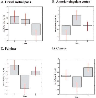
the data (Fig. 6) at voxels of interest. ranging from 7 months to 3 years, averaging over 1.5 years. In
The association between the pain score ratings and stimulator- all patients the initial bene®cial response has been maintained
induced paraesthesia was determined using Pearson's correlation throughout the follow-up period.
coef®cient and standard linear regression.
Psychophysical data
Results Figure 1 shows the results of the pain assessment session on
Suboccipital stimulators in chronic migraine the day prior to scanning. On switching off the stimulator, the
patients headache recurred immediately and peaked within 20 min.
Eight patients underwent 12 implant procedures for the On switching on the stimulator, the headache began to
treatment of chronic migraine. The only immediate post- improve immediately and within 30 min rendered six patients
operative complication reported was the development of an pain-free while the other two had only mild residual pain. All
abdominal haematoma at the implantation site of the receiver/ patients reported that the effect of suboccipital stimulation on
generator. Three patients have needed revisions after the the time course of pain recurrence and relief was highly
initial implantation because of lead migration; one of these consistent.
patients has needed two revisions. All patients reported that Pain and paraesthesia scores obtained during the PET scans
switching on the stimulator rapidly suppressed the pain but it were negatively correlated. Pearson's correlation coef®cient
recurred instantaneously when the stimulator was switched was ±0.61 (P < 0.001). Figure 2 shows the scatter plot of pain
off. and paraesthesia scores, and the plot of standard linear
Four patients have had an excellent response; their regression. Figure 3 shows the mean pain and paraesthesia
headaches are completely suppressed and they rarely have score by the scanning state.
breakthrough headaches. Two patients described their
response to stimulation as very good; the headaches are
completely suppressed most of the time, though they have PET data
breakthrough headaches on about 10 days per month, for Pain analysis
which they either have to take analgesics or increase the The regions in which rCBF covaried with pain scores were
stimulation amplitude. The other two patients continued to the dorsal rostral pons, right lentiform nucleus, head of the
have constant headaches but both reported their response to right caudate nucleus, ACC, postcentral gyrus [Brodmann
stimulation as good, with the severity of the headaches area (BA) 1/2/3], left frontal cortex (BA 11), right frontal
reduced by 50±75%; one of these patients had derived further cortex (BA 44), right temporal cortex (BA 22), cuneus,
bene®t from implanted bilateral supraorbital stimulators. All precuneus, right occipital cortex (BA 18), left occipital cortex
patients reported that they had managed to either completely (BA19) and cerebellum (P < 0.001 uncorrected for multiple
stop or considerably reduce the headache medications they comparisons). The coordinates and the Z scores of these
were taking. regions are listed in Table 2.Chronic migraine and the brainstem 225
Fig. 2 Graph of pain scores (VRS: 0 = no pain; 10 = most severe
pain) versus stimulator-induced paraesthesia (0 = no paraesthesia,
5 = strongest paraesthesia) in chronic migraine patients (n = 8).
Each symbol represents the results from an individual patient.
Dashed line is the plot of standard linear regression (intercept =
5.4 6 0.3; regression coef®cient = ±1.2 6 0.2; P < 0.05; r2 = 0.38).
4.90) was signi®cant (P = 0.026). Parameter estimates
revealed that activity in the cuneus was decreased in state 2
and increased in state 3 relative to state 1 (Figs 4 and 5).
Paraesthesia analysis
The regions in which rCBF covaried with paraesthesia scores
were the dorsal rostral pons, left pulvinar, bilateral insulae,
ACC, left frontal cortex (BA 11), right frontal cortex (BA 11),
left temporal cortex (BA 11, 21, 41), right temporal cortex
(BA 20/37), parahippocampal gyrus (BA 35), cuneus,
precuneus and right occipital cortex (BA 19) (P < 0.001
uncorrected for multiple comparisons). The coordinates and
Fig. 1 Time course of the pain scores reported by chronic the Z scores of these regions are listed in Table 3.
migraine patients (n = 8) when the suboccipital stimulator is Small volume corrections applied to the regions identi®ed
(A) switched off and (B) switched on. The pain intensity is
according to our prior hypothesis showed signi®cant voxels
expressed on the VRS (0 = no pain; 10 = most severe pain).
(P < 0.05, corrected for multiple comparisons) at the left
pulvinar (x = ±16, y = ±32, z = 10) and the ACC (x = ±2, y =
24, z = 18, Z score = 4.23, Z score = 3.49). Activity in the left
Small volume corrections applied to the regions identi®ed pulvinar was increased in state 1 and decreased in state 2
according to our prior hypothesis showed signi®cant voxels relative to state 3. The ACC coordinates are the same as those
(P < 0.05, corrected for multiple comparisons) at the dorsal obtained in the pain analysis and, hence, the activation pattern
rostral pons (x = 0, y = ±26, z = ±24, Z score = 3.65) and the is the same (Figs 5 and 6).
ACC (x = ±2, y = 24, z = 18, Z score = 4.09). In the dorsal
rostral pons, activity was increased in states 1 and 2 relative to
state 3; there was no signi®cant difference in activity between
states 1 and 2. Activity in the ACC was decreased in state 1 Discussion
and increased in state 2 relative to state 3 (Figs 4 and 6). Suboccipital stimulation in chronic migraine
The volume summary generated by SPM99 for an analysis We have described eight cases of chronic migraine who have
lists both the whole-volume corrected and uncorrected responded very well to implanted bilateral suboccipital
signi®cance level at each voxel. An inspection of the stimulation. All patients report that stimulation rapidly
whole-volume corrected signi®cance levels revealed that suppresses the pain and that it recurs instantaneously when
the activation at the cuneus (x = 2, y = ±84, z = 34, Z score = stimulation is ceased, necessitating continuous use of the226 M. S. Matharu et al.
Table 2 Talairach coordinates and Z scores of areas with
rCBF changes covariate with pain scores
Region Coordinates Z score
(P < 0.001)
x y z
Dorsal rostral ponsa 0 ±26 ±24 3.65
Lentiform nucleus 22 6 ±2 3.70
Caudate nucleus (head of) 12 ±2 16 3.57
ACC (BA 24a) ±2 24 18 4.09
Left frontal cortex (BA11) ±24 32 ±18 3.82
Right frontal cortex (BA44) 40 18 12 4.12
Postcentral gyrus (BA 1/2/3) ±36 ±22 52 4.18
Right temporal cortex (BA 22) 58 ±44 20 3.81
Cuneusb 2 ±84 34 4.90
Precuneus 8 ±46 62 4.17
Right occipital cortex (BA 18) 18 ±92 0 4.11
Left occipital cortex BA19 ±8 ±66 4 3.85
Cerebellum 0 ±58 ±16 4.03
Right cerebellum 28 ±48 ±48 3.70
Right cerebellum 34 ±76 ±28 3.69
Left cerebellum ±52 ±60 ±30 3.55
Z score P < 0.001 uncorrected for multiple comparisons. Each
location is a peak within a cluster (de®ned as the voxel with the
highest Z score). aRegions signi®cant after small volume
corrections (P < 0.05, corrected for multiple comparisons).
bRegion signi®cant with whole volume correction (P < 0.05).
Fig. 3 Graphs showing (A) mean pain scores and (B) mean scores
of stimulator-induced paraesthesia by scanning states. The
scanning states were: (1) stimulator at optimum settings: patient
pain-free or with mild pain and paraesthesia; (2) stimulator off:
patient in pain and no paraesthesia; (3) stimulator partially
activated; patient with intermediate levels of pain and
paraesthesia. Fig. 4 Statistical parametric map (SPM{F}) showing brain regions
in which rCBF correlates (positively or negatively) with pain
scores, in particular the dorsal rostral pons, ACC and cuneus
(voxels signi®cant at P < 0.001, uncorrected for display purposes).
device. The patients have been able to either completely
discontinue or markedly reduce the intake of abortive and
preventative medications for headaches. This group of
equivalent for the paraesthesia that accompanies stimulation.
patients have displayed sustained ef®cacy with this treatment
Any credible sham device would, therefore, be required to
over an average follow-up period of 1.5 years. Besides
produce a discernible stimulus, which could then be criticized
electrode migration, which occurred in three patients and
for providing neurostimulation. The results reported here are
required re-implantation, and the development of a minor,
very encouraging, given the very clearly intractable nature of
self-limiting abdominal haematoma in one patient, there have
these cases.
been no other complications. Hence, implanted suboccipital
stimulation is a new and effective therapeutic option in the
management of medically intractable chronic migraine.
The ultimate con®rmation of the utility of a new PET study
therapeutic modality should come from randomized, dou- Methodological considerations
ble-blind, placebo-controlled trials. In various pain condi- Ideally, the design of the study would have included a fourth
tions, the placebo effect explains up to 35% of the pain state, namely stimulation off and the patient pain-free. This
reduction during treatment (Hrobjartsson and Gotzsche, would have allowed us to determine the effects of pain, by a
2001). This poses a special problem in designing blinded contrast with state 1 (stimulation off and the patient in pain),
studies of treatment with stimulation since there is no placebo and the effects of paraesthesia, by a contrast with state 2Chronic migraine and the brainstem 227
Table 3 Table showing the Talairach coordinates and Z
scores of areas with rCBF changes covariate with
paraesthesia scores
Region Coordinates Z score
(P < 0.001)
x y z
Dorsal rostral pons ±2 ±26 ±24 3.41
Fig. 5 SPM{F} showing brain regions in which rCBF correlates Pulvinar* ±16 ±32 10 3.49
(positively or negatively) with paraesthesia scores. (A) ACC. Right insula 38 4 ±10 3.58
(B) Pulvinar nucleus of the thalamus. Voxels signi®cant at Left insula ±36 ±16 16 3.54
P < 0.001, uncorrected for display purposes. ACCa ±2 24 18 4.23
Right frontal cortex (BA 11) 18 34 ±18 3.81
Left frontal cortex (BA 11) ±22 32 ±22 3.50
Left temporal cortex (BA 21) ±52 ±46 2 3.99
(stimulation on and the patient pain-free). This would have Left temporal cortex (BA 41) ±46 ±28 6 3.90
been possible if the patients had remained pain-free for a Right temporal cortex (BA 20/37) 58 ±34 ±26 3.80
period of time after stimulation was switched off. Parahippocampal gyrus (BA 35) ±12 4 ±34 4.37
Unfortunately, in our patient group the pain recurred Cuneus 0 ±84 36 4.07
immediately and was already at a moderate intensity within Precuneus 8 ±46 64 3.44
Right occipital cortex (BA 19) 52 ±62 ±10 3.50
5 min, thus making it impossible to obtain scans in this state. Cerebellum 2 ±58 ±18 3.79
Consequently, we used a parametric approach in an attempt to Left cerebellum ±52 ±60 ±26 4.51
delineate the brain regions involved in pain and with Left cerebellum ±28 ±86 ±28 4.16
stimulation-induced paraesthesia. As pain and paraesthesia Right cerebellum 30 ±44 ±28 3.67
scores in our patients were fairly well correlated, we expected
Z scores P < 0.001 uncorrected for multiple comparisons. Each
the covariate analysis of pain and paraesthesia to show location is a peak within a cluster (de®ned as the voxel with the
common foci of activation. This was borne out in the results highest Z score). aRegions signi®cant after small volume
of the analysis with a threshold of P < 0.001 uncorrected. corrections (P < 0.05, corrected for multiple comparisons).
Changes in rCBF covarying with both pain and paraesthesiae
were observed in the dorsal rostral pons, ACC, left frontal the ACC and left pulvinar. The relative activation of the ACC
cortex, cuneus, precuneus and cerebellum (Tables 2 and 3). is directly correlated to pain scores and inversely correlated to
However, the key issue in the interpretation of the data is the paraesthesia scores. The relative activation of the left
direction of change in rCBF, since pain and paraesthesia pulvinar and the cuneus is directly correlated to paraesthesia
scores are negatively correlated. This was ascertained by scores and inversely correlated to pain scores. The relative
determining the parameter estimates at the peak voxel of the activity of the dorsal rostral pons was increased in states 1 and
signi®cant regions across the three states. 2 relative to state 3; there was no signi®cant difference in the
An inherent problem with PET studies of neurostimulation activity between states 1 and 2. This activation pattern of the
is the timing of the conditions. Poststimulation analgesia has dorsal rostral pons does not simply mirror either pain or
been described in humans (Wall and Sweet, 1967) and has paraesthesia scores. So what is the explanation for this
been attributed to activation of central structures beyond the activation pattern in the dorsal rostral pons?
stimulation period (Roberts and Rees, 1986; Linderoth and
Meyerson, 2002). One approach is to acquire pain scans at the
beginning of the experimental session and the pain-free scans Brainstem and migraine
subsequently (Hautvast et al., 1997; Kupers et al., 2000). It has been postulated that the brainstem plays a pivotal role
However, it could then be argued that the observed rCBF in migraine (Goadsby et al., 1982, 1991; Welch et al., 2001).
changes between the pain and pain-free states might re¯ect The ®rst evidence in humans to indicate a potential role for
monotonic state-independent time effects rather than pain or PAG dysfunction in migraine came from observations that
paraesthesia-related changes. In our group of patients, we did non-migraine patients with PAG electrode implantation for
not observe poststimulation analgesia, and therefore it is chronic pain sometimes developed migraine-like headache
likely that central structures are not modulated by stimulation (Raskin et al., 1987). These observations were subsequently
beyond the stimulation period. For this reason we chose to con®rmed in a larger number of patients (Veloso et al., 1998).
randomize the order of the states. A multiple sclerosis plaque in the PAG (Haas et al., 1993), a
midbrain arteriovenous malformation (Goadsby, 2002) and a
brainstem cavernoma (Afridi and Goadsby, 2003) have been
Pain and paraesthesia-related rCBF changes reported to produce migraine-like headaches. Using func-
We observed signi®cant changes in rCBF correlated to the tional imaging methods, activation in brainstem regions,
pain of chronic migraine at the dorsal rostral pons, ACC and notably the PAG (Weiller et al., 1995) and dorsolateral pons
cuneus and correlated to stimulation-induced paraesthesia at (Bahra et al., 2001), has been reported. This activation pattern228 M. S. Matharu et al.
Pulvinar
Some PET studies of chronic pain have demonstrated
pulvinar activation with pain relief. Hsieh and colleagues
studied a group of neuropathic pain patients, observing an
increase in rCBF in the pulvinar nucleus on alleviation of the
pain by anesthetic blocks (Hsieh et al., 1995). A patient with a
thalamic stimulator for chronic facial pain was reported to
have a relative increase in rCBF in the bilateral pulvinar
nuclei when the pain was abolished by stimulation (Kupers
et al., 2000). Left pulvinar activation with spinal cord
stimulation in patients with refractory angina pectoris has
been noted (Hautvast et al., 1997), although this effect is
secondary to stimulation rather than pain alleviation since the
patients were pain-free throughout the study. In keeping with
these reports, we observed an increase in rCBF in the left
pulvinar nucleus on alleviation of pain of chronic migraine by
suboccipital stimulation.
In cats, pulvinar neurons respond to noxious and non-
noxious peripheral stimuli, but only after relatively long
delays of up to several hundred milliseconds (Kudo et al.,
Fig. 6 Parameter estimates (or average relative changes in rCBF) 1968). In the primate, the pulvinar has been shown to receive
plotted against scanning conditions for the regions of interest: nociceptive input and to project to the sensory cortex and
(A) dorsal rostral pons (x = 0, y = ±26, z = ±24); (B) anterior
cingulate cortex (x = ±2, y = 24, z = 18); (C) pulvinar (x = ±16, posterior parietal cortex (Yeterian and Pandya, 1985;
y = ±32, z = 10); (D) cuneus (x = 2, y = ±84, z = 34). The Friedman and Murray, 1986; Gingold et al., 1991). The
scanning states were: (1) stimulator at optimum settings: patient pulvinar has been suggested to provide the information
pain-free or with mild pain and paraesthesia; (2) stimulator off: necessary for the synthesis of somatosensory input and to
patient in pain and no paraesthesia; (3) stimulator partially participate in affective association (Yeterian and Pandya,
activated; patient with intermediate levels of pain and
paraesthesia. 1988; Schmahmann and Pandya, 1990). Furthermore, pulvi-
notomy and electrical stimulation of the pulvinar have been
used successfully in the treatment of chronic pain in humans
seems speci®c for migraine, given that these areas have not (Gybels and Sweet, 1989). With the available information, it
is dif®cult to determine whether the activation of the pulvinar
been observed in acute cluster headache (May et al., 1998a,
mediates pain relief or is just associated with it.
2000), SUNCT (short-lasting unilateral neuralgiform
headache attacks with conjunctival injection and tearing)
syndrome (May et al., 1999), atypical facial pain (Derbyshire
et al., 1994) and experimentally induced facial pain (May Cuneus
et al., 1998b). An unexpected ®nding was changes in rCBF in the cuneus
A plausible explanation for the changes in the dorsal rostral that were directly correlated to paraesthesia scores and
pons is that they re¯ect a constant dysfunctional activation of inversely correlated to pain scores. A functional MRI study
this structure, as might be predicted in chronic migraine. This investigating the affective dimension of pain reported right
would be consistent with the almost immediate re-emergence cuneus activation, which the authors attributed to the
of the pain when the stimulator is turned off, and is exactly anticipation and subjective experience of pain (Fulbright
what would be predicted from the ongoing activation in the et al., 2001). Perhaps there is a similar anticipatory role here.
brainstem seen after successful treatment of pain in both
reported episodic migraine studies (Weiller et al., 1995;
Bahra et al., 2001). This analysis suggests that the effect of Conclusions
suboccipital stimulation may be at a site other than the dorsal We have reported eight patients with chronic migraine who
rostral pons. This is consistent with our observation of a have shown a marked response with implantation of bilateral
patient with hemicrania continua responding to suboccipital suboccipital stimulators. The bene®cial response has been
stimulation (unpublished case, M. S. Matharu, T. Bartsch, R. maintained for an average follow-up period of 1.5 years. A
Weiner and P. J. Goadsby) and preliminary reports of patients PET study in this group of chronic migraineurs with
with chronic cluster headache responding (Dodick, Late- implanted suboccipital stimulators showed signi®cant
Breaking Abstract, 14th Migraine Trust International changes in rCBF in the dorsal rostral pons, ACC and cuneus
Symposium, London, September 2002; unpublished obser- correlated to pain scores and in the ACC and left pulvinar
vations, P. J. Goadsby and Watkins). correlated to stimulation-induced paraesthesia scores.Chronic migraine and the brainstem 229
Suboccipital stimulation-modulated activity in the left cord stimulation in patients with refractory angina pectoris. Eur J
pulvinar, ACC and cuneus is probably involved in the Neurosci 1997; 9: 1178±83.
Headache Classi®cation Committee of The International Headache Society.
affective dimension of pain. The activation pattern at the The International Classi®cation of Headache Disorders. 2nd ed.
dorsal rostral pons is highly suggestive of a role for this Cephalalgia 2004; 24: 1±160.
structure in the pathophysiology of chronic migraine and Hrobjartsson A, Gotzsche PC. Is the placebo powerless? An analysis of
supports the view that episodic and chronic migraine are on clinical trials comparing placebo with no treatment. New Engl J Med
the same neurobiological spectrum, representing disorders of 2001; 344: 1594±602.
Hsieh JC, Belfrage M, Stone-Elander S, Hansson P, Ingvar M. Central
the brain. representation of chronic ongoing neuropathic pain studied by positron
emission tomography. Pain 1995; 63: 225±36.
Kudo T, Yoshii N, Shimizu S, Aikawa S, Nishioka S, Nakahama H.
Acknowledgements Stereotaxic thalamotomy for pain relief. [Japanese]. Seishin Igaku
Kenkyusho Gyosekishu 1968; 15: 15±30.
M.S.M. is a Migraine Trust Research Fellow, T.B. is
Kupers RC, Gybels JM, Gjedde A. Positron emission tomography study of a
supported by a grant from Deutsche Forschungsgemein- chronic pain patient successfully treated with somatosensory thalamic
schaft and N.W., R.S.J.F. and P.J.G. are supported by the stimulation. Pain 2000; 87: 295±302.
Wellcome Trust. This work was presented in preliminary Lanteri-Minet M, Auray JP, El Hasnaoui A, Dartigues JF, Duru G, Henry P,
form at the 55th Meeting of the American Academy of et al. Prevalence and description of chronic daily headache in the general
population in France. Pain 2003; 102: 143±9.
Neurology, Honolulu, Hawaii, 2 April 2003 (Matharu et al.,
Linderoth B, Meyerson BA. Spinal cord stimulation: mechanism of action.
2003). In: Burchiel K, editor. Surgical management of pain. New York: Thieme;
2002. p. 505±26.
Matharu MS, Bartsch T, Ward N, Frackowiak RSJ, Weiner RL, Goadsby PJ.
References Central neuromodulation in chronic migraine with implanted suboccipital
Afridi S, Goadsby PJ. New onset migraine with a brain stem cavernous stimulators [abstract]. Neurology 2003; 60 (5 Suppl): A404±5.
angioma. J Neurol Neurosurg Psychiatry 2003; 74: 680±2. May A, Bahra A, Buchel C, Frackowiak RSJ, Goadsby PJ. Hypothalamic
Bahra A, Matharu MS, Buchel C, Frackowiak RSJ, Goadsby PJ. Brainstem activation in cluster headache attacks. Lancet 1998a; 352: 275±8.
activation speci®c to migraine headache. Lancet 2001; 357: 1016±7. May A, Kaube H, Buchel C, Eichten C, Rijntjes M, Juptner M, et al.
Castillo J, MunÄoz P, Guitera V, Pascual J. Epidemiology of chronic daily Experimental cranial pain elicited by capsaicin: a PET-study. Pain 1998b;
headache in the general population. Headache 1999; 39: 190±6. 74: 61±6.
Derbyshire SW, Jones AK, Devani P, Friston KJ, Feinmann C, Harris M, May A, Bahra A, Buchel C, Turner R, Goadsby PJ. Functional magnetic
et al. Cerebral responses to pain in patients with atypical facial pain resonance imaging in spontaneous attacks of SUNCT: short-lasting
measured by positron emission tomography. J Neurol Neurosurg neuralgiform headache with conjunctival injection and tearing. Ann
Psychiatry 1994; 57: 1166±72. Neurol 1999; 46: 791±4.
Frackowiak RSJ, Friston KJ. Functional neuroanatomy of the human brain: May A, Bahra A, Buchel C, Frackowiak RSJ, Goadsby PJ. PET and MRA
positron emission tomographyÐa new neuroanatomical technique. J Anat ®ndings in cluster headache and MRA in experimental pain. Neurology
1994; 184: 211±25. 2000; 55: 1328±35.
Friedman DP, Murray EA. Thalamic connectivity of the second Raskin NH, Hosobuchi Y, Lamb S. Headache may arise from perturbation of
somatosensory area and neighboring somatosensory ®elds of the lateral brain. Headache 1987; 27: 416±20.
sulcus of the macaque. J Comp Neurol 1986; 252: 348±73. Roberts MH, Rees H. The antinociceptive effects of stimulating the pretectal
Fulbright RK, Troche CJ, Skudlarski P, Gore JC, Wexler BE. Functional MR nucleus of the rat. Pain 1986; 25: 83±93.
imaging of regional brain activation associated with the affective Scher A, Stewart WF, Liberman J, Lipton RB. Prevalence of frequent
experience of pain. AJR Am J Roentgenol 2001; 177: 1205±10. headache in a population sample. Headache 1998; 38: 497±506.
Gingold SI, Greenspan JD, Apkarian AV. Anatomic evidence of nociceptive Schmahmann JD, Pandya DN. Anatomical investigation of projections from
inputs to primary somatosensory cortex: relationship between thalamus to posterior parietal cortex in the rhesus monkey: a WGA-HRP
spinothalamic terminals and thalamocortical cells in squirrel monkeys. J and ¯uorescent tracer study. J Comp Neurol 1990; 295: 299±326.
Comp Neurol 1991; 308: 467±90. Silberstein SD, Lipton RB. Chronic daily headache. In: Silberstein SD,
Goadsby PJ. Neurovascular headache and a midbrain vascular Lipton RB, Dalessio DJ, editors. Wolff's headache and other head pain.
malformationÐevidence for a role of the brainstem in chronic 7th ed. New York: Oxford University Press; 2001. p. 247±82.
migraine. Cephalalgia 2002; 22: 107±11. Silberstein SD, Lipton RB, Sliwinski M. Classi®cation of daily and near-
Goadsby PJ, Lambert GA, Lance JW. Differential effects on the internal and daily headaches: a ®eld study of revised IHS criteria. Neurology 1996; 47:
external carotid circulation of the monkey evoked by locus coeruleus 871±5.
stimulation. Brain Res 1982; 249: 247±54. Talairach J, Tournoux P. Coplanar stereotaxic atlas of the human brain.
Goadsby PJ, Zagami AS, Lambert GA. Neural processing of craniovascular Stuttgart: Thieme, 1988.
pain: a synthesis of the central structures involved in migraine. Headache Veloso F, Kumar K, Toth C. Headache secondary to deep brain
1991; 31: 365±71. implantation. Headache 1998; 38: 507±15.
Goadsby PJ, Lipton RB, Ferrari MD. Migraine- current understanding and Wall PD, Sweet WH. Temporary abolition of pain in man. Science 1967;
treatment. New Engl J Med 2002; 346: 257±70. 155: 108±9.
Gybels JM, Sweet WH. Neurosurgical treatment of persistent pain. Wang S-J, Fuh J-L, Lu S-R, Liu C-Y, Hsu L-C, Wang P-N, et al. Chronic
Physiological and pathological mechanisms of human pain. Pain daily headache in Chinese elderly. Prevalence, risk factors, and biannual
Headache 1989; 11: 1±402. follow-up. Neurology 2000; 54: 314±9.
Haas DC, Kent PF, Friedman DI. Headache caused by a single lesion of Weiller C, May A, Limmroth V, Juptner M, Kaube H, Schayck RV, et al.
multiple sclerosis in the periaqueductal gray area. Headache 1993; 33: Brain stem activation in spontaneous human migraine attacks. Nat Med
452±5. 1995; 1: 658±60.
Hautvast RW, Ter Horst GJ, DeJong BM, DeJongste MJ, Blanksma PK, Weiner R. Peripheral nerve stimulation. In: Burchiel K, editor. Surgical
Paans AM. Relative changes in regional cerebral blood ¯ow during spinal management of pain. New York: Thieme; 2002. p. 496±504.230 M. S. Matharu et al. Weiner RL, Reed KL. Peripheral neurostimulation for control of intractable Yeterian EH, Pandya DN. Corticothalamic connections of the posterior occipital neuralgia. Neuromodulation 1999; 2: 217±21. parietal cortex in the rhesus monkey. J Comp Neurol 1985; 237: 408±26. Welch KMA, Goadsby PJ. Chronic daily headache: nosology and Yeterian EH, Pandya DN. Corticothalamic connections of paralimbic pathophysiology. Curr Opin Neurol 2002; 15: 287±95. regions in the rhesus monkey. J Comp Neurol 1988; 269: 130±46. Welch KM, Nagesh V, Aurora S, Gelman N. Periaqueductal grey matter dysfunction in migraine: cause or the burden of illness? Headache 2001; Received April 15, 2003. Revised August 24, 2003. 41: 629±37. Accepted August 25, 2003
You can also read