Coronavirus Disease 2019 (COVID-19) and Cardiovascular Disease
←
→
Page content transcription
If your browser does not render page correctly, please read the page content below
10.1161/CIRCULATIONAHA.120.046941

                                                                    Coronavirus Disease 2019 (COVID-19) and Cardiovascular Disease

                                                                          Running Title: Clerkin et al.; COVID-19 and Cardiovascular Disease

                                                               Kevin J. Clerkin, MD, MSc; Justin A. Fried, MD; Jayant Raikhelkar, MD; Gabriel Sayer, MD;
                                                                          Jan M. Griffin, MD; Amirali Masoumi, MD; Sneha S. Jain, MD, MBA;
                                                                     Daniel Burkhoff, MD, PhD; Deepa Kumaraiah, MD, MBA; LeRoy Rabbani, MD;
                                                                                        Allan Schwartz, MD; Nir Uriel, MD, MSc

                                                                 Department of Medicine, Division of Cardiology, Columbia University Vagelos College of
                                                                                         Physicians and Surgeons, New York, NY

                                                              Addresses for Correspondence:
                                                              Nir Uriel, MD, MSc, FACC
                                                              Professor of Medicine
                                                              Director of NYP Heart Failure, Heart Transplant
                                                              & Mechanical Circulatory Support Programs
Downloaded from http://ahajournals.org by on March 23, 2020

                                                              Columbia University Irving Medical Center
                                                              Weill Cornell Medicine
                                                              622 West 168th street, PH4-129
                                                              New York, NY 10032
                                                              Tel: 1-212-342-3259
                                                              Fax: 1-212-305-7439
                                                              Twitter: @nyphospital/@nypcucvi

                                                              Kevin Clerkin, MD, MSc, FACC
                                                              Assistant Professor of Medicine
                                                              Columbia University Vagelos College of Physicians and Surgeons
                                                              Center for Advanced Cardiac Care
                                                              Division of Cardiology
                                                              Columbia University Irving Medical Center
                                                              Address: 622 W 168th Street, PH 4-129
                                                              New York, NY 10032
                                                              Tel: 646.317.5589
                                                              Fax: 1-212-305-7439
                                                              Email: kjc2142@cumc.columbia.edu
                                                              Twitter: @nyphospital/@nypcucvi@NirUrielMD

                                                                                                            1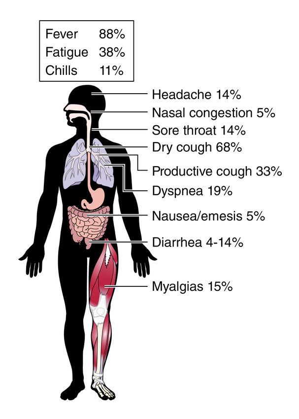
10.1161/CIRCULATIONAHA.120.046941

                                                              Abstract

                                                              Coronavirus disease 2019 (COVID-19) is a global pandemic impacting nearly 170
                                                              countries/regions and more than 285,000 patients worldwide. COVID-19 is caused by the Severe
                                                              Acute Respiratory Syndrome Coronavirus-2 (SARS-CoV-2), which invades cells through the
                                                              angiotensin converting enzyme 2 (ACE2) receptor. Among those with COVID-19, there is a
                                                              higher prevalence of cardiovascular disease and more than 7% of patients suffer myocardial
                                                              injury from the infection (22% of the critically ill). Despite ACE2 serving as the portal for
                                                              infection, the role of ACE inhibitors or angiotensin receptor blockers requires further
                                                              investigation. COVID-19 poses a challenge for heart transplantation, impacting donor selection,
                                                              immunosuppression, and post-transplant management. Thankfully there are a number of
                                                              promising therapies under active investigation to both treat and prevent COVID-19.

                                                              Key Words: COVID-19; myocardial injury; pandemic; heart transplant
Downloaded from http://ahajournals.org by on March 23, 2020

                                                                                                             2
10.1161/CIRCULATIONAHA.120.046941
Coronavirus disease 2019 (COVID-19) is a global pandemic. As of March 21st, 2020
infected patients were present in 167 countries/regions around the world and there were more
than 285,000 cases worldwide with nearly 12,000 fatalities.1 While the outbreak began in China,
the number of cases outside of China exceeded those in China on March 15th, 2020 and are
currently rising at an exponential rate. Furthermore, the number of fatalities in Italy now exceeds
the total in China. COVID-19 interacts with cardiovascular system on multiple levels, increasing
morbidity in patients with underlying cardiovascular conditions and provoking myocardial injury
and dysfunction.
COVID-19 is caused by the Severe Acute Respiratory Syndrome Coronavirus-2 (SARS-
CoV-2). This novel single-stranded enveloped RNA virus is the seventh known human
coronavirus. SARS-CoV-2 is unlike the other coronaviruses known to cause the common cold
(229E, OC43, NL63, and HKU1), but similar to the zoonotic severe acute respiratory syndrome
Downloaded from http://ahajournals.org by on March 23, 2020
coronavirus (SARS-CoV) from 2002 (SARS)2 and the Middle East respiratory syndrome
coronavirus (MERS-CoV) from 2012 (MERS).3 SARS-CoV-2 is believed to have originated in
bats, similar to many other coronaviruses, as it shares 89-96% nucleotide identity with bat
coronaviruses.4 Like SARS and MERS, it is believed SARS-CoV-2 moved from bats to an
intermediate host (possibly a Malayan Pangolin, which shares 91% nucleotide identity) and then
to humans.5 (Figure 1)
SARS-CoV-2 infection is caused by binding of the viral surface spike protein to the
human angiotensin-converting enzyme 2 (ACE2) receptor following activation of the spike
protein by transmembrane protease serine 2 (TMPRSS2).6 ACE2 is expressed in the lung
(principally Type II alveolar cells7) and appears to be the predominant portal of entry. ACE2 is
highly expressed in the heart as well, counteracting the effects of angiotensin II in states with
310.1161/CIRCULATIONAHA.120.046941
excessive activation of the renin-angiotensin system such as hypertension (HTN), congestive
heart failure (CHF), and atherosclerosis.8 In addition to the heart and lung, ACE2 is expressed in
the intestinal epithelium, vascular endothelium, and the kidneys, providing a mechanism for the
multi-organ dysfunction that can be seen with SARS-CoV-2 infection.8, 9 There is increasing
evidence linking COVID-19 with increased morbidity and mortality from cardiovascular disease.
In this review, we will summarize the rapidly evolving data in this field.
Clinical Presentation
SARS-CoV-2 is spread predominantly via respiratory droplets, but also can be aerosolized or
detected in the stool. Transmission may occur from both symptomatic and asymptomatic
patients, with secondary infection rates ranging 0.5-5%.10, 11 SARS-CoV-2 has been
demonstrated to remain stable for up to 3 hours in the aerosolized form, up to 24 hours on
cardboard, and as many as three days on plastic or stainless steel.12 The median incubation time
Downloaded from http://ahajournals.org by on March 23, 2020
is 4-5 days and 97.5% will experience symptoms within 11.5 days of exposure.13, 14
Early reports suggest the most common symptoms are fever (88%) and dry cough
(67.7%), which are shared with many other viral syndromes (Figure 2). Conspicuously,
rhinorrhea (4.8%) and gastrointestinal symptoms (diarrhea 4-14%, nausea/emesis 5%) appear to
be less frequent with COVID-19.11 Reports from China demonstrate that a significant majority of
patients (81%) had mild symptoms (no pneumonia or mild pneumonia) from COVID-19. Among
those with more significant symptoms, 14% experienced severe symptoms (dyspnea, respiratory
rate ≥30/min, blood oxygen saturation ≤93%, partial pressure of arterial oxygen to fraction of
inspired oxygen ratio 50% within 24 to 48 hours) and 5% were
critical (respiratory failure, septic shock, and/or multiple organ dysfunction or failure).9
410.1161/CIRCULATIONAHA.120.046941
The case fatality rate (CFR, number of deaths/number of those diagnosed) has differed
significantly around the world. The original reports from China suggested a CFR of 2.3%15 with
subsequent reports estimating the symptomatic case fatality risk (the probability of dying after
developing symptoms) was lower at 1.4%16, which contrasts with influenza (0.1%), MERS
(34%), and SARS (10%).17 Based on reported data from March 21st, 2020, the CFR varies
significantly by country: China 4.0% (81,304 cases), Italy 8.6% (47,021 cases), Iran 7.5%
(20,610 cases), Spain 5.4% (25,374 cases), South Korea 1.2% (8,779 cases), Germany 0.3%
(21,828 cases), and the United States 1.3% (22,043 cases).1 The CFR rises rapidly with
increasing age; the CFR is less than 1% for those under 50 years of age, rising to 1.3% for 50
year olds, 3.6% for 60 year olds, 8% for septuagenarians, and 14.8% for octogenarians.15
Similarly, the CFR increases with disease severity, as there were no deaths reported among the
mild or severe cases in the Chinese cohort; however, the CFR was 49% among critical patients.
Downloaded from http://ahajournals.org by on March 23, 2020
Furthermore, compared to patients with no comorbidities in whom the CFR is 0.9%, patients
with medical comorbidities have a significantly increased CFR: 10.5% for cardiovascular disease
(CVD); 7.3% for diabetes mellitus (DM); 6.3% for COPD; 6% for HTN; and 5.6% for cancer.15
COVID-19 in Patients with Cardiovascular Disease
Cardiovascular disease was a common comorbidity in patients with COVID-19 predecessors
SARS and MERS. In SARS, the prevalence of DM and CVD was 11% and 8% respectively, and
the presence of either comorbidity increased the risk of death twelvefold.2, 18 DM and HTN were
prevalent in about 50% of cases of MERS, while CVD was present in approximately 30% of
patients.19 The increased presence of cardiovascular comorbidities holds true for COVID-19 as
well, most notably among those with more severe disease. In one cohort of 191 patients from
Wuhan, China, any comorbidity was present in 48% (67% of non-survivors), HTN was present
510.1161/CIRCULATIONAHA.120.046941
in 30% (48% of non-survivors), DM in 19% (31% of non-survivors), and CVD in 8% (13% of
non-survivors).20 In a cohort of 138 hospitalized patients with COVID-19, comorbidities were
similarly prevalent (46% overall, 72% in patients requiring an ICU), as were cardiovascular
comorbidities: HTN in 31% (58% in patients requiring an ICU), CVD in 15% (25% in patients
requiring an ICU), and DM in 10% (22% in patients requiring an ICU).21 Analysis of an
outpatient and inpatient cohort of 1,099 patients with COVID-19 reported that 24% had any
comorbidity (58% among those with intubation or death), with 15% having HTN (36% among
those with intubation or death), 7.4% with DM (27% among those with intubation or death), and
2.5% with coronary heart disease (9% among those with intubation or death).13 Data from the
National Health Commission (NHC) of China demonstrated that 35% of patients diagnosed with
COVID-19 had HTN and 17% had coronary heart disease.22 Lastly, a recent meta-analysis of
eight studies from China including 46,248 infected patients showed the most prevalent
Downloaded from http://ahajournals.org by on March 23, 2020
comorbidities were HTN (17±7%, 95% CI 14-22%) and DM (8±6%, 95% CI 6-11%), followed
by cardiovascular diseases (5±4%, 95% CI 4-7%).23 The mechanism of these associations
remains unclear at this time. Potential explanations include CVD being more prevalent in those
with advancing age, a functionally impaired immune system, elevated levels of ACE2, or a
predisposition to COVID-19 for those with CVD.
COVID-19 and Myocardial Injury
Myocardial injury, evidenced by elevated cardiac biomarkers, was recognized among early cases
in China. In the aforementioned study of 138 hospitalized patients with COVID-19 in Wuhan
China, cardiac injury (elevated high sensitivity Troponin I [hs-cTnI] or new ECG or
echocardiographic abnormalities) was present in 7.2% of patients overall, and 22% that required
ICU care.21 The report from the NHC of China reported that almost 12% of patients without
610.1161/CIRCULATIONAHA.120.046941
known CVD had elevated troponin levels or cardiac arrest during the hospitalization.22 Notably,
hs-cTnI was above the 99th percentile upper reference limit in 46% of non-survivors as opposed
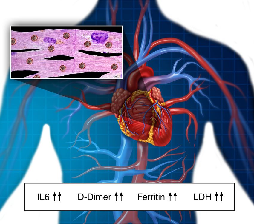
to 1% of survivors.20 (Figure 3)
Early reports indicate that there are two patterns of myocardial injury with COVID-19.
One study demonstrated that at 4 days following symptom onset, median hs-cTnI levels were 8.8
pg/mL in non-survivors vs. 2.5 pg/mL in survivors. During follow-up, the median hs-cTnI
among survivors did not change significantly (2.5-4.4 pg/mL), whereas it rose to 24.7 pg/mL on
Day 7, to 55.7 pg/mL on Day 13, to 134.5 pg/mL on Day 19, and to 290.6 pg/mL on Day 22 for
non-survivors.20 Notably, the median time to death from the onset of symptoms was 18.5 days
(IQR 15 – 20 days). The rise in hs-cTnI tracks with other inflammatory biomarkers (D-dimer,
ferritin, interleukin-6 (IL-6), lactate dehydrogenase), raising the possibility that this reflects
cytokine storm or secondary hemophagocytic lymphohistiocytosis more than isolated myocardial
Downloaded from http://ahajournals.org by on March 23, 2020
injury. In contrast, reports of patients presenting with predominantly cardiac symptoms suggest a
different pattern, potentially viral myocarditis or stress cardiomyopathy. For example, one case
recently published involved a man presenting with chest pain and ST-segment elevation on his
electrocardiogram, but without coronary obstruction. An echocardiogram noted left ventricular
dysfunction (ejection fraction 27%, LVEDD 5.8cm) and elevated cardiac biomarkers (troponin T
>10 ng/mL, NT-proBNP>21,000 pg/mL).24 Following a therapeutic approach which included
intravenous immunoglobulin and steroids, ejection fraction and cardiac biomarkers normalized
within three weeks. In another report from China, a 63 year old man with no past cardiac history
presented with both severe respiratory manifestation and evidence of fulminant myocarditis with
an enlarged left ventricle (LVEDD 6.1 cm) and depressed left ventricular function (ejection
fraction 32%). The patient had an elevated troponin-I (>11 ng/mL) and NT-proBNP (>22,000
710.1161/CIRCULATIONAHA.120.046941
pg/ml). Given the severity of his cardiogenic shock, he was placed on extracorporeal membrane
oxygenation and was treated with intravenous immunoglobulin, steroids, anti-viral therapy and
renal replacement therapy. The patient ultimately showed recovery of his ventricular function
within 2 weeks.25 While both of these patients were treated with glucocorticoids it is unclear the
impact of this therapy, as both the WHO and CDC currently do not recommend glucocorticoid
use unless otherwise indicated (e.g. chronic obstructive pulmonary disease or asthma
exacerbation).26, 27 Similarly a report from the NHC of China comments that a subset of patients
presented with palpitations and chest pain, not the typical fever and cough.22 Based on available
but limited data, it appears that the incidence of fulminant myocarditis and profound cardiogenic
shock is low; however, the rate of recovery and mode of treatment are yet to be determined.
The exact mechanism of cardiac involvement in COVID-19 remains under investigation.
One potential mechanism is direct myocardial involvement mediated via ACE2. A murine model
Downloaded from http://ahajournals.org by on March 23, 2020
demonstrated pulmonary infection with SARS-CoV also precipitated an ACE2‐dependent
myocardial infection.28 Among humans, during the Toronto SARS outbreak, SARS-CoV viral
RNA was detected in 35% of autopsied hearts.29 Other suggested mechanisms of COVID-19
related cardiac involvement include a cytokine storm, mediated by an imbalanced response
among subtypes of T helper cells20, and hypoxia induced excessive intracellular calcium leading
to cardiac myocyte apoptosis.22
Role of angiotensin converting enzyme inhibitors (ACEi) and angiotensin receptor blockers
(ARB)
ACE2 is a homolog of ACE that converts angiotensin II to angiotensin 1-7, thereby diminishing
vasoconstriction mediated by the renin angiotensin system. The use of ACEi and ARB are
common in cardiovascular disorders (HTN, coronary artery disease, congestive heart failure, and
810.1161/CIRCULATIONAHA.120.046941
DM). There are conflicting data from studies demonstrating whether these drugs increase30-32 or
have minimal effect on ACE2 levels33-36. SARS-CoV-2 entry into cells is ACE2 dependent
(Figure 4); however, ACE2 appears to be protective against acute lung injury. In a murine model,
binding of the SARS-CoV spike protein to ACE2 caused ACE2 downregulation, leading to an
increase in angiotensin II and ultimately increased pulmonary vascular permeability, inducing
pulmonary edema, and reduced lung function. However, treatment with recombinant ACE237 and
losartan38 mitigated the degree of lung injury. On this basis losartan is being studied for potential
mitigation of lung injury among both inpatients and outpatients with COVID-19.39, 40 At this time
nearly all major societies have recommended against adding or stopping ACEi, ARB, or other
RAAS antagonists in this setting, unless done on clinical grounds independently of COVID-19,
given the lack of evidence currently available on their potential benefit or harm.
Heart Transplantation in the Era of COVID-19
Downloaded from http://ahajournals.org by on March 23, 2020
During prior coronavirus epidemics (SARS & MERS), transplant patients presented with similar
symptoms as the general population.41, 42 In the current pandemic, a case study described the
clinical courses of two heart transplant recipients from the Hubei province of China. Both
patients presented with fevers and had laboratory and CT scans that were similar to non-
immunosuppressed individuals with bilateral ground glass opacities in a peripheral distribution.
One had relatively mild disease, while the other required hospitalization requiring supplemental
oxygen.43 Both survived and were treated with antibiotics and antivirals, while the more ill
patient also required cessation of immunosuppression, along with treatment with
methylprednisolone and IVIG. A survey of 87 heart transplant recipients in Wuhan, China did
not find a higher risk of infection with SARS-CoV-2 if routine preventive measures were used,
and while encouraging, this needs to be confirmed in larger populations.44 Further, we do not
910.1161/CIRCULATIONAHA.120.046941
know if transplant patients will experience differences in disease severity or duration compared
with the non-immunosuppressed population.
The ongoing pandemic has raised the question of whether to continue offering heart
transplantation due to concerns about a risk for exposure to COVID-19 during hospitalization, as
well as challenges in controlling the infection in the context of high levels of
immunosuppression. Current recommendations are to continue heart transplantation without
changes in immunosuppression provided the recipient has not tested positive for SARS-CoV-2
and has not had exposure to or symptoms of COVID-19 in the prior two to four weeks.45, 46
Major societal recommendations include avoiding donors with known or suspected COVID-19
and if a donor had COVID-19, they should be COVID-19 free (by PCR) for at least 14 days
(owing to the incubation period of ~5 days and onset of symptoms in ~11.5 days).14, 47 We
recognize the difficulties of this decision with increasing prevalence of COVID-19 in the donor
Downloaded from http://ahajournals.org by on March 23, 2020
population, that may be asymptomatic, especially if the donor cannot be tested for COVID-19.
Recommended management of transplant recipients who developed COVID-19, based on very
limited data to date, is supportive care and continuation of immunosuppression for mild COVID-
19 with reduction of the anti-metabolite (mycophenolate or azathioprine) and further treatment
based on disease severity and drug availability.45 Notably, one potential treatment option for
COVID-19 are protease inhibitors, which will increase calcineurin inhibitor levels.
Treatment
Preventive measures are the best strategy for COVID-19 at this time. While vaccines and
monoclonal antibodies against SARS-CoV-2 are in development, a number of other
investigational therapies, using repurposed clinically approved drugs targeting SARS-CoV-2 cell
invasion and replication, may be considered. Recombinant human ACE2 (APN01) was
1010.1161/CIRCULATIONAHA.120.046941
developed in 2010 and could potentially both neutralize the virus and protect against acute lung
injury. It has been demonstrated to be safe and reduce levels of both angiotensin II and IL-6 in a
Phase II study of acute respiratory distress syndrome48. It is currently under investigation in
China in severe COVID-19. The serine protease inhibitor camostat mesylate, which is approved
in Japan for chronic pancreatitis and postoperative reflux esophagitis among other indications,
has been shown to block TMPRSS2 activity and inhibit SARS-CoV entry into cells.49 This well
tolerated therapy has been proposed as a treatment to prevent SARS-CoV-2 spike protein
activation, thereby preventing cell entry and controlling infection. Remdesevir is a broad
spectrum antiviral that interrupts RNA replication by acting as an nucleotide analog.50 Initially
developed to treat Ebola, it has been demonstrated to have both in vitro activity against SARS-
CoV-2 and prevent and reduce disease severity in MERS-CoV in primates.51, 52 It has been safe
in prior trials and currently is enrolling in clinical trials in China53, 54 and the United States.55, 56
Downloaded from http://ahajournals.org by on March 23, 2020
Chloroquine (anti-malarial drug) and hydroxychloroquine (rheumatoid arthritis or systemic lupus
erythematosus treatment) block SARS-CoV-2 cell entry in vitro at similar concentrations that are
achieved with treatment for rheumatoid arthritis (500 mg twice daily for chloroquine & 600 mg
twice a day loading followed by 400-600 mg a day for hydroxychloroquine) and trials with these
agents are ongoing.52, 57-60 Additionally, early studies suggest clinical benefit in COVID-19 with
reduction in pneumonia severity, decreased length of hospitalization, and earlier viral
clearance.61 The combination protease inhibitor lopinavir/ritonavir used in HIV was
demonstrated to have in vitro activity against SARS-CoV and improved clinical outcomes when
used in combination with ribavirin for SARS.62 There have been reports of its success in treating
SARS-CoV-2, though the first randomized control trial did not demonstrate statistically
significant benefit among hospitalized patients with COVID-19.63 In this study of 199 patients,
1110.1161/CIRCULATIONAHA.120.046941
28 day mortality was 5.8% lower (95% CI -17.3-5.7%) for the lopinavir/ritonavir treated patients
and the median time to improvement was 1 day shorter; further data are needed to determine the
role of lopinavir/ritonavir. Antiviral medications typically used for influenza (oseltamivir and
arbidol) have been applied, without clinical efficacy data available. Another drug approved for
influenza, favipiravir, is considered promising as it inhibits RNA polymerase and is being
studied in a clinical trial in China.64 Other proposed strategies include interferon and
convalescent serum.
Tocilizumab and sarilumab are IL-6 receptor antagonists used in the treatment of
rheumatoid arthritis, while tocilizumab also has the indication for the treatment of cytokine
release syndrome as is seen with chimeric antigen receptor-T cell (CAR-T) therapy. These may
be potential therapies for COVID-19 patients that display elements of cytokine storm or
secondary hemophagocytic lymphohistiocytosis with markedly elevated IL-6, ferritin, D-dimer,
Downloaded from http://ahajournals.org by on March 23, 2020
and hs-cTnI levels. Tocilizumab has been used with reported success for patients with severe
COVID-19 and there are ongoing clinical trials65-67 and a trial of sarilumab just launched in the
United States.68
Conclusion
COVID-19, caused by SARS-CoV-2, is a global pandemic evolving in real time. Cardiovascular
comorbidities are common in patients with COVID-19 and such patients are at higher risk of
morbidity and mortality. However, it is not known if the presence of cardiovascular comorbid
conditions pose independent risk or whether this is mediated by other factors (e.g. age).
Myocardial injury is present in more than a quarter of critical cases and presents in two patterns:
acute myocardial injury and dysfunction on presentation and myocardial injury that develops as
illness severity intensifies. The continuation of clinically indicated ACEi and ARB medications
1210.1161/CIRCULATIONAHA.120.046941
is recommended based on the available evidence at this time. There are a number of promising
treatments under investigation, but none with proven clinical efficacy to date.
Acknowledgments
We would like thank Deborah Burkhoff for graphical support.
Sources of Funding
None
Disclosures
None of the authors have disclosures pertinent to this work.
Downloaded from http://ahajournals.org by on March 23, 2020
References
1. Dong E, Du H and Gardner L. An interactive web-based dashboard to track COVID-19 in
real time. Lancet Infect Dis. Feb 19, 2020. doi: 10.1016/S1473-3099(20)30120-1. [epub ahead of
print] Accessed 3/21/20.
2. Chan JWM, Ng CK, Chan YH, Mok TYW, Lee S, Chu SYY, Law WL, Lee MP and Li
PCK. Short term outcome and risk factors for adverse clinical outcomes in adults with severe
acute respiratory syndrome (SARS). Thorax. 2003;58:686-689.
3. Badawi A and Ryoo SG. Prevalence of comorbidities in the Middle East respiratory
syndrome coronavirus (MERS-CoV): a systematic review and meta-analysis. Int J Infect Dis.
2016;49:129-133.
4. Andersen KG, Rambaut A, Lipkin WI, Holmes EC and Garry RF. The proximal origin of
SARS-CoV-2. Nat Med. March 17, 2020. doi: 10.1038/s41591-020-0820-9. [epub ahead of
print].
5. Zhang T WQ, Zhang Z. Probable pangolin origin of SARS-CoV-2 associated with the
COVID-19 outbreak. Curr Biol. March 13, 2020. doi: 10.1016/j.cub.2020.03.022. [epub ahead of
print]
6. Hoffmann M, Kleine-Weber H, Schroeder S, Krüger N, Herrler T, Erichsen S, Schiergens
TS, Herrler G, Wu NH, Nitsche A, Müller MA, Drosten C and Pöhlmann S. SARS-CoV-2 Cell
Entry Depends on ACE2 and TMPRSS2 and Is Blocked by a Clinically Proven Protease
Inhibitor. Cell. March 5, 2020. doi: 10.1016/j.cell.2020.02.052. [epub ahead of print].
1310.1161/CIRCULATIONAHA.120.046941
7. Zhao Y, Zhao Z, Wang Y, Zhou Y, Ma Y and Zuo W. Single-cell RNA expression
profiling of ACE2, the putative receptor of Wuhan 2019-nCov. bioRxiv. January 26, 2020. doi:
10.1101/2020.01.26.919985.
8. Tikellis C and Thomas MC. Angiotensin-Converting Enzyme 2 (ACE2) Is a Key
Modulator of the Renin Angiotensin System in Health and Disease. Int J Pept.
2012;2012:256294-256294.
9. Zhang H, Penninger JM, Li Y, Zhong N and Slutsky AS. Angiotensin-converting enzyme
2 (ACE2) as a SARS-CoV-2 receptor: molecular mechanisms and potential therapeutic target.
Intensive Care Med. March 3, 2020. doi: 10.1007/s00134-020-05985-9. [epub ahead of print].
10. Zou L, Ruan F, Huang M, Liang L, Huang H, Hong Z, Yu J, Kang M, Song Y, Xia J,
Guo Q, Song T, He J, Yen HL, Peiris M and Wu J. SARS-CoV-2 Viral Load in Upper
Respiratory Specimens of Infected Patients. New Eng J Med. 2020;382:1177-1179.
11. World Health Organization. Report of the WHO-China Joint Mission on Coronavirus
Disease 2019 (COVID-19). February 28, 2020. https://www.who.int/publications-detail/report-
of-the-who-china-joint-mission-on-coronavirus-disease-2019-(covid-19).
12. van Doremalen N, Bushmaker T, Morris DH, Holbrook MG, Gamble A, Williamson BN,
Tamin A, Harcourt JL, Thornburg NJ, Gerber SI, Lloyd-Smith JO, de Wit E and Munster VJ.
Aerosol and Surface Stability of SARS-CoV-2 as Compared with SARS-CoV-1. New Eng J
Med. March 17, 2020. doi: 10.1056/NEJMc2004973. [epub ahead of print].
13. Guan W-j, Ni Z-y, Hu Y, Liang W-h, Ou C-q, He J-x, Liu L, Shan H, Lei C-l, Hui DSC,
Du B, Li L-j, Zeng G, Yuen K-Y, Chen R-c, Tang C-l, Wang T, Chen P-y, Xiang J, Li S-y,
Wang J-l, Liang Z-j, Peng Y-x, Wei L, Liu Y, Hu Y-h, Peng P, Wang J-m, Liu J-y, Chen Z, Li
G, Zheng Z-j, Qiu S-q, Luo J, Ye C-j, Zhu S-y and Zhong N-s. Clinical Characteristics of
Coronavirus Disease 2019 in China. New Eng J Med. February 28, 2020. doi:
Downloaded from http://ahajournals.org by on March 23, 2020
10.1056/NEJMoa2002032. [epub ahead of print].
14. Lauer SA, Grantz KH, Bi Q, Jones FK, Zheng Q, Meredith HR, Azman AS, Reich NG
and Lessler J. The Incubation Period of Coronavirus Disease 2019 (COVID-19) From Publicly
Reported Confirmed Cases: Estimation and Application. Ann Intern Med. March 10, 2020. doi:
10.7326/M20-0504. [epub ahead of print].
15. Wu Z and McGoogan JM. Characteristics of and Important Lessons From the
Coronavirus Disease 2019 (COVID-19) Outbreak in China: Summary of a Report of 72 314
Cases From the Chinese Center for Disease Control and Prevention. JAMA. Feb 24, 2020. doi:
10.1001/jama.2020.2648. [epub ahead of print].
16. Wu JT, Leung K, Bushman M, Kishore N, Niehus R, de Salazar PM, Cowling BJ,
Lipsitch M and Leung GM. Estimating clinical severity of COVID-19 from the transmission
dynamics in Wuhan, China. Nat Med. March 19, 2020. doi: 10.1038/s41591-020-0822-7. [epub
ahead of print].
17. Munster VJ, Koopmans M, van Doremalen N, van Riel D and de Wit E. A Novel
Coronavirus Emerging in China — Key Questions for Impact Assessment. New Eng J Med.
2020;382:692-694.
18. Booth CM, Matukas LM, Tomlinson GA, Rachlis AR, Rose DB, Dwosh HA, Walmsley
SL, Mazzulli T, Avendano M, Derkach P, Ephtimios IE, Kitai I, Mederski BD, Shadowitz SB,
Gold WL, Hawryluck LA, Rea E, Chenkin JS, Cescon DW, Poutanen SM and Detsky AS.
Clinical features and short-term outcomes of 144 patients with SARS in the greater Toronto area.
Jama. 2003;289:2801-2809.
1410.1161/CIRCULATIONAHA.120.046941
19. Badawi A and Ryoo SG. Prevalence of comorbidities in the Middle East respiratory
syndrome coronavirus (MERS-CoV): a systematic review and meta-analysis. Int J Infect Dis.
2016;49:129-133.
20. Zhou F, Yu T, Du R, Fan G, Liu Y, Liu Z, Xiang J, Wang Y, Song B, Gu X, Guan L,
Wei Y, Li H, Wu X, Xu J, Tu S, Zhang Y, Chen H and Cao B. Clinical course and risk factors
for mortality of adult inpatients with COVID-19 in Wuhan, China: a retrospective cohort study.
The Lancet. March 11, 2020. doi: 10.1016/S0140-6736(20)30566-3. [epub ahead of print].
21. Wang D, Hu B, Hu C, Zhu F, Liu X, Zhang J, Wang B, Xiang H, Cheng Z, Xiong Y,
Zhao Y, Li Y, Wang X and Peng Z. Clinical Characteristics of 138 Hospitalized Patients With
2019 Novel Coronavirus–Infected Pneumonia in Wuhan, China. JAMA. 2020;323:1061-1069.
22. Zheng Y-Y, Ma Y-T, Zhang J-Y and Xie X. COVID-19 and the cardiovascular system.
Nat Rev Cardiol. March 5, 2020. doi: 10.1038/s41569-020-0360-5. [epub ahead of print].
23. Yang J, Zheng Y, Gou X, Pu K, Chen Z, Guo Q, Ji R, Wang H, Wang Y and Zhou Y.
Prevalence of comorbidities in the novel Wuhan coronavirus (COVID-19) infection: a systematic
review and meta-analysis. Int J Infect Dis. March 12, 2020. doi: 10.1016/j.ijid.2020.03.017.
[epub ahead of print].
24. Hu H, Ma F, Wei X and Fang Y. Coronavirus fulminant myocarditis saved with
glucocorticoid and human immunoglobulin. Eur Heart J. March 16, 2020. doi:
10.1093/eurheartj/ehaa190. [epub ahead of print].
25. Zeng JH, Liu YX, Yuan J, Wang FX, Wu WB, Li JX, Wang LF, Gao H, Wang Y, Dong
CF, Li YJ, Xie XJ, Feng C and Liu L. First Case of COVID-19 Infection with Fulminant
Myocarditis Complication: Case Report and Insights. preprints. 2020; 2020030180. doi:
10.20944/preprints202003.0180.v1.
26. Centers for Disease Control and Prevention. Interim Clinical Guidance for Management
Downloaded from http://ahajournals.org by on March 23, 2020
of Patients with Confirmed Coronavirus Disease (COVID-19). Revised March 7th, 2020.
https://www.cdc.gov/coronavirus/2019-ncov/hcp/clinical-guidance-management-patients.html.
27. World Health Organization. Clinical management of severe acute respiratory infection
(SARI) when COVID-19 disease is suspected. March 13, 2020.
https://www.who.int/publications-detail/clinical-management-of-severe-acute-respiratory-
infection-when-novel-coronavirus-(ncov)-infection-is-suspected.
28. Oudit GY, Kassiri Z, Jiang C, Liu PP, Poutanen SM, Penninger JM and Butany J. SARS-
coronavirus modulation of myocardial ACE2 expression and inflammation in patients with
SARS. Eur J Clin Invest. 2009;39:618-625.
29. Booth CM, Matukas LM, Tomlinson GA, Rachlis AR, Rose DB, Dwosh HA, Walmsley
SL, Mazzulli T, Avendano M, Derkach P, Ephtimios IE, Kitai I, Mederski BD, Shadowitz SB,
Gold WL, Hawryluck LA, Rea E, Chenkin JS, Cescon DW, Poutanen SM and Detsky AS.
Clinical Features and Short-term Outcomes of 144 Patients With SARS in the Greater Toronto
Area. JAMA. 2003;289:2801-2809.
30. Ishiyama Y, Gallagher PE, Averill DB, Tallant EA, Brosnihan KB and Ferrario CM.
Upregulation of Angiotensin-Converting Enzyme 2 After Myocardial Infarction by Blockade of
Angiotensin II Receptors. 2004;43:970-976.
31. Ferrario CM, Jessup J, Chappell MC, Averill DB, Brosnihan KB, Tallant EA, Diz DI and
Gallagher PE. Effect of angiotensin-converting enzyme inhibition and angiotensin II receptor
blockers on cardiac angiotensin-converting enzyme 2. Circulation. 2005;111:2605-2610.
1510.1161/CIRCULATIONAHA.120.046941
32. Ocaranza MP, Palomera C, Román M, Bargetto J, Lavandero S and Jalil JE. Effect of
hypertension on angiotensin-(1-7) levels in rats with different angiotensin-I converting enzyme
polymorphism. Life sciences. 2006;78:1535-1542.
33. Klimas J, Olvedy M, Ochodnicka-Mackovicova K, Kruzliak P, Cacanyiova S, Kristek F,
Krenek P and Ochodnicky P. Perinatally administered losartan augments renal ACE2 expression
but not cardiac or renal Mas receptor in spontaneously hypertensive rats. J Cell Mol Med.
2015;19:1965-1974.
34. Walters TE, Kalman JM, Patel SK, Mearns M, Velkoska E and Burrell LM. Angiotensin
converting enzyme 2 activity and human atrial fibrillation: increased plasma angiotensin
converting enzyme 2 activity is associated with atrial fibrillation and more advanced left atrial
structural remodelling. EP Europace. 2016;19:1280-1287.
35. Burchill LJ, Velkoska E, Dean RG, Griggs K, Patel SK and Burrell LM. Combination
renin-angiotensin system blockade and angiotensin-converting enzyme 2 in experimental
myocardial infarction: implications for future therapeutic directions. Clinical Science.
2012;123:649-658.
36. Burrell LM, Risvanis J, Kubota E, Dean RG, MacDonald PS, Lu S, Tikellis C, Grant SL,
Lew RA, Smith AI, Cooper ME and Johnston CI. Myocardial infarction increases ACE2
expression in rat and humans. Eur Heart J. 2005;26:369-75; discussion 322-324.
37. Imai Y, Kuba K, Rao S, Huan Y, Guo F, Guan B, Yang P, Sarao R, Wada T, Leong-Poi
H, Crackower MA, Fukamizu A, Hui C-C, Hein L, Uhlig S, Slutsky AS, Jiang C and Penninger
JM. Angiotensin-converting enzyme 2 protects from severe acute lung failure. Nature.
2005;436:112-116.
38. Kuba K, Imai Y, Rao S, Gao H, Guo F, Guan B, Huan Y, Yang P, Zhang Y, Deng W,
Bao L, Zhang B, Liu G, Wang Z, Chappell M, Liu Y, Zheng D, Leibbrandt A, Wada T, Slutsky
Downloaded from http://ahajournals.org by on March 23, 2020
AS, Liu D, Qin C, Jiang C and Penninger JM. A crucial role of angiotensin converting enzyme 2
(ACE2) in SARS coronavirus–induced lung injury. Nat Med. 2005;11:875-879.
39. ClinicalTrials.gov. Randomized Controlled Trial of Losartan for Patients With COVID-
19 Not Requiring Hospitalization. Identifier: NCT04311177. March 17, 2020.
https://clinicaltrials.gov/ct2/show/NCT04311177.
40. ClinicalTrials.gov. Randomized Controlled Trial of Losartan for Patients With COVID-
19 Requiring Hospitalization. Identifier: NCT04312009. March 17, 2020.
https://clinicaltrials.gov/ct2/show/NCT04312009.
41. AlGhamdi M, Mushtaq F, Awn N and Shalhoub S. MERS CoV Infection in Two Renal
Transplant Recipients: Case Report. Am J Transplant. 2015;15:1101-1104.
42. Kumar D, Tellier R, Draker R, Levy G and Humar A. Severe Acute Respiratory
Syndrome (SARS) in a Liver Transplant Recipient and Guidelines for Donor SARS Screening.
Am J Transplant. 2003;3:977-981.
43. Li F, Cai J and Dong N. First Cases of COVID-19 in Heart Transplantation From China.
J Heart Lung Transplant. 2020. doi: 10.1016/j.healun.2020.03.006. [epub ahead of print].
44. Zong-Li Ren RH, Zhi-Wei Wang, Min Zhang, Yong-Le Ruan , Zhi-Yong Wu , Hong-
Bing Wu , Xiao-Ping Hu, Zhi-Peng Hu, Wei Ren, Luo-Cheng Li, Fei-Feng Dai, Huan Liu, Xin
Cai. Epidemiological and Clinical Characteristics of Heart Transplant Recipients During the
2019 Coronavirus Outbreak in Wuhan, China: A Descriptive Survey Report. J Heart Lung
Transplant. 2020. doi: 10.1016/j.healun.2020.03.008. [epub ahead of print].
45. Guidance for Cardiothoracic Transplant and Mechanical Circulatory Support Centers
regarding SARS CoV-2 infection and COVID-19: March 17, 2020.
1610.1161/CIRCULATIONAHA.120.046941
https://community.ishlt.org/HigherLogic/System/DownloadDocumentFile.ashx?DocumentFileK
ey=afb06f06-5d63-13d4-c107-d152a9f6cd46.
46. American Society of Transplantation. 2019-nCoV (Coronavirus): FAQs for Organ
Transplantation. Updated Feb 29, 2020.
https://www.myast.org/sites/default/files/COVID19%20FAQ%20Tx%20Centers%20030220-
1.pdf.
47. Li Q, Guan X, Wu P, Wang X, Zhou L, Tong Y, Ren R, Leung KSM, Lau EHY, Wong
JY, Xing X, Xiang N, Wu Y, Li C, Chen Q, Li D, Liu T, Zhao J, Liu M, Tu W, Chen C, Jin L,
Yang R, Wang Q, Zhou S, Wang R, Liu H, Luo Y, Liu Y, Shao G, Li H, Tao Z, Yang Y, Deng
Z, Liu B, Ma Z, Zhang Y, Shi G, Lam TTY, Wu JT, Gao GF, Cowling BJ, Yang B, Leung GM
and Feng Z. Early Transmission Dynamics in Wuhan, China, of Novel Coronavirus–Infected
Pneumonia. New Eng J Med. January 29, 2020. doi: 10.1056/NEJMoa2001316. [epub ahead of
print].
48. Khan A, Benthin C, Zeno B, Albertson TE, Boyd J, Christie JD, Hall R, Poirier G, Ronco
JJ, Tidswell M, Hardes K, Powley WM, Wright TJ, Siederer SK, Fairman DA, Lipson DA,
Bayliffe AI and Lazaar AL. A pilot clinical trial of recombinant human angiotensin-converting
enzyme 2 in acute respiratory distress syndrome. Critical Care. 2017;21:234.
49. Kawase M, Shirato K, van der Hoek L, Taguchi F and Matsuyama S. Simultaneous
Treatment of Human Bronchial Epithelial Cells with Serine and Cysteine Protease Inhibitors
Prevents Severe Acute Respiratory Syndrome Coronavirus Entry. J Virol. 2012;86:6537-6545.
50. Gordon CJ, Tchesnokov EP, Feng JY, Porter DP and Gotte M. The antiviral compound
remdesivir potently inhibits RNA-dependent RNA polymerase from Middle East respiratory
syndrome coronavirus. J Biol Chem. Feb 24, 2020. doi: 10.1074/jbc.AC120.013056 [epub ahead
of print].
Downloaded from http://ahajournals.org by on March 23, 2020
51. de Wit E, Feldmann F, Cronin J, Jordan R, Okumura A, Thomas T, Scott D, Cihlar T and
Feldmann H. Prophylactic and therapeutic remdesivir (GS-5734) treatment in the rhesus
macaque model of MERS-CoV infection. PNAS. Feb 13, 2020;201922083. doi:
10.1073/pnas.1922083117. [epub ahead of print].
52. Wang M, Cao R, Zhang L, Yang X, Liu J, Xu M, Shi Z, Hu Z, Zhong W and Xiao G.
Remdesivir and chloroquine effectively inhibit the recently emerged novel coronavirus (2019-
nCoV) in vitro. Cell Res. 2020;30:269-271.
53. ClinicalTrials.gov. Severe 2019-nCoV Remdesivir RCT. Identifier: NCT04257656. Last
Updated Feb 24, 2020. https://clinicaltrials.gov/ct2/show/NCT04257656.
54. ClinicalTrials.gov. Mild/Moderate 2019-nCoV Remdesivir RCT. Identifier:
NCT04252664. Last Updated Feb 24, 2020. https://clinicaltrials.gov/ct2/show/NCT04252664.
55. ClinicalTrials.gov. Study to Evaluate the Safety and Antiviral Activity of Remdesivir
(GS-5734™) in Participants With Severe Coronavirus Disease (COVID-19). Identifier:
NCT04292899. Updated March 19, 2020. https://clinicaltrials.gov/ct2/show/NCT04292899.
56. ClinicalTrials.gov. Study to Evaluate the Safety and Antiviral Activity of Remdesivir
(GS-5734™) in Participants With Moderate Coronavirus Disease (COVID-19) Compared to
Standard of Care Treatment. Identifier: NCT04292730. Updated March 19, 2020.
https://clinicaltrials.gov/ct2/show/NCT04292730.
57. ClinicalTrials.gov. Comparison of Lopinavir/Ritonavir or Hydroxychloroquine in
Patients With Mild Coronavirus Disease (COVID-19). Identifier: NCT04307693. Updated
March 13, 2020. https://clinicaltrials.gov/ct2/show/NCT04307693.
1710.1161/CIRCULATIONAHA.120.046941
58. ClinicalTrials.gov. Efficacy and Safety of Hydroxychloroquine for Treatment of
Pneumonia Caused by 2019-nCoV ( HC-nCoV ). Identifier: NCT04261517. Updated March 4,
2020. https://clinicaltrials.gov/ct2/show/NCT04261517.
59. Clinical Trials.gov. Post-exposure Prophylaxis for SARS-Coronavirus-2. Identifier:
NCT04308668. Updated March 19, 2020. https://clinicaltrials.gov/ct2/show/NCT04308668.
60. ClinicalTrials.gov. Chloroquine Prevention of Coronavirus Disease (COVID-19) in the
Healthcare Setting (COPCOV). Identifier: NCT04303507. Updated March 11, 2020.
https://clinicaltrials.gov/ct2/show/NCT04303507
61. Gao J, Tian Z and Yang X. Breakthrough: Chloroquine phosphate has shown apparent
efficacy in treatment of COVID-19 associated pneumonia in clinical studies. BioSci Trends.
2020;14:72-73.
62. Chu CM, Cheng VC, Hung IF, Wong MM, Chan KH, Chan KS, Kao RY, Poon LL,
Wong CL, Guan Y, Peiris JS and Yuen KY. Role of lopinavir/ritonavir in the treatment of
SARS: initial virological and clinical findings. Thorax. 2004;59:252-256.
63. Cao B, Wang Y, Wen D, Liu W, Wang J, Fan G, Ruan L, Song B, Cai Y, Wei M, Li X,
Xia J, Chen N, Xiang J, Yu T, Bai T, Xie X, Zhang L, Li C, Yuan Y, Chen H, Li H, Huang H,
Tu S, Gong F, Liu Y, Wei Y, Dong C, Zhou F, Gu X, Xu J, Liu Z, Zhang Y, Li H, Shang L,
Wang K, Li K, Zhou X, Dong X, Qu Z, Lu S, Hu X, Ruan S, Luo S, Wu J, Peng L, Cheng F, Pan
L, Zou J, Jia C, Wang J, Liu X, Wang S, Wu X, Ge Q, He J, Zhan H, Qiu F, Guo L, Huang C,
Jaki T, Hayden FG, Horby PW, Zhang D and Wang C. A Trial of Lopinavir–Ritonavir in Adults
Hospitalized with Severe Covid-19. New Eng J Med. March 18, 2020. doi:
10.1056/NEJMoa2001282. [epub ahead of print].
64. Furuta Y, Komeno T and Nakamura T. Favipiravir (T-705), a broad spectrum inhibitor of
viral RNA polymerase. Proc Jpn Acad Ser B Phys biol sci. 2017;93:449-463.
Downloaded from http://ahajournals.org by on March 23, 2020
65. ClinicalTrials.gov. Tocilizumab in COVID-19 Pneumonia (TOCIVID-19) (TOCIVID-
19). Identifier: NCT04317092. Updated March 20, 2020.
https://www.clinicaltrials.gov/ct2/show/NCT04317092.
66. ClinicalTrials.gov.Tocilizumab vs CRRT in Management of Cytokine Release Syndrome
(CRS) in COVID-19 (TACOS). Identifier: NCT04306705. Updated March 17, 2020.
https://clinicaltrials.gov/ct2/show/NCT04306705.
67. ClinicalTrials.gov. Tocilizumab for SARS-CoV2 Severe Pneumonitis. Identifier:
NCT04315480. Updated March 19, 2020. https://clinicaltrials.gov/ct2/show/NCT04315480.
68. ClinicalTrials.gov. Evaluation of the Efficacy and Safety of Sarilumab in Hospitalized
Patients With COVID-19. Identifier: NCT04315298. Updated March 19, 2020.
https://www.clinicaltrials.gov/ct2/show/NCT04315298.
1810.1161/CIRCULATIONAHA.120.046941
Figure Legends
Figure 1. Suspected transmission pathway of SARS-CoV-2 to humans
Figure 2. Symptoms of COVID-19
Figure 3. Myocardial injury during COVID-19 can be explained by two mechanisms.1) From
the associated cytokine storm manifested by elevated levels of IL-6, ferritin, LDH, and D-dimer.
2) Myocardial dysfunction from the direct effect of SARS-CoV-2 on the heart.
Figure 4. SARS-CoV-2 binds to the ACE2 receptor following activation of the spike protein by
TMPRSS2
Downloaded from http://ahajournals.org by on March 23, 2020
19Downloaded from http://ahajournals.org by on March 23, 2020
Downloaded from http://ahajournals.org by on March 23, 2020
Downloaded from http://ahajournals.org by on
Downloaded from http://ahajournals.org by on March 23, 2020
You can also read