Influence of dosimetry method on bone lesion absorbed dose estimates in PSMA therapy: application to mCRPC patients receiving Lu-177-PSMA-I&T
←
→
Page content transcription
If your browser does not render page correctly, please read the page content below
Brosch-Lenz et al. EJNMMI Physics     (2021) 8:26
https://doi.org/10.1186/s40658-021-00369-4
                                                                                                                                       EJNMMI Physics

 ORIGINAL RESEARCH                                                                                                                              Open Access

Influence of dosimetry method on bone
lesion absorbed dose estimates in PSMA
therapy: application to mCRPC patients
receiving Lu-177-PSMA-I&T
Julia Brosch-Lenz1* , Carlos Uribe2,3, Astrid Gosewisch1, Lena Kaiser1, Andrei Todica1, Harun Ilhan1,
Franz Josef Gildehaus1, Peter Bartenstein1, Arman Rahmim2,3,4, Anna Celler3, Sibylle Ziegler1 and Guido Böning1

* Correspondence: Julia.Brosch-
Lenz@med.uni-muenchen.de                 Abstract
1
 Department of Nuclear Medicine,
University Hospital, LMU Munich,         Background: Patients with metastatic, castration-resistant prostate cancer (mCRPC)
Marchioninistrasse 15, 81377             present with an increased tumor burden in the skeleton. For these patients, Lutetium-
Munich, Germany                          177 (Lu-177) radioligand therapy targeting the prostate-specific membrane antigen
Full list of author information is
available at the end of the article      (PSMA) has gained increasing interest with promising outcome data. Patient-
                                         individualized dosimetry enables improvement of therapy success with the aim of
                                         minimizing absorbed dose to organs at risk while maximizing absorbed dose to
                                         tumors. Different dosimetric approaches with varying complexity and accuracy exist for
                                         this purpose. The Medical Internal Radiation Dose (MIRD) formalism applied to tumors
                                         assumes a homogeneous activity distribution in a sphere with unit density for
                                         derivation of tumor S values (TSV). Voxel S value (VSV) approaches can account for
                                         heterogeneous activities but are simulated for a specific tissue. Full patient-individual
                                         Monte Carlo (MC) absorbed dose simulation addresses both, heterogeneous activity
                                         and density distributions. Subsequent CT-based density weighting has the potential to
                                         overcome the assumption of homogeneous density in the MIRD formalism with TSV
                                         and VSV methods, which could be a major limitation for the application in bone
                                         metastases with heterogeneous density. The aim of this investigation is a comparison
                                         of these methods for bone lesion dosimetry in mCRPC patients receiving Lu-177-PSMA
                                         therapy.
                                         Results: In total, 289 bone lesions in 15 mCRPC patients were analyzed. Percentage
                                         difference (PD) of average absorbed dose per lesion compared to MC, averaged over all
                                         lesions, was + 14 ± 10% (min: − 21%; max: + 56%) for TSVs. With lesion-individual
                                         density weighting using Hounsfield Unit (HU)-to-density conversion on the patient’s CT
                                         image, PD was reduced to − 8 ± 1% (min: − 10%; max: − 3%). PD on a voxel level for
                                         three-dimensional (3D) voxel-wise dosimetry methods, averaged per lesion, revealed
                                         large PDs of + 18 ± 11% (min: − 27%; max: + 58%) for a soft tissue VSV approach
                                         compared to MC; after voxel-wise density correction, this was reduced to − 5 ± 1%
                                         (min: − 12%; max: − 2%).
                                         (Continued on next page)

                                      © The Author(s). 2021, corrected publication 2021. Open Access This article is licensed under a Creative Commons Attribution 4.0
                                      International License, which permits use, sharing, adaptation, distribution and reproduction in any medium or format, as long as you
                                      give appropriate credit to the original author(s) and the source, provide a link to the Creative Commons licence, and indicate if
                                      changes were made. The images or other third party material in this article are included in the article's Creative Commons licence,
                                      unless indicated otherwise in a credit line to the material. If material is not included in the article's Creative Commons licence and
                                      your intended use is not permitted by statutory regulation or exceeds the permitted use, you will need to obtain permission directly
                                      from the copyright holder. To view a copy of this licence, visit http://creativecommons.org/licenses/by/4.0/.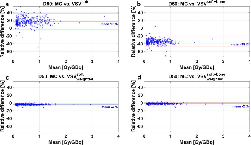
Brosch-Lenz et al. EJNMMI Physics     (2021) 8:26                                                              Page 2 of 17

                   (Continued from previous page)
                   Conclusion: Patient-individual MC absorbed dose simulation is capable to account for
                   heterogeneous densities in bone lesions. Since the computational effort prevents its
                   routine clinical application, TSV or VSV dosimetry approaches are used. This study
                   showed the necessity of lesion-individual density weighting for TSV or VSV in Lu-177-
                   PSMA therapy bone lesion dosimetry.
                   Keywords: Radioligand therapy, mCRPC, PSMA, Lutetium-177, 3D dosimetry, Tumor
                   dosimetry, OLINDA/EXM®, Voxel S value, Monte Carlo simulation

                 Background
                 The incidence of prostate cancer has been steadily increasing over the past decades in
                 western populations [1, 2]. Patients with castration-resistant prostate cancer (mCRPC)
                 typically present a large metastatic tumor burden in the bones [3]. Radioligand therap-
                 ies (RLT) targeting the prostate-specific membrane antigen (PSMA) such as Lutetium-
                 177-PSMA (Lu-177-PSMA) and Actinium-225-PSMA have shown promising results in
                 patients ineligible for other therapies or have shown progress after receiving other sys-
                 temic treatment options [4]. The clinical value of personalized dosimetry in RLT lies in
                 a possible increase of the therapeutic window by limiting absorbed dose to organs at
                 risk while maximizing absorbed dose to tumors. Thus, personalized dosimetry is indis-
                 pensable for correlation with therapy response and patient outcome, enabling adjust-
                 ments for subsequent therapy cycles. The first Lu-177-DKFZ-PSMA-617 absorbed dose
                 estimates were published in 2015 [5]. Nonetheless, up to now, there are still few publi-
                 cations addressing the absorbed doses delivered to tumors after Lu-177-PSMA therapy
                 [5–11]. While there is a clear definition of absorbed dose D as “the quotient of dε by
                 dm, where dε is the mean energy imparted by ionizing radiation to matter of mass dm”
                 in Report 85 of the International Commission on Radiation Units and Measurements
                 (ICRU) [12], there are, however, different approaches for estimation of absorbed dose
                 for internal radionuclide therapies, each with varying complexity and accuracy.
                   The use of pre-calculated organ-specific S values according to the Medical Internal
                 Radiation Dose (MIRD) Committee formalism [13] has become more prevalent using
                 the OLINDA/EXM® 2.0 software (HERMES Medical Solutions, Sweden) [14]. However,
                 for the particular situation of tumor absorbed dose estimation, this approach relies on
                 the unit density sphere model for calculation of tumor S values (TSV) that assumes
                 homogeneous activity distribution within the spherical tumor and a tumor density of 1
                 g/cm3 (i.e., soft tissue). Thus, this fast and simple approach has limited applicability to
                 bone lesions with higher densities and non-uniform activity distributions. Mass scaling
                 of TSVs has been applied to include patient-specific density variations [15, 16], though
                 the lesion-individual density in mCRPC patients may still limit the value of mass scal-
                 ing of TSV. A three-dimensional (3D) voxel-wise dosimetry approach includes
                 radionuclide-specific absorbed dose kernels or voxel S values (VSVs), which are pre-
                 simulated for a specific tissue type and voxel size [17]. The use of VSVs accounts for
                 heterogeneous activity distributions under the assumption of a homogeneous material
                 and density [17]. Monte Carlo (MC) absorbed dose simulations based on SPECT/CT
                 data include patient-individual, heterogeneous density, and activity distributions, yield-
                 ing 3D voxel-wise absorbed dose estimations.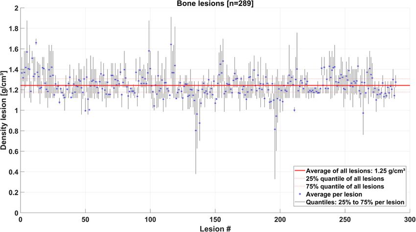
Brosch-Lenz et al. EJNMMI Physics   (2021) 8:26                                                                      Page 3 of 17

                   The aim of this work is to investigate various dosimetry techniques for accurate bone
                 lesion absorbed dose estimation in Lu-177-PSMA therapy of mCRPC. The unit density
                 sphere model for TSVs for volume of interest (VOI)-based dosimetry, and VSVs for dif-
                 ferent tissue types for 3D voxel-based dosimetry, without and with a tissue-specific
                 density weighting were compared to patient-individual dosimetry by Monte Carlo
                 simulations.

                 Methods
                 Patients
                 The study was conducted retrospectively on anonymized data and was approved by the
                 local ethics committee of our institution. Fifteen patients with metastatic, castration-
                 resistant prostate cancer (mCRPC) and pronounced metastases in the skeleton were in-
                 cluded in this study. Table 1 presents the detailed patient characteristics. Patients re-
                 ceived a first cycle of radioligand therapy using Lu-177-PSMA-I&T with activities of
                 7.4 GBq (10 patients) and 9.0 GBq (5 patients). The higher initial therapy activities were
                 used in case of severe burden of bone metastases and/or presence of visceral
                 metastases.

                 Image acquisition and reconstruction
                 Following the standard clinical routine imaging protocol of our institution, patients
                 underwent quantitative Lu-177 SPECT/CT imaging (Symbia IntevoTM T16 SPECT/CT,
                 3/8" crystal, medium-energy low-penetration collimator, Siemens Healthcare, Germany)
                 at 24 h, 48 h, and 72 h post injection (p.i.). At least two SPECT bed positions were

                 Table 1 Summary of patients being included. Previous treatment (1: yes; 0: no): OP surgery, RTx
                 radiotherapy, AHT anti-hormonal therapy (including second line AHT with bicalutamide,
                 enzalutamide, abiraterone acetate), CTx chemotherapy (docetaxel, cabazitaxel), Ra-223 radium-223
                 dichloride
                 Patient      Age    Activity     PSA        Gleason       Previous treatment
                                     (GBq)        (ng/ml)    score
                                                                           OP     RTx     AHT      CTx      Ra-223
                                                  prior to
                                                  therapy
                 1            61     7.44         25.9       9             0      1       1        1        0
                 2            75     7.46         38.4       9             1      0       1        1        0
                 3            75     7.44         1070       8             1      1       1        1        1
                 4            78     9.04         570        9             0      0       1        1        0
                 5            62     7.47         848        -             0      1       1        0        0
                 6            59     7.47         5.38       7b            0      1       1        1        0
                 7            74     9.19         1696       -             1      1       1        0        0
                 8            63     7.46         149        8             0      1       1        1        0
                 9            82     7.44         20.2       9             1      1       1        0        0
                 10           70     7.42         127        9             1      1       1        1        1
                 11           75     9.05         436        9             0      1       1        1        0
                 12           49     9.00         121        9             1      1       1        1        1
                 13           64     7.47         1268       8             0      1       1        1        0
                 14           79     7.46         72.7       7b            0      0       1        0        0
                 15           73     9.04         19.6       9             1      0       1        1        0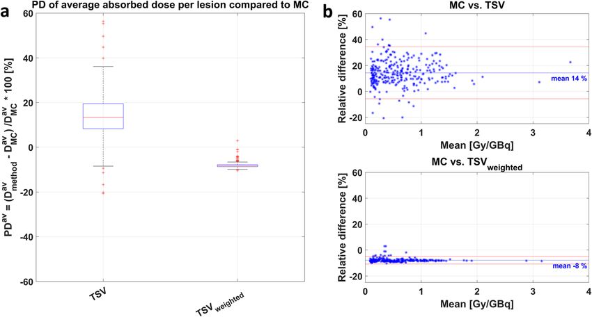
Brosch-Lenz et al. EJNMMI Physics           (2021) 8:26                                                          Page 4 of 17

                 acquired in auto-contour mode followed by a low-dose CT. Image acquisition parame-
                 ters included a 128 × 128 matrix with 64 angular steps and a duration of 5 s per step.
                 These parameters were chosen as a compromise between covering the extended axial
                 field of view (FOV) and patient comfort. The imaging energy window was centered at
                 the energy of the upper photo peak of Lu-177 at 208 keV (width 15%). Quantitative
                 SPECT reconstruction was performed with the clinically established Hermes Hybrid
                 Recon v.2.1.1 reconstruction, which represents an ordered-subset ordinary-Poisson
                 maximum a priori expectation maximization (OS-MAP-EM) reconstruction algorithm
                 with a one-step late weighted quadratic penalty function and collimator-specific depth-
                 dependent detector response modelling (16 MAP iterations, 8 subsets, Bayesian weight
                 0.01, HERMES Medical Solutions, Sweden) [18, 19]. CT-based attenuation correction
                 and model-based scatter estimation as described by Sohlberg et al. [18] were used. The
                 SPECT images were calibrated with a system-specific calibration factor, which was ob-
                 tained using the same SPECT image acquisition and reconstruction parameters for a
                 cylinder phantom (20 cm diameter), homogeneously filled with a known Lu-177 activity
                 concentration [5, 20, 21].

                 Image processing
                 All images were processed with PMOD (v4.005; PMOD Technologies LLC). Rigid co-
                 registration of all CT and SPECT volumes was performed onto the SPECT/CT image
                 data at 24 h p.i., which served as reference. An individual bone map and a whole-body
                 VOI were derived from the reference CT by threshold-based segmentation (Hounsfield
                 Unit (HU) threshold of 200 for bone map [3], HU threshold − 200 to − 100 for the
                 whole body), and kidney VOIs were defined by manual delineation. To further segment
                 individual bone lesions within the skeletal bone map, the semi-automatic k-means clus-
                 ter segmentation of PMOD 3D tool was used on the 24-h SPECT [3]. All VOIs were
                 copied to the co-registered SPECT data sets. Since image artifacts and noise impact
                 voxel-wise fitting, time-activity curves were fitted in pre-defined VOIs to determine
                 VOI-wise effective half-lives. VOI activities for the kidneys, tumor lesions, and remain-
                 der of the body (whole-body minus the kidneys and tumor lesions) were fitted using a
                 mono-exponential fit model. A hybrid VOI/voxel-wise approach was used for gener-
                 ation of time-integrated activity images to partially maintain the voxel-wise activity dis-
                 tribution information. The time-integrated activity images per patient were generated
                 with MATLAB (R2019b, The MathWorks, Inc. Natick, MA) based on the reference
                 SPECT at 24 h p.i. and the individual VOI map:

                                    voxel
                      ~ voxel ¼ At¼0
                      A                                                                                   ð1Þ
                                λVOI

                 where A~ voxel denotes the time-integrated activity per voxel, Avoxel is the activity at time
                                                                                    t¼0
                                                            .
                 point zero in a voxel, and λVOI ¼     ln 2         uses the effective half-life obtained from
                                                              t 1=2
                 mono-exponential fitting in the related VOI. Avoxel
                                                               t¼0 was computed as:

                              voxel λVOI ∙t
                       t¼0 ¼ At
                      Avoxel       ∙e                                                                     ð2Þ
Brosch-Lenz et al. EJNMMI Physics (2021) 8:26 Page 5 of 17
with the time t being the exact time point of the individual 24 h p.i. SPECT
acquisition.
Dosimetry calculations
We investigated 7 different dosimetry approaches by utilizing the aforementioned time-
integrated activity images and the reference CT of each patient.
MC method: Patient-specific Monte Carlo (MC) absorbed dose simulation
Patient-specific MC absorbed dose simulation accounts for the patient’s anatomy by
using the geometry and density information from the patient’s CT image [22]. The
radioactive decay, the interactions of the ionizing radiation with matter, and conse-
quently the absorbed dose are simulated based on the patient-individual time-
integrated activity distribution. Hence, MC absorbed dose simulations contain the high-
est level of complexity for modelling of radiation transport and interactions of ionizing
radiation with matter with associated energy deposition among all other applied
methods in this study. In concordance with inter alia Dieudonné et al. [23] and Grimes
et al. [24], we considered MC dosimetry as the reference method assessing the other
methods for bone lesion dosimetry. MC simulations in this study were performed using
the GATE MC code version 8.2, based on GEANT4 version 10.5.1. This code has previ-
ously been validated for use in nuclear medicine therapies [25–27]. The radionuclide
data were based on the Nuclear Data Sheets of Kondev et al. [28] and are the same as
in OLINDA/EXM® [29]. A CT scan of a Gammex tissue characterization phantom
(Gammex 467; Gammex Inc., Middleton, WI) using the same imaging parameters from
the patient scans was performed, which confirmed the HU-to-density relationship of
our CT device with that implemented in GATE. GATE converts HU-to-density values
with internal tables based on Schneider et al. [22]. The time-integrated activity image
of each patient was normalized with its total number of decays and used as the input
for the simulations. The total number of 109 primary decays per patient simulation was
divided into 20 sub-simulations for parallel execution on separate CPUs to increase
simulation speed (dual CPU system with 2 INTEL XEON 4114 CPUs, 10 cores each,
2.2 GHz, 192 GB RAM, running on Linux). The relative statistical uncertainty in the
absorbed dose per voxel was calculated as described by Chetty et al. [30]. The voxel size
of the simulation was (4.7952 mm)3 corresponding to the voxel sizes of the SPECT acqui-
sitions. All particle range thresholds were set to 0.1 mm.
TSV method: Tumor S values (TSV) according to the unit density sphere model
The tumor S values from the uniform and unit density sphere model of OLINDA/
EXM® 2.0 (HERMES Medical Solutions, Sweden) were used. This method represents
the model with the lowest level of complexity and can be considered as the most simple
and applicable method, yet clinically available. Since the total time-integrated activity
per lesion and the lesion volume were known from the processing steps described
above, the average lesion absorbed dose was calculated following the MIRD formalism
[13] by multiplication of the tumor S value for the selected tumor volume with the
tumor time-integrated activity. This approach is assuming that the lesion mass is com-
parable to the lesion volume at a tissue density of 1 g/cm3. TSVs are available for aBrosch-Lenz et al. EJNMMI Physics (2021) 8:26 Page 6 of 17
limited number of sphere volumes/masses. Hence, the TSV per lesion was obtained by
fitting the available TSVs within OLINDA/EXM®, and subsequent calculation of the
TSV for the lesion mass m with the fit parameters (TSV(m) = 2.19 ∙ 10−5 ∙ m−0.99). This
method includes solely the tumor self-dose [31] and is further based on the assumption
that lesions were all of spherical shape with unit density and uniform activity distribu-
tion [32].
TSVweighted method: TSV according to the unit density sphere model with additional lesion-
individual density weighting
A simple method aiming to improve this absorbed dose estimate and to account for
the tissue-specific tumor density is to convert the patient CT-image voxel-wise to dens-
ities using the HU-to-density conversion table, followed by the extraction of average
lesion-individual density using the lesion VOI. The absorbed dose estimate is subse-
quently adjusted by weighting the lesion absorbed dose value Dlesion with the ratio of
unit density and the average lesion-individual density ρlesion , being equivalent to the
mass scaling of S values [16]. This method takes into account the actual average lesion
density ρlesion rather than assuming a fixed density for all lesions.
1g
Dlesion ¼Dlesion
∙ cm3 : ð3Þ
weighted
ρlesion
VSVsoft method: Absorbed dose convolution model using voxel S values (VSVs) based on
International Commission On Radiological Protection (ICRP) soft tissue
To account for the non-uniform activity distribution in 3D voxel-wise dosimetry, the
use of VSVs for dosimetry has gained increasing interest [17]. For this purpose, GATE
MC code was used for the simulation of Lu-177 VSVs using the voxel size of the time-
integrated activity images, namely (4.7952 mm)3. The simulation used the soft tissue
composition according to the ICRP [33, 34]. The central voxel of the ICRP soft tissue
medium in a 51 × 51 × 51 matrix was set as Lu-177 source voxel, and 108 primaries
were simulated. The VSVs represent the absorbed dose distribution per decay such that
when convolved with the time-integrated activity image this results in a patient-specific
3D voxel-wise absorbed dose map.
VSV soft
weighted method: Absorbed dose convolution model using VSVs based on ICRP soft tissue
with additional density weighting
A limitation of the VSVsoft method was that the VSVs were simulated exclusively for
soft tissue, and hence, the applicability for bone lesion dosimetry is hindered. Similar to
the density weighting presented in the TSVweighted method, it is possible to adjust for
the different densities of the patient-individual anatomy and the density of the simu-
lated VSVs. For this, the HUs of the patients’ CT were voxel-wise converted into dens-
ity values. Consequently, the 3D voxel-wise absorbed dose map from the VSVsoft
method is voxel-wise weighted with the ratio of the VSV density of ICRP soft tissue
ρICRP to the actual voxel density ρvoxel [23]:Brosch-Lenz et al. EJNMMI Physics (2021) 8:26 Page 7 of 17
ρICRP
weighted ¼ D
Dvoxel ∙ : ð4Þ
voxel
ρvoxel
VSVsoft+bone method: Absorbed dose convolution model using VSVs based on ICRP soft tissue
and VSVs based on ICRP cortical bone
We extended the VSVsoft method by simulation of cortical bone VSVs using a standard
ICRP cortical bone composition [33, 34] with the same simulation setup as for the
ICRP soft tissue VSVs in the VSVsoft method. Similar to Lee et al. [35] who used mul-
tiple VSVs for regions with different tissues and densities, the combination of VSVsoft
and VSVbone was tested. For this, the patient’s bone map was used to distinguish be-
tween regions containing bone or soft tissue. The corresponding tissue-specific VSVs
were applied in their respective regions. Subsequently, to obtain a total 3D voxel-wise
absorbed dose map, the soft tissue 3D voxel-wise absorbed dose map (in soft tissue re-
gions) and the cortical bone 3D voxel-wise absorbed dose map (in bone regions) are
combined into a single image.
VSV softþbone
weighted method: Absorbed dose convolution model using VSVs based on ICRP soft
tissue and VSVs based on ICRP cortical bone with additional density weighting
The skeleton itself is not merely composed of the cortical bone and shows a heteroge-
neous composition of tissues with varying densities. Therefore, to further account for
the variations in bone composition, beyond the above-mentioned standard cortical
model, a similar voxel-wise density weighting as in Eq. (4) is applied to the combined
3D voxel-wise absorbed dose map obtained from the VSVsoft+bone method in order to
correct for differences in density per voxel.
Comparisons
The TSV and TSVweighted yield average lesion absorbed doses in agreement with the
definition of average absorbed dose Dav in a chosen region of a specific tissue with total
mass mt as defined by Eq. 6.3 in the ICRU Report 86 [36]. To enable a comparison of
this average absorbed dose per lesion Dav for the TSV approaches with the 3D MC
voxel-wise absorbed dose maps, the average was formed accordingly, yielding Dav MC . The
percentage difference PDav was calculated:
method − DMC
Dav av
PDav ¼ ∙100 ð7Þ
Dav
MC
To evaluate the 3D voxel-wise absorbed dose maps obtained from MC, VSVsoft,
soft+bone
VSVsoft
weighted , VSV , and VSVsoftþbone
weighted , the minimum absorbed dose within 25%,
50%, and 75% of the VOI volume per lesion was calculated, giving D25, D50, and D75.
For the assessment of the agreement of the different investigated 3D voxel-wise
absorbed dose estimation methods, PDvox was calculated on a voxel level for VSVsoft,
soft+bone
VSVsoft
weighted , VSV , and VSVsoftþbone
weighted compared with MC:
method − DMC
Dvox vox
PDvox ¼ vox ∙100 ð9Þ
DMCBrosch-Lenz et al. EJNMMI Physics (2021) 8:26 Page 8 of 17
Bland-Altman plots [37, 38] were used to compare the absorbed dose estimation
methods.
Results
In total, 289 bone lesions in the 15 mCRPC patients were evaluated. The segmented le-
sion volumes were on average 19.1 ml (range: 1.1 to 453.2 ml). The bone lesions were
distributed within the whole skeleton. The majority of lesions were situated in the ver-
tebrae (106), followed by the ribs (68), the extremities (64), and the pelvis (51). The
average lesion density was 1.25 ± 0.11 g/cm3 (min: 0.80 g/cm3; max: 1.66 g/cm3), aver-
aged over all 289 bone lesions. The density variation within each bone lesion is dis-
played for all lesions in Fig. 1.
MC simulations
The overall simulation time per patient for the MC method was less than 4.5 h. The
maximum relative statistical uncertainty in absorbed dose simulations was below 2.4 %
for all voxels in all lesions and below 0.9 % on average over all lesion voxels. The max-
imum statistical uncertainty in the absorbed dose for the target region of ICRP soft tis-
sue and ICRP cortical bone VSVs of the VSVsoft, VSVsoft
weighted , VSV
soft+bone
, and
VSVsoftþbone
weighted methods was below 3.2%. This was for the most distant voxel from the
source voxel. The average over all target voxels was below 2.0%.
Comparison of dosimetry methods
The percentage difference PDav of average lesion absorbed dose estimates for the un-
altered TSV was + 14 ± 10% (min: − 21%; max: + 56%) compared to MC, averaged over
all lesions. The lesion-individual density weighting reduced the PDav of TSVweighted to
− 8 ± 1% (min: − 10 %; max: − 3%). Figure 2a illustrates the decrease in range of PDav
for TSVweighted compared to TSV, further supported by the Bland-Altman plot in Fig.
2b, showing the mean value of both methods compared to their relative difference.
Fig. 1 Density variation per lesion, given for all 289 bone lesionsBrosch-Lenz et al. EJNMMI Physics (2021) 8:26 Page 9 of 17
Fig. 2 a Boxplot of PDav per bone lesion of TSV and TSVweighted compared to MC. b Bland-Altman plot
The percentage difference (PD) of D25, D50, and D75 of VSVsoft, VSVsoft
weighted ,
VSVsoft+bone, and VSVsoftþbone
weighted methods compared to MC are given in Table 2, averaged
over all lesions. The density weighting of VSV reduced the PD compared to the un-
weighted methods. The smallest PD of − 2% for D25, D50, and D75 was found for
VSVsoftþbone
weighted . The evaluation on a voxel level revealed PD
vox
of + 18 ± 11% (min: − 27%;
max: + 58%) for VSVsoft, averaged per VOI and over all lesions. This was reduced to −
5 ± 1% (min: − 12 %; max: − 2%) after voxel-wise density weighting for VSVsoft
weighted .
VSVsoft+bone showed PDvox of − 34 ± 6% (min: − 60%; max: + 5%). VSVsoftþbone
weighted showed
the smallest PDvox of − 2 ± 1% (min: − 9%; max: 0%). These observations are summa-
rized in Fig. 3. The additional density weighting of VSVsoft softþbone
weighted , and VSV weighted , led to
an overall smaller range of percentage differences than the associated method without
weighting.
Figure 4 shows low bias for D50 compared to MC for the bone lesion absorbed dose
estimates achieved with the density weighted VSVsoft softþbone
weighted (Fig. 4c) and VSV weighted
(Fig. 4d). Furthermore, their corresponding limits of agreement and mean relative
difference were the smallest with fewest outliers of all investigated 3D voxel-wise
dosimetry methods. The Bland-Altman plots in Fig. 4a and b demonstrate the larger
variations in lesion absorbed doses of the unweighted dosimetry methods compared to
MC dosimetry.
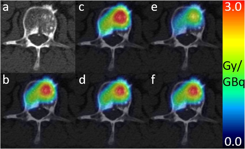
Figure 5 visualizes a patient example showing the same transversal slice of 3D voxel
absorbed dose maps from the 3D voxel-wise dosimetry methods fused with the corre-
sponding slice of the patient`s CT (Fig. 5a). The 3D absorbed dose maps for the dis-
played bone lesion obtained from MC (Fig. 5b), VSVsoft softþbone
weighted (Fig. 5d), and VSVweighted
(Fig. 5f) are comparable. The 3D absorbed dose map of VSVsoft (Fig. 5c) generally over-
estimates and VSVsoft+bone (Fig. 5e) underestimates the 3D absorbed dose map obtained
from MC (Fig. 5b).Brosch-Lenz et al. EJNMMI Physics (2021) 8:26 Page 10 of 17
Table 2 PD in minimum absorbed doses to 25%, 50%, and 75% (D25, D50, D75) of the lesion VOI
volume compared against MC. The PD was formed per lesion and then averaged over all lesion
giving the presented value
Method VSVsoft VSVsoft
weighted
VSVsoft+bone VSVsoftþbone
weighted
Mean ± SD Mean ± SD Mean ± SD Mean ± SD
PD of D25 [%] 15 ± 14 −4±2 − 36 ± 8 −2±2
PD of D50 [%] 17 ± 11 −4±2 − 35 ± 6 −2±2
PD of D75 [%] 18 ± 10 −5±1 − 34 ± 6 −2±1
Discussion
Patients with advanced mCRPC present with a considerably high tumor burden in the
bone. Furthermore, osteosclerotic bone metastases can develop an increased number of
osteoblasts leading to an elevated bone mass and increased density in the bone lesions
[39]. Consequently, bone lesion absorbed dose estimates in Lu-177-PSMA therapy are
affected by regional variations in bone tissue density, as observed in our investigations
(Fig. 1). The absorbed dose estimates may depend on the strategy to account for these
Fig. 3 Boxplot of PDvox of the 3D voxel-wise dosimetry methods compared against MC. PDvox was averaged
per lesionBrosch-Lenz et al. EJNMMI Physics (2021) 8:26 Page 11 of 17
Fig. 4 Bland-Altman plots of D50 compared against MC for a VSVsoft, b VSVsoft+bone, c VSVsoft
weighted , and d
softþbone
VSVweighted . The mean value of both methods was plotted against the relative difference of both methods.
The blue line gives the mean relative differences and the red lines indicate the 95% limits of agreement
local changes. In this study, different techniques for VOI-wise and 3D voxel-wise dos-
imetry with varying complexity were compared. Simplified methods were tested against
absorbed dose estimation by full Monte Carlo simulation. For this purpose, dosimetry
results of 289 bone lesions of 15 mCRPC patients receiving their first cycle of Lu-177-
PSMA-I&T therapy were assessed. To our knowledge, this study is the first to analyze
and compare different dosimetric approaches for absorbed dose estimation in a high
number of bone lesions in Lu-177-PSMA therapy.
The first method was based on the application of OLINDA/EXM®, which is widely
clinically available and has been commonly used for dosimetry estimations in Lu-177-
PSMA therapies [5–7, 9–11]. The percentage difference PDav of average lesion
Fig. 5 Patient example showing the transversal slice of 3D voxel-wise absorbed dose maps, fused with the
patient’s CT image in a. Maps in units of Gy/GBq were achieved with methods: b MC, c VSVsoft, d VSVsoft
weighted ,
e VSVsoft+bone, and f VSVweighted
softþbone
. Average density of the displayed lesion was 1.20 g/cm3Brosch-Lenz et al. EJNMMI Physics (2021) 8:26 Page 12 of 17
absorbed doses compared to the MC average absorbed dose Dav MC ranged from an
underestimation of − 21% to an overestimation by + 56%, yielding an averaged overesti-
mation of + 14 ± 10% in all lesions. The broad spread of relative differences can partly
be explained by the different assumptions made within this approach, i.e., spherical
shape, uniform activity distribution, and unit density of the tumor. The latter may have
the greatest impact for bone lesions with increased density. Using the VOI-based
method TSVweighted, we hence attempted to correct for the different density of bone le-
sions compared to the unit density sphere model of TSVs by using the average lesion-
individual density obtained from the patient’s CT scan. The mass scaling of the TSV
with the lesion-individual average density addresses this assumption, yielding a reduced
PDav compared to MC as highlighted in Fig. 2. The spread of the PDav of average lesion
absorbed dose estimates was reduced to − 10 to − 3% with an average absorbed dose
underestimation of − 8 ± 1%. This remaining difference may be associated to the as-
sumptions that the tumor has only contributions of self-dose and is having a spherical
shape in the TSV methods. Previous studies assessed the accuracy of absorbed dose es-
timation in soft tissue lesion against MC. Howard et al. [40] compared lesion absorbed
dose estimates from the unit density sphere model of OLINDA/EXM® against MC
simulation for Iodine-131 (I-131) radioimmunotherapy of lymphoma patients and con-
cluded that the lesion shape has a minor impact when comparing the self-dose compo-
nent. Their investigations revealed an absorbed dose underestimation compared to MC
absorbed dose with a range of − 2 to − 31% PD, with average − 15 ± 8%. Grimes et al.
[24] found good agreement of neuroendocrine tumor absorbed doses for Lu-177 from
the unit density sphere model of OLINDA/EXM® and MC simulations with average
percentage differences of − 3.8% ± 5.2%. Similar results with differences of − 5% were
found by Divoli et al. [41], comparing absorbed doses of OLINDA/EXM® and MC for
artificial spherical tumors in liver and lung. Our work assessed bone lesion absorbed
dose estimation and the mass scaling of TSVweighted with lesion-individual average
density as described herein revealed PDav compared to MC in the range of those re-
ported in the literature [24, 40–42]. Pacilio et al. [43] investigated absorbed dose esti-
mates for bone metastases of patients receiving Radium-223 (Ra-223) dichloride
therapy. This publication used a fixed density of 1.4 g/cm3 for density weighting of the
unit density sphere model of OLINDA/EXM®. If no lesion-individual density can be ob-
tained using the patient CT image, this approach may result in more realistic values.
However, the average lesion density for all 289 bone lesions investigated in this study
was 1.25 ± 0.11 g/cm3, being lower than the proposed density of the skeleton of 1.4 g/
cm3 [44]. The inter-lesion density variation displayed in Fig. 1 further supports the use
of lesion-individual densities for mass scaling of TSV.
So far, 3D voxel-wise dosimetry calculations using VSVs were mainly applied in settings
with heterogeneous activity distributions in homogeneous density distributions. For these
implementations, a high agreement for tumor absorbed doses obtained from VSVs for soft tis-
sue and MC simulation for soft tissue lesions was reported. Grimes et al. [24] reported only −
1.5 % ± 4.6% difference for Lu-177, and Dieudonné et al. [45] stated − 0.33% difference for
Yttrium-90 (Y-90) and − 0.15% difference for I-131 for a hepatic tumor phantom. In general,
VSV dosimetry calculations can account for heterogeneous activity distributions but not for
density differences since they were simulated for a single homogeneous medium. For theBrosch-Lenz et al. EJNMMI Physics (2021) 8:26 Page 13 of 17
majority of organs and lesions in the abdomen, only small density variations are assumed and
a VSVsoft approach can therefore be safely used in the clinical setting. However, the assump-
tion mentioned above has to be questioned in situations with large local tissue density
changes. Thus, an adapted absorbed dose estimation approach becomes necessary for bone le-
sions in mCRPC patients. Based on our results for 3D voxel-wise absorbed dose calculations,
we observed that both approaches, the utilization of single soft tissue VSVs (VSVsoft) and of
separate VSVs for soft tissue and bone (VSVsoft+bone), reveal limitations in estimation of
absorbed dose in bone lesions. Investigating the PDvox revealed on average a strong overesti-
mation by + 18 ± 11% (min: − 27 %; max: + 58%) for VSVsoft. VSVsoft+bone on the other hand
still showed limited capability of adequately estimating the absorbed dose per bone lesion; it
exhibited a large underestimation of absorbed dose by − 34 ± 6% (min: − 60%; max: + 5%).
These observations may be explained by the underestimated tissue density, which is an inher-
ent characteristic of the soft tissue voxel absorbed dose kernel VSVsoft, compared to the actual
bone lesion density. Therefore, this underestimation of voxel density results in an underesti-
mation of the voxel’s mass and consequently in an overestimation of the absorbed dose per
voxel. On the other hand, VSVsoft+bone relies on the assumption that bone lesions consist
merely out of the cortical bone, although a bone lesion can have different components and
densities [46]. In this case, a larger mass than the actual lesion mass is assumed, and conse-
quently, the observed absorbed dose is artificially smaller.
The VSV dosimetry methods with subsequent density weighting, as investigated
in our study, seem to better address voxel-wise density changes and may therefore
yield improved comparability with MC simulation. The proposed methods
vox
VSVsoft softþbone
weighted and VSVweighted led to significantly reduced PD compared to Monte
Carlo simulation, with an underestimation of on average − 5 ± 1% (min: − 12%;
max: − 2%) and − 2 ± 1% (min: − 9%; max: 0%), respectively. These findings are
supported by the Bland-Altman plots for D50 in Fig. 4c and d, obeying the smal-
lest spread of data points and smallest mean relative difference compared to the
MC method. Further, the majority of data points is within the 95% limits of agree-
ment, given by the red lines. This observation is in concordance with Dieudonné
et al. [23], who reported improved absorbed dose agreement for a density corrected
VSV approach compared to full MC 3D voxel-wise dosimetry for three clinical
cases with focus on soft tissue. Dieudonné et al. observed a lesion absorbed dose
difference for a I-131-Tositumomab case of − 3.1%, an organ absorbed dose differ-
ence of maximum − 1.1% for a Lu-177-peptide case, and an organ absorbed dose
difference of maximum + 0.8 % for a Y-90-microspheres case. Besides, Lee et al.
[36] noted an overall improvement of whole-body absorbed dose estimates when
introducing multiple tissue-specific VSVs, when compared to the utilization of a
single tissue VSV. However, our results for bone lesion dosimetry indicate that the
effect of additional density weighting onto a single VSV (VSVsoft
weighted compared to
VSVsoft) outperformed the effect of adding multiple VSVs for various tissues with-
out density weighting (VSVsoft+bone compared to VSVsoft). In this work, VSVs were
derived for a homogenous tissue. Hence, the application of absorbed dose kernel
convolution approaches has limitations if neighboring voxels consist of different
tissues. Due to the small maximum range of the β- particles of Lu-177 in soft tis-
sue of 2 mm [15], and given the voxel size of (4.7952 mm)3 in this investigation,Brosch-Lenz et al. EJNMMI Physics (2021) 8:26 Page 14 of 17
we expected this effect to be small when compared to the other effects investigated
herein. In our study, we attempted to compensate for tissue differences with the
proposed voxel-wise density weighting. Nevertheless, the magnitude of absorbed
dose variations related to particle transport across tissue borders with respect to
VSV methods requires further investigation.
3D voxel-wise dosimetry offers the visualization of regional variations in lesion
absorbed dose estimates on a voxel level. The drawback of 3D voxel-wise dosimetry
methods is that individual voxels can be influenced by image artifacts and noise. Fur-
ther, the limited resolution of SPECT imaging leads to a spill-over of reconstructed ac-
tivity between structures. Thus, the reconstructed 3D activity distribution does not
fully represent a purely physiological activity distribution pattern and has to be inter-
preted with care. The development and potential amelioration to handle intra-skeletal
partial-volume and spill-over compensation techniques should therefore be subject for
future investigations. Within this work, we aimed at reducing the impact of the afore-
mentioned effects by using quantitative SPECT reconstruction including distant-
dependent point spread function of the detector and a hybrid VOI/voxel-wise approach
to reduce the impact of noise and image artifacts on the determination of the time-
integrated activity images which serve as an input for the 3D voxel-wise dosimetry
methods. The applicability of density weighting is further limited to the CT resolution,
and is thus not capable to account for heterogeneities on the sub-millimeter scale. In
addition, co-registration of the 24 h, 48 h, and 72 h SPECT and CT images could po-
tentially influence the absorbed dose estimates. This becomes relevant with regard to
the outliers with small average lesion densities in Fig. 1, which represent lesions located
in the ribs, with challenging co-registration due to breathing, patient’s motion, and less
reproducible patient positioning between the image acquisitions from day to day. The
different steps required for dosimetry include quantitative patient imaging, co-
registration, segmentation, fitting, and time-integrated activity assessment, before any
absorbed dose estimation can be made [47]. This work concentrated solely on this last
step of absorbed dose estimation. The pre-processing was the same for all herein pre-
sented dosimetry methods, and thus, possible mistakes in the pre-processing would im-
pact all methods equally.
Conclusions
In our study of 289 bone lesions in mCRPC patients receiving Lu-177-PSMA-I&T ther-
apy, the proposed voxel S value dosimetry approach with subsequent voxel-wise density
weighting was associated with comparable absorbed dose estimates for bone lesions as
obtained with full patient-individual Monte Carlo absorbed dose simulation. It there-
fore has the potential to enable routine patient-individual 3D voxel-wise dosimetry
evaluations. Further, TSV approaches using lesion-individual average density for mass
scaling provide fast and accurate average bone lesion absorbed dose estimates.
Abbreviations
mCRPC: Metastatic, castration-resistant prostate carcinoma; PSMA: Prostate-specific membrane antigen; Lu-
177: Lutetium-177; TSV: Tumor S value; VSV: Voxel S value; 3D: Three-dimensional; MC: Monte Carlo; p.i.: Post injection;
FOV: Field of view; VOI: Volume of interest; HU: Hounsfield Unit; MIRD: Medical Internal Radiation Dose;
ICRP: International Commission On Radiological Protection; PD: Percentage difference; I-131: Iodine-131; Y-90: Yttrium-
90; Ra-223: Radium-223; D25: Minimum absorbed dose to 25 % of lesion VOI volume; D50: Minimum absorbed dose to
50 % of lesion VOI volume; D75: Minimum absorbed dose to 75 % of lesion VOI volumeBrosch-Lenz et al. EJNMMI Physics (2021) 8:26 Page 15 of 17
Acknowledgements
We would like to thank Dr. François Bénard for facilitating the collaboration between LMU and BC Cancer (Vancouver,
Canada). Special thanks to all technologists at the Department of Nuclear Medicine, University Hospital, LMU Munich
for their participation in data collection. This is in memoriam of Dr. Anna Celler who sadly passed away during
finalization of this publication. She was a pioneer in quantitative and dynamic SPECT imaging and further was
considered as a leading expert in dosimetry. She was not only an outstanding researcher in her field; she was also an
excellent teacher and took incomparable care of those whom she supervised. By personally caring for her students,
she had and will continuously have great impact on their personal and professional lives.
Authors’ contributions
JB, CU, AG, LK, AT, HI, PB, AR, AC, SZ, and GB designed the concept of the study. FG was responsible for the
radiopharmaceutical production. JB, AG, AT, HI, and GB reviewed the clinical data for dosimetry. All data analysis was
carried out by JB, CU, AG, and GB. All authors contributed to the drafting of the manuscript, and all authors read and
approved the final manuscript.
Funding
This work was partly funded by the German Research Foundation (DFG) within the Research Training Group GRK 2274.
Open Access funding enabled and organized by Projekt DEAL.
Availability of data and materials
Please contact author for data requests.
Declarations
Ethics approval and consent to participate
This study is based on retrospective and anonymized data, which was acquired for routine clinical dosimetry (Ethics
Committee of LMU Munich 20-520).
Consent for publication
Not applicable.
Competing interests
The authors declare that they have no competing interest.
Author details
1
Department of Nuclear Medicine, University Hospital, LMU Munich, Marchioninistrasse 15, 81377 Munich, Germany.
2
PET Functional Imaging, BC Cancer, 600 West 10th Avenue, Vancouver, BC V5Z 4E6, Canada. 3Department of
Radiology, University of British Columbia, 2775 Laurel Street, Vancouver, BC V5Z 1M9, Canada. 4Department of
Integrative Oncology, BC Cancer Research Centre, 675 West 10th Avenue, Vancouver, BC V5Z 1L3, Canada.
Received: 23 October 2020 Accepted: 23 February 2021
References
1. Torre LA, Siegel RL, Ward EM, Jemal A. Global cancer incidence and mortality rates and trends—an update. Cancer
Epidemiol Prevent Biomark. 2016;25(1):16–27.
2. Zhou CK, Check DP, Lortet-Tieulent J, Laversanne M, Jemal A, Ferlay J, et al. Prostate cancer incidence in 43 populations
worldwide: an analysis of time trends overall and by age group. Int J Cancer. 2016;138(6):1388–400.
3. Gosewisch A, Ilhan H, Tattenberg S, Mairani A, Parodi K, Brosch J, et al. 3D Monte Carlo bone marrow dosimetry for
Lu-177-PSMA therapy with guidance of non-invasive 3D localization of active bone marrow via Tc-99m-anti-granulocyte
antibody SPECT/CT. EJNMMI Res. 2019;9(1):76.
4. Kratochwil C, Fendler WP, Eiber M, Baum R, Bozkurt MF, Czernin J, et al. EANM procedure guidelines for
radionuclide therapy with 177 Lu-labelled PSMA-ligands (177 Lu-PSMA-RLT). Eur J Nuclear Med Mol Imaging.
2019;46(12):2536–44.
5. Delker A, Fendler WP, Kratochwil C, Brunegraf A, Gosewisch A, Gildehaus FJ, et al. Dosimetry for 177
Lu-DKFZ-PSMA-617: a new radiopharmaceutical for the treatment of metastatic prostate cancer. Eur J Nuclear
Med Mol Imaging. 2016;43(1):42–51.
6. Okamoto S, Thieme A, Allmann J, D’Alessandria C, Maurer T, Retz M, et al. Radiation dosimetry for 177Lu-PSMA I&T in
metastatic castration-resistant prostate cancer: absorbed dose in normal organs and tumor lesions. J Nuclear Med.
2017;58(3):445–50.
7. Fendler WP, Reinhardt S, Ilhan H, Delker A, Böning G, Gildehaus FJ, et al. Preliminary experience with dosimetry,
response and patient reported outcome after 177Lu-PSMA-617 therapy for metastatic castration-resistant prostate
cancer. Oncotarget. 2017;8(2):3581.
8. Violet J, Jackson P, Ferdinandus J, Sandhu S, Akhurst T, Iravani A, et al. Dosimetry of 177Lu-PSMA-617 in metastatic
castration-resistant prostate cancer: correlations between pretherapeutic imaging and whole-body tumor dosimetry
with treatment outcomes. J Nuclear Med. 2019;60(4):517–23.
9. Baum RP, Kulkarni HR, Schuchardt C, Singh A, Wirtz M, Wiessalla S, et al. Lutetium-177 PSMA radioligand therapy of
metastatic castration-resistant prostate cancer: safety and efficacy. J Nuclear Med. 2016;115:168443.
10. Kratochwil C, Giesel FL, Stefanova M, Benešová M, Bronzel M, Afshar-Oromieh A, et al. PSMA-targeted radionuclide therapy of
metastatic castration-resistant prostate cancer with 177Lu-labeled PSMA-617. J Nuclear Med. 2016;57(8):1170–6.Brosch-Lenz et al. EJNMMI Physics (2021) 8:26 Page 16 of 17
11. Yadav MP, Ballal S, Tripathi M, Damle NA, Sahoo RK, Seth A, et al. Post-therapeutic dosimetry of 177Lu-DKFZ-
PSMA-617 in the treatment of patients with metastatic castration-resistant prostate cancer. Nuclear Med
Commun. 2017;38(1):91–8.
12. Seltzer S, Bartlett D, Burns D, Dietze G, Menzel H, Paretzke H, et al. ICRU report 85 fundamental quantities and units for
ionizing radiation. J ICRU. 2011;11(1):1–31.
13. Snyder W, Ford M, Warner G, Watson S. MIRD pamphlet no. 11. The Society of Nuclear Medicine, New York; 1975. p. 92–3.
14. Stabin MG, Siegel JA. RADAR dose estimate report: a compendium of radiopharmaceutical dose estimates based on
OLINDA/EXM version 2.0. J Nuclear Med. 2018;59(1):154–60.
15. Cremonesi M, Ferrari M, Bodei L, Tosi G, Paganelli G. Dosimetry in peptide radionuclide receptor therapy: a review. J
Nuclear Med. 2006;47(9):1467–75.
16. Howell RW, Wessels BW, Loevinger R, Committee M. The MIRD perspective 1999. J Nuclear Med. 1999;40(1):3S–10S.
17. Bolch WE, Bouchet LG, Robertson JS, Wessels BW, Siegel JA, Howell RW, et al. MIRD pamphlet no. 17: the
dosimetry of nonuniform activity distributions—radionuclide S values at the voxel level. J Nuclear Med. 1999;
40(1):11S–36S.
18. Sohlberg A, Watabe H, Iida H. Acceleration of Monte Carlo-based scatter compensation for cardiac SPECT. Phys Med
Biol. 2008;53(14):N277.
19. Kangasmaa T, Sohlberg A, Kuikka JT. Reduction of collimator correction artefacts with bayesian reconstruction in spect.
Int J Mol Imaging. 2011;2011(Article ID 630813):6. https://doi.org/10.1155/2011/630813.
20. Ljungberg M, Celler A, Konijnenberg MW, Eckerman KF, Dewaraja YK, Sjögreen-Gleisner K. MIRD pamphlet no. 26: joint
EANM/MIRD guidelines for quantitative 177Lu SPECT applied for dosimetry of radiopharmaceutical therapy. J Nuclear
Med. 2016;57(1):151–62.
21. Dewaraja YK, Frey EC, Sgouros G, Brill AB, Roberson P, Zanzonico PB, et al. MIRD pamphlet no. 23: quantitative SPECT for
patient-specific 3-dimensional dosimetry in internal radionuclide therapy. J Nuclear Med. 2012;53(8):1310–25.
22. Schneider W, Bortfeld T, Schlegel W. Correlation between CT numbers and tissue parameters needed for Monte Carlo
simulations of clinical dose distributions. Phys Med Biol. 2000;45(2):459.
23. Dieudonné A, Hobbs RF, Lebtahi R, Maurel F, Baechler S, Wahl RL, et al. Study of the impact of tissue density
heterogeneities on 3-dimensional abdominal dosimetry: comparison between dose kernel convolution and direct
Monte Carlo methods. J Nuclear Med. 2013;54(2):236–43.
24. Grimes J, Celler A. Comparison of internal dose estimates obtained using organ-level, voxel S value, and Monte Carlo
techniques. Med Phys. 2014;41(9):092501.
25. Papadimitroulas P. Dosimetry applications in GATE Monte Carlo toolkit. Phys Med. 2017;41:136–40.
26. Papadimitroulas P, Loudos G, Nikiforidis GC, Kagadis GC. A dose point kernel database using GATE Monte Carlo
simulation toolkit for nuclear medicine applications: Comparison with other Monte Carlo codes. Med Phys.
2012;39(8):5238–47.
27. Sarrut D, Bardiès M, Boussion N, Freud N, Jan S, Létang JM, et al. A review of the use and potential of the GATE Monte
Carlo simulation code for radiation therapy and dosimetry applications. Med Phys. 2014;41(6Part1):064031.
28. Kondev F. Nuclear Data Sheets for A= 177. Nuclear Data Sheets. 2019;159:1–412.
29. Stabin MG, da Luz LC. Decay data for internal and external dose assessment. Health Phys. 2002;83(4):471–5.
30. Chetty IJ, Rosu M, Kessler ML, Fraass BA, Ten Haken RK, McShan DL. Reporting and analyzing statistical uncertainties in
Monte Carlo–based treatment planning. Int J Radiation Oncol Biol Phys. 2006;65(4):1249–59.
31. Lassmann M, Chiesa C, Flux G, Bardiès M. EANM Dosimetry Committee guidance document: good practice of clinical
dosimetry reporting. Eur J Nuclear Med Mol Imaging. 2011;38(1):192–200.
32. Stabin MG, Sparks RB, Crowe E. OLINDA/EXM: the second-generation personal computer software for internal dose
assessment in nuclear medicine. J Nuclear Med. 2005;46(6):1023–7.
33. NIST National Institute of Standards and Technonolgy. https://physics.nist.gov/cgi-bin/Star/compos.pl. Accessed 30 Mar 2020
34. McConn RJ, Gesh CJ, Pagh RT, Rucker RA, Williams R III. Compendium of material composition data for radiation
transport modeling. Richland: Pacific Northwest National Lab.(PNNL); 2011.
35. Lee MS, Kim JH, Paeng JC, Kang KW, Jeong JM, Lee DS, et al. Whole-body voxel-based personalized dosimetry: the
multiple voxel S-value approach for heterogeneous media with nonuniform activity distributions. J Nuclear Med.
2018;59(7):1133–9.
36. International Commission on Radiation Units and Measurements. Quantification and Reporting of Low-dose and Other
Heterogeneous Exposures. Oxford: Oxford University Press; 2011;11(2011):1–77.
37. Bland JM, Altman DG. Statistical methods for assessing agreement between two methods of clinical measurement. Int J
Nurs Stud. 2010;47(8):931–6.
38. Bland JM, Altman DG. Measuring agreement in method comparison studies. Stat Methods Med Res. 1999;8(2):135–60.
39. Ibrahim T, Flamini E, Mercatali L, Sacanna E, Serra P, Amadori D. Pathogenesis of osteoblastic bone metastases from
prostate cancer. Cancer. 2010;116(6):1406–18.
40. Howard DM, Kearfott KJ, Wilderman SJ, Dewaraja YK. Comparison of I-131 radioimmunotherapy tumor dosimetry:
unit density sphere model versus patient-specific Monte Carlo calculations. Cancer Biother Radiopharm.
2011;26(5):615–21.
41. Divoli A, Chiavassa S, Ferrer L, Barbet J, Flux GD, Bardies M. Effect of patient morphology on dosimetric calculations for
internal irradiation as assessed by comparisons of Monte Carlo versus conventional methodologies. J Nuclear Med.
2009;50(2):316–23.
42. Amato E, Cicone F, Auditore L, Baldari S, Prior JO, Gnesin S. A Monte Carlo model for the internal dosimetry of choroid
plexuses in nuclear medicine procedures. Phys Med. 2018;49:52–7.
43. Pacilio M, Ventroni G, De Vincentis G, Cassano B, Pellegrini R, Di Castro E, et al. Dosimetry of bone metastases in
targeted radionuclide therapy with alpha-emitting 223 Ra-dichloride. Eur J Nuclear Med Mol Imaging.
2016;43(1):21–33.
44. Cristy M, Eckerman K. Specific Absorbed fractions of energy at various ages from internal photon sources: 1, Methods.
TN (USA): Oak Ridge National Lab.; 1987.Brosch-Lenz et al. EJNMMI Physics (2021) 8:26 Page 17 of 17
45. Dieudonné A, Hobbs RF, Bolch WE, Sgouros G, Gardin I. Fine-resolution voxel S values for constructing absorbed dose
distributions at variable voxel size. J Nuclear Med. 2010;51(10):1600–7.
46. Hough M, Johnson P, Rajon D, Jokisch D, Lee C, Bolch W. An image-based skeletal dosimetry model for the ICRP
reference adult male—internal electron sources. Phys Med Biol. 2011;56(8):2309.
47. Mora-Ramirez E, Santoro L, Cassol E, Ocampo-Ramos JC, Clayton N, Kayal G, et al. Comparison of commercial dosimetric
software platforms in patients treated with 177Lu-DOTATATE for peptide receptor radionuclide therapy. Med Phys. 2020;
47(9):4602–15.
Publisher’s Note
Springer Nature remains neutral with regard to jurisdictional claims in published maps and institutional affiliations.You can also read