Syngeneic homograft of framework regions enhances the affinity of the mouse anti human epidermal receptor 2 single chain antibody e23sFv
←
→
Page content transcription
If your browser does not render page correctly, please read the page content below
EXPERIMENTAL AND THERAPEUTIC MEDICINE 21: 136, 2021

                 Syngeneic homograft of framework regions enhances
                   the affinity of the mouse anti‑human epidermal
                       receptor 2 single‑chain antibody e23sFv
        QING OU‑YANG1,2*, JUN‑LIN REN3*, BO YAN4*, JIAN‑NAN FENG5, AN‑GANG YANG1 and JING ZHAO4

             1
             State Key Laboratory of Cancer Biology, Department of Immunology, Fourth Military Medical University,
                  Xi'an, Shaanxi 710032; 2State Key Laboratory of Kidney Diseases, Department of Nephrology,
        Chinese PLA General Hospital & Chinese PLA Medical School, Beijing 100853; 3Department of Infectious Diseases,
                PLA Navy General Hospital, Beijing 100142; 4Department of Biochemistry and Molecular Biology,
                    Fourth Military Medical University, Xi'an, Shaanxi 710032; 5Department of Immunology,
                             Beijing Institute of Basic Medical Sciences, Beijing 100850, P.R. China

                                    Received September 26, 2019; Accepted October 30, 2020

                                                    DOI: 10.3892/etm.2020.9568

Abstract. e23sFv is a HER2‑targeted single‑chain variable            potential compared with EX1. The 3D structure of EX1 and the
fragment (scFV) that was characterized as the targeting portion      HER2‑EX1 complex was acquired using molecular homology
of a HER2‑targeted tumour proapoptotic molecule in our               modelling and docking and the HER2 epitopes of EX1 and
previous study. In vitro antibody affinity maturation is a method    the molecular interaction energy of the EX1‑HER2 complex
to enhance antibody affinity either by complementarity‑deter‑        were predicted. In the present study, it was demonstrated that
mining region (CDR) mutagenesis or by framework region (FR)          scFv affinity improvement based on sequence alignment was
engraftment. In the present study, the affinity of e23sFv was        feasible and effective. Moreover, the FR grafting strategy
enhanced using two strategies. In one approach, site‑directed        was indicated to be more effective and simple compared with
mutations were introduced into the FRs of e23sFv (designated         site‑directed mutagenesis to improve e23sFv affinity. In conclu‑
EMEY), and in the other approach e23sFv FRs were substi‑             sion, it was indicated that the affinity‑improved candidate EX1
tuted with FRs from the most homologous screened antibodies          may present a great potential for the diagnosis and treatment of
(designated EX1 and EX2). Notably, EX1 derived from the              HER2‑overexpressing tumours.
FR engraftment strategy demonstrated a 4‑fold higher affinity
for HER2 compared with e23sFv and was internalized into              Introduction
HER2‑overexpressing cells; however, EMEY and EX2 exhib‑
ited reduced affinity for HER2 and decreased internalization         HER2 is a member of the human EGFR family and a well‑estab‑
                                                                     lished target for HER2‑overexpressing tumour treatment (1). E23
                                                                     is a monoclonal antibody derived from mice immunized with
                                                                     HER2‑overexpressing cell components and has been indicated
Correspondence to: Dr An‑Gang Yang, State Key Laboratory             to specifically bind to the extracellular domain of HER2 (2). In
of Cancer Biology, Department of Immunology, Fourth Military         addition, e23 was generated by fusing the variable region of the
Medical University, 169 Changle West Road, Xi'an, Shaanxi 710032,    light chain (VL) with that of the heavy chain (VH) to form a
P.R. China                                                           single‑chain variable fragment (scFv) named e23sFv (3). e23sFv
E‑mail: agyang@fmmu.edu.cn                                           has been indicated to exhibit reduced molecular weight and
Professor Jing Zhao, Department of Biochemistry and Molecular        more efficient penetration into solid tumours compared with
Biology, Fourth Military Medical University, 169 Changle West        that of e23; therefore, it has been utilized in immunoconjugates,
Road, Xi'an, Shaanxi 710032, P.R. China                              such as immunotoxin or immune‑proapoptotic molecules, as
E‑mail: zhaojing@fmmu.edu.cn                                         the portion targeted to HER2 (3,4). Despite its advantages in
                                                                     terms of size and trafficking, e23sFv exhibits a 4‑fold reduced
*
    Contributed equally
                                                                     binding affinity for HER2 compared with that of e23 or the
Abbreviations: scFv, single‑chain variable fragment; VL,             corresponding antigen‑binding fragment (Fab) (5). To achieve
light‑chain variable region; VH, heavy‑chain variable region; CDR,   equivalent effectiveness, a higher dose is required for antibodies
complementarity‑determining region; FR, framework region             with less affinity; however, an increased dose of therapeutic
                                                                     antibodies can result in more intensive immunogenic reactions
Key words: antibody affinity improvement, scFv, site‑directed        and possibly unexpected toxicities (6). To ensure the safety and
mutation, engraftment                                                efficacy of e23sFv‑derived immuno‑proapoptotic molecules in
                                                                     HER2‑targeted tumour treatment, it is necessary to enhance the
                                                                     affinity of e23sFv for the HER2 extracellular domain (ECD) (5).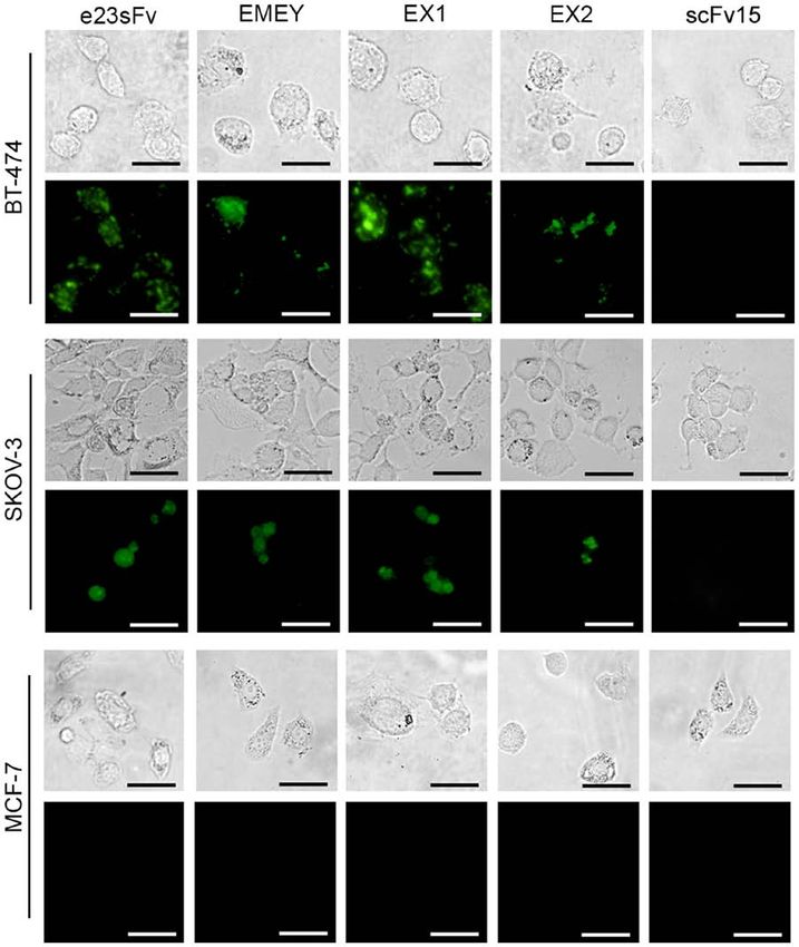
2                     OU-YANG et al: NOVEL APPROACHES FOR E23SFV ANTIBODY AFFINITY IMPROVEMENT

    In vitro antibody affinity maturation enhances the antibody    commercially (Beijing Augct DNA‑Syn Biotechnology Co.,
affinity using genetic engineering (7). Several approaches         Ltd.) and designated EMEY (Fig. 1B).
have been developed to improve the antibody affinity, the              For the CDR engraftment, FR scaffolds were selected
majority of which have focused on the mutagenesis of the           from the top two VL and VH homologous domains (Fig. 1A).
complementarity‑determining region (CDR), because CDRs             The FRs of L1 (the VL of the TN1 Fab) and H1 (the VH of
are directly involved in antibody‑antigen interactions (7). The    FabOX108) were substituted into e23sFv, giving rise to a
crystal structure of antibody‑antigen complexes has revealed       chimaera referred to as EX1 (Fig. 1B and Table I). Another
that specific CDR residues of an antibody directly contact         chimaera, EX2, was generated by substituting the FRs of
antigens and thus determine the affinity and specificity of        L2 (the VL of daclizumab) and H2 (the VH of 5E1 Fab) into
the antibody (8). Affinity improvement in vitro predomi‑           e23sFv (Fig. 1B and Table I). The codons of EX1 and EX2 were
nantly involves inducing random mutagenesis in CDRs and            further optimized for prokaryotic expression and synthesized
screening the mutants for enhanced affinity and site‑directed      by Beijing Augct DNA‑Syn Biotechnology Co., Ltd.
mutagenesis to deliberately enhance the affinity based on anti‑
body conformation (9). In addition to CDR manipulation, the        Prokaryotic expression and purification of the e23sFv
pioneering work of Foote and Winter (10) has suggested that        derivatives. The genes encoding e23sFv‑derived scFvs fused
residues in the β‑sheet structure of framework regions (FRs),      with His‑tag at the 3' end and with 5'‑ and 3'‑flanking NcoI
which support CDRs, serve critical roles in the adjustment of      and NotI restriction sites were cloned into the prokaryotic
the loop structures of CDRs. Although these residues, which        expression vector pET28a (Takara Biotechnology Co.,
are referred to as ‘Vernier zone residues’, do not directly        Ltd.). The resulting recombinant plasmids were verified
interact with the antigen, careful selection of these residues     by agarose gel electrophoresis using 1% gel and ethidium
may prove essential for shaping the diversity of the structures    bromide visualization. The recombinant plasmids were then
in the primary repertoire and affinity maturation (11).            transformed into E. coli BL21 (DE3) (Takara Biotechnology
    In the present study, the affinity of a single variable        Co., Ltd.), and after 3 h of induction with 1 mM isopropyl
fragment, e23sFv, was more improved using FR engineering           β‑d‑1‑thiogalactopyranoside (IPTG) at 37˚C, the bacteria were
compared with CDR mutagenesis. The e23sFv FR was                   harvested and sonicated for 30 min at 2 sec intervals on ice for
substituted with FRs from the two most homologous anti‑            use in SDS‑PAGE analysis to demonstrate the presence of the
bodies in the National Centre for Biotechnology Information        proteins of interest in inclusion bodies. For the purification of
(NCBI) protein database and two candidates named EX1 and           inclusion bodies, the precipitates were dissolved in 8 M urea
EX2 were constructed. Another candidate was constructed            buffer and subjected to Ni 2+ ‑NTA affinity chromatography
by e23sFv FR residue mutation based on the sequence                using Ni‑NTA His•Bind Resins (Novagen; MilliporeSigma)
alignment with the variable region of the homologous anti‑         according to the manufacturer's instructions. After extensive
bodies. All three recombinant scFvs retained the e23sFv            washing with 100 mM imidazole buffer, the bound proteins
CDRs. The affinity assays demonstrated that EX1 exhibited          were eluted at room temperature with 1,000 mM imidazole
the highest homology with e23sFv, thereby significantly            buffer and refolded by linearly decreasing the urea gradient
improving its affinity for HER2, and was internalized into         (7, 6, 4, 2, 1 and 0 M). The renatured proteins were finally
HER2‑overexpressing cells more effectively compared with           dialyzed into PBS (pH 7.4) and quantified by BCA assay
the other candidates.                                              (Pierce; Thermo Fisher Scientific, Inc.).

Materials and methods                                              Identification of the e23sFv derivatives by SDS‑PAGE and
                                                                   western blotting. SDS‑PAGE (12%) was performed using
Framework redesign of e23sFv‑based scFvs by mutagenesis            renatured e23sFv proteins (30 µg per sample). The protein
and engraftment. Two strategies were used to reconstitute the      electrophoresis gel was stained using Coomassie brilliant
FRs of e23sFv, site‑directed mutagenesis and CDR grafting,         blue R‑250 followed by greyscale analysis of the purity
both of which were based on the analysis of the e23sFv amino       of the scFvs with Image J (version 1.44; National Institutes
acid sequence homology of proteins in the NCBI database            of Health). Specifically, the background‑corrected density of
(https://blast.ncbi.nlm.nih.gov/Blast.cgi). Five VL (L1‑L5) and    the protein band was divided by the background‑corrected
five VH (H1‑H5) sequences with the highest similarity to the       density of the entire lane and multiplies by 100 to obtain the
VL and VH domains of e23sFv are presented in Fig. 1A, and          purity percentages. The e23sFv‑based scFvs were identified by
in these candidates all FRs were aligned and comparable to         immunoblotting using nitrocellulose membranes. Membranes
that of e23sFv. Detailed information on the five VLs and five      were blocked with 2% non‑fat milk at room temperature for
VHs selected is presented in Table I.                              1 h and was then incubated with anti‑His antibody conjugated
    For the site‑directed mutations, five residues in the VL and   to horseradish peroxidase (1:1,000 dilution; cat. no. 34460;
six residues in the VH sequences in the e23sFv FRs, which          Qiagen, Inc.) at 4˚C overnight followed by chemiluminescent
differed among the five domains in the homologous FRs, were        detection using Pierce™ ECL Western Blotting Substrate
replaced with the most frequently occurring amino acids in the     (cat. no. 32209; Thermo Fisher Scientific, Inc.) according to
homologous proteins (Fig. 1A), with the exception of the first     the manufacturer's instructions.
S‑T mutation in VL, which was not substituted to ensure that
the chemical characteristics of the domains were retained. The     FITC labelling of the e23sFv derivatives. For the quantification
resulting e23sFv derivative with 11 point mutations was geneti‑    of cellular binding and visualization of the subsequent
cally optimized with E. coli‑preferred codons, synthesized         internalization by HER2‑overexpressing cells, the purified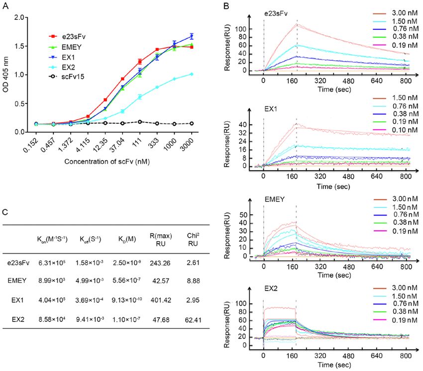
EXPERIMENTAL AND THERAPEUTIC MEDICINE 21: 136, 2021                                                                3

Figure 1. Design of FR‑engineered e23sFv derivatives. (A) Amino acid sequence alignment of the VL and VH domains of mouse anti‑HER2 single‑chain vari‑
able fragment, e23sFv, and their five most homologous counterparts identified in the National Centre for Biotechnology Information protein database. L1‑L5
represent VL homologous sequences and H1‑H5 represent VH homologous sequences. CDRs and FRs are indicated in columns. The residues that are identical
to those of e23sFv are indicated with dashed lines, and missing residues in the CDRs are indicated with asterisks. Non‑identical FR residues in e23sFv and all
their five homologs in the VL or VH collection are in red. Introduced site‑directed mutations are indicated by blue triangles, above which are the corresponding
substituted residues. (B) The schematic structure of three e23sFv derivatives. EMEY includes 11 mutated residues in the FRs of e23sFv, as indicated by
triangles. EX1 and EX2 represent CDR grafts of e23sFv in the L1‑H1 and L2‑H2 FR scaffolds, respectively. FR, framework region; VL, light‑chain variable
region; VH, heavy‑chain variable region; CDR, complementarity‑determining region.

e23sFv‑derived scFvs were conjugated with FITC using Hook™                        Measurement of the affinity constant by surface plasmon reso‑
dye labelling kit (G‑Biosciences). The FITC‑labelled scFvs were                   nance (SPR). An SPR assay was used for the kinetic analysis of
sterilized using 0.22 µm centrifugal filters (EMD Millipore).                     the binding of the e23sFv‑based scFvs to recombinant HER2
The absorption of fluorescein at 495 nm was detected by a                         in vitro. A ProteOn™ GLC sensor chip (Bio‑Rad Laboratories,
UV‑2450 spectrophotometer (Shimadzu Corporation), and the                         Inc.) was immobilized with 5 µg/ml HER2 in 10 mM acetate
molar ratio of FITC to scFv was ~2.5:1.                                           (pH 5.5). Five solutions in PBS‑T with two‑fold serial dilutions
                                                                                  of the e23sFv derivatives were injected and run through the
Binding affinity to recombinant HER2 as determined by                             sensor chip at a flow rate of 50 µl/min. The analyte injection
ELISA. Recombinant human HER2 (Sino Biological; 500 ng                            programme included a 180 sec association phase followed by
per well) was immobilized on 96‑well plates at 4˚C for 16 h. The                  a 600 sec dissociation phase. The data were analysed in a 1:1
HER2‑coated plates were blocked with 1% BSA (Sigma‑Aldrich;                       Langmuir binding model (12) using ProteOn Manager software
Merck KGaA) at room temperature for 1 h and incubated with                        (Bio‑Rad Laboratories, Inc.). The equilibrium constant (K D)
the three‑fold‑serially diluted e23sFv derivatives from 3 µM                      was calculated as the ratio of the dissociation rate constant (Koff )
for 4 h at room temperature. ScFv15 served as the negative                        to the association rate constant (Kon).
control. After washing with PBS‑Tween‑20 (0.05%; PBS‑T), the
bound scFvs were incubated with an HRP‑conjugated anti‑His                        Binding affinity for cellular HER2 as determined by flow
antibody (1:2,000 dilution; cat. no. 34460; Qiagen, Inc.) for                     cytometry. HER2‑positive cells (BT‑474 and SKOV‑3 cells)
1 h at room temperature. Following extensive washing with                         and HER2‑negative cells (MCF‑7 cells) were purchased
PBS‑T, the microplates were incubated with a 2,2'‑azino‑bis                       from American Type Culture Collection and cultured in
(3‑ethylbenzothiazoline‑6‑sulfonic acid) diammonium salt                          RPMI‑1640 medium (cat. no. 10‑040‑CV; Corning, Inc.)
(7 mM) substrate at 37˚C for 20 min, and the absorbance                           with 10% FBS (cat. no. 10099; Gibco; Thermo Fisher
was measured at 409 nm using a Sunrise microplate reader                          Scientific, Inc.) at 37˚C and 5% CO2. After blocking in 5%
(Tecan Group, Ltd.).                                                              BSA (cat. no. A1933; Sigma‑Aldrich; Merck KGaA) at 4˚C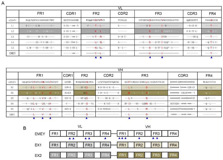
4                      OU-YANG et al: NOVEL APPROACHES FOR E23SFV ANTIBODY AFFINITY IMPROVEMENT

Table I. Detailed information of selected framework region scaffolds by homologous analysis.

Chain                                    Name                                         Origin              Entry ID          (Refs.)

L1         Chain L, human thrombopoietin neutralizing antibody TN1 Fab   Mus musculus pdb|2ZKH|                                16
L2         Chain B, crystal structure of the Fab fragment of therapeutic Mus musculus pdb|3NFP|                                17
           antibody daclizumab
L3         Chain A, Fab fragment of the engineered human monoclonal      Mus musculus pdb|1AD0|                                18
           antibody A5B7
L4         Immunoglobulin H23 light chain kappa variable region          Humanized    gb|AAB38288.1|                           19
L5         Chain L, structure of CD40L in complex with the Fab fragment  Humanized    pdb|1I9R|                                20
           of the humanized 5C8 antibody
H1         Chain B, crystal structure of FabOX108                        Mus musculus pdb|3DGG|                                21
H2         Crystal structure of sonic hedgehog bound to the 5E1 Fab      Mus musculus pdb|3MXW|                                22
           fragment
H3         Chain B, crystal structure of IL‑23 in complex with           Mus musculus pdb|3D85|                                23
           neutralizing Fab
H4         Immunoglobulin heavy chain V region humanized                 Humanized    gb|AAB24133.1|                           24
           bispecific antibody
H5         Immunoglobulin heavy chain V region precursor                 Homo sapiens pir|PN0444|                              25

Fab, antigen‑binding fragment.

for 30 min, 1x10⁶ HER2‑positive cells (BT‑474 and SKOV‑3            with the most homologous antibodies based on the homolo‑
cells) and HER2‑negative cells (MCF‑7 cells) were incubated         gous protein structures obtained from the Protein Data Bank
with 125 nM FITC‑conjugated e23sFv derivatives on ice for           (PDB) (https://www.rcsb.org/) using the Homology module in
1 h. For the negative control, a non‑specific scFv, scFv15          Discovery Studio 4.5 software (BIOVIA; Dassault Systèmes).
against hepatitis B virus surface antigen (HbsAg), which            The predicted structures of e23sFv and EX1 were subsequently
was purified and labelled with FITC as previously described,        docked to the crystal structure of the human HER2 ECD using
was used at 125 nM to exclude nonspecific binding (13). In          ZDOCK Server (http://zdock.umassmed.edu.).
addition, a FITC‑labelled anti‑HER2 antibody (1:2,000;
cat. no. 10004‑R511‑F; Sino Biological) was used as a positive      Statistical analysis. All assays were performed in triplicate on
control. After antibody incubation at 4˚C for 30 min, the cells     three independent occasions unless otherwise stated. The data
were rinsed with PBS, and FITC intensities were quantified          are presented as the mean ± SD. ANOVA followed by Tukey's
using a FACSCalibur flow cytometer (BD Biosciences) with            post hoc test was used for the statistical analyses, which were
CellQuest software (BD Biosciences).                                performed with SPSS v15.0 software (SPSS, Inc.). P
EXPERIMENTAL AND THERAPEUTIC MEDICINE 21: 136, 2021 5
Figure 2. Prokaryotic expression and purification of the e23sFv‑derived scFvs. (A) Identification of the insertion of the e23sFv‑based scFv genes into the
pET28a prokaryotic expression plasmid by double digestion with NcoI and NotI. Arrows indicate the bands of e23sFv and its derivatives. (B and C) Expression
and purification of the e23sFv derivatives in E. coli BL21 (DE3). The purified e23sFv derivatives were verified by (B) SDS‑PAGE and (C) western blotting with
an anti‑His antibody as indicated by arrows. scFv, single‑chain variable fragment.
performed to avoid the risk of structurally interfering with KD of 2.5 nM (Fig. 3C). Given that the Kon of EX1 was only
antibody‑antigen recognition. e23sFv was engrafted with ~0.6‑fold higher compared with that of e23sFv, the enhanced
L1‑H1 and L2‑H2 as FR acceptors, and these constructs were affinity of EX1 was mainly attributed to a ~4.3‑fold lower Koff.
referred to as EX1 and EX2, respectively (Fig. 1B). The binding capabilities of the other two scFvs, EMEY and
These three His‑tagged‑FR‑engineered e23sFv‑based EX2, were diminished with a KD value of ~0.5 and ~0.1 µM,
scFv genes were cloned into a pET28a prokaryotic expression respectively (Fig. 3C). These results indicated that EX1, when
plasmid (Fig. 2A), followed by transformation into E. coli BL21 bound to recombinant HER2, exhibited a slightly faster on‑rate
(DE3). IPTG induction resulted in a robust expression of the and a slower off‑rate compared with those of parental e23sFv,
engineered scFvs in the inclusion bodies of the bacteria, which resulting in a stronger antibody‑antigen interaction.
were denatured and purified by Ni2+‑NTA affinity chromatog‑
raphy. The yield of the refolded scFvs was 1 mg/l for e23sFv Cellular binding and internalization of the FR‑engineered
and 3‑4 mg/l for the three derivatives, as quantified by a BCA anti‑HER2 scFvs. Subsequently, the potential of the e23sFv
assay, suggesting improved protein production presumably due derivatives to recognize endogenous HER2 was evaluated.
to FR engineering. The purity of both e23sFv and EMEY was Three cell lines with differential HER2 expression levels were
approximately 93%, as quantified by SDS‑PAGE, slightly less incubated with FITC‑labelled e23sFv derivatives, and the
than the 98% purity of EX1 and EX2, which indicated a subtle percentages of the bound cells were quantified by flow cytom‑
difference in nickel‑binding dynamics resulting from the FR etry. As demonstrated in Fig. 4, in the BT‑474 breast cancer
substitutions (Fig. 2B). All purified scFvs were confirmed by cells that express high levels of HER2 (~100% in the case of the
western blotting with an anti‑His antibody (Fig. 2C). positive binding control), EX1 bound to >90% of the cell popu‑
lation, comparable to the binding capacity of e23sFv, whereas
In vitro binding affinity of the FR‑engineered anti‑HER2 the cellular binding of EMEY and EX2 decreased to less than
scFvs. Since all the original CDRs of e23sFv were maintained 30% of the cell population. Similar results were observed in
intact in the three FR‑engineered scFvs, their HER2‑binding the SKOV‑3 ovarian cancer cells with moderate HER2 expres‑
activities were theoretically expected to be retained. To address sion, demonstrating comparable binding activities between
this hypothesis, in vitro ELISA was performed to detect the EX1 and e23sFv and impaired binding of EMEY and EX2.
binding capacities of the e23sFv derivatives to recombinant Fluorescent HER2‑negative MCF‑7 breast cancer cells were
HER2 that was immobilized on the microplates. As illustrated undetected, as was the non‑specific HbsAg‑targeting scFv15,
in Fig. 3A, EMEY and EX1 exhibited binding patterns similar indicating the specificity of this cell‑based binding assay.
to that of parental e23sFv, but in contrast to e23sFv, they did not It was also determined whether interaction with cell
reach binding saturation at the highest dose. By contrast, the surface HER2 enabled the e23sFv derivatives to enter the
binding curve of EX2 indicated that the maximal binding level cells. Intracellular fluorescence of the FITC‑labelled e23sFv
of EX2 was only 68.2% compared with e23sFv, suggesting the derivatives was monitored in HER2‑positive cell lines 4 h
considerably decreased ability of EX2 to bind HER2. post‑incubation. In contrast to the negative controls consisting
To further dissect the association and dissociation properties of MCF‑7 cells and scFv15, a patchy distribution of EX1
of the e23sFv‑derived scFvs with recombinant HER2, SPR was fluorescence was observed in both BT‑474 and SKOV‑3 cells,
performed to monitor the binding kinetics based on five sensor‑ indicating endosomal localization similar to the pattern of
grams in which different concentrations of scFvs run through a e23sFv (Fig. 5). However, the fluorescence of the other two
HER2‑coated sensor chip. An increase in the SPR signal was variants was faint compared with that of e23sFv and EX1,
observed in the nanomolar range for EX1 and e23sFv, differing which indicated a weaker HER2 binding and internalization.
from the micromolar range obtained for EMEY and EX2
(Fig. 3B). The KD of EX1 was ~0.9 nM, exhibiting nearly a 3‑fold Docking mechanism of the enhanced EX1‑HER2 interaction.
increased binding affinity compared with that of e23sFv, with a The in silico docking analysis revealed that both e23sFv and6 OU-YANG et al: NOVEL APPROACHES FOR E23SFV ANTIBODY AFFINITY IMPROVEMENT
Figure 3. In vitro binding of the e23sFv‑derived scFvs to recombinant HER2. (A) Affinity measurement by ELISA. HER2‑coated microplates were incubated
with the e23sFv derivatives at various concentrations, and the bound scFvs were detected using an anti‑His antibody. scFv15 served as the negative control.
(B) One‑shot kinetics of SPR. Five sensorgrams indicated the response of HER2‑immobilized sensor chips with five diluted concentrations of the e23sFv
derivatives. (C) Comparison of Kon, Koff and K D of the e23sFv derivatives calculated from SPR sensing. scFv, single‑chain variable fragment; SPR, surface
plasmon resonance; RU, resonance unit; Kon, association rate constant; Koff, dissociation rate constant, K D, equilibrium constant; Chi2, goodness‑of‑fit between
the binding model and theoretical affinity; OD, optical density.
the three mutants bound to domain IV of the HER2 ECD, been lessened (4). Therefore, improving the affinity of e23sFv
and several residues were identified as putatively important is of great importance to maintain the targeting function of
for these binding interfaces (Fig. 6A‑D). All the scFv variants e23sFv. The decreased affinity of e23sFv compared with that
formed distinct but overlapping interfaces with domain IV of e23 has been primarily attributed to conformational altera‑
of the HER2 ECD (Fig. 6E). The HER2 binding energy of tions after its reconstruction (4). Restoration of the affinity
EX1 was predicted to be slightly higher compared with that of e23sFv may be effectively accomplished by mutagenesis
of e23sFv while EMEY and EX2 showed decreased binding approaches to manipulate the interface of antibody‑antigen
energy compared with e23sFv (Fig. 6E). contact.
CDRs in the antibody variable region are well‑known
Discussion as the antibody repertoire‑determining regions and are
responsible for antigen‑antibody interactions (7,8). Hotspots
e23sFv is an scFv derived from the HER2‑targeted monoclonal of somatic mutations during the process of antibody affinity
antibody e23 by fusing its VL with its VF (3). e23sFv exhibits maturation are predominantly located in CDRs, which indi‑
a decreased molecular size compared with its IgG counterpart cates that the diversity of the amino acids in CDRs mainly
e23 and can therefore penetrate solid tumours more effec‑ accounts for antibody specificity and affinity (7,8). Based on
tively (3). However, e23sFv has been indicated to exhibit a the critical role that CDRs serve in antigen binding, mutations
decreased affinity for HER2 by a factor of 4 compared with are frequently introduced into CDRs to obtain high affinity
that of e23 or the corresponding e23 Fab, according to the variants in the practice of in vitro antibody affinity improve‑
results of a competitive binding assay (4). With a decreased ment (15). However, β ‑sheets in the FRs of the antibody
affinity, the function of e23sFv as the targeting moiety has variable region serve a scaffold role for the loop structure ofEXPERIMENTAL AND THERAPEUTIC MEDICINE 21: 136, 2021 7 Figure 4. Binding of the e23sFv‑derived single‑chain variable fragments to HER2 on the cell surface. HER2‑positive cells (BT‑474 and SKOV‑3 cells) and HER2‑negative cells (MCF‑7 cells) were incubated with FITC‑labelled e23sFv derivatives and subjected to flow cytometry analysis. (A) Representative dataset of one‑parameter histograms. For the isotype control, FITC‑labelled scFv15 against HBsAg was used. For the positive control, a commercial FITC‑conjugated anti‑HER2 antibody was used. (B) Statistical analysis from three independent parallel experiments. *P
8                         OU-YANG et al: NOVEL APPROACHES FOR E23SFV ANTIBODY AFFINITY IMPROVEMENT

Figure 5. Internalization of the e23sFv‑derived single‑chain variable fragments by HER2‑positive cells. Following incubation with FITC‑labelled e23sFv
derivatives, BT‑474, SKOV‑3 and MCF‑7 cells were observed under fluorescence microscopy. MCF‑7 cells served as the HER2‑negative cell controls and
scFv15 was the non‑specific binding control. Scale bar, 100 µm. The data are representative of at least three independent experiments.

    A potent application of scFvs in tumour diagnosis and treat‑             or caspase‑6 among others, can be fused to the C‑terminus of
ment requires the administration of scFvs at a dose sufficient               EX1 as in our previous studies, and be internalized with scFv
for tumour targeting with minimal off‑tumour effects (5). The                into tumour cells (4). The internalized molecules are presumed
improved affinity of EX1 for efficient HER2 binding required                 to enter endosomes with HER2 molecules and be carried and
a small dose, which indicated that it poses a reduced risk of                released to kill tumour cells, which guarantees the success of
off‑target effects and is less likely to induce toxicity in organs,          subsequently applied antibody‑based therapeutics. The inter‑
such as the liver and kidney. Moreover, a small dose may                     nalized proapoptotic molecules are subsequently translocated
result in less intense immunogenic reactions and lower costs                 to the cytoplasm where they execute proapoptotic activity.
for treatment, which are also important issues to consider.                  The construction of an EX1‑proapoptotic fusion protein and
Immunofluorescence experiments indicated that EX1 can be                     analysis of its antitumour toxicity are ongoing projects.
internalized into cells after binding to HER2 on the cell surface,               The approach of the present study for in vitro antibody affinity
which may suggest that proapoptotic molecules, such as tBid,                 maturation was based on protein sequence alignment and domain
EXPERIMENTAL AND THERAPEUTIC MEDICINE 21: 136, 2021 9 Figure 6. Docking mechanism of the enhanced EX1‑HER2 interaction. (A‑D) In silico docking of e23sFv, EMEY, EX1 and EX2 and their interactions with the predicted surface models of the HER2 ECD. All the scFv fragments form distinct but overlapping interfaces with domain IV of the HER2 ECD. The 3D structures of (A) e23sFv, (B) EMEY, (C) EX1 and (D) EX2 are presented as coloured ribbons. (E) Binding energy with HER2 and the predicted binding epitopes of all the scFv fragments. ECD, extracellular domain; scFv, single‑chain variable fragment. grafting. Affinity‑improved candidates were achieved by retaining e23sFv. One plausible explanation may be that the alterations in the CDRs of the parental scFv and substituting the FRs with the FR core residues failed to fully contribute to antibody folding and counterparts from a homologous antibody. This approach main‑ resulted in functional loss. Nonetheless, the methodology used in tained the conformational structure of the parental scFv and the the current study was indicated to be of satisfactory efficiency maximal binding specificity to the antigen. Moreover, FR grafting and can be widely applied for antibody affinity improvement. In is more straightforward compared with site‑directed mutagenesis conclusion, the FR grafting strategy was indicated to be more at specific residues because the mutations are frequently based effective and simple compared with site‑directed mutagenesis on the crystal structure of the antigen‑antibody complex (7). to improve e23sFv affinity. Moreover, it was indicated that the However, several candidates in the present study were indicated affinity‑improved candidate EX1 may be used for the diagnosis to exhibit a partially decreased affinity for HER2 compared with and treatment of HER2‑overexpressing tumours.
10                     OU-YANG et al: NOVEL APPROACHES FOR E23SFV ANTIBODY AFFINITY IMPROVEMENT

Acknowledgements                                                      4. Jia LT, Zhang LH, Yu CJ, Zhao J, Xu YM, Gui JH, Jin M, Ji ZL,
                                                                         Wen WH, Wang CJ, et al: Specific tumoricidal activity of a
                                                                         secreted proapoptotic protein consisting of HER2 antibody and
Teh authors would like to thank Dr Dandan Chai from the                  constitutively active caspase‑3. Cancer Res 63: 3257‑3262, 2003.
Department of Immunology, Fourth Military Medical                     5. Ou‑Yang Q, Yan B, Li A, Hu ZS, Feng JN, Lun XX, Zhang MM,
                                                                         Zhang MD, Wu KC, Xue FF, et al: Construction of human‑
University (Xi'an, China) for technical assistance.                      ized anti‑HER2 single‑chain variable fragments (husFvs) and
                                                                         achievement of potent tumor suppression with the reconstituted
Funding                                                                  husFv‑Fdt‑tBid immunoapoptotin. Biomaterials 178: 170‑182,
                                                                         2018.
                                                                      6. Mazor R, Onda M and Pastan I: Immunogenicity of therapeutic
The present study was funded by National Natural Science                 recombinant immunotoxins. Immunol Rev 270: 152‑164, 2016.
Foundation of China (grant nos. 81630069, 81421003, 81172147,         7. Sheedy C, Mackenzie CR and Hall JC: Isolation and affinity
                                                                         maturation of hapten‑specific antibodies. Biotechnol Adv 25:
81372459 and 81972871).                                                  333‑352, 2007.
                                                                      8. Briney B, Sok D, Jardine JG, Kulp DW, Skog P, Menis S,
Availability of data and materials                                       Jacak R, Kalyuzhniy O, de Val N, Sesterhenn F, et al: Tailored
                                                                         immunogens direct affinity maturation toward HIV neutralizing.
                                                                         Cell 166: 1459‑1464.e11, 2016.
The datasets used and/or analysed during the current study            9. Prassler J, Steidl S and Urlinger S: In vitro affinity maturation
are not publicly available because they are part of an ongoing           of HuCAL antibodies: Complementarity determining region
                                                                         exchange and RapMAT technology. Immunotherapy 1: 571‑583,
project but are available from the corresponding author on               2009.
reasonable request.                                                  10. Foote J and Winter G: Antibody framework residues affecting
                                                                         the conformation of the hypervariable loops. J Mol Biol 224:
                                                                         487‑499, 1992.
Authors' contributions                                               11. Teplyakov A, Obmolova G, Malia TJ, Raghunathan G,
                                                                         Martinez C, Fransson J, Edwards W, Connor J, Husovsky M,
JLR, JZ and AGY designed the research. QOY and BY                        Beck H, et al: Structural insights into humanization of anti‑tissue
                                                                         factor antibody 10H10. MAbs 10: 269‑277, 2018.
performed all the experiments. JNF performed the molecular           12. Zhang L, Cai QY, Cai ZX, Fang Y, Zheng CS, Wang LL, Lin S,
modelling analysis. JZ and AGY wrote the manuscript. All                 Chen DX and Peng J: Interactions of bovine serum albumin with
author read and approved the final manuscript.                           anti‑cancer compounds using a ProteOn XPR36 array biosensor
                                                                         and molecular docking. Molecules 21: 1706, 2016.
                                                                     13. Yan B, Ouyang Q, Zhao Z, Cao F, Wang T, Jia X, Meng Y, Jiang S,
Ethics approval and consent to participate                               Liu J, Chen R, et al: Potent killing of HBV‑related hepatocellular
                                                                         carcinoma by a chimeric protein of anti‑HBsAg single‑chain
                                                                         antibody and truncated Bid. Biomaterials 34: 4880‑4889, 2013.
Not applicable.                                                      14. LuCore SD, Litman JM, Powers KT, Gao S, Lynn AM,
                                                                         Tollefson WT, Fenn TD, Washington MT and Schnieders MJ:
Patient consent for publication                                          Dead‑end elimination with a polarizable force field repacks
                                                                         PCNA structures. Biophys J 109: 816‑826, 2015.
                                                                     15. Tiller KE, Chowdhury R, Li T, Ludwig SD, Sen S, Henry KA and
Not applicable.                                                          Tessier PM: Facile affinity maturation of antibody variable domains
                                                                         using natural diversity mutagenesis. Front Immunol 8: 986, 2017.
                                                                     16. Chothia C and Lesk AM: Canonical structures for the hypervari‑
Competing interests                                                      able regions of immunoglobulins. J Mol Biol 196: 901‑917, 1987.
                                                                     17. Moreira GMSG, Fuhner V and Hust M: Epitope mapping by
The authors declare that they have no competing interests.               phage display. Methods Mol Biol 1701: 497‑518, 2018.
                                                                     18. Tahara T, Kuwaki T, Matsumoto A, Morita H, Watarai H,
                                                                         Inagaki Y, Ohashi H, Ogami K, Miyazaki H and Kato T:
References                                                               Neutralization of biological activity and inhibition of receptor
                                                                         binding by antibodies against human thrombopoietin. Stem
                                                                         Cells 16: 54‑60, 1998.
 1. Rimawi MF, Schiff R and Osborne CK: Targeting HER2 for the       19. Wright GJ, Cherwinski H, Foster‑Cuevas M, Brooke G,
    Treatment of Breast Cancer. Annu Rev Med 66: 111‑128, 2015.          Puklavec MJ, Bigler M, Song Y, Jenmalm M, Gorman D,
 2. Kasprzyk PG, Song SU, Di Fiore PP and King CR: Therapy of            McClanahan T, et al: Characterization of the CD200 receptor
    an animal model of human gastric cancer using a combination of       family in mice and humans and their interactions with CD200.
    anti‑erbB‑2 monoclonal antibodies. Cancer Res 52: 2771‑2776,         J Immunol 171: 3034‑3046, 2013.
    1992.
 3. Batra JK, Kasprzyk PG, Bird RE, Pastan I and King CR:
    Recombinant anti‑erbB2 immunotoxins containing Pseudomonas                        This work is licensed under a Creative Commons
    exotoxin. Proc Natl Acad Sci USA 89: 5867‑5871, 1992.                             Attribution-NonCommercial-NoDerivatives 4.0
                                                                                      International (CC BY-NC-ND 4.0) License.
You can also read