Regulation of HoxA expression in developing and regenerating axolotl limbs
←
→
Page content transcription
If your browser does not render page correctly, please read the page content below
Development 121, 17311741 (1995)                                                                                                   1731
Printed in Great Britain © The Company of Biologists Limited 1995

Regulation of HoxA expression in developing and regenerating axolotl limbs

David M. Gardiner1,*, Bruce Blumberg2, Yuriko Komine1 and Susan V. Bryant1
1Developmental Biology Center and Department of Developmental and Cell Biology, University of California Irvine, Irvine,
CA 92717, USA
2The Salk Institute for Biological Studies, La Jolla, CA 92037, USA

*Author for correspondence

SUMMARY

Homeobox genes are important in the regulation of                   during regeneration, HoxA13 and HoxA9 do not follow the
outgrowth and pattern formation during limb develop-                rule of spatial colinearity observed in developing limbs.
ment. It is likely that homeobox genes play an equally              Instead, both genes are initially expressed in the same pop-
important role during limb regeneration. We have isolated           ulation of stump cells, giving them a distal Hox code
and identified 17 different homeobox-containing genes               regardless of the level of amputation. In addition, both are
expressed by cells of regenerating axolotl limbs. Of these,         reexpressed within 24 hours after amputation, suggesting
nearly half of the clones represent genes belonging to the          that reexpression may be synchronous rather than tempo-
HoxA complex, which are thought to be involved in pattern           rally colinear. Treatment with retinoic acid alters this Hox
formation along the proximal-distal limb axis. In this paper        code to that of a more proximal region by the rapid and
we report on the expression patterns of two 5′ members of           differential downregulation of HoxA13, at the same time
this complex, HoxA13 and HoxA9. These genes are                     that expression of HoxA9 is unaffected. HoxA reexpression
expressed in cells of developing limb buds and regenerat-           occurs prior to blastema formation, 24-48 hours after
ing blastemas. The pattern of expression in developing              amputation, and is an early molecular marker for dedif-
axolotl limb buds is comparable to that in mouse and chick          ferentiation.
limb buds; the expression domain of HoxA13 is more
distally restricted than that of HoxA9. As in developing
mouse and chick limbs, HoxA13 likely functions in the               Key words: homeobox, HoxA13, HoxA9, urodele, axolotl, limb
specification of distal limb structures, and HoxA9 in the           development, pattern formation, limb regeneration, retinoic acid,
specification of more proximal structures. In contrast,             dedifferentiation

INTRODUCTION                                                        and Gruss, 1993; Yokouchi et al., 1991) and 5′ HoxD genes in
                                                                    patterning of the anterior-posterior limb axis (Dollé et al.,
Several lines of evidence support the hypothesis that genes of      1989; Izpisúa-Belmonte et al., 1991; Nohno et al., 1991;
the Hox complexes are involved in pattern formation of ver-         Yokouchi et al., 1991). Overexpression of HoxD11 in chick leg
tebrate embryos. Among this evidence is the colinearity             buds results in digit one sometimes developing with the mor-
between the position along the rostrocaudal axis at which a par-    phology of digit 2, presumably because of a change in the Hox
ticular Hox gene is expressed and the physical location of that     code (Morgan et al., 1992). Misexpression of HoxB8 in mice,
Hox gene within the complex (Duboule and Dollé, 1989;               under the control of an RARβ promoter, leads to major limb
Graham et al., 1989). Hence, 3′ genes are expressed rostrally       duplications (Charité et al., 1994). Although loss of function
and early, whereas more 5′ genes are expressed more caudally        experiments have yielded less dramatic phenotypes, possibly
and later. The domains of Hox gene expression overlap,              due to redundancy in function between paralogs, the location
leading to characteristic combinations of Hox gene products in      of the defects have been consistent with their domains of
particular segments of the body (Kessel and Gruss, 1991).           expression. Hence, in limbs lacking HoxA11 (Small and Potter,
Support for the idea that combinations of Hox genes specify         1993) or HoxD11 (Davis and Capecchi, 1994) function, defects
positional identity comes from experiments in transgenic mice.      are observed in the forearm and wrist (and for HoxA11, equiv-
When Hox gene expression is forced in ectopic locations or          alent regions of the hind limb), which corresponds to the region
eliminated by gene knockout, mice often develop predictable         between the proximal border of normal expression and the
transformations in segment identity (see Krumlauf, 1993,            proximal boundary of the next most 5′ gene. In HoxD13
1994).                                                              knockout mice, defects are observed in the hand/foot regions
   Hox genes are also involved in pattern formation in devel-       where this gene is normally expressed (Dollé et al., 1993).
oping limbs. Expression patterns suggest an involvement of 5′          Retinoids, retinoic acid (RA) in particular, are powerful
HoxA genes in specification of the proximal-distal axis (Haack      experimental tools for studying the mechanisms of pattern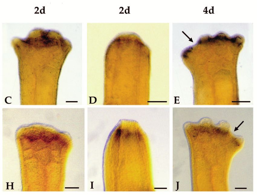
1732 D. M. Gardiner and others

formation. When developing embryos are treated with RA,                  detailed in Blumberg et al. (1991), with the following variations. Total
dramatic alterations in pattern of the main body axis, as well           RNA was isolated from the mesenchymal component of medium bud
as the limbs, are induced (see Bryant and Gardiner, 1992). In            forelimb blastemas after the wound epidermis was removed manually.
some cases it has been shown that RA treatment leads to                  Each library was constructed from 2 µg of poly(A)+ RNA selected by
changes in Hox gene expression, further evidence of the rela-            standard oligo(dT) chromatography. The proximal-blastema library
                                                                         contained 8×10⁷ independent clones and the distal-blastema library
tionship between Hox genes and pattern formation. For                    contained 2×10⁸ independent clones.
example, when mouse embryos are treated with RA, changes
in segment identity are accompanied by changes in the com-               Isolation and sequencing of axolotl Hox genes
bination of Hox genes expressed (Kessel and Gruss, 1991).                We screened 5×10⁵ unamplified clones from the distal-blastema
Equivalent evidence from developing limbs has been reported;             library and 106 unamplified clones from the proximal-blastema library
when anterior chick limb bud cells are exposed to RA released            with a mixture of 1024 oligonucleotides [C(G,T)(A,C,G,T)C(G,T)-
from an implanted bead, ectopic expression of posterior-distal           (A,G)TT(C,T)T(G,T)(A,G)AACCA(A,G)-AT(C,T)TT] that are com-
Hox genes accompany changes in the limb pattern (Izpisúa-                plementary to all possible variations of the DNA sequence encoding
Belmonte et al., 1991; Nohno et al., 1991; Hayamizu and                  the conserved amino acid sequence KIWF(Q/K)NRR. We used the
Bryant, 1994).                                                           tetramethylammonium chloride method for degenerate oligonu-
                                                                         cleotide screening (Burglin et al., 1989) , and plaque-purified the
   We are interested in the remarkable ability of urodele                clones. The inserts were excised as subclones in the Bluescript SK−
amphibians to regenerate perfect replacement limbs after ampu-           phagemid vector according to the manufacturer’s protocol (Strata-
tation. Among the many questions regarding how regeneration              gene). We sequenced plasmid DNA with the use of a degenerate
occurs, the role of Hox genes is of particular importance con-           oligonucleotide      [C(G,T)(A,C,G,T)C(G,T)(A,G)TT(C,T)T(G,T)-
sidering the role of these genes in pattern formation during             (A,G)AACCA] corresponding to the amino acid sequence
embryogenesis and limb development. In addition, because                 WF(Q/K)NRR as a primer. In addition we used a T3 primer to obtain
retinoids have such dramatic effects on pattern formation during         sequence information from the 5′ end of the directionally cloned
regeneration, it is important to understand the relationship             inserts. Sequence data were analyzed using the GCG Sequence
between pattern alterations induced by retinoids and the corre-          Analysis Software Package; similarity searches were performed using
sponding changes in Hox gene expression. In this paper we                the Blast Programs, NCBI.
report on the isolation and identification of a large number of          Northern hybridization analysis
homeobox genes expressed in regeneration blastemas. We also              For RNA-blot analysis, total RNA (5 µg, 10 µg or 20 µg) from limbs
have characterized the expression of two 5′ members of the               at various stages of regeneration were separated by electrophoresis in
HoxA complex, HoxA13 and HoxA9. We report that both genes                1% agarose-0.66 M formaldehyde gels, and transferred to nylon
are reexpressed early in the regeneration cascade and are among          membranes (Hybond-N, Amersham) according to the manufacturer’s
the earliest molecular markers for dedifferentiation of limb             protocol. The amount of RNA loaded was quantitated spectrophoto-
stump cells. Their initial expression does not conform to the            metrically. To check that equal amounts of RNA were loaded, we
usual pattern of temporal and spatial colinearity. It is not until       visualized the 18S and 28S ribosomal RNA bands by either UV
later blastemal stages that they show differential expression            shadowing or by ethidium bromide staining of the gels. Blots were
patterns along the proximal-distal axis. We also report that the         hybridized with 32P-labeled probes in 50% formamide, 5× SSPE, 5×
                                                                         Denhardt’s solution, 0.5% SDS and 20 µg/ml sonicated salmon sperm
two genes differ in their response to retinoid treatment.                DNA at 42°C for 48 hours. Filters were washed at 65°C in 0.1 × SSPE,
                                                                         0.1% SDS and autoradiographed at −70°C with intensifying screens.
                                                                         Autoradiographs were digitized (HP Scanjet IICX) and quantitated
MATERIALS AND METHODS                                                    using Scan Analysis software (Biosoft).
Preparation of blastemas                                                 Preparation of digoxigenin-labeled RNA probes
Experiments were performed on axolotls (Ambystoma mexicanum)             Digoxigenin-labeled RNA probes for whole-mount in situ hybridiz-
spawned at either UCI or the Axolotl Colony, Indiana University. For     ation were synthesized according to the manufacturer’s protocol
isolation of RNA, blastemas were generated on animals measuring          (Boehringer). The 5′ HoxA13 probes were transcribed from the 430-
10-15 cm, snout to tail tip. Animals measuring 4-5 cm were used to       bp EcoRI fragment that contains 320 bp of coding region (including
generate blastemas for whole-mount in situ hybridization. To initiate    the entire homeobox) and 110 bp of 3′ untranslated region (UTR) (Fig.
regeneration, we anesthetized animals in a 0.1% solution of MS222        1A). The 3′ probe was transcribed from the 1100-bp EcoRI-XhoI 3′
(Sigma) and amputated limbs either at proximal (mid-humerus),            UTR fragment (Fig. 1A). The HoxA9 probe was transcribed from the
middle (mid-radius/ulna) or distal (carpals) levels. The amputation      620-bp EcoRI-BglII fragment from clone Hp14 that contains 140 bp
surface was trimmed flat.                                                of the homeobox and 580 bp of coding region 5′ to the homeobox
                                                                         (Fig. 1C). The probes were not hydrolyzed.
RNA isolation
Total RNA was extracted using urea/LiCl (Auffray and Rougeon,            Whole-mount in situ hybridization
1980). Tissues were homogenized in 3 M LiCl/6 M urea/0.1% SDS            Our procedure for whole-mount in situ hybridization to axolotl
and stored at 4°C for 2-5 days to precipitate RNA. Insoluble materials   blastemas and limb buds is based largely on the protocol of Harland
were collected by centrifugation at 10,000 g and rinsed twice with 3     (1991) with the modifications reported in Lamb et al. (1993).
M LiCl to remove contaminants. The pellet was solubilized in 10 mM          Tissues were fixed overnight at room temperature in MEMFA (0.1
Tris-HCl (pH 8.0)/0.5% SDS, centrifuged at 10,000 g and the RNA-         M MOPS, pH 7.4, 2 mM EGTA, 1 mM MgSO4, 3.7% formaldehyde)
containing supernatant was phenol-chloroform extracted and precipi-      with gentle agitation and then stored at −20°C in 100% methanol.
tated with 1/10 volume 5 N NaCl and 2 volumes ethanol.                   Tissues were rehydrated to PTw (PBS with 0.1% Tween-20) and
                                                                         treated with proteinase K (20 µg/ml) at 37°C for 30 minutes for
Construction of cDNA libraries                                           blastemas or 15 minutes for limb buds. Tissues were acetylated with
Blastema cDNA libraries were constructed in λZAPII (Stratagene) as       0.5% acetic anhydride in 0.1M triethanolamine (pH 7.8) for 10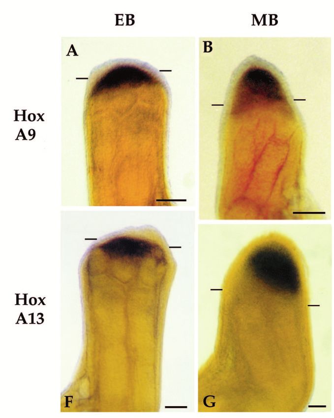
Homeobox genes and limb regeneration 1733

minutes, rinsed with PTw, refixed in 4% formaldehyde in PTw for 20     of RA (all trans, Sigma) in DMSO for 20 minutes and rinsed twice,
minutes and rinsed in PTw.                                             briefly, in PBS. Beads were implanted into the anterior distal-most
   Tissues were prehybridized overnight in hybridization solution      region of blastemas, adjacent to the wound epithelium by making a
(50% formamide, 5× SSC, 1 mg/ml yeast RNA, 100 µg/ml heparin,          tunnel with a 27G hypodermic needle and inserting a single bead
1× Denhardt’s solution, 0.1% Tween-20, 0.1% CHAPS, 5 mM                using watchmaker forceps. Animals that had retained the bead were
EDTA) at 50°C. The digoxigenin-labeled probe (10 µg/ml in hybrid-      fixed for analysis by whole-mount in situ hybridization.
ization solution) was heated to 95-98°C for 30 minutes, diluted to 1
µg/ml in hybridization solution and added to samples for hybridiz-
ation at 50°C for 48-72 hours. Following hybridization the tissues     RESULTS
were washed once with hybridization solution without probe, three
times with 2× SSC (20 minutes each) and twice with 0.2× SSC (30        Isolation and identification of axolotl homeobox-
minutes each); all these washes were done at 60°C.                     containing cDNAs
   Tissues were rinsed twice with maleic acid buffer (MAB; 100 mM
maleic acid, 150 mM NaCl, pH 7.5), and then rinsed with MAB-B          We used a degenerate oligonucleotide complementary to the
(MAB with 2 mg/ml BSA). Tissues were treated with antibody-            conserved third helix of the homeobox [KIWF(Q/K)NRR] to
blocking solution (20% heat-inactivated sheep serum in MAB-B)          screen axolotl blastema cDNA libraries for homeobox-con-
overnight at 4°C. At the same time, the alkaline-phosphatase (AP)      taining clones. From that screen we isolated and determined
conjugated anti-digoxigenin antibody (Boehringer) was diluted 1:400    the nucleotide sequence of 105 clones, of which 80 had an open
in blocking solution and preabsorbed overnight at 4°C with 10 mg/ml    reading frame with a high degree of deduced amino acid
axolotl powder (acetone extracted limb and blastema tissues). The      identity to known vertebrate homeobox genes. These 80
next day, the antibody solution was diluted to 1:1000 with blocking    homeobox containing clones represent the axolotl homologs of
solution and added to the samples. After an overnight incubation at    17 different homeobox genes (Table 1). Twenty five clones
4°C, the tissues were rinsed 10 times with MAB, and twice with AP
buffer (100 mM Tris, pH 9.5, 100 mM NaCl, 50 mM MgCl2). Tissues
                                                                       contained nucleotide sequences complementary to the probe,
were then incubated in AP substrate: 340 µg/ml NBT and 175 µg/ml       but did not contain a long open reading frame and were not
BCIP (Boehringer) in AP buffer with 1 mM levamisol (Sigma). After      very similar to any sequences in the data bases.
the chromogenic reaction was complete (1 to 3 hours), tissues were        The two most abundant axolotl homeobox genes isolated
postfixed overnight in Bouin’s fixative, rinsed in 70% ethanol, and    were HoxA9 and HoxD10, which together accounted for one
stored in methanol at −20°C. Tissues were cleared in methyl salicy-    third of all the homeobox-containing clones. The HoxA
late for photography.                                                  complex was the most complete of the four vertebrate Hox
Retinoid treatment                                                     complexes both in terms of total number of clones (49% of the
For systemic retinoid treatment, 4-6 cm animals that had regenerated
                                                                       total) and the number of members (7 of 11 total, and 7 of the
limbs to an early to medium bud stage (7 days post amputation) were    8 most 5′ members of the complex). Only one clone (en/Msx)
treated by adding retinol palmitate (all trans type VII, SIGMA) at a   did not have a very high degree of amino acid identity with
concentration of 50 IU/ml to the aquarium water for 1 to 5 days.       any single homeobox gene. It encodes an homeobox-contain-
Blastema tissues were treated locally with RA by implanting 200 µm     ing protein that is equally similar to both engrailed proteins and
beads (AG1-X2, Bio-Rad) that had been soaked in a 1 mg/ml solution     Msx proteins. We are investigating the possibility that this

             Table 1. Identification of homeobox-containing genes isolated from axolotl blastema cDNA libraries
                                                                                                                  No. of clones
                                                                                            Probable
      Deduced homeobox AA sequence                                                          homolog        Prox       Dist        Total
      PKRSRTAYTRQQVLELEKEFHFNRYLTRRRRIEIAHTLCLSERQVKIWFQNRRMKWKKDH                           HoxA4           1          1           2
      GKRARTAYTRYQTLELEKEFHFNRYLTRR***************IKIWFQNRRMKWKKDN                           HoxA5           3          −           3
      RKRGRQTYTRYQTLELEKEFHFNRYLTRRRRIEIAHALCLSERQIKIWFQNRRMKWKKEH                           HoxA7           4          −           4
      TRKKRCPYTKHQTLELEKEFLFNMYLTRDRRYEVARLLNLTERQVKIWFQNRRMKMKKIN                           HoxA9          12          4          16
      GRKKRCPYTKHQTLELEKEFLFNMYLTRERRLEISRS***********************                           HoxA10          3          4           7
      TRKKRCPYAKYQIRELEREFFFSIYINKEKRLQLSRMLNLTDRQVKIWFQNRRMKEKKIN                           HoxA11          1          4           5
      GRKKRVPYTKVQLKELEREYATNKFITKDKRRRISATTNLSERQVTIWFQNRRVKEKKVI                           HoxA13          −          2           2
      SKRARTAYTSAQLVELEKEFHFNRYLCRPRRVEMANLLNLTERQIDIWFQNRRMKYKKDQ                           HoxB3           5          2           7
      GRRGRQTYTRYQTLELEKEFHYNRYLTRRRRIEIAHALCLTERQIKIWFQNRRMKWKKEN                           HoxB6           1          2           3
      DRKKRVPYSKGQLRELEKEYASSKFITKDRRRQIATATNLSERQITIWFQNRRVKEKKVF                           HoxC13          −          2           2
      RRRGRQTYSRFQTLELEKEFLFNPYLTRKRRIEVSHALGLTERQVKIWFQNRRMKWKKEN                           HoxD8           3          −           3
      GRKKRCPYTKHQTLELEKEFLFNMYLTRERRLEISKSVNLTDRQVKIWFQNRRMKLKKMS                           HoxD10          8          3          11
      SRKKRCPYTKYQIRELEREFFFNVYINKEKRLQLSRMLNLSDRQVKIWFQNRRMREKKLN                           HoxD11          3          2           5
      NRKPRTPFTTSQLLALERKFRQKQYLSIAERAEFSNSLALTETQVKIWFQNRRAKAKRLQ                           Msx2            4          3           7
      IRKPRTIYSSYQLAALQRRFQKAQYLALPERAELAAQLGLTQTQVKIWFQNRRSKFKKLY                           Dlx3            −          1           1
      KRKRSWSRAVFSNLQRKGLEKRFEIQKYVTKPDRKQLA**********************                           Hlx             1          −           1
      RARPRTKFSTEQLQELERSFQEQRYIGVAEKRRLARELNLSELRIKTWFQNRRMKFNGSE                           en/Msx          1          −           1

****Regions of incomplete sequence information.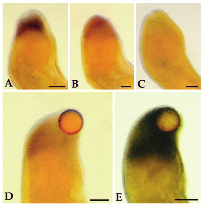
1734 D. M. Gardiner and others

novel homeobox gene may be specific to regenerating                      identical to mouse, human, chick, guinea pig and frog
blastemas.                                                               sequences (see Gehring et al., 1994). This conservation of
                                                                         sequence extends beyond the homeobox; 20 of the next 21
Identification of the axolotl homologs of HoxA13                         more 5′ amino acids and 10 of the 11 more 3′ amino acids are
and HoxA9                                                                identical to mouse and guinea pig sequences (data not shown).
Two clones had identical nucleotide sequences between an                 There is an in-frame stop codon 11 amino acids 3′ to the home-
internal EcoRI site and the vector EcoRI site at the 5′ end of           odomain and a 3′ UTR of approximately 900 nucleotides.
the inserts (Fig. 1A,B), and were identified as the axolotl
homolog of HoxA13 (Table 2). Comparison of the deduced                   Northern hybridization analysis of HoxA13
amino acid sequence within the homeodomain with sequence                 expression
data from other vertebrate species (Table 2) indicates that              Axolotl HoxA13 expression is detected as a single transcript of
axolotl HoxA13 is completely identical to both mouse and                 approximately 2.3 kb on northern blots of total RNA from
human sequences (see Gehring et al., 1994). Sequence data                regenerating blastemas (Fig. 2A-D). Probes from both the 5′
have not been reported for regions outside the homeobox for              region of the transcript, which includes the homeobox, and the
the mouse homolog. Data for the human homolog include only               3′ region (Fig. 1A) detect the same size transcript and reveal
the homeodomain and five amino acids 3′ to the homeodomain.              the same expression pattern. The blots illustrated in Fig. 2A-
The axolotl and human genes are identical in this short 3′               D have been probed with the 3′ probe.
region also (data not shown). There is an in-frame stop codon               HoxA13 is expressed in developing limb buds (Fig. 2B), is
six amino acids 3′ to the homeodomain and a 3′ UTR of                    not detected in mature limbs (Fig. 2A), and is reexpressed (or
approximately 1200 nucleotides.                                          dramatically upregulated) during regeneration of forelimbs,
   Sixteen clones had identical nucleotide sequences within the          hind limbs, and tails (Fig. 2B). Low levels of HoxA13 tran-
homeobox and in regions of overlap 5′ to the homeobox (Fig.              scripts are first detected within a few days post-amputation,
1C,D) and were identified as the axolotl homolog of HoxA9.               coincident with the period of dedifferentiation (Fig. 2C).
Comparison of the deduced amino acid sequence within the                 Higher levels of transcription are detected several days after
homeodomain with sequence data from other vertebrate                     amputation (early bud stages) when blastemal cells are first
species (Table 2) indicates that axolotl HoxA9 is completely             present as an observable accumulation of undifferentiated

                                A) Axolotl HoxA13
Fig. 1. Schematic map and
                                EcoRI                  EcoRI                                                      XhoI
sequence of the axolotl
                               5'                                                                                    3'
HoxA13 and HoxA9 cDNAs.
                                         5' probe                                 3' probe
(A) Schematic map
                                                                                                             100 bp
indicating the coding region
(box) and homeodomain          B) Axolotl HoxA13
(hatched box) of HoxA13.
Two fragments obtained by      ACCAATGGCTGGAATGGGCAAGTGTACTGTTCCAAGGAGCAAGGGCAGCCCCCGCACCTCTGGAAGTCCTCTCTGCCGGACGTGGTTTGG 90
                               T N G W N G Q V Y C S K E Q G Q P P H L W K S S L P D V V W
digestion with EcoRI and       CATCCCTCGGATGCGAACTCGTACAGGCGAGGCCGGAAGAAGCGCGTGCCGTACACCAAGGTCCAGCTGAAGGAACTGGAGCGCGAGTAC 180
XhoI were used as probes.      H P S D A N S Y R R G R K K R V P Y T K V Q L K E L E R E Y
The 5′ EcoRI and 3′ XhoI       GCCACGAATAAGTTCATTACCAAGGACAAACGGAGGCGGATATCGGCCACCACCAACCTCTCCGAGCGCCAGGTCACAATTTGGTTCCAA 270
sites were created during      A T N K F I T K D K R R R I S A T T N L S E R Q V T I W F Q
                               AACAGGAGGGTCAAAGAGAAGAAGGTCATCAACAAACTCAAGACCACCAGCTAAtggactccccgcctcctttttttttggtctcaacaa 360
library construction and are   N R R V K E K K V I N K L K T T S *
the sites at which the insert  aaacctaagccgaagctaaaagaaaactgacagttacaaggaaatgaagctgttcattggtcaccagaattc                                 432
was cloned into Bluescript
SK−. (B) The nucleotide
                               C) Axolotl HoxA9
sequence and conceptual
amino acid translation of the          Hp14          Hp20                 BglII
                               EcoRI                                                  BamHI                                 XhoI
axolotl HoxA13 cDNA from       5'                                                                                              3'
the 5′ EcoRI site to the
                                                      5' probe                                          3' probe
internal EcoRI site located in                                                                                         100 bp
the 3′UTR. The
homeodomain is underlined.     D) Axolotl HoxA9
(C) Schematic map of
HoxA9 indicating the           AGGCACTACGGCATCAAGCCCGAGCCGCTGCCGCCGGGGACCCGCCGGGGGGACTGCACCACCTTCGACAGCAGCCACACGCTCTCCCTG 90
                               R H Y G I K P E P L P P G T R R G D C T T F D S S H T L S L
position of the two            TCCGACTACGGCTCCTCTCCCGCCGACAAGCAGAGCAGCGAAGGGGCTTTCCCCGAGGCCCCCGCCGAGACCGAGGCCAGCGGAGACAAG 180
fragments used as probes.      S D Y G S S P A D K Q S S E G A F P E A P A E T E A S G D K
The 5′ probe contains          CCTGCCATTGACCCAAACAACCCGGCTGCGAACTGGCTGCACGCGAGGTCGACGCGCAAGAAGCGCTGCCCTTACACCAAGCACCAGACC 270
                               P A I D P N N P A A N W L H A R S T R K K R C P Y T K H Q T
nucleotides from the coding    CTGGAGCTGGAGAAGGAGTTCCTCTTCAACATGTACCTGACGCGGGACCGCAGGTACGAGGTGGCCCGGCTGCTGAACCTGACCGAGCGG 360
region (box) and extends       L E L E K E F L F N M Y L T R D R R Y E V A R L L N L T E R
from the 5′ end of clone       CAGGTCAAGATCTGGTTCCAGAACCGGCGCATGAAGATGAAAAAAATCAACAAGGACCGGCCTAAGGAGTGAaagggggccgctggcggc 450
Hp14 to the BglII site within  Q V K I W F Q N R R M K M K K I N K D R P K E *
the homeobox (hatched
box). The EcoRI and XhoI sites were created during library construction. (D) The nucleotide sequence and conceptual amino acid translation of
the axolotl HoxA9 cDNA from the 5′ end of clone Hp20 to the end of the coding region. The homeodomain is underlined. Genbank accession
numbers: HoxA9, U20941; HoxA13, U20942.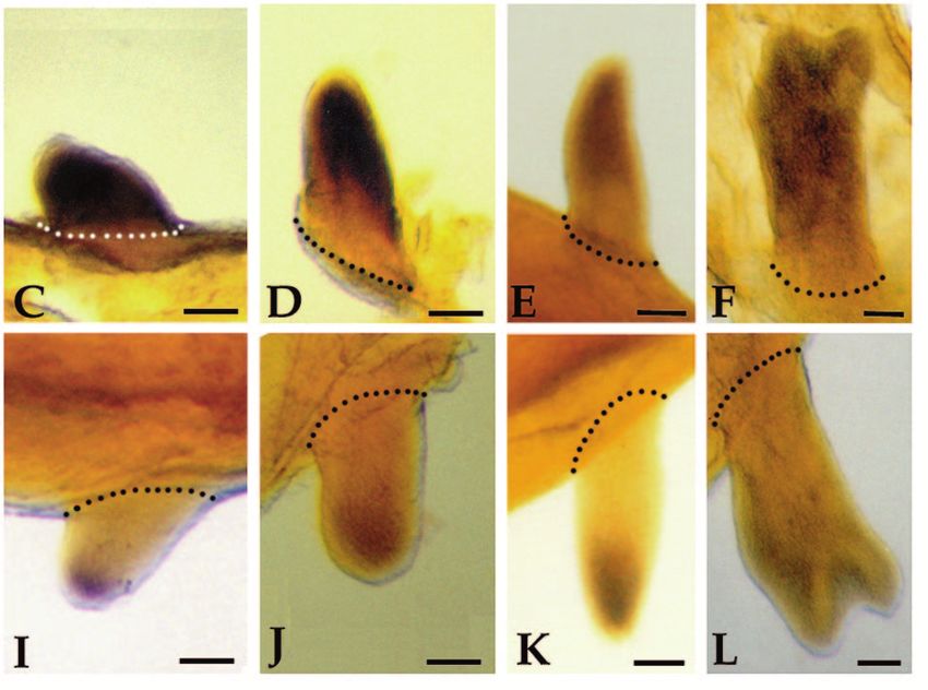
Homeobox genes and limb regeneration 1735

Table 2. Comparison of deduced amino acid sequence for the homeodomain of axolotl HoxA9 and HoxA13 to Drosophila
                                   Abd-B and to homologs from other species
              Animal                   Gene                                         Amino acid sequence
              Drosophila              Abd-B       VRKKRKPYSKFQTLELEKEFLFNAYVSKQKRWELARNLQLTERQVKIWFQNRRMKNKKNS

              Axolotl                 HoxA9...T----C--T-H------------M-LTRDR-Y-V--L-N----------------M--IN
              Human                   HoxA9       T----C--T-H------------M-LTRDR-Y-V--L-N----------------M--IN
              Mouse                   HoxA9       T----C--T-H------------M-LTRDR-Y-V--L-N----------------M--IN
              Chick                   HoxA9       T----C--T-H------------M-LTRDR-Y-V--L-N----------------M--IN
              Guinea pig              HoxA9       T----C--T-H------------M-LTRDR-Y-V--L-N----------------M--IN
              Xenopus b.              HoxA9       T----C--T-H------------M-LTRDR-Y-V--L-N----------------M--IN

              Axolotl                 HoxA13      G----V--T-V-LK---R-YAT-KFIT-D--RRISATTN-S----T-------V-E--VI
              Human                   HoxA13      G----V--T-V-LK---R-YAT-KFIT-D--RRISATTN-S----T-------V-E--VI
              Mouse                   HoxA13      G----V--T-V-LK---R-YAT-KFIT-D--RRISATTN-S----T-------V-E--VI

  Dashes (---) represent amino acids that are identical to the Drosophila Abd-B sequence at that position.

cells. Expression remains high over the next several days as                      level of expression is apparent from analysis of the whole-
the blastema increases in size, then decreases as differentiation                 mount in situ hybridization results reported below.
begins (early digit stage, Fig. 2C), and eventually is unde-                         Expression of HoxA13 decreases in response to treatment
tectable when the limb is fully regenerated (as in Fig. 2A).                      with retinoids that cause pattern duplications along the
   HoxA13 is not uniformly expressed in blastemas. Expression                     proximal-distal limb axis (Table 3). Regenerating forelimbs
is restricted to the mesenchymal tissues; transcripts are not                     treated for as little as 1 day with retinol palmitate at the
detected in RNA samples from epidermal tissues separated                          medium bud stage of regeneration exhibit some degree of
from the underlying mesenchyme (Fig. 2A). Expression is dif-                      proximal-distal duplication. The proportion and degree of
ferentially regulated along the proximal-distal axis; expression                  duplicated limbs increases after 2 days of treatment, and all
is about 30% greater in middle level blastemas as compared to                     limbs are duplicated after 3 days of treatment. The level of
proximal blastemas (Fig. 2B). The reason for this difference in                   HoxA13 expression in blastemas collected after 3 days of

Fig. 2. Expression of HoxA13 and HoxA9 in developing, regenerating and mature axolotl limbs as visualized by northern blot analysis. The
sizes of the transcripts were determined relative to the mobility of axolotl 28S rRNA (3.9 kb) and 18S rRNA (1.95 kb). The position of the 18S
rRNA band is indicated by the hash mark on the left. Equal amounts of total RNA were loaded for each blot as determined
spectrophotometrically and verified by either UV shadowing or ethidium bromide staining of the gels. The blots in A and E were also
hybridized with a probe for axolotl Dlx3 (inserts) to confirm that transcripts could be detected in those lanes that are negative for HoxA13 and
HoxA9. (A) Expression of HoxA13 in mature limbs (Mature) and medium bud forelimb blastemas (MB FL). Tissues were from either humerus
levels (P) or mid radius/ulna levels (D). In the MB FL samples, blastemas were separated into epidermal fractions (Epid) and mesenchymal
fractions (Mes) prior to RNA extraction. 10 µg total RNA/lane. (B) Expression of HoxA13 in medium bud blastemas (MB Regenerate) from
forelimbs (FL), hindlimbs (HL), and tails (T) and in developing forelimb buds. 5 µg total RNA/lane. (C) Expression of HoxA13 at different
stages of regeneration from radius/ulna level amputations of forelimbs (FL). Stages are dedifferentiation (DD), early bud (EB), medium bud
(MB), late bud (LB), palette (PAL) and early digits (ED). 20 µg total RNA/lane. (D) The effect of retinol palmitate (RP) treatment on HoxA13
expression. RNA was extracted from control medium bud (MB) blastemas (Cont) that had not been treated with retinoids and medium bud
blastemas from animals that had been treated with retinol palmitate for 3 days (3d). 5 µg total RNA/lane. (E) Expression of HoxA9 in mature
distal limbs (Dist Mat) and in epidermal (Epid) and mesenchymal fractions (Mes) of medium bud forelimb blastemas (MB). 10 µg total
RNA/lane. (F) Expression of HoxA9 in developing forelimb buds and at different stages of regeneration from humerus level amputations of
forelimbs (FL). Stages are as in C. 5 µg total RNA/lane.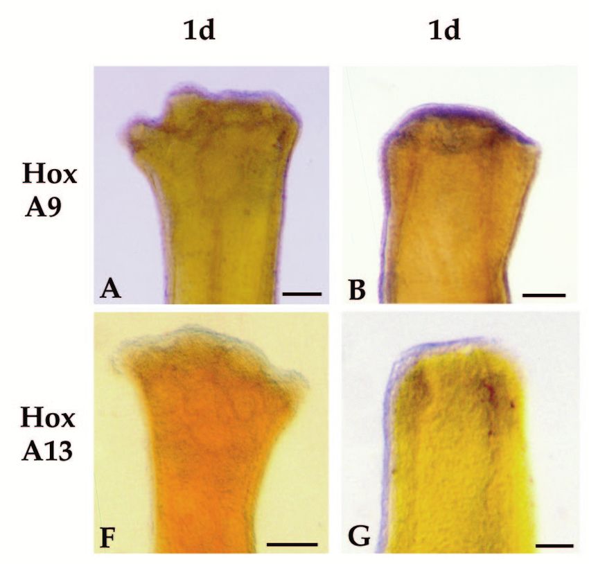
1736 D. M. Gardiner and others

 Table 3. Pattern duplicating activity of retinol palmitate                     mal cells but not epidermal cells (Fig. 2E). We analyzed the
    on axolotl distal forelimb medium bud blastemas                             response of HoxA9 to retinoids by whole-mount in situ hybrid-
     Treatment                 No. of          No. of       Duplication         ization as described below.
     duration         n        normal        duplicated       index*
                                                                                In situ hybridization analysis of HoxA expression in
     1 day            5           1              4              2.4
     2 day            4           1              3              3.25            developing limb buds
     3 day            4           0              4              4.25            We used whole-mount in situ hybridization to analyze the
                                                                                patterns of HoxA13 and HoxA9 expression in limb buds
   *Duplication index is from Maden (1983). The maximum proximal-distal
duplication in which an entire limb,including a pectoral girdle forms from a    ranging from the earliest stage of forelimb outgrowth (stage
distal amputation stump has a score of 5; the index is the mean score for all   36) to later stages when digits one and two have formed (stages
limbs.                                                                          41/42) (Fig. 3). Stages are based on the comparable limb bud
                                                                                morphologies for Ambystoma punctatum (Harrison, 1969).
                                                                                HoxA13 probes from both the 5′ and 3′ regions of the transcript
treatment is less than half (45%) the level in equivalent staged                (Fig. 1A) revealed the same expression pattern; the 5′ probe
blastemas not treated with retinoids (Fig. 2D). We have                         was used because it resulted in a more intense signal. HoxA9
analyzed retinoid-induced downregulation of HoxA13                              transcripts were localized with a 5′ probe (Fig. 1C).
expression further by whole-mount in situ hybridization as                         HoxA9 is expressed early in limb development (stage 36)
described below.                                                                throughout the limb bud mesenchyme and in the adjacent flank
                                                                                region (Fig. 3A). At stage 37, the intensity of staining
Northern hybridization analysis of axolotl HoxA9                                increases, but the pattern is the same (Fig. 3B). At stage 38,
expression                                                                      expression is no longer detected in the flank and there is a small
Axolotl HoxA9 expression is detected as a single transcript of                  region of cells at the base of the limb bud that does not express
approximately 2 kb on northern blots of total RNA from regen-                   HoxA9 (Fig. 3C). At later stages, HoxA9 continues to be
erating blastemas (Fig. 2E-F). Probes from both the 5′ region of                expressed throughout the limb bud except in the proximal-most
the transcript, which includes most of the homeobox, and the 3′                 region (Fig. 3C,D). There are no fate maps for axolotl limb
UTR (Fig. 1C) detect the same size transcript and reveal the                    buds to compare with these expression patterns; however,
same expression patterns. The blot illustrated in Fig. 2E has been              expression is still evident during differentiation of skeletal
probed with the 5′ probe and that in Fig. 2F with the 3′ probe.                 elements, at which time HoxA9 expression extends from the
   The pattern of HoxA9 expression is similar to HoxA13. It is                  distal third of the humerus through the lower arm and hand
expressed in developing limb buds (Fig. 2F), is not detected in                 (Fig. 3F).
mature limbs (Fig. 2E), and is reexpressed during regeneration                     HoxA13 expression is first detected later and in a more
(Fig. 2F). The maximal level of expression occurs during the                    distally restricted population of limb bud cells as compared to
blastemal stages, then decreases during redifferentiation (Fig.                 HoxA9. Transcripts are not detected at stage 36 (Fig. 3G), and
2F). Within the blastema, HoxA9 is expressed by mesenchy-                       staining is limited to the distal region of stage 37 limb buds

Fig. 3. Expression of HoxA13 and HoxA9 in developing axolotl forelimbs as visualized by whole-mount in situ hybridization. The yellow
coloration of the limbs is due to the Bouin’s postfixative, which we do not completely remove so as to provide enhanced contrast with the
blue reaction product. Anterior is to the right in all limb buds. (A-F) Hox A9 expression; (G-L) HoxA13 expression; (A,G) stage 36; (B,H)
stage 37; (C,I) stage 38; (D,J) stage 39; (E,K) stage 40; (F, L) stage 41/42 (stages after Harrison, 1969). Large dots indicate the base of the
bud; small dots in A and H outline the edge of the bud where it overlaps the body. Scale bar, 100 µm.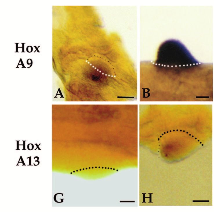
Homeobox genes and limb regeneration 1737

Fig. 4. Expression of HoxA13 and HoxA9 during early stages of axolotl limb regeneration as visualized by whole-mount in situ hybridization.
All samples are forelimbs amputated at either the level of the humerus (B,D,G and I) or the level of the carpals (A,C,E,F,H and J). The limbs
illustrated were collected 1 day (1d), 2 days (2d) or 4 days (4d) after amputation. Limbs are viewed from the dorsal side and are matched left
limbs (HoxA9, anterior to the right) and right limbs (HoxA13, anterior to the left) from the same animal. Arrows in E and J indicate regions of
stump tissues not expressing HoxA9 or HoxA13. Scale bar, 200 µm.

(Fig. 3H). HoxA13 expression remains distally restricted at                 In situ hybridization analysis of HoxA expression
later stages (Fig. 3I-K), and is localized to the region in which           during limb regeneration
hand structures differentiate (Fig. 3L).                                    Reexpression of both HoxA9 and HoxA13 is initiated at a very

Fig. 5. Expression of HoxA13 and HoxA9 during blastema stages of axolotl limb regeneration as visualized by whole-mount in situ
hybridization. All samples are forelimbs amputated at either the level of the humerus (C,E,H and J) or the level of the radius/ulna (A,B,D,F,G
and I). The amputation plane is indicated by hash marks. All limbs are viewed from the dorsal side and are either left limbs with anterior to the
right (HoxA9) or right limbs with anterior to the left (HoxA13). The limbs illustrated represent progressively later stages of regeneration; early
bud (EB), medium bud (MB), late bud (LB) and palette (PAL). Scale bar, 200 µm.
1738 D. M. Gardiner and others

early stage of regeneration in internal tissues of the stump, but    amputation. This difference accounts for the results from
not in epidermal cells (Fig. 4). Activation of this early            northern analysis indicating a higher level of HoxA13
expression domain precedes the accumulation of blastema cells,       expression in distal blastemas (greater proportion of express-
and is associated with the stump tissues that will dedifferenti-     ing cells) as compared to proximal blastemas (lesser propor-
ate to form the blastema. We observed a similar pattern of early     tion of expressing cells; Fig. 2B).
expression at each of the three levels of amputation along the          The expression domain of HoxA13 is asymmetric with
proximal-distal limb axis (mid-stylopod, mid-zeugopod and            respect to the anterior-posterior axis of the limb. The
distal-carpals). Expression of both genes is detectable 24 hours     expression domain extends further proximally in the posterior
post-amputation in most limbs (Fig. 4A,B,F,G). Expression            region and is more distally restricted in the anterior region.
increases such that it is readily detectable in all limbs by 48      This pattern is observed in both distal (Fig. 5G) and proximal
hours (Fig. 4C,D,H,I). In comparing the expression of HoxA9          (Fig. 5H) blastemas. A similar anterior-posterior asymmetry
and HoxA13 in left and right limbs of the same animal, we have       has been observed in HoxA13 expression during limb devel-
not detected a difference in the time of onset of expression.        opment in the chick and mouse (Haack and Gruss, 1993;
Expression increases over the next few days in a stripe 100-150      Yokouchi et al., 1991). HoxA9 expression does not exhibit
µm wide, localized immediately proximal to the amputation            such an asymmetrical pattern, and both genes are expressed
surface (Figs 4D,J, 5A,F). Activation is localized to regions        uniformly with respect to the dorsal-ventral limb axis. A
adjacent to the wound epidermis. In some limbs the amputation        dorsal-ventral asymmetry in HoxA13 expression has been
plane occurred distal to a digit base (Fig. 4E,J), and HoxA          observed in developing mouse limbs (Haack and Gruss, 1993),
expression is not activated in the region covered by mature,         but not in developing chick limbs (Yokouchi et al., 1991).
interdigital epidermis (arrows).
   At later stages of regeneration, when a blastema is present       HoxA13 and HoxA9 respond differently to retinoids
distal to the amputation plane, mesenchymal cells of the             The expression of HoxA13 during regeneration is downregu-
blastema express both HoxA9 and HoxA13 (Fig. 5); expression          lated in response to exposure to retinoids. After one day of
is not detected in epidermal cells. Dedifferentiating stump cells    systemic treatment with retinol palmitate, HoxA13 expression
(proximal to the amputation plane) continue to express HoxA9         is decreased at the distal tip of the blastema in some but not
as the blastema elongates (Fig. 5A-C); however, expression           all samples (e.g. Fig. 6A). By 3 days of treatment, the level of
becomes restricted to the blastema at later stages when dedif-       HoxA13 expression ranged from not detectable (not illustrated)
ferentiation has ceased (Fig. 5D,E). In contrast, HoxA13 is not      to noticeably downregulated and uniformly expressed with
expressed in dedifferentiating stump cells beyond the early bud      respect to the anterior-posterior limb axis (Fig. 6B). Corre-
stage of blastema formation (Fig. 5F-J).                             sponding to this inhibition of HoxA13 expression, the
   In contrast to the preblastema stages in which HoxA9 and          frequency and degree of pattern duplication increased. After 1
HoxA13 transcripts are colocalized in both proximal and distal       day of treatment, a few of the limbs exhibited pattern alter-
stump cells, the expression domains of these two genes within        ations; whereas, after 3 days of treatment all limbs exhibited
the blastema become spatially distinct as growth progresses          some degree of pattern alteration (Table 3).
(Fig. 5). HoxA9 is expressed throughout both proximal and               HoxA13 expression is downregulated in less than 24 hours
distal blastemas at all stages, and at late stages is expressed in   when blastema cells are treated directly with RA released from
the distal third of the redifferentiating humerus, the lower arm,    beads implanted into the blastema (Fig. 6D). The level of
and hand (Fig. 5E). HoxA13 expression is detected throughout         expression is lowest adjacent to the bead (implanted into the
the early bud blastema (Fig. 5F), but subsequently becomes           anterior-distal region) and is higher in the posterior-proximal
restricted to the distal region of the blastema (Fig. 5G-J). Thus    region away from the RA bead. As with systemic treatment of
the HoxA13 expression domain becomes nested within the               regenerating limbs, RA bead implants into blastemas result in
HoxA9 expression domain. At late stages of regeneration, the         pattern duplications at high frequency (Sessions, Wanek and
distal domain of HoxA13 expression corresponds to the differ-        Bryant, unpublished observations).
entiating digits (Fig. 5J); HoxA13 is not expressed in more             In contrast to HoxA13, the expression of HoxA9 is not
proximal regions of the regenerate, such as the distal radius        noticeably altered in response to RA released from beads.
and ulna reformed from a distal amputation. Work is in               Expression in both proximal and distal RA-treated blastemas
progress to develop blastema fate maps, which will allow for         does not appear to differ from control blastemas at either 17
a more precise correlation between expression domains and            hours or 24 hours after bead implantation (Fig. 6E).
structures regenerated.
   The size of the HoxA13 expression domain relative to the
total size of the blastema differs between distal and proximal       DISCUSSION
blastemas. In distal blastemas (Fig. 5G), cells throughout most
of the blastema express HoxA13; only a narrow zone of cells          Regulation of homeobox gene expression during
at the base and in the anterior-proximal region of the blastema      limb regeneration is complex
do not express HoxA13. In proximal blastemas (Fig. 5H) the           As a result of a screen for homeobox genes expressed in regen-
zone of non-expressing cells at the base of the blastema is          erating limb blastemas, we have identified a total of 17
much larger than in distal blastemas; thus the distal expression     different axolotl genes with homology to known homeobox
domain represents a smaller proportion of the blastema. This         genes. Previous screens of cDNA libraries from regenerating
difference is probably a consequence of the fact that only distal    newt limbs have resulted in the identification of nine
structures are regenerated from a distal amputation, but both        homeobox genes expressed during limb regeneration: HoxA11
proximal and distal structures are regenerated from a proximal       and Hox B3 (Beauchemin and Savard, 1993); HoxC6 (Savard
Homeobox genes and limb regeneration 1739

et al., 1988; Tabin, 1989); HoxC10 and HoxD10 (Simon and             region that forms the hand, as in chick and mouse limbs (Haack
Tabin, 1993); HoxD11 (Brown and Brockes, 1991) ; Dlx1,               and Gruss, 1993; Yokouchi et al., 1991). This domain is nested
Dlx3 and Emx2 (Beauchemin and Savard, 1993). We isolated             within the HoxA9 domain, which at early stages includes the
the axolotl homologs of five of these newt genes in our screen       entire limb bud as well as the adjacent flank. As the limb
(HoxA11, HoxB3, HoxD10, HoxD11, and Dlx3). We have sub-              grows, the proximal border of expression moves out onto the
sequently isolated the axolotl homolog of HoxC10 from axolotl        bud, and staining remains intense from there to the tip of the
limb blastema RNA by RT-PCR (Komine, Gardiner and                    bud. At later stages, HoxA9 is expressed throughout the region
Bryant, unpublished data). Thus, to date, a total of 21 different    that will form the hand, the forearm and the distal third of the
homeobox genes are known to be expressed in regenerating             humerus. HoxA9 expression does not appear to be less intense
limb blastemas. It is now apparent that there is not a single, or    distally in regions of overlap with HoxA13 expression, as has
even just a few, but many homeobox genes that might be               been reported for HoxA11 in mouse and chick (Haack and
involved in regulating growth and pattern formation during           Gruss, 1993; Yokouchi et al., 1991).
limb regeneration. In addition, several of these genes are
expressed as multiple transcripts with spatially distinct            Reexpression of HoxA genes is not colinear during
expression patterns (Beauchemin and Savard, 1993; Savard et          initiation of regeneration
al., 1988; Torok, Gardiner and Bryant, unpublished data), indi-      The pattern of HoxA gene reexpression during regeneration
cating an even more complex role in regeneration.                    departs markedly from the spatial and temporal colinearity
   Almost all of the homeobox genes expressed in regenerat-          characteristic of developing limbs of axolotls and other verte-
ing blastemas are also expressed in developing limb buds of          brates. Neither gene is expressed in mature limbs, but both can
other vertebrates (see Izpisúa-Belmonte and Duboule, 1992).          be detected within 1-2 days after amputation in a stripe of mes-
The broad overlap in the homeobox genes expressed during             enchymal cells immediately beneath the wound epidermis.
limb development and regeneration is supportive of the view          Expression is similar, regardless of the proximal-distal level of
that these two developmental processes involve common                the amputation. The exact time of onset of expression is
mechanisms of growth regulation and pattern formation (see           somewhat variable within the first 24-48 hours, with some but
Bryant and Gardiner, 1992; Muneoka and Sassoon, 1992).               not all limbs showing clear expression at 24 hours. However,
Although most, perhaps even all, of the same homeobox genes          in matched contralateral limbs, whenever HoxA9 was detected
are expressed, the ways in which their expression is regulated       in one limb, HoxA13 was detected in the other. While these
differs (as discussed below), and the events involved in the         results suggest that the reexpression of HoxA9 and HoxA13 is
initiation of outgrowth are different (see Bryant and Gardiner,      synchronous rather than colinear, further experiments are
1992; Muneoka and Sassoon, 1992). It may prove to be the             necessary to rule out a very rapid activation via the canonical
case that some homeobox genes are uniquely expressed in              HoxA9 – HoxA10 – HoxA11 – HoxA13 sequence.
either developing or regenerating limbs. We note that the novel         Both HoxA9 and HoxA13 continue to be colocalized during
axolotl en/Msx gene, though clearly a homeobox gene (50%             the early stages of blastema formation, at both proximal and
amino acid identity to both engrailed and msh proteins within        distal limb levels. Spatially distinct domains of expression
the homeodomain), may be unique to regeneration.                     emerge during growth of the blastema as HoxA13 expression
   A striking result of our screen is the relative abundance and     becomes confined to a distal subset of the cells that also express
complexity of members of the HoxA complex expressed during           HoxA9. During these later stages of regeneration, the relation-
limb regeneration. Studies of developing mouse and chick             ship of HoxA expression patterns is the same as in developing
limbs indicate that the 5′ HoxA genes function in specifying         limbs, with HoxA13 expression correlated with regeneration of
the proximal-distal limb pattern. Since regeneration is essen-       the hand, and HoxA9 with regeneration of the hand, lower arm
tially reforming the proximal-distal axis, it is perhaps not sur-    and distal humerus.
prising that HoxA genes are so abundantly expressed.                    According to the hypothesis that segmental identity is based
                                                                     on combinatory expression of Hox genes (Kessel and Gruss,
HoxA genes are expressed in a colinear sequence in                   1991), the most distal part of the limb pattern (hand) would be
axolotl limb buds                                                    characterized by the overlapping expression of HoxA9 and
The expression of HoxA13 and HoxA9 during axolotl limb               HoxA13, whereas more proximal regions would express
development is consistent with the principle of temporal and         HoxA9 but not HoxA13. The HoxA expression patterns in
spatial colinearity as described for other vertebrate limbs          developing limbs and blastema stage regenerating limbs are
(Izpisúa-Belmonte and Duboule, 1992). The more 3′ gene,              consistent with this hypothesis. Given that the coexpression of
HoxA9, is first expressed at an earlier stage of limb develop-       HoxA13 and HoxA9 is characteristic of the distal limb pattern,
ment than is the more 5′ gene, HoxA13. Similarly, HoxA9 has          then the distal part of the limb pattern is the first to be respec-
a more extensive expression domain with a more proximal              ified during regeneration, regardless of the level of amputation.
boundary than HoxA13, which has a more distally restricted           The regions that are intermediate between the stump and the
expression domain that is nested within the HoxA9 domain.            newly formed distal tip appear to arise later, during growth of
Thus HoxA gene expression in developing axolotl limbs                the blastema, as the domains of HoxA9 and HoxA13 become
appears to be regulated the same as in developing limbs of           spatially distinct. Whether the cells for this intermediate zone
other vertebrates.                                                   originate from the distally specified cells of the early blastema,
   Although presently there are no fate maps for developing          or from the stump, remains to be determined. It is generally
axolotl limbs, it is possible to correlate expression domains        thought that the pattern of both developing and regenerating
with structures that differentiate during the later stages of limb   limbs is specified in a strict proximal to distal sequence. We
development. At these stages, HoxA13 is expressed in the             conclude that for regeneration at least, this is not the case.
1740 D. M. Gardiner and others
have been well characterized at the level of cell biology;
whereas, effects on gene expression have been less well char-
acterized, especially for regenerating limbs. Retinoids cause a
distal blastema to regenerate as if it had been transformed to a
proximal blastema, leading to the formation of duplicated
pattern along the proximal-distal axis (Maden, 1982). It is
presumed that the changes in positional information are a con-
sequence of retinoid-induced changes in gene expression.
The finding that proximalization of the blastema by retinoid
treatment is associated with an early downregulation of
HoxA13, but not of HoxA9 expression, is consistent with the
idea that the Hox code of the treated cells is changed to that of
a more proximal region. However, as discussed above,
blastemas initially express a distal Hox code, and thus a
retinoid-treated blastema, with a proximal Hox code, is not the
same as a blastema arising at a proximal limb level. The steps
leading from a retinoid-proximalized blastema to the final
duplicated limb pattern have not yet been studied. Despite this
gap in our knowledge, the coincidence between altered
HoxA13 expression and altered pattern, provides further
evidence of the importance of HoxA genes in limb pattern
formation. In addition, we note that this is the second report of
retinoid-responsive Hox gene expression during regeneration
(see Simon and Tabin, 1993). Taken together with our finding
Fig. 6. Effects of retinoid treatment on the expression of HoxA13 and that stump cells initially acquire a distal Hox code regardless
HoxA9 as visualized by whole-mount in situ hybridization. of their position along the proximal-distal axis, regenerating
(A) HoxA13 expression in a medium bud blastema on an animal amphibian limbs no longer provide support of the hypothesis
treated with retinol palmitate for 1 day. (B) HoxA13 expression in a that Hox gene expression patterns become permanently
medium bud blastema on an animal treated with retinol palmitate for imprinted and cannot be modified (Mavilio, 1993).
3 days. (C) A medium bud blastema that was hybridized with the The response of HoxA9 and HoxA13 to retinoid treatment of
sense probe from the 5′ region of HoxA13. (D) HoxA13 expression in regenerating limbs in vivo is consistent with that of teratocar-
a medium bud blastema into which a retinoic acid-containing bead
cinoma cells in vitro (Simeone et al., 1991). Genes at the 3′
was implanted 23 hours earlier. (E) HoxA9 expression in a medium
bud blastema into which a retinoic acid-containing bead was end of the Hox complexes are activated by RA, genes located
implanted 23 hours earlier. Scale bar, 200 µm in the middle of the complex do not react strongly to RA, and
genes at the 5′ end are either not affected, inhibited or strongly
downregulated. In blastemas, HoxA9, which is in the middle
In addition to providing insights as to how the proximal- of the complex is relatively unresponsive to retinoids; whereas,
distal limb axis is reestablished during regeneration, HoxA9 HoxA13, at the 5′ most end of the complex, is inhibited. In
and HoxA13 expression are early molecular markers for the addition, another 5′ Hox gene, HoxD13, is inhibited by RA in
process referred to as ‘dedifferentiation’. This term refers to vivo during chick limb development (Hayamizu and Bryant,
events occurring in the transition zone between mature stump 1994). The coordinated upregulation of 3′ Hox genes and
tissues proximal to the amputation surface and the blastema downregulation of 5′ Hox genes could cause positional identity
that forms distally (see Wallace, 1981). Dedifferentiation to be shifted to a more rostral position along the rostrocaudal
begins soon after amputation and is considered to be critical axis, and a more proximal position along the limb axis.
for the initiation of regeneration. Considering how rapidly An intriguing feature of limb regeneration is that the regen-
HoxA reexpression is induced in the stump, it must be situated erate is always appropriate to the level of amputation and
close to the beginning of the regeneration cascade. As precisely matches the stump. This implies that mature limb
discussed elsewhere (Bryant and Gardiner, 1992; Muneoka cells retain a memory of their identity along the proximal-distal
and Sassoon, 1992), the key developmental processes that dis- limb axis. This memory is not simply that of upper arm versus
tinguish limb regeneration from limb development are those lower arm or hand, but of a precise position within a particu-
associated with the initiation of the regeneration cascade; lar limb segment. Discovering how limb cells store and
induction of HoxA reexpression during dedifferentiation reaccess this information is one of the challenges of regenera-
appears to be one such process. The possibility that regener- tion research that is of importance to understanding both limb
ation is initiated with a generalized activation of gene regeneration and limb development. Were it not for animals
expression, of which the HoxA genes described here are only that can regenerate so flawlessly, we would not appreciate that
a part, will become clear as more of the genes expressed in positional information is so fine-grained, that this information
regeneration are studied. is stored by differentiated cells, and that it can be accessed.
The observations that HoxA genes are expressed in specific
Hox A13 and HoxA9 respond differently to retinoid patterns along the proximal-distal limb axis, and that alter-
treatments that cause proximalization of blastemas ations in these patterns coincide with changes in the positional
The effects of retinoids on developing and regenerating limbs identity of blastema cells, suggest that understanding howHomeobox genes and limb regeneration 1741
HoxA genes are regulated is an important step in meeting this method for Xenopus embryos. In Methods in Cell Biology, Volume 36,
challenge. Xenopus laevis: Practical Uses in Cell and Molecular Biology (eds. B. K.
Kay and H. B. Peng), pp. 685-695. San Diego: Academic Press Inc.
Harrison, R. G. (1969). Organization and Development of the Embryo New
We wish to thank Dr Juan-Carlos Izpisúa-Belmonte, Lina Mullen,
Haven: Yale University Press.
Maureen Torok and Dr Eric Yang for helpful comments on the man- Hayamizu, T. F. and Bryant, S. V. (1994). Reciprocal changes in Hox D13
uscript, and Dr Eddy De Robertis for his warm hospitality at UCLA and RAR-β2 expression in response to retinoic acid in chick limb buds. Dev.
where this project was initiated. Research supported by PHS grant Biol. 166, 123-132.
HD25620, ONR grant N00014-92-J-1967, and an allocation of Izpisúa-Belmonte, J.-C. and Duboule, D. (1992). Homeobox genes and
computer resources by the University of California Irvine. pattern formation in the vertebrate limb. Dev. Biol. 152, 26-36.
Izpisúa-Belmonte, J.-C., Tickle, C., Dollé, P., Wolpert, L. and Duboule, D.
(1991). Expression of the homeobox Hox-4 genes and the specification of
position in chick wing development. Nature 350, 585-589.
REFERENCES Kessel, M. and Gruss, P. (1991). Homeotic transformations of murine
vertebrae and concomitant alteration of Hox codes induced by retinoic acid.
Auffray, C. and Rougeon, F. (1980). Purification of mouse immunoglobulin Cell 67, 89-104.
heavy-chain messenger RNAs from total myeloma tumor RNA. Eur. J. Krumlauf, R. (1993). Mouse Hox genetic functions. Curr. Opin. Genet. Dev. 3,
Biochem. 107, 303-314. 621-625.
Beauchemin, M. and Savard, P. (1993). Expression of five homeobox genes Krumlauf, R. (1994). Hox genes in vertebrate development. Cell 78, 191-201.
in the adult newt appendages and regeneration blastemas. In Limb Lamb, T. M., Knecht, A. K., Smith, W. C., Stachel, S. E., Economides, A.
Development and Regeneration (eds. J. F. Fallon, P. F. Goetinck, R. O. N., Stahl, N., Yancopolous, G. D. and Harland, R. M. (1993). Neural
Kelley and D. L. Stocum), pp. 41-50. New York: Wiley-Liss Inc. induction by the secreted polypeptide noggin. Science 262, 713-718.
Blumberg, B., Wright, C. V. E., De Robertis, E. M. and Cho, K. W. Y. Maden, M. (1982). Vitamin A and pattern formation in the regenerating limb.
(1991). Organizer-specific homeobox genes in Xenopus laevis embryos. Nature 295, 672-675.
Science 253, 194-196. Mavilio, F. (1993). Regulation of vertebrate homeobox-containing genes by
Brown, R. and Brockes, J. P. (1991). Identification and expression of a morphogens. Europ. J. Biochem. 212, 273-288.
regeneration-specific homeobox gene in the newt limb blastema. Morgan, B. A., Izpisúa-Belmonte, J.-C., Duboule, D. and Tabin, C. J.
Development 111, 489-496. (1992). Targeted misexpression of Hox-4.6 in the avian limb bud causes
Bryant, S. V. and Gardiner, D. M. (1992). Retinoic acid, local cell-cell apparent homeotic transformations. Nature 358, 236-239.
interactions, and pattern formation in vertebrate limbs. Dev. Biol. 152, 1-25. Muneoka, K. and Sassoon, D. (1992). Limb development and regeneration.
Burglin, T. R., Finney, M., Coulson, A. and Ruvkun, G. (1989). Dev. Biol. 152, 37-49.
Caenorhabditis elegans has scores of homoeobox-containing genes. Nature Nohno, T., Noji, S., Koyama, E., Ohyama, K., Myokai, F., Kuroiwa, A.,
341, 239-243. Saito, T. and Taniguchi, S. (1991). Involvement of the Chox-4 chicken
Charité, J., de Graaff, W., Shen, S. and Deschamps, J. (1994). Ectopic homeobox genes in determination of anteroposterior axial polarity during
expression of Hoxb-8 causes duplication of the ZPA in the forelimb and limb development. Cell 64, 1197-1205.
homeotic transformation of axial structures. Cell 78, 589-601. Savard, P., Gates, P. B. and Brockes, J. P. (1988). Position dependent
Davis, A. P. and Capecchi, M. R. (1994). Axial homeosis and appendicular expression of a homeobox gene transcript in relation to amphibian limb
skeleton defects in mice with a targeted disruption of Hoxd-11. Development regeneration. EMBO J. 7, 4275-4282.
120, 2187-2198. Simeone, A., Acampora, D., Nigro, V., Faiella, A., D’Esposito, M.,
Dollé, P., Dierich, A., LeMeur, M., Schimmang, T., Schuhbaur, B., Stornaiuolo, A., Mavilio, F. and Boncinelli, E. (1991). Differential
Chambon, P. and Duboule, D. (1993). Disruption of Hoxd-13 gene induces regulation by retinoic acid of the homeobox genes of the four HOX loci in
localized heterochrony leading to mice with neotenic limbs. Cell 75, 431- human embryonal carcinoma cells. Mech. Dev. 33, 215-228.
441. Simon, H.-G. and Tabin, C. J. (1993). Analysis of Hox-4.5 and Hox-3.6
Dollé, P., Izpisúa-Belmonte, J.-C., Falkenstein, H., Renucci, A. and expression during newt limb regeneration: differential regulation of
Duboule, D. (1989). Coordinate expression of the murine Hox-5 complex paralagous Hox genes suggest different roles for members of different Hox
homoeobox-containing genes during limb pattern formation. Nature 342, clusters. Development 117, 1397-1407.
767-772. Small, K. M. and Potter, S. S. (1993). Homeotic transformations and limb
Duboule, D. and Dollé, P. (1989). The structural and functional organization of defects in Hox A11 mutant mice. Genes Dev. 7, 2318-2328.
the murine HOX gene family resembles that of Drosophila homeotic genes. Tabin, C. J. (1989). Isolation of potential vertebrate limb-identity genes.
EMBO J. 8, 1497-1505. Development 105, 813-820.
Gehring, W. J., Affolter, M. and Burglin, T. (1994). Homeodomain proteins. Wallace, H. (1981). Vertebrate Limb Regeneration Chichester: John Wiley and
Ann. Rev. Biochem. 63, 487-526. Sons.
Graham, A., Papalopulu, N. and Krumlauf, R. (1989). The murine and Yokouchi, Y., Sasaki, H. and Kuroiwa, A. (1991). Homeobox gene
Drosophila homeobox gene complexes have common features of expression correlated with the bifurcation process of limb cartilage
organization and expression. Cell 57, 367-378. development. Nature 353, 443-445.
Haack, H. and Gruss, P. (1993). The establishment of murine Hox-1
expression domains during patterning of the limb. Dev. Biol. 157, 410-422.
Harland, R. M. (1991). In situ hybridization: An improved whole-mound (Accepted 13 February 1995)You can also read