Newly identified viral genomes in pangolins with fatal disease
←
→
Page content transcription
If your browser does not render page correctly, please read the page content below
Virus Evolution, 2020, 6(1): veaa020

                                                                             doi: 10.1093/ve/veaa020
                                                                             Research article

Newly identified viral genomes in pangolins with fatal
disease

                                                                                                                                                         Downloaded from https://academic.oup.com/ve/article/6/1/veaa020/5819286 by guest on 02 November 2020
Wen-Hua Gao,1,† Xian-Dan Lin,2,† Yan-Mei Chen,1,3,† Chun-Gang Xie,4
Zhi-Zhou Tan,1,‡ Jia-Jun Zhou,5 Shuai Chen,2 Edward C. Holmes,3,6,§ and
Yong-Zhen Zhang1,3,*
1
 Department of Zoonosis, National Institute for Communicable Disease Control and Prevention, Chinese
Center for Disease Control and Prevention, Beijing, 102206, China, 2Wenzhou Center for Disease Control and
Prevention, Wenzhou, 325001, Zhejiang, China, 3Shanghai Public Health Clinical Center & School of Life
Science, Fudan University, Shanghai, 201052, China, 4Jinhua Wildlife Protection Station, Jinhua Forestry
Bureau, Jinhua, 321000, Zhejiang, China, 5Zhejiang Forest Resource Monitoring Center, Hangzhou, 310020,
Zhejiang, China and 6Marie Bashir Institute for Infectious Diseases and Biosecurity, School of Life and
Environmental Sciences and School of Medical Sciences, The University of Sydney, Sydney, NSW 2006, Australia
*Corresponding author: E-mail: zhangyongzhen@shphc.org.cn or zhangyongzhen@icdc.cn
†
These authors contributed equally to this work.

‡
https://orcid.org/0000-0002-2844-9673
§
https://orcid.org/0000-0001-9596-3552

Abstract
Epizootic pathogens pose a major threat to many wildlife species, particularly in the context of rapidly changing environ-
ments. Pangolins (order Pholidota) are highly threatened mammals, in large part due to the trade in illegal wildlife. During
July to August 2018 four sick wild pangolins (three Manis javanica and one Manis pentadactyla) exhibiting a variety of clinical
symptoms were rescued by the Jinhua Wildlife Protection Station in Zhejiang province, China. Although three of these
animals died, fortunately one recovered after 2 weeks of symptomatic treatment. Using meta-transcriptomics combined
with reverse transcription polymerase chain reaction (RT-PCR), we identified two novel RNA viruses in two of the dead pan-
golins. Genomic analysis revealed that these viruses were most closely related to pestiviruses and coltiviruses, although
still highly genetically distinct, with more than 48 and 25 per cent sequence divergence at the amino acid level, respectively.
We named these Dongyang pangolin virus (DYPV) and Lishui pangolin virus (LSPV) based on the sampling site and hosts.
Although coltiviruses (LSPV) are known to be transmitted by ticks, we found no evidence of LSPV in ticks sampled close to
where the pangolins were collected. In addition, although DYPV was present in nymph ticks (Amblyomma javanense) col-
lected from a diseased pangolin, they were not found in the local tick population. Epidemiological investigation revealed
that both novel viruses might have been imported following the illegal international trade of pangolins. Hence, these data
indicate that illegal wildlife trafficking not only threatens the status of pangolin populations, but may also spread epizootic
pathogens.

Key words: Pangolins; fatal disease; pestivirus; coltivirus; illegal wildlife trade.

C The Author(s) 2020. Published by Oxford University Press.
V
This is an Open Access article distributed under the terms of the Creative Commons Attribution Non-Commercial License (http://creativecommons.org/
licenses/by-nc/4.0/), which permits non-commercial re-use, distribution, and reproduction in any medium, provided the original work is properly cited.
For commercial re-use, please contact journals.permissions@oup.com

                                                                                                                                                1
2   | Virus Evolution, 2020, Vol. 6, No. 1

1. Introduction                                                         2. Materials and methods
Pangolins, also known as scaly anteaters (family: Manidae, or-          2.1 Sick pangolins and sample collection
der: Pholidota), are unique among the mammals because of
                                                                        During July to August 2018, four sick wild pangolins were sent
their overlapping scales (made of keratin) and have a geo-
                                                                        to the Jinhua Wildlife Rescue Station of Zhejiang province,
graphic distribution that includes parts of Asia and Africa
                                                                        China (Fig. 1). At the station, these pangolins received labora-
(Gaudin et al. 2009). Only eight pangolin species are present
                                                                        tory and clinical examination and subsequent treatment.
worldwide, four in Asia including China, and four in Africa.
                                                                        Although three of the animals died, one recovered following 2
There is a growing interest in pangolin welfare, including the
                                                                        weeks of treatment. Blood and tissue samples were collected
threats posed by poaching and the deforestation of their natural
                                                                        from all four animals, and ticks were collected from two
habitats (Hua et al. 2015). Most importantly, pangolins have be-
                                                                        (Supplementary Table S1). Pangolins and ticks were initially
come unfortunate icons for the international and domestic ille-
                                                                        identified to the species level by experienced field biologists
gal trade in wildlife, and are the most trafficked mammal
                                                                        and later confirmed by analyzing sequences of the mitochon-
(Gaubert et al. 2018). Of the eight species, four are listed as vul-

                                                                                                                                                           Downloaded from https://academic.oup.com/ve/article/6/1/veaa020/5819286 by guest on 02 November 2020
                                                                        drial cytochrome b (mt-cyt b) gene or mitochondrial 16S rDNA
nerable, two are endangered, and the remaining two are criti-
                                                                        gene as described previously (Chen et al. 2012; Guo et al. 2013).
cally endangered on the International Union for Conservation
                                                                           This study was reviewed and approved by the ethics
of Nature Red List of Threatened Species (du Toit et al. 2017).
                                                                        committee of the National Institute for Communicable Disease
    Pestiviruses (family: Flaviviridae) are enveloped RNA viruses
                                                                        Control and Prevention of the China Center for Disease Control
with highly variable single-stranded positive-sense RNA
                                                                        and Prevention (CDC). All procedures for autopsy and
genomes of 12.3 kb that comprises a single large open reading
                                                                        sample collection were in strict according to the guidelines
frame (ORF) encoding a polyprotein of about 3,900 amino acids
                                                                        for the Laboratory Animal Use and Care from the China CDC
(aa) in length (Postel et al. 2015; Tautz et al. 2015). Some pestivi-
                                                                        (SYXK(Jing)2017-0021).
ruses (e.g. bovine viral diarrhea virus, border disease virus, clas-
sical swine fever virus) are well-known animal pathogens that
cause severe disease including contagious hemorrhagic disease
                                                                        2.2 RNA library construction, sequencing, and data
in pigs and respiratory and reproductive disease in cattle and          analysis
sheep (Vilcek and Nettleton 2006; Valdazo-Gonzalez et al. 2007;         Total RNA was extracted from blood, organ tissue, and fecal
Moennig and Becher 2015). Wildlife disease (e.g. wild boar and          samples, as well as ticks, using Nucleo Spin RNA Blood (MN,
deer) due to pestiviruses has also been reported (Ridpath et al.        Düren, Germany), RNeasy Plus Mini Kit (Qiagen, Valencia,
2008; Blome et al. 2017). Following the application of molecular        California USA), RNeasy Plus Universal Mini Kit (Qiagen) and
and genomics methods of virus discovery, a number of novel              TRIzol LS Reagent (Invitrogen, Carlsbad, California, USA), re-
pestiviruses have been identified in recent years, including            spectively, following the manufacturer’s instructions. DNA was
those from bats, rodents, and harbor porpoises (Wu et al. 2012;         extracted using the DNeasy Blood and Tissue kit (Qiagen). For
Firth et al. 2014; Smith et al. 2017; Jo et al. 2019). Finally,         RNA library construction, aliquots of RNA solution were pooled
pestivirus-like viruses have also identified in arthropods (Shi         in equal quantity (Supplementary Table S2). The SMARTer
et al. 2016a) and arthropods collected from mammals (Harvey             Stranded Total RNA-Seq Kit v2 (TaKaRa, Dalian, China) and
et al. 2018). Together, these data highlight the circulation of a di-   KAPA RNA HyperPrep Kit with RiboErase (HMR; KAPA,
verse range of pestiviruses in a broad range of animal hosts.           Wilmington, Massachusetts, USA) was used to construct RNA li-
    Coltiviruses (genus: Coltivirus, family: Reoviridae) are double-    braries from blood, organ tissue, and fecal samples, respec-
strand segmented RNA viruses. Currently, only two viruses               tively. Ribosomal (r) RNA was removed using the Ribo-Zero-
have been defined in the genus: Colorado tick fever virus (CTFV)        Gold (HMR) Kit (Illumina, San Diego, California, USA). Paired-
found in North America, and Eyach virus (EYAV) from Europe.             end (150 bp) sequencing of each RNA library was performed on
Their RNA genome comprises twelve segments. Both viruses                the HiSeqX10 platform (Illumina).
can cause severe disease in humans (Attoui et al. 2005;                     Bioinformatic analyses of the sequencing reads were under-
Moutailler et al. 2016), and are transmitted by ticks (Attoui et al.    taken as described previously (Shi et al. 2016a). In brief, adaptor-
2002). Recently, several novel coltiviruses have been character-        and quality-filtered sequencing reads were assembled de novo
ized, including Tarumizu tick virus from Haemaphysalis flava            using the Trinity program (version 2.5.1). Viral contigs were
ticks in Japan (Fujita et al. 2017), Kundal virus from Hyalomma         identified by comparison (using blastn and Diamond blastx) to
anatolicum ticks in India (Yadav et al. 2019), Shelly headland
virus (SHLV) from Ixodes holocyclus ticks in Australia (Harvey
et al. 2018), and Tai Forest reovirus (TFRV) from free-tailed bats
(Chaereophon aloysiisabaudiae) in Côte d’Ivoire (Weiss et al. 2017).
Hence, there is evidently a high diversity of coltiviruses in
nature and many more are likely to be identified. To date, how-
ever, it is unknown whether coltiviruses are associated with
wildlife disease.
    Herein, we performed a meta-transcriptomic (i.e. total RNA
Sequencing) analysis of four diseased pangolins rescued by the
Jinhua Wildlife Protection Station of Zhejiang province of China
in 2018. From these animals, we identified and characterized
the novel viral agents—a pestivirus and a coltivirus—from two
pangolins. Additionally, we analyzed the clinical features and          Figure 1. Sampling locations (red circles) of sick pangolins from Zhejiang prov-
pathological changes associated with disease in these animals.          ince, China.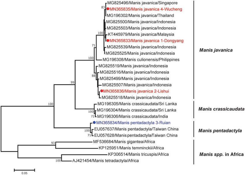
W.-H. Gao et al.    |   3

the NCBI non-redundant nucleotide (nt) and protein (nr) data-                        MegAlign program implemented in the Lasergene software
base with e-values set to 1  1010 and 1  104, respectively.                      package v5.0 (DNAstar).
Likely contaminating viral sequences were excluded from the
meta-transcriptomic data (Supplementary Table S2) using
                                                                                     3. Results
methods described previously (Asplund et al. 2019). In addition,
the high frequency of retrovirus sequences was also excluded,                        3.1 Illness and clinical features of sick pangolins
as the majority of them probably were host genes, and some of                        Between July and August in 2018, four sick wild pangolins were
them might be contaminating viral sequences, such as alphare-                        sent to the Jinhua Wildlife Rescue Station of Zhejiang province,
troviral and gammaretroviral sequences probably linked to lab-                       China (Fig. 1). For simplicity, they were referred to here as
oratory components (Asplund et al. 2019). Finally, the putative                      ‘1-Dongyang’, ‘2-Lishui’, ‘3-Ruian’, and ‘4-Wucheng’, according
viruses present in the blood, liver, spleen, lung, and kidney                        to the location found. Morphological examination and molecu-
samples were confirmed by PCR. The quantity of the transcripts                       lar identification revealed that these pangolins belong to the
mapped to each viral contig was determined using the RSEM                            Sunda pangolin, Manis javanica (1-Dongyang, 2-Lishui, and

                                                                                                                                                                    Downloaded from https://academic.oup.com/ve/article/6/1/veaa020/5819286 by guest on 02 November 2020
program (Li et al. 2010) implemented in Trinity.                                     4-Wucheng) and the Chinese pangolin, Manis pentadactyla
                                                                                     (3-Ruian). The details and clinical signs exhibited by these
2.3 PCR and sequencing                                                               pangolins are described in Table 1. All four pangolins exhibited
                                                                                     anorexia when sent to the rescue station, and twitching and
Total RNA was reverse transcribed using a one-step RT-PCR
                                                                                     slobbering behavior were observed in 1-Dongyang and 2-Lishui.
kit (TaKaRa). The viral RNA in ticks was detected by nested PCR
                                                                                     Hemorrhage and skin lesions were obvious in 1-Dongyang,
targeting the conserved regions of the RNA-dependent RNA po-
                                                                                     whereas edema on the front legs was found in 2-Lishui. Finally,
lymerase (RdRp) gene of both the pestivirus and coltivirus. To
                                                                                     pangolin 4-Wucheng appeared febrile and contained skin
recover complete viral genomes, primers were designed based
                                                                                     lesions, whereas pangolin 3-Ruian exhibited relatively mild
on the assembled pestivirus and coltivirus contigs obtained by
                                                                                     clinical signs.
meta-transcriptomics (Supplementary Table S3). The genome
                                                                                         X-ray tests revealed a large area of shadow in the left lung of
termini were determined by 50 /30 RACE kits (TaKaRa).
                                                                                     pangolin 2-Lishui, suggestive of pneumonia (Supplementary
    The QIAquick Gel Extraction kit (Qiagen) was used to purify
                                                                                     Fig. S1). Due to the lack of healthy pangolin as control in this
the PCR products before sequencing. Purified DNA 70 per cent were considered significant.                           1-Dongyang following autopsy, although no obvious pathologi-
Identities among nt and aa sequences were calculated using the                       cal changes were observed in other inner organs. Consistent

Table 1. Background information, clinical features, and examination of the rescued pangolins.

Pangolin ID        Species           Gender/age           State    Clinical features                       Clinical examination

                                                                                                           X-ray                         Hematological analysis

1-Dongyang         M. javanica       Male/adult           Dead     Anorexia, twitch, slobber, hem-         Normal                        PLT #, GLU #, choles-
                                                                     orrhage,a and skin lesionsb                                           terol #, and amylase "
2-Lishui           M. javanica       Female/adult         Dead     Anorexia, twitch, slobber, and          Large area of shadow          ALT ", AST ", amylase ",
                                                                     edemac                                  on the left lung              and total bilirubin #
3-Ruian            M. pentadactyla   Female/juvenile      Alive    Anorexia, anxiety, and edemac           –                             –
4-Wucheng          M. javanica       Female/adult         Dead     Anorexia, fever, and skin               Stones in the stomach         PLT #, ALP ", BUN "
                                                                     lesionsb

a
    On the left ear.
b
    On the face and paws.
c
On the front legs.
PLT, platelet count; GLU, glucose; AST, aspartate aminotransferase; ALT, alanine aminotransferase; BUN, blood urea nitrogen; ALP, alkaline phosphatase.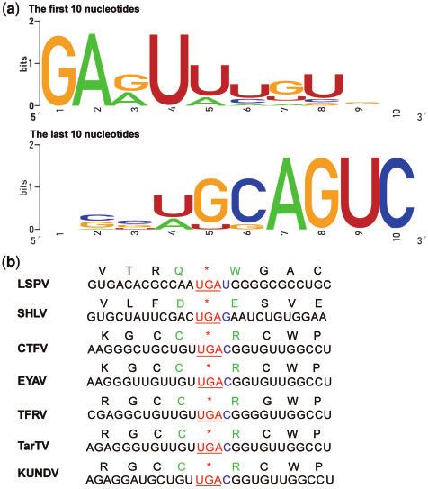
4 | Virus Evolution, 2020, Vol. 6, No. 1
with the obvious congestion, histological tests revealed hemor- 3.4 Genetic features of DYPV and LSPV
rhage in the liver and lung (Supplementary Fig. S2). In addition,
To further characterize both viruses, primers were designed
pyknotic nucleus of the hepatic cells, lymphocyte pyknosis
based on the sequences obtained here and those of related vi-
in spleen, necrosis of the hepatic plate, and glomerular
ruses described previously. In this manner we were able to re-
necrosis were observed. Finally, collagen fiber degeneration
cover the complete genomes of DYPV from 1-Dongyang and the
was observed in the liver, spleen, and lung (Supplementary
ticks (A. javanense) collected from this animal. The genetic fea-
Fig. S2).
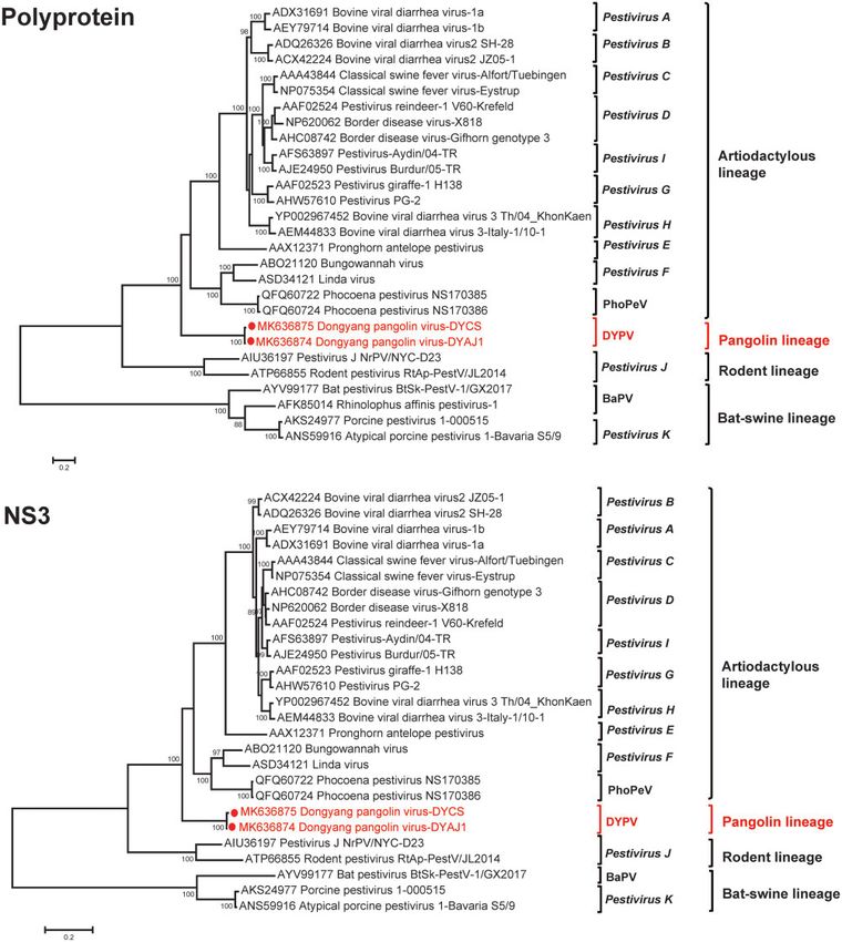
tures of DYPV are described in Fig. 2a. Notably, viruses obtained
For pangolin 2-Lishui, obvious necrosis was observed in the
from the pangolin and ticks were closely related to each other,
lung, kidney, and spleen, and congestion was observed in the
although still exhibited almost 2 per cent nt sequence differ-
liver (Supplementary Fig. S2) in addition to mesenteric lymph-
ence. In addition, both viruses were genetically and phylogenet-
adenopathy. Notably, many milky white lesions were present in
ically distinct from known viruses of the genus Pestivirus, with
both lungs, especially in the left lower lobe (1 2 cm).
>43 per cent nt and >48 per cent aa differences (Supplementary
Histological tests revealed necrosis in the liver, spleen, lung,
Table S5). As with classical pestiviruses, the genome of DYPV
Downloaded from https://academic.oup.com/ve/article/6/1/veaa020/5819286 by guest on 02 November 2020
kidney, trachea, and small intestinal, as well as hemorrhage in
encodes twelve proteins. All known cleavage sites of the NS3
the lung (Supplementary Fig. S2).
protease were observed in DYPV (Supplementary Fig. S3). In ad-
dition, the NS4A-NS4B cleavage site, located at Position L2426/
3.3 Identification of viral agents by A2427 in BVDV1 and L2336/A2337 in CSFV, appeared at Position
meta-transcriptomics and PCR L2315/K2316 site in DYPV (Supplementary Fig.S3).
To identify the possible etiologic agents of disease in the four Notably, despite the use of both meta-transcriptomic and
pangolins, eight meta-transcriptomic libraries from blood, PCR methods, only nine genome segments (1–5 and 8–11) were
liver, spleen, lung, kidney, and fecal samples were constructed, obtained from the novel coltivirus—in animal 2-Lishui—and our
generating a total of 306,908,179 paired-end sequence reads. De attempt to recover segments 6, 7, and 12 failed. Details of the
novo assembly revealed the high abundance of a pestivirus- genetic features of LSPV are described in Fig. 2b. All recovered
and coltivirus-like virus in all the meta-transcriptomic libraries segments had the consensus sequences (GAG/AUU/A) at the
of the pangolin 1-Dongyang and 2-Lishui, representing 6–80 50 -terminus and (G/CAGUC) and the 30 -terminus (Fig. 3a), respec-
and 1–29 per cent of total viral contigs, respectively tively. However, the genome sequences of LSPV were distinct
(Supplementary Table S2). Notably, despite the presence of from those of recognized coltiviruses, with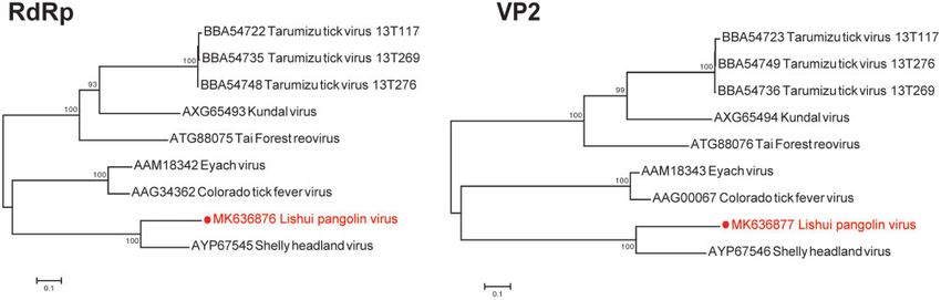
W.-H. Gao et al.    |   5

                                                                                                                                                            Downloaded from https://academic.oup.com/ve/article/6/1/veaa020/5819286 by guest on 02 November 2020
Figure 2. Schematic of the annotated DYPV and LSPV genomes. (a) Genome comparison between DYPV and known pestiviruses. (b) Genome comparison between LSPV
and known coltiviruses.

To determine the likely geographic origin of these sick pango-                 3.6 Molecular investigation of DYPV and LSPV in local
lins, sequences of mt-cyt b gene were amplified from their tis-                ticks
sue. Genetic analysis revealed that all the sequences obtained
                                                                               As DYPV was identified in ticks (A. javanense) collected from
from Sunda pangolins in this study fell into the M. javanica
                                                                               pangolin 1-Dongyang, and LSPV was closely related to SHLV
group, while the Chinese pangolins clustered with those of M.
                                                                               also identified in ticks (I. holocyclus) from Australia, we collected
pentadactyla. Notably, three Sunda pangolins sampled here
were very closely related to those from Indonesia, Malaysia,                   ticks at the locations from where the sick pangolins were found.
and Thailand and Singapore, with 99.9, 99.7, and 99.9 per cent                 Consequently, 452 ticks representing 7 species were collected,
nt sequence identity, respectively, indicating that they were                  including 220 Haemaphysalis hystricis, 147 Rhipicephalus microplus,
most likely (illegally) imported into China from abroad. In                    49 Haemaphysalis longicornis, 11 Ixodes granulatus, 11 Rhipicephalus
contrast, the pangolin 3-Ruian (M. pentadactyla) clustered to-                 haemaphysaloides, 9 Rhipicephalus sanguineus, and 5 Haemaphysalis
gether with those from Taiwan/China, with 99.1 per cent nt                     mageshimaensis. Unfortunately, neither DYPV nor LSPV were
sequence identity, suggesting that this animal was not                         identified in these ticks by meta-transcriptomics and nested
imported. As no sequences related to the A. javanense mito-                    RT-PCR.
chondrial 16S rDNA were available, we could not determine
the origin of the A. javanense ticks collected from sick Sunda
                                                                               4. Discussion
pangolins. Hence, a systemic effort should be considered to
establish comprehensive databases for the speciation of ar-                    Pangolins are insectivorous, predominantly nocturnal, and pre-
thropod vectors and as a tool for determining the geographic                   date almost exclusively on ants and termites, with a strong
origin of the collected arthropods.                                            preference for particular insect species (Lin et al. 2015;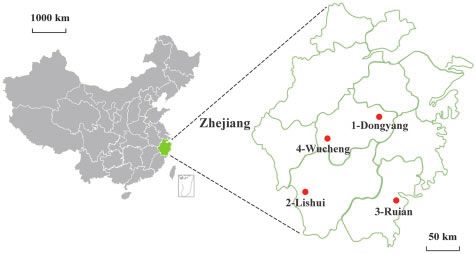
6    | Virus Evolution, 2020, Vol. 6, No. 1

                                                                                     Pestiviruses are well-known animal pathogens that cause
                                                                                 significant economic loss, infecting both domestic (e.g. pigs, cat-
                                                                                 tle, sheep, and goats) and wild (e.g. wild boars and ruminants)
                                                                                 animals (Vilcek and Nettleton 2006). Pestivirus infections may
                                                                                 be subclinical or cause a range of clinical signs including acute
                                                                                 diarrhea, acute hemorrhagic syndrome, acute fatal disease, as
                                                                                 well as a wasting disease. Herein, a novel pestivirus, designated
                                                                                 DYPV, was identified in multiple organs in one of the sick pan-
                                                                                 golins [1-Dongyang (M. javanica)] that had clear pathological
                                                                                 changes. Phylogenetic analysis indicates that it represents a
                                                                                 novel member of the genus Pestivirus. Although more detailed
                                                                                 confirmatory results are required, these data suggest that
                                                                                 DYPV-like pestiviruses may be responsible for the hemorrhagic
                                                                                 disease observed in the pangolins.

                                                                                                                                                           Downloaded from https://academic.oup.com/ve/article/6/1/veaa020/5819286 by guest on 02 November 2020
                                                                                     To date, it is commonly believed that vertebrates, rather
                                                                                 than invertebrates, are the main hosts of pestiviruses (Smith
                                                                                 et al. 2017). Recently, however, a novel pestivirus, named
                                                                                 Fairfax Lookout virus, was identified in ticks (Ixodes trichosuri).
                                                                                 However, given its phylogenetic position next to mammalian
                                                                                 pestiviruses, as well as its the extremely low abundance, it
                                                                                 was proposed that the virus was associated with the verte-
                                                                                 brate host rather than from the tick itself (Harvey et al. 2018).
                                                                                 In this study, DYPV was not only identified in the pangolins,
                                                                                 but also in nymph ticks collected from the pangolins.
                                                                                 However, DYPV was not found in the adult ticks also sampled
                                                                                 from pangolin 1-Dongyang. Interestingly, although DYPV from
Figure 3. Characteristics of the LSPV genome. (a) Sequence conservation of the
50 - and 30 -terminal 10 nts in genomic segments of the LSPV genome were ana-    ticks was closely related to that sampled from pangolins, there
lyzed and visualized using Weblogo. (b) Sequence information around the stop     were 2 per cent nt differences across the viral genome sug-
codon of segment VP9 of LSPV and known coltivirus.                               gesting that they are separated by multiple transmission
                                                                                 events. Phylogenetic analysis indicated that Sunda pangolins,
Choo et al. 2016). Eight species of pangolins are present in Africa              including the pangolin 1-Dongyang, were most likely imported
and Asia, with habitat loss and changes in their living environ-                 into China from Indonesia, Malaysia, and the Philippines
ment seriously affecting their population status. Most impor-                    (Fig. 6). In addition, A. javanense ticks have not found in
tantly, pangolins have been greatly impacted by the illegal                      Zhejiang province (Chen et al. 2010), DYPV was not identified
international and domestic wildlife trade for traditional medi-                  in locally collected ticks, and we did not observe this virus in
cine or meat (Zhang et al. 2015). Hence, pangolins are listed as                 previous large scale tick sampling studies (Li et al. 2015; Shi
vulnerable, endangered or critically endangered on the IUCN                      et al. 2016a,b, 2018). Together, these observations suggest that
                                                                                 the virus was probably imported from abroad with the illegal
Red List of Threatened Species (du Toit et al. 2017). Notably,
                                                                                 trafficked pangolin.
pangolins may also be threatened by epizootic pathogens, ei-
                                                                                     Coltiviruses are well-known tick-borne pathogens that can
ther present in their original habitats or in their new environ-
                                                                                 cause human disease. For example, CTFV causes mild febrile ill-
ments following translocation. Indeed, the translocation or
                                                                                 ness or more severe disease including infection of the central ner-
trafficking of domestic and wild animals plays an important
                                                                                 vous system, and/or hemorrhagic fever (Goodpasture et al. 1978;
role in the spread of many epizootic pathogens (Lin et al. 2012;
                                                                                 Attoui et al. 2005). Additionally, EYAV infections have been asso-
Kosmider et al. 2013; Peeler et al. 2015).
                                                                                 ciated with human neurological disease (Moutailler et al. 2016).
    In this study, four pangolins—three M. javanica likely from
                                                                                 Notably, although some coltiviruses (e.g. CTFV, EYAV, TFRV) have
Indonesia, Malaysia, and Thailand, and one M. pentadactyla
                                                                                 been detected in wildlife (such as bats and rodents), there is no
probably of local origin—were found to be suffering disease
                                                                                 clear evidence that these viruses can cause disease in animals
and were sent to a local rescue station for treatment.
                                                                                 (Moutailler et al. 2016; Weiss et al. 2017; Williamson et al. 2019). In
Unfortunately, three animals died, whereas one recovered. To                     this study, a novel coltivirus, named LSPV, was identified in one
date, reports on pangolin disease have been rare and mainly                      of the sick pangolins [2-Lishui (M. javanica)]. Given the high abun-
limited to those caused by bacteria and parasites (Mohapatra                     dance of LSPV in the meta-transcriptomic data, combined with
et al. 2016; Jabin et al. 2019). There is no available literature on vi-         the clinical features and pathologic changes appeared in the pan-
ral infections of pangolins until the recent identification of                   golin (2-Lishui), as well as the detection of LSPV in several organs,
Parainfluenza Virus 5, sendai virus, and coronavirus from Sunda                  our data suggest that LSPV may have caused systemic infection
pangolins in China (Liu et al. 2019; Wang et al. 2019). Using a com-             and the death of the pangolin in question, although this will need
bination of meta-transcriptomic and PCR methods, we identified                   to be confirmed with additional data.
two novel RNA viruses—a pestivirus and a coltivirus—in two                           It is generally recognized that the coltivirus genome com-
dead pangolins. Based on their clinical signs (Table 1), all these               prises twelve segments of linear double-stranded RNA (Fujita
four pangolins appeared to suffer infectious disease. However, as                et al. 2017). Recently, a novel coltivirus, Forest reovirus (TFRV),
no complete clinical data and laboratory parameters were                         was identified in free-tailed bats (C. aloysiisabaudiae; Weiss et al.
obtained from these animals, we could not clearly define the dis-                2017). Interestingly, the virus genome lacks segments 6, 7, and
ease they suffered.                                                              12, and it was proposed that these missing segments were not
W.-H. Gao et al. | 7
Downloaded from https://academic.oup.com/ve/article/6/1/veaa020/5819286 by guest on 02 November 2020
Figure 4. ML trees based on aa sequences of the entire coding sequences (polyprotein) and NS3 genes of DYPV and other known pestiviruses. The numbers at nodes
indicate bootstrap support values after 1,000 replications. Bootstrap values higher than 70 per cent were considered significant and shown on the trees.
identified probably due to low similarities between TFRV and imported from Indonesia (Fig. 6) it is possible that LSPV, like
known coltiviruses (Weiss et al. 2017). Strikingly, as with TFRV, DYPV, was also imported into China.
we were unable to identify viral segments 6, 7, and 12 in LSPV, Finally, despite some discussion concerning the role of pango-
despite the use of methods that previously obtained the com- lins in the emergence of the novel coronavirus (severe acute respi-
plete genome of the highly divergent Jingmen tick virus (Qin ratory syndrome coronavirus 2, SARS-CoV-2;Wahba et al. 2020;
et al. 2014). It is therefore clear that additional studies are Wong et al. 2020), the cause of the corona virus disease 2019
needed to infer whether these segments are indeed absent from ( COVID-19) outbreak (Wu et al. 2020; Zhou et al. 2020), we have not
LSPV and TFRV. Finally, as pangolin 2-Lishui may have been found coronaviruses in these pangolins by meta-transcriptomic8 | Virus Evolution, 2020, Vol. 6, No. 1
Downloaded from https://academic.oup.com/ve/article/6/1/veaa020/5819286 by guest on 02 November 2020
Figure 5. ML trees based on aa sequences of the RdRp genes and putative RNA methyltransferase (VP2) genes of LSPV and other known coltiviruses. The numbers at
nodes indicate bootstrap support values after 1,000 replications. Bootstrap values higher than 70 per cent were considered significant and shown on the trees.
Figure 6. ML tree based on nt sequences of the mt-cyt b gene of four pangolins and other known pangolins. The numbers at nodes indicate bootstrap support values af-
ter 1,000 replications. Bootstrap values higher than 70 per cent were considered significant and shown in the trees.
and PCR methods. Therefore, more efforts are needed to infer the GenBank under the accession numbers MK636874–MK636884,
role of pangolins in the transmission of SARS-CoV-2. MN365828–MN365832, and MN365833–MN365836.
Supplementary data
Supplementary data are available at Virus Evolution online. Funding
This work was financially supported by National Natural
Data Availability Science Foundation of China (81861138003, 31930001, and
All viral genome sequences, 16S rDNA and mt-cyt b gene 81672057) and the Special National Project on investigation
sequences generated in this study have been deposited in of basic resources of China (2019FY)101500 . E.C.H. wasW.-H. Gao et al. | 9
funded by an ARC Australian Laureate Fellowship Barcode, Phylogenetic Status and Its Implication in Wildlife
(FL170100022). Forensics’, Experimental and Applied Acarology, 78: 461–7.
Jo, W. K. et al. (2019) ‘An Evolutionary Divergent Pestivirus
Conflict of interest: No conflicts of interest. Lacking the N(Pro) Gene Systemically Infects a Whale Species’,
Emerging Microbes & Infections, 8: 1383–92.
Katoh, K., and Standley, D. M. (2013) ‘MAFFT Multiple Sequence
References
Alignment Software Version 7: Improvements in Performance
and Usability’, Molecular Biology and Evolution, 30: 772–80.
Asplund, M. et al. (2019) ‘Contaminating Viral Sequences in High-
Kosmider, R. et al. (2013) ‘Echinococcus multilocularis Introduction
Throughput Sequencing Viromics: A Linkage Study of 700
and Establishment in Wildlife via Imported Beavers’,
Sequencing Libraries’, Clinical Microbiology and Infection, 25: 1277–85.
Attoui, H. et al. (2002) ‘Genus Coltivirus (Family Reoviridae): Veterinary Record, 172: 606.2.
Kumar, S. et al. (2016) ‘MEGA7: Molecular Evolutionary Genetics
Genomic and Morphologic Characterization of Old World
Analysis Version 7.0 for Bigger Datasets’, Molecular Biology and
and New World Viruses’, Archives of Virology, 147: 533–61.
Downloaded from https://academic.oup.com/ve/article/6/1/veaa020/5819286 by guest on 02 November 2020
Attoui, H. et al. (2005) ‘Coltiviruses and Seadornaviruses in Evolution, 33: 1870–4.
North America, Europe, and Asia’, Emerging Infectious Diseases, Li, B. et al. (2010) ‘RNA-Seq Gene Expression Estimation with
11: 1673–9. Read Mapping Uncertainty’, Bioinformatics, 26: 493–500.
Blome, S. et al. (2017) ‘Classical Swine Fever-an Updated Review’, Li, C. X. et al. (2015) ‘Unprecedented Genomic Diversity of RNA
Viruses, 9: 86. Viruses in Arthropods Reveals the Ancestry of Negative-Sense
Capella-Gutierrez, S. et al. (2009) ‘trimAl: A Tool for Automated RNA Viruses’, eLife, 4: e05378.
Alignment Trimming in Large-Scale Phylogenetic Analyses’, Lin, M. F. et al. (2015) ‘Aspects of Digestive Anatomy, Feed Intake
Bioinformatics, 25: 1972–3. and Digestion in the Chinese Pangolin (Manis pentadactyla) at
Chen, Z. et al. (2010) ‘Ticks (Acari: Ixodoidea: Argasidae, Ixodidae) Taipei Zoo’, Zoo Biology, 34: 262–70.
of China’, Experimental and Applied Acarology, 51: 393–404. Lin, X. D. et al. (2012) ‘Migration of Norway Rats Resulted in the
et al. (2012) ‘Morphological, Biological and Molecular Worldwide Distribution of Seoul Hantavirus Today’, Journal of
Characteristics of Bisexual and Parthenogenetic Haemaphysalis Virology, 86: 972–81.
longicornis’, Veterinary Parasitology, 189: 344–52. Liu, P. et al. (2019) ‘Viral Metagenomics Revealed Sendai Virus
Chin, S. C. et al. (2015) ‘Hematologic and Serum Biochemical and Coronavirus Infection of Malayan Pangolins (Manis javan-
Parameters of Apparently Healthy Rescued Formosan ica)’, Viruses, 11: 979.
Pangolins (Manis pentadactyla pentadactyla)’, Journal of Zoo and Moennig, V., and Becher, P. (2015) ‘Pestivirus Control Programs:
Wildlife Medicine, 46: 68–76. How Far Have we Come and Where Are we Going?’, Animal
Choo, S. W. et al. (2016) ‘Pangolin Genomes and the Evolution of Health Research Reviews, 16: 83–7.
Mammalian Scales and Immunity’, Genome Research, 26: 1312–22. Mohapatra, R. K. et al. (2016) ‘Check List of Parasites and Bacteria
du Toit, Z. et al. (2017) ‘Mitochondrial Genomes of African Recorded from Pangolins (Manis sp.)’, Journal of Parasitic
Pangolins and Insights into Evolutionary Patterns and Diseases, 40: 1109–15.
Phylogeny of the Family Manidae’, BMC Genomics, 18: 746. Moutailler, S. et al. (2016) ‘Diversity of Viruses in Ixodes ricinus,
Firth, C. et al. (2014) ‘Detection of Zoonotic Pathogens and and Characterization of a Neurotropic Strain of Eyach Virus’,
Characterization of Novel Viruses Carried by Commensal New Microbes and New Infections, 11: 71–81.
Rattus norvegicus in New York City’, mBio, 5: e01933–01914. Peeler, E. J. et al. (2015) ‘Animal Disease Import Risk Analysis–A
Fujita, R. et al. (2017) ‘Isolation and Characterization of Review of Current Methods and Practice’, Transboundary and
Tarumizu Tick Virus: A New Coltivirus from Haemaphysalis Emerging Diseases, 62: 480–90.
flava Ticks in Japan’, Virus Research, 242: 131–40. Postel, A. et al. (2015) ‘Close Relationship of Ruminant
Gaubert, P. et al. (2018) ‘The Complete Phylogeny of Pangolins: Pestiviruses and Classical Swine Fever Virus’, Emerging
Scaling up Resources for the Molecular Tracing of the Most Infectious Diseases, 21: 668–72.
Trafficked Mammals on Earth’, Journal of Heredity, 109: 347–59. Qin, X. C. et al. (2014) ‘A Tick-Borne Segmented RNA Virus
Gaudin, T. J. et al. (2009) ‘The Phylogeny of Living and Extinct Contains Genome Segments Derived from Unsegmented Viral
Pangolins (Mammalia, Pholidota) and Associated Taxa: A Ancestors’, Proceedings of the National Academy of Sciences of the
Morphology Based Analysis’, Journal of Mammalian Evolution, 16: United States of America, 111: 6744–9.
235–305. Ridpath, J. F. et al. (2008) ‘Reproductive Tract Disease Associated
Goodpasture, H. C. (1978) ‘Colorado Tick Fever: Clinical, with Inoculation of Pregnant White-Tailed Deer with Bovine
Epidemiologic, and Laboratory Aspects of 228 Cases in Viral Diarrhea Virus’, American Journal of Veterinary Research, 69:
Colorado in 1973-1974’, Annals of Internal Medicine, 88: 303–10. 1630–6.
Guindon, S. et al. (2010) ‘New Algorithms and Methods to Shi, M. et al. (2016a) ‘Divergent Viruses Discovered in Arthropods
Estimate Maximum-Likelihood Phylogenies: Assessing the and Vertebrates Revise the Evolutionary History of the
Performance of PhyML 3.0’, Systematic Biology, 59: 307–21. Flaviviridae and Related Viruses’, Journal of Virology, 90: 659–69.
Guo, W. P. et al. (2013) ‘Phylogeny and Origins of Hantaviruses et al. (2016b) ‘Redefining the Invertebrate RNA Virosphere’,
Harbored by Bats, Insectivores, and Rodents’, PLoS Pathogens, 9: Nature, 540: 539–43.
e1003159. et al. (2018) ‘The Evolutionary History of Vertebrate RNA
Harvey, E. et al. (2018) ‘Extensive Diversity of RNA Viruses in Viruses’, Nature, 556: 197–202.
Australian Ticks’, Journal of Virology, 93: e01358–18. Smith, D. B. et al. (2017) ‘Proposed Revision to the Taxonomy of
Hua, L. et al. (2015) ‘Captive Breeding of Pangolins: Current the Genus Pestivirus, Family Flaviviridae’, Journal of General
Status, Problems and Future Prospects’, ZooKeys, 507: 99–114. Virology, 98: 2106–12.
Jabin, G. et al. (2019) ‘Identifying the Tick Amblyomma javanense Tautz, N. et al. (2015) ‘The Molecular Biology of Pestiviruses’,
(Acari: Ixodidae) from Chinese Pangolin: Generating Species Advances in Virus Research, 93: 47–160.10 | Virus Evolution, 2020, Vol. 6, No. 1
Valdazo-Gonzalez, B. et al. (2007) ‘Genetic and Antigenic Typing Wong, M. et al. (2020) ‘Evidence of Recombination in
of Border Disease Virus Isolates in Sheep from the Iberian Coronaviruses Implicating Pangolin Origins of nCoV-2019’,
Peninsula’, The Veterinary Journal, 174: 316–24. bioRxiv, doi: 10.1101/2020.02.07.939207.
Vilcek, S., and Nettleton, P. F. (2006) ‘Pestiviruses in Wild Wu, F. et al. (2020) ‘A New Coronavirus Associated with Human
Animals’, Veterinary Microbiology, 116: 1–12. Respiratory Disease in China’, Nature, 579: 265–9.
Wahba, L. et al. (2020) ‘Identification of a Pangolin Niche Wu, Z. et al. (2012) ‘Virome Analysis for Identification of Novel
for a 2019-nCoV-Like Coronavirus via an Extensive Mammalian Viruses in Bat Species from Chinese Provinces’,
Meta-Metagenomic Search’, bioRxiv, doi: 10.1101/2020. Journal of Virology, 86: 10999–1012.
02.08.939660. Yadav, P. D. et al. (2019) ‘Characterization of Novel Reoviruses
Wang, X. et al. (2019) ‘Complete Genome Sequence of Wad Medani Virus (Orbivirus) and Kundal Virus (Coltivirus)
Parainfluenza Virus 5 (PIV5) from a Sunda Pangolin (Manis Collected from Hyalomma anatolicum Ticks in India during
Javanica) in China’, Journal of Wildlife Diseases, 55: 947–50. Surveillance for Crimean Congo Hemorrhagic Fever’, Journal of
Weiss, S. et al. (2017) ‘A Novel Coltivirus-Related Virus Isolated Virology, 93: e00106–19.
from Free-Tailed Bats from Cote D’Ivoire Is Able to Infect Zhang, H. et al. (2015) ‘Molecular Tracing of Confiscated Pangolin
Downloaded from https://academic.oup.com/ve/article/6/1/veaa020/5819286 by guest on 02 November 2020
Human Cells in Vitro’, Virology Journal, 14: 181. Scales for Conservation and Illegal Trade Monitoring in
Williamson, B. N. et al. (2019) ‘Prevalence and Strains of Colorado Southeast Asia’, Global Ecology and Conservation, 4: 414–22.
Tick Fever Virus in Rocky Mountain Wood Ticks in the Bitterroot Zhou, P. et al. (2020) ‘A Pneumonia Outbreak Associated with a
Valley, Montana’, Vector-Borne and Zoonotic Diseases, 19: 694–702. New Coronavirus of Probable Bat Origin’, Nature, 579: 270–3.You can also read