CONSEQUENCE OF HISTOINCOMPATIBILITY BEYOND GVH-REACTION IN CYTOMEGALOVIRUS DISEASE ASSOCIATED WITH ALLOGENEIC HEMATOPOIETIC CELL TRANSPLANTATION: ...
←
→
Page content transcription
If your browser does not render page correctly, please read the page content below
viruses
Review
Consequence of Histoincompatibility beyond GvH-Reaction
in Cytomegalovirus Disease Associated with Allogeneic
Hematopoietic Cell Transplantation: Change of Paradigm
Matthias J. Reddehase *, Rafaela Holtappels                      and Niels A. W. Lemmermann

                                          Institute for Virology and Research Center for Immunotherapy (FZI), University Medical Center
                                          of the Johannes Gutenberg-University Mainz, 55131 Mainz, Germany; r.holtappels@uni-mainz.de (R.H.);
                                          lemmermann@uni-mainz.de (N.A.W.L.)
                                          * Correspondence: matthias.reddehase@uni-mainz.de

                                          Abstract: Hematopoietic cell (HC) transplantation (HCT) is the last resort to cure hematopoietic
                                          malignancies that are refractory to standard therapies. Hematoablative treatment aims at wiping
                                          out tumor cells as completely as possible to avoid leukemia/lymphoma relapse. This treatment
                                          inevitably co-depletes cells of hematopoietic cell lineages, including differentiated cells that constitute
                                          the immune system. HCT reconstitutes hematopoiesis and thus, eventually, also antiviral effector
                                          cells. In cases of an unrelated donor, that is, in allogeneic HCT, HLA-matching is performed to
                                          minimize the risk of graft-versus-host reaction and disease (GvHR/D), but a mismatch in minor
                                          histocompatibility antigens (minor HAg) is unavoidable. The transient immunodeficiency in the

                                   period between hematoablative treatment and reconstitution by HCT gives latent cytomegalovirus
                                          (CMV) the chance to reactivate from latently infected donor HC or from latently infected organs of
Citation: Reddehase, M.J.;
                                          the recipient, or from both. Clinical experience shows that HLA and/or minor-HAg mismatches
Holtappels, R.; Lemmermann, N.A.W.
                                          increase the risk of complications from CMV. Recent results challenge the widespread, though never
Consequence of Histoincompatibility
beyond GvH-Reaction in
                                          proven, view of a mechanistic link between GvHR/D and CMV. Instead, new evidence suggests that
Cytomegalovirus Disease Associated        histoincompatibility promotes CMV disease by inducing non-cognate transplantation tolerance that
with Allogeneic Hematopoietic Cell        inhibits an efficient reconstitution of high-avidity CD8+ T cells capable of recognizing and resolving
Transplantation: Change of Paradigm.      cytopathogenic tissue infection.
Viruses 2021, 13, 1530. https://
doi.org/10.3390/v13081530                 Keywords: avidity; antigen presentation; CD8 T cells; cytomegalovirus; cytomegalovirus disease; graft-
                                          versus-host disease (GvHD); hematopoietic cell transplantation (HCT); hematopoietic reconstitution
Academic Editor: Anna Cliffe

Received: 16 June 2021
Accepted: 28 July 2021
                                          1. Introduction
Published: 3 August 2021
                                                Human cytomegalovirus (hCMV) is the prototype member of the β-subfamily of
                                          the herpes virus family [1]. Public awareness is low, because an intact immune system
Publisher’s Note: MDPI stays neutral
                                          efficiently resolves primary infection. Infection can principally occur in all age groups, but
with regard to jurisdictional claims in
published maps and institutional affil-
                                          the virus is often acquired in early childhood and may be viewed epidemiologically as a
iations.
                                          “daycare center infection” that usually passes with only mild, feverish symptoms. CMV
                                          is thus rarely considered in a differential diagnosis of age-typical, harmless infections [2].
                                          Upon contact between the asymptomatically infected child and a CMV-naïve expectant
                                          mother in secondary pregnancy, there exists a risk of primary infection and dia-placental
                                          transmission [3], resulting in congenital infection of the immunological immature fetus with
Copyright: © 2021 by the authors.
                                          a range of birth defects collectively known as cytomegalic inclusion disease (CID) [2,4–7].
Licensee MDPI, Basel, Switzerland.
                                                Other risk groups of significant clinical relevance are patients who undergo an im-
This article is an open access article
distributed under the terms and
                                          munocompromising therapy of unrelated primary diseases. This includes recipients of
conditions of the Creative Commons
                                          solid organ transplantation (SOT), who receive immunosuppressive treatment to prevent
Attribution (CC BY) license (https://     graft rejection by a host-versus-graft (HvG) reaction (for clinical overviews, see [5,8,9]).
creativecommons.org/licenses/by/          The focus of this brief review is on hematopoietic (stem) cell (HC) transplantation (HCT),
4.0/).                                    which is the last therapeutic option to cure aggressive types of hematopoietic malignan-

Viruses 2021, 13, 1530. https://doi.org/10.3390/v13081530                                                   https://www.mdpi.com/journal/viruses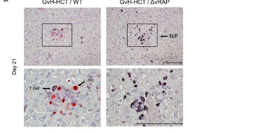
Viruses 2021, 13, 1530                                                                                           2 of 10

                         cies that are refractory to standard anti-tumor therapies. In essence, malignant cells are
                         wiped out by hematoablative treatment, which, unavoidably, also depletes non-malignant
                         hematopoietic cells of all differentiation lineages, including mature cells that mediate
                         innate and adaptive immunity. HCT is the means to repopulate the bone marrow stroma
                         with hematopoietic stem and progenitor cells and thus to reconstitute the immune system.
                         The phase of transient immunodeficiency after HCT is a “window of opportunity” for
                         hCMV to reactivate to productive infection within the latently infected donor HC or within
                         latently infected cells residing in organs of the recipient, or both (reviewed and discussed
                         in [10]). Lack of immune control leads to an unrestricted inter- and intra-tissue virus spread
                         with histopathological lesions that can lead to multiple-organ CMV disease. Interstitial
                         pneumonia represents the most feared disease manifestation with often lethal outcome,
                         particularly when the clinical hCMV variant is resistant to common antiviral drugs [11–13].
                         In such cases, adoptive immunotherapy by transfer of antiviral CD8+ T cells is the last
                         resort to close the “gap of risk” between hematoablative treatment of the primary disease
                         and complete immune reconstitution by HCT [14–21]. Follow-up monitoring of HCT recip-
                         ients by PCR to detect virus reactivation with high sensitivity is routine at transplantation
                         centers worldwide to initiate antiviral therapies at the earliest possible moment, a strategy
                         known as “pre-emptive” therapy [12].
                               It is longstanding clinical experience that HCT-associated CMV disease, when com-
                         pared to syngeneic HCT with identical twins as donor and recipient [22,23] or even autolo-
                         gous HCT [24], is of higher incidence and often more severe when a family or unrelated HC
                         donor and the recipient differ in major histocompatibility (MHC/HLA) or minor histocom-
                         patibility (minor-H) loci [24–26]. Such clinical settings are referred to as “allogeneic” HCT.
                         Specifically, HCT with an HLA-identical sibling donor and recipient pair is an allogeneic
                         HCT based on differences in minor-H loci [26]. As such immunogenetic mismatches are
                         the basis for an immunological graft-versus-host (GvH) reaction and disease (GvHR/D)
                         mediated primarily by donor T cells specific for non-shared histocompatibility antigens, the
                         donor and recipients are HLA type-matched as close as practicable for risk management.
                         Despite this, differences in minor-H antigens (minor-HAg) are unavoidable and bear a
                         risk of GvHD. Stern and colleagues [27] recently reviewed the issue of hCMV latency and
                         reactivation in recipients of allogeneic HCT. Here we focus on the specific question of
                         whether post-HCT GvH/D and CMV disease are mechanistically linked, or if both are
                         independent consequences of the underlying histoincompatibility.
                               In a superficial view, immunogenetic mismatches and GvHR/D are used as if they
                         were synonyms. Clinical treatment regimens leave an uncertainty, however, if GvHR/D
                         after allogeneic HCT is caused by effector functions of host-reactive donor-type effector
                         cells derived from lymphoid-lineage hematopoietic differentiation and thymic education,
                         or rather from mature donor T cells already present in the HC transplant. In fact, the
                         HC transplant is often deliberately left undepleted of mature T cells, accepting adverse
                         effects of GvHR to maintain a beneficial graft-versus-leukemia/lymphoma (GvL) reaction
                         for reducing the risk of tumor relapse from minimal residual leukemia/lymphoma. This,
                         next to GvHD and CMV disease, is a major, if not the major, concern in tumor therapy by
                         HCT. Accordingly, separating GvH-reactive from GvL-reactive cells is a topic of intense
                         research ([28,29], reviewed in [30]). In addition, in the case of a CMV+ donor, mature
                         antiviral T cells in the HC transplant can also exert a beneficial graft-versus-infection
                         (GvI) effect [31]. Therefore, the precise and highly individualized regimen of allogeneic
                         HCT always demands a benefit–risk assessment depending on the individual donor–
                         recipient constellation. In fact, hematopoietic reconstitution originating from allogeneic
                         hematopoietic stem cells is expected to lead to “transplantation tolerance”. A conditioning
                         HCT performed with HC from an SOT donor has even been discussed as a potential
                         strategy to prevent graft rejection by tolerizing SOT recipients against MHC/HLA antigens
                         and/or minor-HAg expressed by the donor tissue (for an overview, see [32]).
                               Despite all undeniable genetic differences between the host species adapted human
                         and animal CMVs, as well as between their respective hosts, the mouse model has, in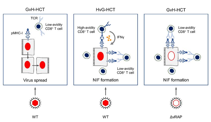
Viruses 2021, 13, 1530                                                                                            3 of 10

                         the past, already proven its validity for human CMV disease by identifying fundamental,
                         CMV-common principles of pathogenesis, immune control, and therapeutic intervention
                         (reviewed in [33]). We have explained this with biological convergence during virus–host
                         co-evolution [33]. Specifically, immunotherapy of post-HCT CMV disease by adoptive
                         transfer of CD8+ T cells in the mouse model based on murine CMV (mCMV) ([34,35],
                         reviewed in [36–38]) has been successfully translated to preemptive immunotherapy of
                         CMV disease in clinical HCT settings (see above). In the weakness of any reductionistic
                         approach in animal models to never reproduce the clinical reality in all its complexity
                         lies also the strength of defined conditions. In fact, human CMV disease cannot be fully
                         reproduced by any model because it is never a predictable, uniform entity. It depends
                         on the individual’s genetic constitution and infection history that defines the latent CMV
                         genome load and incidence of reactivation (discussed in [39]), and on genetic differences in
                         hCMV variants/strains. These likely differ between donor and recipient and can have a
                         fundamental impact on cell-type tropism and thus on pathogenicity, as well as on immuno-
                         logical marks in terms of antigens and immune evasion proteins expressed [1,40,41]. In
                         addition, the primary malignancy and its treatment history are also determinants for the
                         outcome of HCT in clinical real life. Thus, no model will ever perfectly suit human CMV
                         disease in any individual HCT recipient.
                               Here we summarize and interpret two recent reports on CMV disease in mouse models
                         of allogeneic HCT performed in an immunogenetic GvH transplantation direction across a
                         single MHC class-I difference [42], as well as across a difference in a minor-H locus [43].
                         Combined, both models led us to the conclusion that lethal CMV disease occurs despite
                         the absence of GvH-reactive effector cells and, instead, can be caused by non-cognate
                         transplantation tolerance that inhibits the efficient reconstitution of antiviral CD8+ T cells,
                         thereby leading to unrestricted virus spread and histopathology.

                         2. Key Results from Mouse Models of Allogeneic HCT
                         2.1. Lethality from CMV Infection after HCT in Immunogenetic GvH Transplantation Direction
                              The impact of mCMV infection on the outcome of syngeneic experimental HCT, with
                         sex-matched BALB/c mice as donors and recipients, has been studied extensively (for
                         reviews, see [37,38]). In this model, only epigenetic differences between individual mice
                         might induce GvHR/D, which is not the case only in autologous HCT. In essence, control
                         of mCMV infection and prevention of lethality was found to depend on the number of
                         transplanted HCs and correlated with the efficient and timely reconstitution of high-avidity
                         antiviral CD8+ T cells [37,38].
                              A more recently studied model of allogeneic HCT across an MHC class-I dispar-
                         ity used BALB/c mice and mutant BALB/c-H-2dm2 mice alternatingly as HCT donors
                         and recipients [42]. BALB/c mice express the full set of MHC class-I molecules of the
                         H-2d haplotype, namely Kd , Dd , and Ld . Based on a spontaneous genetic deletion, the
                         Ld molecule is absent in the otherwise genetically identical strain BALB/c-H-2dm2 . This
                         special feature provided the chance to perform HCT selectively as either HvG-HCT or
                         GvH-HCT. Theoretically, the transplantation direction in HvG-HCT allows only a unidi-
                         rectional response of recipient–resident CD8+ T cells against the Ld molecule expressed
                         on transplanted donor-derived HC, whereas the transplantation direction in GvH-HCT
                         allows only a unidirectional response of reconstituted donor-genotype CD8+ T cells against
                         the Ld molecule expressed on all cell types of the recipient (Figure 1).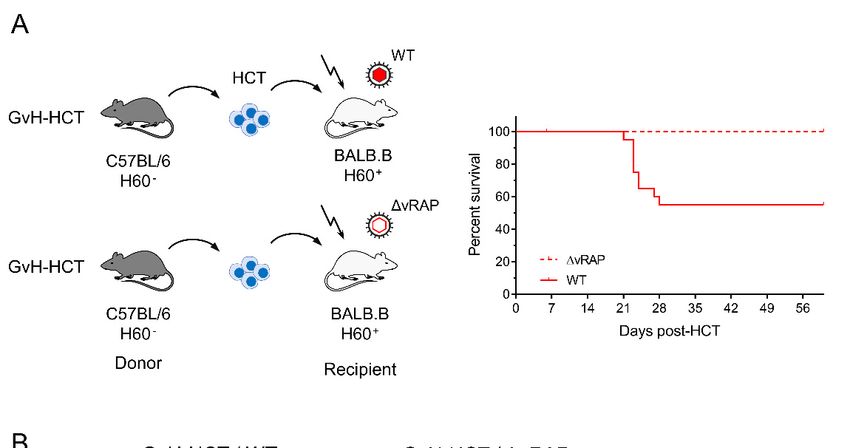
Viruses2021,
Viruses 13,x1530
2021,13, FOR PEER REVIEW 44ofof11
10
Figure 1. Comparison
Figure 1. Comparison ofoflethality
lethality and
and viral
viral histopathology
histopathology in mCMV-infected
in mCMV-infected recipients
recipients of HCTofperformed
HCT performed
in immuno- in
immunogenetic HvG or GvH direction. (A) Model and Kaplan-Meier survival curves. The flash symbol indicates
genetic HvG or GvH direction. (A) Model and Kaplan-Meier survival curves. The flash symbol indicates hematoablative
hematoablative total-body γ-irradiation. The dotted lines mark the 50% survival time. (B) 2-color immunohistological
total-body γ-irradiation. The dotted lines mark the 50% survival time. (B) 2-color immunohistological images of liver
images of liver tissue sections (upper panels, overview; lower panels, resolved to greater detail) showing tissue infiltration
tissue
by sections
T cells (black(upper panels,
staining) and overview; lower panels,
infected hepatocytes resolved
(iHC, to greater
red staining). detail)
Frames showing regions
demarcate tissue infiltration
resolved tobygreater
T cells
detail in the corresponding lower panel images. NIF, nodular inflammatory focus. Bar markers: 100 μm. Reproduced in
(black staining) and infected hepatocytes (iHC, red staining). Frames demarcate regions resolved to greater detail the
from
corresponding
reference [42] inlower
a newpanel images. NIF, nodular inflammatory focus. Bar markers: 100 µm. Reproduced from reference [42]
arrangement.
in a new arrangement.
The immunogenetic direction made a fundamental difference in the outcome of HCT.
WhereasThe most
immunogenetic
recipients of direction
HvG-HCT made a fundamental
survived difference
the infection, none inofthe
theoutcome
recipientsof
HCT. Whereas
survived undermost recipients
conditions of HvG-HCT
of GvH-HCT survived
(Figure 1A).the
Asinfection, none offor
clearly shown the the
recipients
liver,
survived under conditions of GvH-HCT (Figure 1A). As clearly shown for the
survival of HvG-HCT recipients correlates with tissue infiltration by T cells that cluster liver, survival
of HvG-HCT
around recipients
the few correlates
remaining withhepatocytes,
infected tissue infiltration by Tforming
thereby cells that cluster around
so-called “nodularthe
few remaining infected hepatocytes, thereby forming so-called “nodular
inflammatory foci” (NIF) confining, and eventually resolving, productive infection. In inflammatory
foci” contrast,
sharp (NIF) confining, andTeventually
infiltrating resolving,
cells are scarce productive
and NIF infection.
are barely formed In sharp
after contrast,
GvH-HCT.
infiltrating T cells are scarce and NIF are barely formed after GvH-HCT. As
As a consequence of missing immune control, the infection spreads unhindered, resulting a consequence
of missing immune control, the infection spreads unhindered, resulting in extended viral
in extended viral histopathology (Figure 1B). Notably, evidence for GvHD-typical
histopathology (Figure 1B). Notably, evidence for GvHD-typical histopathology is missing.
histopathology is missing.
2.2. Failure in the Reconstitution of High Avidity Virus-Specific CD8+ T Cells after GvH-HCT
For explaining the selective failure of antiviral control in GvH-HCT recipients com-
pared to efficient antiviral control in HvG-HCT recipients, CD8+ T cells were isolated from2.2. Failure in the Reconstitution of High Avidity Virus-Specific CD8+ T Cells after GvH-HCT
For explaining the selective failure of antiviral control in GvH-HCT recipients
Viruses 2021, 13, 1530 5 of 10
compared to efficient antiviral control in HvG-HCT recipients, CD8+ T cells were isolated
from the livers of both groups of HCT recipients. Viral epitope-specific IFNγ+CD8+ T cells
were quantitated depending on their functional avidity in recognizing cell surface
the livers of both
peptide-MHC groups
class-I of HCT recipients.
complexes m164-DViral epitope-specific
d, M105-K IFNγ+ CD8
d, and m145-K
+ T cells were
d (Figure 2) [42].
quantitated depending on their functional avidity in recognizing cell surface peptide-MHC
Responding cells were categorized into non-protective “low avidity” cells and protective
class-I complexes m164-Dd , M105-Kd , and m145-Kd (Figure 2) [42]. Responding cells were
“high avidity” cells based on the previous finding that an avidity corresponding to an
categorized into non-protective “low avidity” cells and protective “high avidity” cells
exogenous
based onpeptide loading
the previous concentration
finding that an avidityof ≤10 −9 M is needed for the recognition of
corresponding to an exogenous peptide
antigenic peptides presented by
− 9infected cells following endogenous
loading concentration of ≤10 M is needed for the recognition of antigenic antigen processing
peptides
[36,44].
presented by infected cells following endogenous antigen processing [36,44]. At a glance,cells
At a glance, compared to HvG-HCT, frequencies of viral epitope-specific
were generally
compared low, and were
to HvG-HCT, even of
frequencies lower
viral in the high-avidity
epitope-specific compartment,
cells were after
generally low, and GvH-
HCT.were
Thiseven lower
result in the high-avidity
identified compartment,
an insufficient after GvH-HCT.
reconstitution This result
of antiviral CD8+ identified
T cells as the
an insufficient reconstitution of antiviral + T cells as the reason for unrestricted virus
reason for unrestricted virus spread and CD8
extensive viral histopathology after GvH-HCT.
spread and extensive viral histopathology after GvH-HCT.
+ CD8+ T+ cells after GvH-HCT
FigureFigure
2. Frequencies and and
2. Frequencies functional avidities
functional ofofliver-derived,
avidities viral epitope-specific
liver-derived, viral epitope-specific IFNγ
IFNγ +CD8 T cells after GvH-HCT
compared to HvG-HCT.
compared Reproduced
to HvG-HCT. from
Reproduced reference
from reference[42]
[42]in
in aa modified graphicalpresentation.
modified graphical presentation.
2.3. 2.3. Enhancement
Enhancement of of AntigenPresentation
Antigen Presentation Restores
Restores Antiviral
AntiviralProtection after
Protection GvH-HCT
after GvH-HCT
A second modelofofTTcell-depleted
cell-depleted allogeneic b
A second model allogeneicHCT
HCTused C57BL/6
used C57BL/6 (H-2(H-2
haplotype)
b haplotype)
mice as donors and BALB.B mice as recipients in a GvH-HCT [43]. These two mouse strains
mice as donors and BALB.B mice as recipients in a GvH-HCT [43]. These two mouse
share the major histocompatibility complex and are thus identical in MHC class-I as well as
strains share the major histocompatibility complex and are thus identical in MHC class-I
class-II antigens, while differing in genetic background, including minor histocompatibility
as well
loci. Aaswell-studied
class-II antigens, while differing
and particularly in geneticisbackground,
strong minor-HAg including
H60 (for a review, minor
see [45]),
histocompatibility
which is not expressed in C57BL/6 donors of GvH-HCT but is expressed in the BALB.B(for a
loci. A well-studied and particularly strong minor-HAg is H60
review, see [45]),
recipients which
(Figure 3A). is not expressed in C57BL/6 donors of GvH-HCT but is expressed
In the context
in the BALB.B of mCMV
recipients (Figureinfection,
3A). it is worth noting that H60 is an activatory ligand
of the natural killer (NK) cell receptor NKG2D, and is downregulated in infected cells
by the viral m155 gene product [46]. As NKG2D is expressed also on activated CD8+ T
cells, serving as a costimulatory receptor, H60-NKG2D ligation could even enhance GvH-
reactivity against H60-expressing uninfected cells.
Based on the experience of poor reconstitution of high-avidity epitope-specific CD8+ T
cells in the MHC class-I mismatch model of GvH-HCT ([42], see above), BALB.B recipients
were infected either with wild-type (WT) virus or with a mutant deleted in viral genes that
encode immunoevasive regulators of antigen presentation (∆vRAP).
The idea behind infection with ∆vRAP virus was to relieve peptide-MHC class-I
complexes of the vRAP-mediated inhibition of their transport to the cell surface, and thus
to enhance antigen presentation for recruiting also low-avidity virus-specific CD8+ T cells
into NIF for the recognition of infected cells ([42,43] and referencing of vRAP functions
therein). As was the case in the MHC class-I mismatch model (recall Figure 1), GvH-HCT
was associated with significant lethality from infection with WT virus expressing vRAPs
that inhibit antigen presentation. In contrast, despite a genetic predisposition to GvHR
directed against the minor-HAg H60, all recipients survived when antigen presentation was
not inhibited by vRAP (Figure 3A). Notably, there was no evidence for a GvHR occurring
at all in the BALB.B recipients regardless of the type of infecting virus, as CD8+ T cellsViruses 2021, 13, 1530 6 of 10
es 2021, 13, x FOR PEER REVIEW 6 of 11
specific for the H60-derived antigenic peptide LTFNYRNL (also known as LYL8) were
not detected [43]. This indicated an absence of GvHR/D due to the establishment of
transplantation tolerance (for reviews, see [32,47]).
Figure 3. of
Figure 3. Comparison Comparison
lethality andof viral
lethality and viral histopathology
histopathology in minor-HAg in minor-HAg H60-mismatched
H60-mismatched GvH- infected
GvH-HCT recipients
with wild-typeHCT
(WT)recipients
mCMV or infected
a mutantwithof wild-type
mCMV deleted(WT) formCMV or a mutant
the genes that codeof for
mCMV deleted for “viral
immunoevasive the genes
regulators of
that code for immunoevasive “viral regulators of antigen presentation” (ΔvRAP). (A) Model and
antigen presentation” (∆vRAP). (A) Model and Kaplan–Meier survival curves. The flash symbol indicates hematoablative
Kaplan–Meier survival curves. The flash symbol indicates hematoablative total-body γ-irradiation.
total-body γ-irradiation. (B) 2-color immunohistological images of liver tissue sections (upper panels, overview; lower
(B) 2-color immunohistological images of liver tissue sections (upper panels, overview; lower
panels, resolved to greater
panels, detail)
resolved showing
to greater tissue
detail) infiltration
showing byinfiltration
tissue T cells (black
by Tstaining) and staining)
cells (black infected hepatocytes
and infected(iHC, red
staining). Frames
hepatocytes (iHC, red staining). Frames demarcate regions resolved to greater detail in NIF,
demarcate regions resolved to greater detail in the corresponding lower panel images. the nodular
inflammatorycorresponding
focus. Bar markers: 100 µm. Reproduced from reference [43] in new arrangement.
lower panel images. NIF, nodular inflammatory focus. Bar markers: 100 μm.
Reproduced from reference [43] in new arrangement.
The histopathological correlate of lethality from WT virus infection of the minor-
+ GvH-HCT recipients is the overall scarcity of liver tissue-infiltrating T cells,
In the context H60
HAg of mCMV infection, it is worth noting that H60 is an activatory ligand
of the naturalmissing NIF cell
killer (NK) formation, and
receptor extensive
NKG2D, andviral spread, whereasinenhanced
is downregulated presentation
infected cells by of viral
peptides after infection with the ∆vRAP virus restored T-cell infiltration,
the viral m155 gene product [46]. As NKG2D is expressed also on activated CD8+ T cells, NIF formation, and
clearance of productive infection, thus avoiding extensive
serving as a costimulatory receptor, H60-NKG2D ligation could even enhance GvH- viral histopathology (Figure 3B).
These findings are
reactivity against H60-expressing decisivecells.
uninfected for revealing the mechanism that causes lethality in the
GvH-HCT infection model. Enhancement of viral antigen
Based on the experience of poor reconstitution of high-avidity presentation in
epitope-specific infected
CD8 + cells by
vRAP deletion can modulate only the recognition of infected
T cells in the MHC class-I mismatch model of GvH-HCT ([42], see above), BALB.B cells by effector T cells, and
thus can prevent only viral histopathology. It cannot, however,
recipients were infected either with wild-type (WT) virus or with a mutant deleted in viralprevent histopathology
resulting from a GvH T-cell attack against uninfected tissue cells. These data formally
genes that encode immunoevasive regulators of antigen presentation (ΔvRAP).
The idea behind infection with ΔvRAP virus was to relieve peptide-MHC class-I
complexes of the vRAP-mediated inhibition of their transport to the cell surface, and thus
to enhance antigen presentation for recruiting also low-avidity virus-specific CD8+ T cells(Figure 3B).
These findings are decisive for revealing the mechanism that causes lethality in the
GvH-HCT infection model. Enhancement of viral antigen presentation in infected cells by
vRAP deletion can modulate only the recognition of infected cells by effector T cells, and
Viruses 2021, 13, 1530 thus can prevent only viral histopathology. It cannot, however, prevent histopathology
7 of 10
resulting from a GvH T-cell attack against uninfected tissue cells. These data formally
exclude lethal histopathology as resulting from GvHR/D and, instead, provide strong
evidence for death being caused by viral histopathology and consequent organ failure.
exclude lethal histopathology as resulting from GvHR/D and, instead, provide strong
evidence for death being caused by viral histopathology and consequent organ failure.
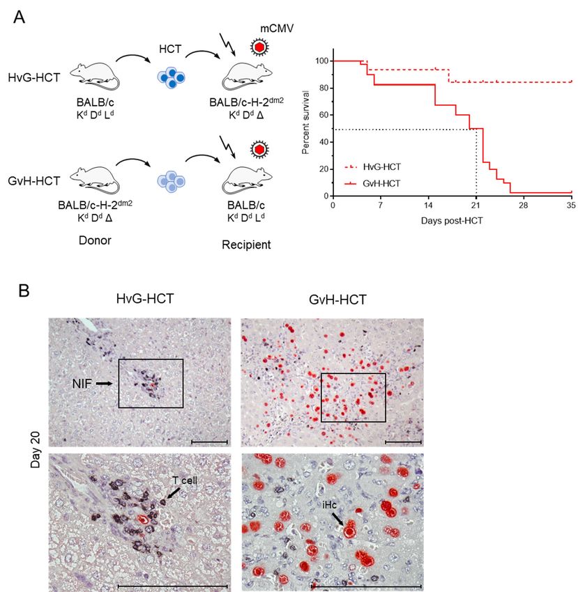
3. Summary
In GvH-HCT and infection with WT virus, reconstitution of viral peptide-specific
3. Summary
CD8+In T cells
GvH-HCTis generally at a low level,
and infection with and
WT the fewreconstitution
virus, cells present are of mostly of a functional
viral peptide-specific
avidity
CD8+ Tthatcellsisisnot high enough
generally forlevel,
at a low recognizing
and the infected
few cellscells, in which
present the presentation
are mostly of
of a functional
antigenic peptides is limited through the action of vRAP. As a result, unhindered
avidity that is not high enough for recognizing infected cells, in which the presentation virus
cell-to-cell
of antigenic spread can is
peptides lead to extensive
limited throughtissue lesions
the action (FigureAs
of vRAP. 4, aleft panel).
result, unhindered virus
cell-to-cell spread can lead to extensive tissue lesions (Figure 4, left panel).
Figure 4.
Figure Graphical abstract.
4. Graphical abstract. pMHC-I,
pMHC-I,peptide-loaded
peptide-loaded MHC-I
MHC-Imolecules.
molecules.TCR,
TCR,TTcell-receptor.
cell-receptor. NIF,
NIF, nodular
nodular inflammatory
inflammatory
focus, aa microanatomical
microanatomical structure
structure formed
formed by
by CD8 +
CD8 TT cells
cells that
that recognize
recognize infected
infected tissue
tissue cells,
cells, here
here represented
represented by
by
focus, +
hepatocytes. WT, wild-type mCMV. ∆vRAP, mCMV with deletion of genes coding for viral regulators of antigen presentation
(vRAP) that inhibit cell surface trafficking of pMHC-I complexes. Adapted from reference [42].
In HvG-HCT and, accordingly, in syngeneic HCT, after infection with WT virus,
reconstitution of viral peptide-specific CD8+ T cells is generally at an elevated level, and
many cells are of a functional avidity that is high enough to recognize infected cells under
conditions of limited peptide presentation. Moreover, once sensitized by TCR signaling,
the high-avidity cells release IFNγ, which is known to oppose vRAP function [48]. This
enhances antigen presentation and recruits even low-avidity CD8+ T cells to infected cells
for recognition. Combined, these mechanisms lead to NIF formation, prevention of viral
spread, and, eventually, to resolution of productive infection (Figure 4, center panel).
In GvH-HCT and infection with ∆vRAP virus, elevated presentation of antigenic pep-
tides can recruit even low-avidity CD8+ T cells into NIF for the recognition of infected cells,
prevention of viral spread, and termination of productive infection (Figure 4, right panel).
4. Conclusions
The data provide reasonable evidence to conclude that lethality associated with CMV
infection in recipients of allogeneic HCT is not caused by virus-mediated aggravation of
GvHD. Instead, it results from enhanced viral pathogenesis that reflects a failure in the
control of infection. Absence of protective antiviral CD8+ T cells does not result from
effector functions of GvH-reactive T cells. Rather, the failure in antiviral control can be
attributed to an inefficient lympho-hematopoietic reconstitution of high-avidity antiviral
CD8+ T cells capable of recognizing limited antigen presentation by infected cells. As the
mechanism of inefficient reconstitution of protective antiviral immunity, we propose thatViruses 2021, 13, 1530 8 of 10
transplantation tolerance against MHC or minor-H mismatch-antigens leads to a bystander,
“non-cognate tolerance” against viral antigens.
5. Lesson Learned for Better Clinical Understanding
In accordance with the two mouse models of allogeneic HCT discussed here, clinical
studies have revealed that a quantitative loss of hCMV-specific CD8+ T cells underlies the
uncontrolled virus replication after allogeneic HCT [49,50]. As discussed above (see the
introduction), no experimental animal model can cover human disease in all its complex-
ity with broad variance between individual recipients of allogeneic HCT and infection
with mostly uncharacterized virus variants. Obviously, in cases of a canonical GvHR/D,
particularly when mature donor T cells are not depleted to maintain a GvL effect for pre-
venting leukemia relapse, elimination of antigen presenting cells of the recipient necessarily
interferes with the reconstitution of protective antiviral T cells. Conversely, immunosup-
pressive treatment for GvHR/D prophylaxis also prevents, or at least critically delays, the
reconstitution of antiviral immunity.
Reducing the complexity and numbers of variables could be seen as a limitation
of experimental animal models, but it offers also the possibility of dissecting different
mechanisms that have a similar outcome and are thus difficult to distinguish by clinical
investigation. These two mouse models of allogeneic HCT contribute the important finding
that mismatches in major or minor histocompatibility antigens result in a failure of antiviral
CD8+ T-cell reconstitution, independent of GvHR/D.
This new insight into the mechanism of CMV disease after allogeneic HCT gives
a further argument for immunotherapy by adoptive transfer of antiviral CD8+ T cells,
ideally not by short-lived effector cells but by long-lived memory cells, to reconstitute
antiviral immunity enduringly. We, like many others, previously thought and argued that
immunotherapy serves, primarily, to bridge the “window of risk” in a transient phase of
immunodeficiency, until endogenous lympho-hematopoietic reconstitution of antiviral
CD8+ T cells takes over. Now we are faced with the possibility that, under conditions
when allogeneic HCT and CMV infection coincide, transplantation tolerance towards the
antigenic mismatch leads to a lasting “non-cognate tolerance” against CMV antigens. The
situation is reminiscent of the idea of using allogeneic HCT for inducing tolerance towards
a subsequent solid organ allograft from the same donor (reviewed in [32]), though with the
difference that non-cognate tolerance towards viral antigens is adverse in its consequences.
We propose that this mechanism might leave recipients, who recovered from allogeneic
HCT, in a state of long-lasting CMV-selective immunodeficiency.
Author Contributions: Writing—original draft preparation, M.J.R.; writing—review and editing,
R.H. and N.A.W.L. All authors have read and agreed to the published version of the manuscript.
Funding: Research from the authors was funded by the Deutsche Forschungsgemeinschaft, Col-
laborative Research Center (CRC) 1292: individual projects TP11 ‘Viral evasion of innate and adap-
tive immune cells and inbetweeners’ (M.J.R. and N.A.W.L.) and TP14 ‘Immunomodulation of cy-
tomegalovirus latency and reactivation by regulatory T cells and dendritic cells’ (R.H.).
Conflicts of Interest: The authors declare no conflict of interest.
References
1. Davison, A.J.; Holton, M.; Dolan, A.; Dargan, D.J.; Gatherer, D.; Hayward, G.S. Comparative genomics of primate cy-
tomegaloviruses. In Cytomegaloviruses: From Molecular Pathogenesis to Intervention; Reddehase, M.J., Ed.; Caister Academic
Press: Norfolk, UK, 2013; Volume I, pp. 1–22.
2. Cannon, M.J.; Grosse, S.D.; Fowler, K.B. The epidemiology and public health impact of congenital cytomegalovirus infection. In
Cytomegaloviruses: From Molecular Pathogenesis to Intervention; Reddehase, M.J., Ed.; Caister Academic Press: Norfolk, UK, 2013;
Volume II, pp. 26–48.
3. Tabata, T.; Petitt, M.; Fang-Hoover, J.; Pereira, L. Survey of cellular immune responses to human cytomegalovirus infection in the
microenvironment of the uterine-placental interface. Med. Microbiol. Immunol. 2019, 208, 475–485. [CrossRef] [PubMed]
4. Britt, W. Manifestations of human cytomegalovirus infection: Proposed mechanisms of acute and chronic disease. Curr. Top.
Microbiol. Immunol. 2008, 325, 417–470.Viruses 2021, 13, 1530 9 of 10
5. Ho, M. The history of cytomegalovirus and its diseases. Med. Microbiol. Immunol. 2008, 197, 65–73. [CrossRef] [PubMed]
6. Boppana, S.B.; Britt, W.J. Synopsis of clinical aspects of human cytomegalovirus disease. In Cytomegaloviruses: From Molecular
Pathogenesis to Intervention; Reddehase, M.J., Ed.; Caister Academic Press: Norfolk, UK, 2013; Volume I, pp. 1–25.
7. Adler, S.P.; Nigro, G. Clinical cytomegalovirus research: Congenital infection. In Cytomegaloviruses: From Molecular Pathogenesis to
Intervention; Reddehase, M.J., Ed.; Caister Academic Press: Norfolk, UK, 2013; Volume II, pp. 55–73.
8. Avery, R.K. Clinical cytomegalovirus research: Thoracic organ transplantation. In Cytomegaloviruses: From Molecular Pathogenesis
to Intervention; Reddehase, M.J., Ed.; Caister Academic Press: Norfolk, UK, 2013; Volume II, pp. 286–300.
9. Emery, V.C.; Milne, R.S.B.; Griffiths, P.D. Clinical cytomegalovirus research: Liver and kidney transplantation. In Cytomegaloviruses:
From Molecular Pathogenesis to Intervention; Reddehase, M.J., Ed.; Caister Academic Press: Norfolk, UK, 2013; Volume II,
pp. 301–311.
10. Reddehase, M.J.; Lemmermann, N.A.W. Cellular reservoirs of latent cytomegaloviruses. Med. Microbiol. Immunol. 2019,
208, 391–403. [CrossRef]
11. Riddell, S.R. Pathogenesis of cytomegalovirus pneumonia in immunocompromised hosts. Semin. Respir. Infect. 1995, 10, 199–208.
[PubMed]
12. Seo, S.; Boeckh, M. Clinical cytomegalovirus research: Hematopoietic cell transplantation. In Cytomegaloviruses: From Molecular
Pathogenesis to Intervention; Reddehase, M.J., Ed.; Caister Academic Press: Norfolk, UK, 2013; Volume II, pp. 337–353.
13. Chemaly, R.F.; Chou, S.; Einsele, H.; Griffiths, P.; Avery, R.; Razonable, R.R.; Mullane, K.M.; Kotton, C.; Lundgren, J.; Komatsu,
T.E.; et al. Definitions of resistant and refractory cytomegalovirus infection and disease in transplant recipients for use in clinical
trials. Clin. Infect. Dis. 2019, 68, 1420–1426. [CrossRef]
14. Riddell, S.R.; Watanabe, K.S.; Goodrich, M.; Li, C.R.; Agha, M.E.; Greenberg, P.D. Restoration of viral immunity in immunodefi-
cient humans by the adoptive transfer of T cell clones. Science 1992, 257, 238–241. [CrossRef] [PubMed]
15. Walter, E.A.; Greenberg, P.D.; Gilbert, M.J.; Finch, R.J.; Watanabe, K.S.; Thomas, D.; Riddell, S.R. Reconstitution of cellular
immunity against cytomegalovirus in recipients of allogeneic bone marrow by transfer of T-cell clones from the donor. N. Engl.
J. Med. 1995, 333, 1038–1044. [CrossRef]
16. Einsele, H.; Roosnek, E.; Rufer, N.; Sinzger, C.; Riegler, S.; Löffler, J.; Grigoleit, U.; Moris, A.; Rammensee, H.G.; Kanz, L.; et al.
Infusion of cytomegalovirus (CMV)-specific T cells for the treatment of CMV infection not responding to antiviral chemotherapy.
Blood 2002, 99, 3916–3922. [CrossRef]
17. Feuchtinger, T.; Opherk, K.; Bethge, W.A.; Topp, M.S.; Schuster, F.R.; Weissinger, E.M.; Mothy, M.; Or, R.; Mashan, M.; Schumm,
M.; et al. Adoptive transfer of pp65-specific T cells for the treatment of chemorefractory cytomegalovirus disease or reactivation
after haploidentical and matched unrelated stem cell transplantation. Blood 2010, 116, 4360–4367. [CrossRef] [PubMed]
18. Bao, L.; Cowan, M.J.; Dunham, K.; Horn, B.; McGuirk, J.; Gilman, A.; Lucas, K.G. Adoptive immunotherapy with CMV-specific
cytotoxic T lymphocytes for stem cell transplant patients with refractory CMV infections. J. Immunother. 2012, 35, 293–298.
[CrossRef] [PubMed]
19. Odendahl, M.; Grigoleit, G.U.; Bönig, H.; Neuenhahn, M.; Albrecht, J.; Anderl, F.; Germeroth, L.; Schmitz, M.; Bornhäuser,
M.; Einsele, H.; et al. Clinical-scale isolation of ’minimally manipulated’ cytomegalovirus-specific donor lymphocytes for the
treatment of refractory cytomegalovirus disease. Cytotherapy 2014, 16, 1245–1256. [CrossRef] [PubMed]
20. Pei, X.Y.; Zhao, X.Y.; Chang, Y.J.; Liu, J.; Xu, L.P.; Wang, Y.; Zhang, X.H.; Han, W.; Chen, Y.H.; Huang, X.J. Cytomegalovirus-
specific T-cell transfer for refractory cytomegalovirus infection after haploidentical stem cell transplantation: The quantitative
and qualitative immune recovery for cytomegalovirus. J. Infect. Dis. 2017, 216, 945–956. [CrossRef] [PubMed]
21. Kaeuferle, T.; Krauss, R.; Blaeschke, F.; Willier, S.; Feuchtinger, T. Strategies of adoptive T -cell transfer to treat refractory viral
infections post allogeneic stem cell transplantation. J. Hematol. Oncol. 2019, 12, 13. [CrossRef] [PubMed]
22. Rappeport, J.; Mihm, M.; Reinherz, E.; Lopansri, S.; Parkman, R. Acute graft-versus-host disease in recipients of bone-marrow
transplants from identical twin donors. Lancet 1979, 2, 717–720. [CrossRef]
23. Applebaum, F.R.; Meyers, J.D.; Fefer, A.; Fluornoy, N.; Cheever, M.A.; Greenberg, P.D.; Hackman, R.; Thomas, E.D. Nonbacterial
nonfungal pneumonia following marrow transplantation in 100 identical twins. Transplantation 1982, 33, 265–268.
24. Wingard, J.R.; Chen, D.Y.; Burns, W.H.; Fuller, D.J.; Braine, H.G.; Yeager, A.M.; Kaiser, H.; Burke, P.J.; Graham, M.L.; Santos,
G.W.; et al. Cytomegalovirus infection after autologous bone marrow transplantation with comparison to infection after allogeneic
bone marrow transplantation. Blood 1988, 71, 1432–1437. [CrossRef] [PubMed]
25. Meyers, J.D.; Flournoy, N.; Thomas, E.D. Nonbacterial pneumonia after allogeneic marrow transplantation: A review of ten years’
experience. Rev. Infect. Dis. 1982, 4, 1119–1132. [CrossRef]
26. Bleakley, M.D.; Brown, M.; Riddell, S. Human CD8+ minor histocompatibility antigen specific cytotoxic T lymphocyte clones
can be generated by primary in vitro stimulation of naїve T cells with dendritic cells from HLA identical siblings. Blood 2004,
104, 2116. [CrossRef]
27. Stern, L.; Withers, B.; Avdic, S.; Gottlieb, D.; Abendroth, A.; Blyth, E.; Slobedman, B. Human cytomegalovirus latency and
reactivation in allogeneic hematopoietic stem cell transplant recipients. Front. Microbiol. 2019, 10, 1186. [CrossRef]
28. Hartwig, U.F.; Nonn, M.; Khan, S.; Link, I.; Huber, C.; Herr, W. Depletion of alloreactive donor T lymphocytes by CD95-mediated
activation-induced cell death retains antileukemic, antiviral, and immunoregulatory T cell immunity. Biol. Blood Marrow
Transplant. 2008, 14, 99–109. [CrossRef] [PubMed]Viruses 2021, 13, 1530 10 of 10
29. Nguyen, H.; Alawieh, A.; Bastian, D.; Kuril, S.; Dai, M.; Daenthanasanmak, A.; Zhang, M.; Iamsawat, S.; Schutt, S.D.; Wu, Y.; et al.
Targeting the complement alternative pathway permits graft versus leukemia activity while preventing graft versus host disease.
Clin. Cancer Res. 2020, 26, 3481–3490. [CrossRef]
30. Deeg, H.J. Chimerism, the microenvironment and control of leukemia. Front. Immunol. 2021, 12, 652105. [CrossRef]
31. Emery, V.C. Relative importance of cytomegalovirus load as a risk factor for cytomegalovirus disease in the immunocompromised
host. Monogr. Virol. 1998, 21, 288–301.
32. Sykes, M. Hematopoietic cell transplantation for tolerance induction: Animal models to clinical trials. Transplantation 2009,
87, 309–316. [CrossRef] [PubMed]
33. Reddehase, M.J.; Lemmermann, N.A.W. Mouse model of cytomegalovirus disease and immunotherapy in the immunocompro-
mised host: Predictions for medical translation that survived the “test of time”. Viruses 2018, 10, 693. [CrossRef] [PubMed]
34. Reddehase, M.J.; Weiland, F.; Münch, K.; Jonjic, S.; Lüske, A.; Koszinowski, U.H. Interstitial murine cytomegalovirus pneumonia
after irradiation: Characterization of cells that limit viral replication during established infection of the lungs. J. Virol. 1985,
55, 264–273. [CrossRef]
35. Reddehase, M.J.; Mutter, W.; Koszinowski, U.H. In vivo application of recombinant interleukin 2 in the immunotherapy of
established cytomegalovirus infection. J. Exp. Med. 1987, 165, 650–656. [CrossRef]
36. Ebert, S.; Podlech, J.; Gillert-Marien, D.; Gergely, K.M.; Büttner, J.K.; Fink, A.; Freitag, K.; Thomas, D.; Reddehase, M.J.; Holtappels,
R. Parameters determining the efficacy of adoptive CD8 T-cell therapy of cytomegalovirus infection. Med. Microbiol. Immunol.
2012, 201, 527–539. [CrossRef] [PubMed]
37. Holtappels, R.; Ebert, S.; Podlech, J.; Fink, A.; Böhm, V.; Lemmermann, N.A.W.; Freitag, K.; Renzaho, A.; Thomas, D.; Reddehase,
M.J. Murine model for cytoimmunotherapy of CMV disease after haematopoietic cell transplantation. In Cytomegaloviruses: From
Molecular Pathogenesis to Intervention; Reddehase, M.J., Ed.; Caister Academic Press: Norfolk, UK, 2013; Volume II, pp. 352–379.
38. Reddehase, M.J. Mutual interference between cytomegalovirus and reconstitution of protective immunity after hematopoietic cell
transplantation. Front. Immunol. 2016, 7, 294. [CrossRef] [PubMed]
39. Adler, S.P.; Reddehase, M.J. Pediatric roots of cytomegalovirus recurrence and memory inflation in the elderly. Med. Microbiol.
Immunol. 2019, 208, 323–328. [CrossRef]
40. Adler, B.; Sinzger, C. Cytomegalovirus interstrain variance in cell type tropism. In Cytomegaloviruses: From Molecular Pathogenesis
to Intervention; Reddehase, M.J., Ed.; Caister Academic Press: Norfolk, UK, 2013; Volume I, pp. 297–321.
41. Wilkinson, G.W.; Davison, A.J.; Tomasec, P.; Fielding, C.A.; Aicheler, R.; Murrell, I.; Seirafian, S.; Wang, E.C.; Weekes, M.; Lehner,
P.J.; et al. Human cytomegalovirus: Taking the strain. Med. Microbiol. Immunol. 2015, 204, 273–284. [CrossRef] [PubMed]
42. Holtappels, R.; Schader, S.I.; Oettel, O.; Podlech, J.; Seckert, C.K.; Reddehase, M.J.; Lemmermann, N.A.W. Insufficient antigen
presentation due to viral immune evasion explains lethal cytomegalovirus organ disease after allogeneic hematopoietic cell
transplantation. Front. Cell. Infect. Microbiol. 2020, 10, 157. [CrossRef]
43. Gezinir, E.; Podlech, J.; Gergely, K.M.; Becker, S.; Reddehase, M.J.; Lemmermann, N.A.W. Enhancement of antigen presentation by
deletion of viral immune evasion genes prevents lethal cytomegalovirus disease in minor histocompatibility antigen-mismatched
hematopoietic cell transplantation. Front. Cell. Infect. Microbiol. 2020, 10, 279. [CrossRef] [PubMed]
44. Holtappels, R.; Freitag, K.; Renzaho, A.; Becker, S.; Lemmermann, N.A.W.; Reddehase, M.J. Revisiting CD8 T-cell ‘memory
inflation’: New insights with implications for cytomegaloviruses as vaccine vectors. Vaccines 2020, 8, 402. [CrossRef]
45. Roopenian, D.; Choi, E.Y.; Brown, A. The immunogenomics of minor histocompatibility antigens. Immunol. Rev. 2002, 190, 86–94.
[CrossRef] [PubMed]
46. Hasan, M.; Krmpotic, A.; Ruzsics, Z.; Bubic, I.; Lenac, T.; Halenius, A.; Loewendorf, A.; Messerle, M.; Hengel, H.; Jonjic, S.; et al.
Selective down-regulation of the NKG2D ligand H60 by mouse cytomegalovirus m155 glycoprotein. J. Virol. 2005, 79, 2920–2930.
[CrossRef] [PubMed]
47. Wood, K.J.; Bushell, A.; Hester, J. Regulatory immune cells in transplantation. Nat. Rev. Immunol. 2012, 12, 417–430. [CrossRef]
48. Fink, A.; Lemmermann, N.A.; Gillert-Marien, D.; Thomas, D.; Freitag, K.; Böhm, V.; Wilhelmi, V.; Reifenberg, K.; Reddehase, M.J.;
Holtappels, R. Antigen presentation under the influence of ’immune evasion’ proteins and its modulation by interferon-gamma:
Implications for immunotherapy of cytomegalovirus infection with antiviral CD8 T cells. Med. Microbiol. Immunol. 2012, 201,
513–525. [CrossRef] [PubMed]
49. Gratama, J.W.; van Esser, J.W.; Lamers, C.H.; Tournay, C.; Löwenberg, B.; Bolhuis, R.L.; Cornelissen, J.J. Tetramer-based
quantification of cytomegalovirus (CMV)-specific CD8+ T lymphocytes in T-cell-depleted stem cell grafts and after transplantation
may identify patients at risk for progressive CMV infection. Blood 2001, 98, 1358–1364. [CrossRef]
50. Cwynarski, K.; Ainsworth, J.; Cobbold, M.; Wagner, S.; Mahendra, P.; Apperley, J.; Goldman, J.; Craddock, C.; Moss, P.A. Direct
visualization of cytomegalovirus-specific T-cell reconstitution after allogeneic stem cell transplantation. Blood 2001, 97, 1232–1240.
[CrossRef] [PubMed]You can also read