ALTERATION OF CLINICAL OUTCOME AND HISTOPATHOLOGY OF YELLOW FEVER VIRUS INFECTION IN A HAMSTER MODEL BY PREVIOUS INFECTION WITH HETEROLOGOUS ...
←
→
Page content transcription
If your browser does not render page correctly, please read the page content below
Am. J. Trop. Med. Hyg., 68(6), 2003, pp. 695–703
Copyright © 2003 by The American Society of Tropical Medicine and Hygiene

          ALTERATION OF CLINICAL OUTCOME AND HISTOPATHOLOGY OF YELLOW
          FEVER VIRUS INFECTION IN A HAMSTER MODEL BY PREVIOUS INFECTION
                          WITH HETEROLOGOUS FLAVIVIRUSES
                SHU-YUAN XIAO, HILDA GUZMAN, AMELIA P. A. TRAVASSOS DA ROSA, HONG-BING ZHU, AND
                                                            ROBERT B. TESH
          Department of Pathology and Center for Tropical Diseases, and Department of Internal Medicine, University of Texas Medical
                                                          Branch, Galveston, Texas

          Abstract. Using a recently described hamster model of severe yellow fever (YF), we examined the hypothesis that
      prior infection with heterologous flaviviruses protects against severe or fatal YF. Hamsters were singly or sequentially
      infected with Japanese encephalitis, St. Louis encephalitis, West Nile, and/or dengue-1 viruses, and then challenged with
      a virulent strain of yellow fever virus (YFV). In contrast to control (naive) hamsters, many of which appeared clinically
      ill or died after YFV infection, the flavivirus-immune animals remained asymptomatic. The flavivirus-immune hamsters
      also had a reduced viremia and lower serum levels of alanine aminotransferase and total bilirubin, compared with naive
      hamsters, following YFV infection. Histologically, livers of animals in the flavivirus-immune and control groups showed
      comparable levels of multifocal necrapoptosis. However, steatosis was not observed in the flavivirus-immune animals,
      whereas naive hamsters developed extensive microvesicular steatosis in the liver following YFV infection. These findings
      suggest that hepatocytic steatosis is an adverse microscopic feature associated with severe disease in YFV infection. Our
      experimental results support earlier anecdotal reports that prior exposure of humans to heterologous flaviviruses
      reduces subsequent risk of fatal YFV infection.

                            INTRODUCTION                                          cine strain of Japanese encephalitis virus (JEV);10 the New
                                                                                  York 385-99 strain of West Nile virus (WNV);11 and the BeAr
   Monath1,2 and others3,4 have suggested that immunologic                        23379 strain of St. Louis encephalitis virus (SLEV).12
cross-protection by heterologous flaviviruses may provide                            The hamsters used were juvenile and adult female Syrian
partial protection against yellow fever (YF). In the 19th cen-                    golden hamsters (Mesocricetus auratus) obtained from Harlan
tury, it was noted that indentured laborers from India and                        Sprague Dawley (Indianapolis, IN). Animals were cared for
British troops who had served in India were less likely to                        in accordance with the guidelines of the Committee on Care
contract severe YF during epidemics of the disease in Africa.3                    and Use of Laboratory Animals (Institute of Laboratory Ani-
Presumably, many of these people had been previously in-                          mal Resources, National Research Council) under an animal
fected with dengue (DEN) viruses, and possibly West Nile                          use protocol approved by the University of Texas Medical
(WN) or Japanese encephalitis (JE) viruses, during their resi-                    Branch. All work with infected animals was carried out in
dence in India. A retrospective investigation of a YF epi-                        biosafety level-3 facilities.
demic in Gambia in 1978 revealed that the inapparent-to-                             Virus assay. Virus levels in blood of hamsters infected with
apparent infection ratio for YF was significantly higher in                       YFV were titrated in cultures of the C6/36 clone of Aedes
individuals with prior exposure to heterologous flaviviruses.5                    albopictus cells, as described previously.7 The presence or
In addition, there is limited experimental evidence from stud-                    absence of viral antigen by immunofluorescence was used as
ies in monkeys supporting the concept that immunity to DEN                        the end-point. The YFV titers were calculated as the tissue
and other flaviviruses may diminish the viremia and severity                      culture infectious dose50 (TCID50) per milliliter of specimen
of yellow fever virus (YFV) infection.4,6                                         by the method of Reed and Muench.13
   To test the hypothesis that prior infection with heterolo-                        Antibody determinations. Serum antibodies to YFV and
gous flaviviruses protects against severe or fatal YF, a series                   the other four flaviviruses were measured by a hemaggluti-
of laboratory experiments were done using a recently de-                          nation inhibition (HI) test. Antigens for the HI test were
scribed hamster model of the disease.7,8 The present paper                        prepared from brains of newborn mice inoculated intracere-
reports our results that support the earlier observations and                     brally with each of the viruses; the infected brains were pro-
hypothesis that heterologous flavivirus antibodies are protec-                    cessed by the sucrose-acetone extraction method.14 Hamster
tive against severe YF. In addition to reduced levels of vire-                    sera were tested by HI at serial two-fold dilutions from 1:20 to
mia and less severe alternation in liver function, the flavivi-                   1:5,120 at pH 6.6 (WNV, JEV, DENV, and SLEV) or 6.4
rus-immune hamsters did not develop microvesicular steato-                        (YFV) with four units of antigen and a 1:200 dilution of goose
sis, one of the classic pathologic features of severe YFV                         erythrocytes, following established protocols.14
infection. The significance of this lack of steatosis in the                         Serum levels of alanine transaminase and bilirubin. Deter-
course of YFV infection is discussed.                                             minations of total bilirubin (TB) and of alanine aminotrans-
                                                                                  ferase (ALT) levels were done on fresh serum from clotted
                  MATERIALS AND METHODS                                           blood, using commercial kits (Total Bilirubin® and Infinity
                                                                                  ALT®; Sigma Diagnostics, St. Louis, MO), according to the
   Viruses and animals. Five flaviviruses were used in this                       manufacturer’s instructions.
study: the Jimenez hamster-virulent (10th passage) strain of                         Experimental infection of animals. Four separate experi-
YFV;7 the Mochizuki high mouse passage (178×) strain of                           ments were done. The viruses used and the sequence in which
dengue-1 virus (DENV);9 the live attenuated SA14-2-8 vac-                         they were administered to the hamster were as follows: Ex-
                                                                            695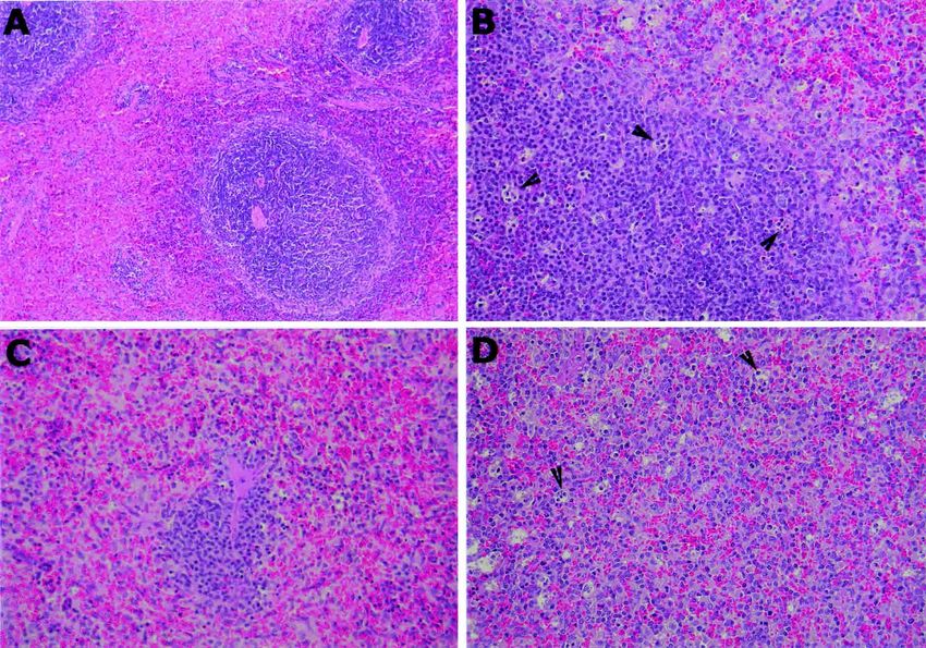
696                                                      XIAO AND OTHERS

periment 1, YFV only (control); Experiment 2, JEV followed                                  RESULTS
by YFV; Experiment 3, DENV, WNV, and YFV in sequence;
and Experiment 4, SLEV, WNV, and YFV in sequence. The                 Establishment of heterologous flavivirus infection(s) in
interval between sequential virus injections ranged from four      hamsters, their antibody response, and subsequent challenge
to six weeks. Hamsters were infected by the intracerebral,         with YFV. Experiment 1. Infection of naive hamsters with
intraperitoneal, or subcutaneous routes, depending on the in-      YFV (Group YF control). Sixteen adult hamsters (11−12
fectivity and virulence of the virus for hamsters. For example,    weeks old) were inoculated intraperitoneally with 106.0
it was necessary to give DENV intracerebrally to immunize          TCID50 of YFV. The animals in this group showed clinical
hamsters, WNV and YFV were given intraperitoneally, and            signs of severe YF (lethargy, anorexia, tremors) on the fifth
JEV and SLEV were inoculated subcutaneously. The sched-            and sixth days after infection, as described previously.7 Eight
ule for each set of infection/challenge experiments are de-        of the animals were bled daily for six consecutive days to
scribed in more detail in the Results. In each experiment,         monitor the level of viremia. On the sixth day after infection,
YFV was the final (challenge) virus administered. Following        these eight animals were exsanguinated to obtain serum for
inoculation of YFV, the hamsters were bled daily for six con-      liver function studies, and tissue samples were taken at nec-
secutive days to monitor the level of viremia. On the sixth day    ropsy for histopathologic examination. The remaining eight
after infection with YFV, the hamsters were exsanguinated          hamsters were observed for 14 days; two of the animals died
and killed. Sera were collected for biochemical studies, in-       on sixth and seventh days, giving a mortality rate (25%) simi-
cluding TB and ALT, and samples of liver, spleen, and for          lar to that previously described.7
                                                                      Experiment 2. Infection with JEV, followed by YFV (Group
some animals, pancreas, were removed for histopathologic
                                                                   JE/YF). Ten subadult hamsters (5−6 weeks old) were inocu-
examination. Day 6 was selected as the time to evaluate the
                                                                   lated subcutaneously with JEV strain SA14-2-8. Twenty-eight
degree of organ pathology following challenge with YFV,
                                                                   days later, the animals were bled to determine the HI anti-
since previous studies8 demonstrated that maximum tissue
                                                                   body titers to JEV and YFV antigens, which are shown in
damage occurs on the sixth post-infection day in the hamster
                                                                   Table 1. The hamsters in this experiment all developed a
model of yellow fever.
                                                                   strong HI antibody response to JEV antigen (1:640−1:1,280),
   Histologic evaluation. Samples of fresh liver, spleen, and
                                                                   but they also had lower levels of cross-reacting antibodies to
pancreas were fixed in 10% neutral-buffered formalin for 24
                                                                   YFV antigen (1:80−1:160). Thirty-one days after JEV infec-
hours and then in 70% alcohol for 1−2 days, before being
                                                                   tion, the 10 hamsters were inoculated intraperitoneally with
processed for routine paraffin embedding. Four to five             106.0 TCID50 of YFV and were then bled for six consecutive
micron−thick sections were made and stained by the hema-           days to determine the level of the resulting YFV viremia. Six
toxylin and eosin method. Some sections were also stained          days after infection with YFV, the hamsters were killed and
using the reticulin and Masson’s trichrome methods. Tissue         necropsied to obtain tissue samples for histopathologic analy-
sections were examined with an Olympus (Melville, NY)              sis. No sera were obtained for liver function tests from this
BX51 microscope equipped with UPlan/Apo lenses. For                group of animals. The animals in this group did not appear
semi-quantitative analysis, a scheme described in a previous       sick when killed on the sixth day of YFV infection.
report was used,8 with modification. In analyzing histopathol-        Experiment 3. Sequential infection with DENV, WNV, and
ogy of the liver, the degree of inflammatory infiltration, hepa-   YFV (Group DEN/WN/YF). Twenty-six juvenile hamsters
tocytic necrapoptosis, and steatosis were evaluated; the sever-    (3−4 weeks old) were inoculated intracerebrally with the Mo-
ity of each parameter was graded on a subjective scale of 0 to     chizuki strain of DENV. This virus strain produces an asymp-
4. For the spleen, a subjective impression of none, minimal,       tomatic infection in juvenile and adult hamsters. Twenty-
mild, moderate, and severe, was graded as 0, 1, 2, 3, and 4,       seven days after DENV infection, 10 of the animals were bled
respectively. The parameters assessed were lymphoid hyper-         to determine their immune status. At this time, all of the
plasia, lymphoid (white pulp) depletion, and macrophage            hamsters had HI antibodies to DENV, with titers ranging
proliferation (activation), as previously described.8 Pancreas     from 1:160 to 1:640. The 26 animals were then inoculated
was also examined in some of the animals; in this organ only       intraperitoneally with 104.0 TCID50 of WNV strain New York
the presence or absence of parenchymal necrosis was re-            385-99. Thirty-one days after WNV infection, 18 of the ham-
corded.                                                            sters were inoculated intraperitoneally with 106.0 TCID50 of
   In situ TUNEL assay. The terminal deoxynucleotidyl trans-       YFV and 10 were bled for six consecutive days to determine
ferase-mediated dUTP nick-end labeling (TUNEL) tech-               the level of viremia. Ten of the 18 YFV-infected animals were
nique was used to assay for apoptosis in sections of hamster       killed on the sixth day for histopathologic analysis; the re-
liver tissue. The ApopTag peroxidase kit (Intergen Company,        maining eight survived and did not appear ill. The other eight
Purchase, NY) was used, following the manufacturer’s in-           DENV/WNV-infected animals did not receive YFV and were
structions, as previously described.8,11,15 For quantification,    used as YFV-negative controls.
10 random high power fields were examined, using a 40×                Experiment 4. Sequential infection with SLEV, WNV, and
objective, and the ApopTag-positive nuclei were counted.           YFV (Group SLE/WN/YF). An additional 26 juvenile ham-
Due to occasional non-specificity of the in situ TUNEL assay,      sters were inoculated subcutaneously with 106.0 TCID50 of
morphologically recognizable necrotic cells or foci were ex-       SLEV strain BeAr 23379. Thirty days later, the animals were
cluded from counting.                                              inoculated intraperitoneally with 104.0 TCID50 of WNV strain
   While conducting the microscopic examinations of the he-        New York 385-99. Forty-five days after WNV infection, nine
matoxylin and eosin−stained sections and the TUNEL-                of the hamsters in this group were bled to determine their
stained sections, the observer was blinded as to the group         flavivirus HI antibody titers. Eighteen of the original 26 ham-
identification of the specimens.                                   sters were then given 106.0 TCID50 of YFV. These animals
HETEROLOGOUS FLAVIVIRUS INFECTIONS                                                      697

                             TABLE 1                                                       and TB levels in the naive animals were markedly elevated
Hemagglutination inhibition (HI) antibody responses of hamsters to                         (Figure 2).
 Japanese encephalitis virus (JEV) and yellow fever virus (YFV)                               Despite the presence of pre-existing antibodies to heterolo-
 antigens 28 days after infection with JEV
                                                                                           gous flavivirus (Tables 1 and 2), the hamsters in Experiments
                                               HI antibody titers* against                 2, 3, and 4 (JE/YF, DEN/WN/YF, and SLE/WN/YF, respec-
       Hamster no.                     JEV antigen                   YFV antigen           tively) still developed a significant YF viremia after challenge
                                                                                           with YFV (Figure 1). The level and pattern of viremia among
          8081                            1,280                          160
          8082                              640                           80               animals in these three experiments were similar; the peak
          8083                              640                          160               virus titers were lower and virus replication was delayed,
          8084                              640                           80               compared with that of naive hamsters after YFV challenge.
          8085                            1,280                          160                  The mean serum ALT and TB levels in each experimental
          8096                              640                          160
          8097                              640                           80               group on the sixth day after YFV infection are shown in
          8098                              640                           80               Figure 2. Following challenge with YFV, hamsters previously
          8099                              640                          160               infected with SLEV and WNV (Experiment 4) developed
          8100                              640                          160               elevated levels of ALT and TB. However, the magnitude of
  * Reciprocal of highest positive serum dilution.
                                                                                           ALT and TB alteration was less than that in naive hamsters
                                                                                           infected with YFV (Figure 2). The mean ALT and TB values
                                                                                           in the DEN/WN/YF group of animals (Experiment 3) were
were bled daily for six days, as described earlier, to determine
                                                                                           also lower than the values obtained in the YFV control ham-
the level and duration of the subsequent YFV viremia. On the
sixth day after inoculation of YFV, 10 of the hamsters were                                sters and were comparable to liver function results obtained
exsanguinated and necropsied. The other eight YFV-infected                                 with the hamsters in Experiment 4 (SLE/WN/YF). In con-
hamsters survived infection and did not appear ill. The eight                              trast, the ALT and TB levels in the eight control animals
remaining hamsters from this group of 26 that had been pre-                                (SLE/WN only) were within normal limits for hamsters, in-
viously infected with SLEV and WNV, but not YFV, were                                      dicating that the alteration of liver function was due to YFV
exsanguinated and necropsied to serve as YFV-negative con-                                 infection and not to the prior SLEV and WNV infections.
trols.                                                                                        Histopathologic changes in hamsters challenged with YFV
   The HI antibody titers in the eight hamsters from Experi-                               after previous infection with heterologous flaviviruses. Ham-
ment 4 immediately before and 6 days after infection with                                  sters infected only by DENV and WNV or by SLEV and
YFV are shown in Table 2. Following immunization with                                      WNV (YFV-negative controls in Experiments 3 and 4, re-
SLEV and WNV, these animals already had HI antibody ti-                                    spectively) showed no histologic abnormalities in the liver,
ters (range ⳱ 1:160−1:640) against YFV antigen. Six days                                   spleen, kidneys, lymph nodes or heart (Figure 3A and B). In
after infection with YFV, the animals had a further increase in                            contrast, the YFV-infected animals in Experiments 3 and 4
their flavivirus HI antibody titers (Table 2), indicating an                               showed mild-to-moderate lobular inflammatory cell infiltra-
amnestic type of antibody response.                                                        tions in the liver on the sixth post-infection day, with scattered
   Reduced YFV viremia and alteration of liver function                                    but frequent hepatocytic necrapoptosis (Figure 3C−F). In
in hamsters previously infected with heterologous flavivi-                                 most of these hamsters, the liver pathology was characterized
ruses. As shown in Figure 1, YFV was present in the blood of                               by evenly distributed (scattered) foci of inflammation in the
the flavivirus-naive (control) hamsters within 24 hours after                              form of microgranulomas, which consisted of small clusters of
infection, peaked on day 3 (mean titer ⳱ 107.5 TCID50/mL),                                 Kupffer cells, lymphocytes, and a few eosinophils. Some of
and had largely disappeared by day 6. The disappearance of                                 these microgranulomas contained an apoptotic hepatocyte,
infectious virus from peripheral blood corresponds with the                                which appeared as condensed pink cytoplasm, with or without
development of HI and IgM antibodies in the hamster model                                  a pyknotic nucleus (Councilman body). In a few cases, the
of yellow fever.7 Six days after YFV infection, the serum ALT                              inflammation was less discrete in distribution but more evenly
                                                                                           scattered in the sinusoidal spaces. Some portal tracts also
                                                                                           had inflammatory cell infiltration, but the piecemeal necro-
                             TABLE 2
                                                                                           sis or interface hepatitis was not noted. The YF control
Hemagglutination-inhibition (HI) antibody titers* in St. Louis en-
 cephalitis virus- and West Nile virus-immune hamsters of experi-                          hamsters also exhibited diffuse microvesicular steatosis six
 ment 4, immediately before and 6 days after yellow fever virus                            days after challenge with YFV (Figure 3G and H), as previ-
 (YFV) challenge                                                                           ously described.8 In contrast, no significant steatosis was
                                                                                           found in livers of hamsters in the DEN/WN/YF and SLE/WN/
                     Before YFV challenge                Six days after YFV challenge
Hamster                                                                                    YF groups (Figures 3C−F and 4). A semi-quantitative histo-
  no.        YFV Ag       WNV Ag        SLEV Ag      YFV Ag       WNV Ag       SLEV Ag     logic scoring showed no difference in severity of inflammation
H-8101         160            640           640         640         1,280       1,280      or necrapoptosis between the YF control hamsters and the
H-8102         160            640           640       1,280         2,560       1,280      YFV-infected hamsters of Experiments 3 and 4 (Figure 4).
H-8103         320          1,280         1,280       2,560         2,560       2,560         Histologic changes in the spleen were similar among ani-
H-8104         640          1,280         1,280       2,560         2,560       2,560
H-8105         320          1,280         1,280       1,280         2,560       2,560      mals in all four experiments, but with certain differences in
H-8107         640          1,280         1,280       1,280         1,280         640      severity. While the non-YFV-challenged hamsters in Experi-
H-8108         320          1,280         1,280       2,560         2,560       2,560      ments 3 and 4 showed no pathology in the spleen (Figure 5A),
H-8109         320          1,280         1,280       2,560         2,560       2,560      all of the animals challenged with YF virus showed hyperpla-
  * Antibody titers against corresponding antigens: YFV Ag ⳱ yellow fever viral antigen;   sia of the lymphoid areas, as characterized by immunoblastic
WNV Ag ⳱ West Nile viral antigen; SLEV Ag ⳱ St. Louis encephalitis viral antigen. HI
antibody titers are expressed as the reciprocal of highest positive serum dilution.        cell proliferation (Figure 5B) or expansion of the lymphoid
698                                                           XIAO AND OTHERS

  FIGURE 1. Mean daily levels of viremia in the four groups of hamsters after challenge with yellow fever (YF) virus. SLE ⳱ St. Louis
encephalitis; WN ⳱ West Nile; D1 ⳱ dengue-1; JE ⳱ Japanese encephalitis; TCID50 ⳱ tissue culture infectious dose50. Bars show the SD.

follicles. At the same time, there was splenic macrophage                  Pancreatic tissue was also examined in some of the ham-
hyperplasia (Figure 5D). Among hamsters in the YF control               sters. As previously reported,8 moderate pancreatic necrosis
group (Experiment 1), white pulp depletion was more promi-              was found in naive hamsters challenged with YFV (Experi-
nent (Figure 5C). As summarized in the semi-quantitative                ment 1), but no necrosis was observed in the pancreas of the
analysis in Figure 4, the flavivirus immune groups (DEN/WN/             flavivirus-immune animals (Experiments 2-4) after YFV chal-
YF and SLE/WN/YF) exhibited more lymphoid hyperplasia,                  lenge.
less severe lymphoid depletion, and similar splenic macro-                 In hamsters previously infected with JEV and then chal-
phage activation compared with the YF control group in Ex-              lenged with YFV, the liver showed mild-to-severe inflamma-
periment 1. The YFV-infected hamsters in Experiment 3                   tory cell infiltration of the portal tracts, as well as in the
showed mild-to-moderate lymphoid hyperplasia in the spleen,             lobular parenchyma, predominantly by activated lympho-
but no significant lymphoid depletion or splenic macrophage             cytes and plasma cells. Occasional eosinophils were also
hyperplasia.                                                            noted. The portal inflammation exhibited a perivascular
                                                                        pattern centered mainly around the portal veins. The bile
                                                                        ducts showed no evidence of injury. There was no interface
                                                                        hepatitis (piecemeal necrosis) in these lesions. The lobular
                                                                        inflammation was characterized by scattered to frequent
                                                                        hepatocytic necrapoptosis, accompanied by the accumulation
                                                                        of lymphocytes and plasma cells in the nearby sinusoidal
                                                                        spaces (Figure 6A). However, no bridging necrosis or large
                                                                        areas of confluent necrosis were noted as observed in the YF
                                                                        control group. Occasionally, prominent peri-central venule
                                                                        inflammation was seen. Again, no significant steatosis was
                                                                        observed in these hamsters. The spleen exhibited marked
                                                                        macrophage proliferation (Figure 6B), as well as lymphoid
                                                                        hyperplasia.
                                                                           In situ TUNEL analysis. An in situ TUNEL stain was per-
                                                                        formed on the liver sections from each group of hamsters, and
   FIGURE 2. Mean serum alanine transaminase (ALT) and total            the apoptotic indices were scored individually, as previously
bilirubin (TB) levels in control and yellow fever (YF) virus−infected   described.8 The mean indices of the groups are shown in Fig-
groups of hamsters. The YF virus-infected hamsters were bled on the     ure 7. No statistically significant difference was found among
sixth day after infection. The differences between the St. Louis en-
cephalitis/West Nile (SL/WN) group and the other groups, and be-
                                                                        any of the groups challenged with YFV (Experiments 1−4).
tween the YF control and the other experimental groups were statis-     No apoptosis was observed in livers of YFV-negative control
tically significant (P < 0.01, by Student’s t-test). DEN ⳱ dengue.      hamsters from Experiments 3 and 4 (Figure 7).
FIGURE 3. A, Normal histology of liver from a hamster previously infected with Saint Louis encephalitis (SLE) and West Nile (WN) viruses.
B, Normal histology of liver from a hamster previously infected with Dengue (DEN) and WN viruses. C and E, Liver from a hamster previously
exposed to SLE and WN viruses six days after challenge with yellow fever (YF) virus. Moderate inflammatory cell infiltration and scattered
Councilman bodies (necrapoptotic bodies) can be seen. D and F, Liver from a hamster previously infected with DEN and WN viruses six days
after challenge with YF virus. Moderate inflammatory cell infiltration and scatted Councilman bodies (necrapoptotic bodies) can be seen. G and
H, Liver from a YF virus-infected control (flavivirus naive) hamster. There is diffuse microvesicular steatosis, and scattered Councilman bodies
can be seen. (Magnifications: ×50 in A, B, C, D, and G; ×100 in E, F, and H.)
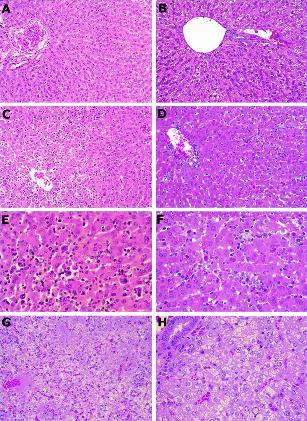
FIGURE 4. Histologic scoring system used to quantitate liver and spleen pathology. SLE ⳱ St. Louis encephalitis; WN ⳱ West Nile; YF ⳱
yellow fever; D1 ⳱ dengue-1; Infl ⳱ inflammation; Necrapop ⳱ necrapoptosis; Steat ⳱ steatosis; Lymph Hyp ⳱ lymphoid hyperplasia; WP Depl
⳱ white pulp depletion; Mac Prolif ⳱ macrophage proliferation. The values for the SLE/WN groups were zero and thus are not visible on the
graph.)

  FIGURE 5. A, Normal histology of spleen from a hamster previously infected with St. Louis encephalitis (SLE) and West Nile (WN) viruses.
Note the distinct border of the lymphoid follicle surrounding a small arteriole (white pulp). B, Spleen from a hamster previously infected with
SLE and WN viruses six days after challenge with yellow fever (YF) virus. Note the lymphoid hyperplasia characterized by the proliferation of
immunoblastic cells, and macrophages (arrowheads). C, White pulp depletion of spleen from a flavivirus-naive (control) hamster six days after
infection with YF virus. D, Splenic macrophage hyperplasia, from a hamster previously infected with dengue-1 and WN viruses six days after
challenge with YF virus. Arrowheads show tangible-body macrophages. (Magnifications: ×25 in A; ×50 in B, C, and D.)
HETEROLOGOUS FLAVIVIRUS INFECTIONS                                                 701

                                                                         correlated with a clinically milder form of YFV infection as
                                                                         shown by lower viremia, less alteration of liver function, and
                                                                         generally asymptomatic infection. Histologic changes in the
                                                                         spleen were also different in the flavivirus-immune hamsters
                                                                         compared with the flavivirus-naive group in that there was
                                                                         more prominent lymphoid hyperplasia and much milder
                                                                         white pulp depletion in the former animals. Another differ-
                                                                         ence noted between the immune hamsters and the naive
                                                                         group was the lack of pancreatic necrosis in the former. In
                                                                         summary, the flavivirus-immune hamsters demonstrated
                                                                         splenic lymphoid hyperplasia, a lack of microvesicular steato-
                                                                         sis in the liver, and an absence of pancreatic necrosis com-
                                                                         pared with the pathology observed in naive animals infected
                                                                         by YFV.
                                                                            In human cases of YF, although abnormalities are noted in
                                                                         multiple organs, the most consistent findings are seen in the
                                                                         liver.16–18 Microvesicular steatosis is a prominent microscopic
                                                                         feature of YFV infection in liver of susceptible vertebrate
                                                                         hosts, including humans, monkeys, or hamsters.8,19–23 In the
                                                                         hamster YF model, small numbers of minute fatty vesicles
                                                                         start to appear in some of the hepatocytes by the third or
                                                                         fourth day after YFV infection; by the fifth or sixth day, the
                                                                         fatty vesicles have spread to involve more cells in larger areas,
                                                                         and they usually occupy the entire cytoplasm (Figure 3G
                                                                         and H).8
                                                                            We have observed the association of liver steatosis with an
                                                                         adverse outcome in other experiments with YFV in the ham-
                                                                         ster model. For example, in studies comparing the virulence
                                                                         (mortality rate) of different YFV strains in hamsters, splenic
                                                                         lymphoid hyperplasia and a lack of liver steatosis were con-
                                                                         sistent findings in hamsters infected by less virulent strains of
                                                                         YFV (Fisher AF and others, unpublished data). These find-
  FIGURE 6. Histopathology of liver and spleen in hamsters previ-
ously infected with JE virus, and challenged with yellow fever virus.    ings suggest that extensive steatosis is related to more severe
A, Liver showing part of a normal bile duct at the right lower corner    clinical disease in YFV infection. However, steatosis may not
of the field, foci of lobular inflammatory cell infiltration, and many   be the cause for the adverse outcome of YFV infection in
Councilman bodies (arrowheads). Note the lack of steatosis. B,           these hamsters; instead it may be simply a marker for severe
Spleen showing marked splenic macrophage proliferation, intermixed
                                                                         liver dysfunction.
with prominent plasmacytoid immunoblasts on the right side edge of
the field. (Magnification ×50.)                                             Massive hepatic steatosis (particularly the microvesicular
                                                                         type) occurs in other acute liver diseases, such as Reye’s syn-
                                                                         drome.24,25 Its presence and rapid onset may indicate severe
                          DISCUSSION
                                                                         functional “shut-down” of the liver, and may explain the high
   The results of this study support previous observations and           mortality seen in diseases such as YF and Reye’s syndrome.
speculation1–6 that immunity to heterologous flavivirus re-              This acutely developing phenomenon suggests dysfunction of
duces the severity of YFV infection. In the present series of            mitochondria, since at the early stages, overt cellular degen-
experiments, prior infection of hamsters with JEV, SLEV,                 eration is rarely observed. Mitochondrial changes also prob-
WNV and DENV protected the animals from fatal disease                    ably play an important role in the activation of apoptosis,26,27
after challenge with YFV. The protective effect of prior het-            or necrapoptosis.28,29 The latter is a major component in YF
erologous flavivirus infection was demonstrated by lower lev-            liver pathology.
els of viremia, less severe alteration of liver function (as mea-           The hepatic necrapoptosis that occurs in YF ranges from
sured by levels of TB and ALT), and an absence of hepatic                scattered, individual Councilman bodies to foci of multiple
steatosis in the flavivirus immune groups (Experiments 2−4)              hepatocytes forming coagula, and diffuse mid-zonal necrosis.
compared with the flavivirus-naive (YF control) group. A                 In advanced or severe YF cases, the mid-zonal pattern can be
similar protective effect by prior heterologous flavivirus in-           obscured by the involvement of zones 1 and 3 as well. The
fection has been reported in a hamster model of WN en-                   pathologic features of necrapoptosis observed in the hamster
cephalitis.12                                                            model of YF8 suggest that a spectrum of changes occurs dur-
   Our studies showed that hamsters previously exposed to                ing the process. This hypothesis is supported by in situ
other flaviviruses developed inflammation and hepatic                    TUNEL staining,8 which revealed nuclear DNA fragmenta-
necrapoptosis similar in extent to that observed in naive ham-           tion in the liver before discernable cell lysis occurred. In ad-
sters after YFV challenge. However, in the flavivirus-immune             dition, typical apoptotic bodies of different stages were seen
animals, no steatosis was found, in contrast to the extensive            in livers of YFV-infected hamsters, using electron microscopy
microvesicular steatosis observed in the flavivirus-naive ham-           (Xiao SY, Popov VL, Tesh RB, unpublished data). We sus-
sters following infection with YFV. The lack of steatosis was            pect that liver cell necrosis in human cases of YF also begins
702 XIAO AND OTHERS
FIGURE 7. The terminal deoxynucleotidyl transferase-mediated dUTP nick-end labeling (TUNEL) index in livers of hamsters infected by
various combinations of flaviviruses. The average number of positively stained cells per 40× high-power field (HPF) of each group is shown.
SLE/WN ⳱ infected by St. Louis encephalitis (SLE) and West Nile (WN) viruses only; SLE/WN/YF ⳱ prior infection with SLE and WN virus,
followed by challenge with yellow fever (YF) virus; DEN/WN/YF ⳱ prior infection with dengue-1 (DEN) and WN viruses, followed by challenge
with YF virus; JE/YF ⳱ prior infection with Japanese encephalitis (JE) virus, followed by challenge with YF virus; YF ⳱ infection with only YF
virus. The differences shown among the YF-challenged groups are not statistically significant (P > 0.05, by Student’s t-test).
as apoptosis, directly induced by virus infection, and not by rus infection.33–35 Most of epidemiologic and laboratory stud-
necroinflammatory injuries. This view is based on the lack of ies of this phenomenon have focused on DENV infection and
consistent inflammatory cell infiltration in the disease, and an attempt to explain why some individuals develop dengue
the fact that the Councilman body, the morphologic evidence hemorrhagic fever (DHF) and dengue shock syndrome
of apoptosis, was first described in YF livers. When viewed in (DSS). One dominant theory is that individuals with pre-
the later stages of the disease, such as at autopsy, the necrosis existing antibody to one DENV serotype are predisposed to
is so extensive and advanced, and may be complicated by the more severe DHF/DSS when subsequently infected by a
other secondary changes, that the process can no longer be different DENV serotype.35 However, our experimental stud-
simply attributed to apoptosis. ies with hamster models of YF and WN encephalitis12 suggest
We suspect that YFV causes morphologic and functional that pre-existing flavivirus antibodies are protective and that
compromise to the mitochondria of the hepatocytes, which they do not increase the severity of these two diseases. Thus,
contributes to cell apoptosis, and at the same time causes immune enhancement does not appear to be a general phe-
abnormal lipid peroxidation and microvesicular fatty changes. nomenon with all flavivirus infections.
The pathogenesis of YFV-induced hepatic necrosis is largely
unknown; but our future research will focus on how YFV Received December 19, 2002. Accepted for publication March 13,
triggers the cellular apoptosis pathways, as well as steatosis. 2003.
Although no studies have been reported in examining the Financial support: This study was supported by the National Insti-
relationship between mitochondrial dysfunction and YFV in- tutes of Health (grants AI-10984 and AI-50175, NO1-AI-25489) and
fection, studies with another flavivirus, hepatitis C virus, in- the Centers for Disease Control and Prevention (grant U50/CCU 620541).
dicate that it induces steatosis in human patients and trans- Authors’ addresses: Shu-Yuan Xiao, Department of Pathology and
genic mice30 by the binding of hepatitis C virus core protein Center for Tropical Diseases, and Department of Internal Medicine,
University of Texas Medical Branch, 301 University Boulevard,
with host cell mitochondrial protein.31 More recently, using
Galveston, TX 77555-0588, Telephone: 409-772-8447, Fax: 409-772-
immunogold electron microscopy, we have found YF viral 4676, E-mail: syxiao@utmb.edu. Hilda Guzman, Amelia P. A. Travas-
antigen to be associated with hepatocyte mitochondria, in sos da Rosa, Hong-Bing Zhu, and Robert B. Tesh, Department of
addition to rough endoplasmic reticulum (Xiao SY, Popov Pathology and Center for Tropical Diseases, University of Texas
VL, Tesh RB, unpublished data). The functional compromise Medical Branch, 301 University Boulevard, Galveston, TX 77555-
0588.
of the hepatocytes may also affect the synthetic functions of
the liver, thus contributing to the bleeding diathesis observed REFERENCES
in patients with YF.32 1. Monath T, 1989. Yellow fever. Monath T, ed. The Arboviruses:
Much has been written during the past three decades on the Epidemiology and Ecology. Boca Raton, FL: CRC Press, 139–
phenomenon of immune enhancement with sequential flavivi- 231.HETEROLOGOUS FLAVIVIRUS INFECTIONS 703
2. Monath T, 1997. Epidemiology of yellow fever: current status and 17. Elton N, Romero A, Trejos A, 1955. Clinical pathology of yellow
speculations on future trends. Saluzzo J, Dodet B, eds. Factors fever. Am J Clin Pathol 25: 135–146.
in the Emergence of Arbovirus Diseases. Paris: Elsevier, 143– 18. Bugher J, 1951. The pathology of yellow fever. Strode G, ed.
156. Yellow Fever. New York: McGraw-Hill, 137–163.
3. Ashcroft MT, 1979. Historical evidence of resistance to yellow 19. Hudson N, 1928. The pathology of experimental yellow fever in
fever acquired by residence in India. Trans R Soc Trop Med the Macacus rhesus. I. Gross pathology. Am J Pathol 4: 395–
Hyg 73: 247–248. 407.
4. Theiler M, Anderson CR, 1975. The relative resistance of den- 20. Hudson N, 1928. The pathology of experimental yellow fever in
gue-immune monkeys to yellow fever virus. Am J Trop Med the Macacus rhesus. II. Microscopic pathology. Am J Pathol 4:
Hyg 24: 115–117. 407–418.
5. Monath TP, Craven RB, Adjukiewicz A, Germain M, Francy 21. Hudson N, 1928. The pathology of experimental yellow fever in
DB, Ferrara L, Samba EM, N’Jie H, Cham K, Fitzgerald SA, the Macacus rhesus. III. Comparison with the pathology of
Crippen PH, Simpson DI, Bowen ET, Fabiyi A, Salaun JJ, yellow fever in man. Am J Pathol 4: 419–439.
1980. Yellow fever in the Gambia, 1978–1979: epidemiologic 22. Bearcroft W, 1957. The histopathology of the liver of yellow
aspects with observations on the occurrence of Orungo virus fever-infected rhesus monkeys. J Pathol Bacteriol 74: 295–303.
infections. Am J Trop Med Hyg 29: 912–928. 23. Tigertt W, Berge T, Gochenour WS, Gleiser C, 1960. Experimen-
6. Henderson BE, Cheshire PP, Kirya GB, Lule M, 1970. Immuno- tal yellow fever. Trans NY Acad Sci 22: 323–333.
logic studies with yellow fever and selected African group B 24. La Montaque J, 1983. Summary of a workshop on disease mecha-
arboviruses in rhesus and vervet monkeys. Am J TropMed nisms and prospects for prevention of Reye’s syndrome. J In-
Hyg19: 110–118. fect Dis 148: 943–950.
7. Tesh RB, Guzman H, da Rosa AP, Vasconcelos PF, Dias LB, 25. Craighead J, 2000. Pathology and Pathogenesis of Human Viral
Bunnell JE, Zhang H, Xiao SY, 2001. Experimental yellow
Disease. San Diego: Academic Press.
fever virus infection in the golden hamster (Mesocricetus au-
26. Fromenty B, Berson A, Pessayre D, 1997. Microvesicular steato-
ratus): I. virologic, biochemical and immunologic studies. J In-
sis an steatohepatitis: role of mitochondrial dysfunction and
fect Dis 183: 1431–1436.
lipid peroxidation. J Hepatol 26: 13–22.
8. Xiao SY, Zhang H, Guzman H, Tesh R, 2001. Experimental yel-
low fever virus infection in the golden hamster (Mesocricetus 27. Rosser B, Gores GJ, 1995. Liver cell necrosis: cellular mecha-
nisms and clinical implications. Gastroenterology 108: 252–275.
auratus): II. Pathology. J Infect Dis 183: 1437–1444.
9. Hotta S, 1969. Dengue and Related Hemorrhagic Diseases. St. 28. Lemasters J, Nieminen A, 1997. Mitochondrial oxygen radical
Louis, MO: Warren H. Green. formation during reductive and oxidative stress to intact hepa-
10. Chen B, Wang I, 1974. Studies on attenuated Japanese B en- tocytes. Biosci Rep 17: 281–291.
cephalitis virus vaccine. 1. Method for obtaining the attenuated 29. Lemasters J, 1999. Mechanisms of hepatic toxicity. V. Necrapo-
2-8 strain and its biological characteristics. Acta Microbiol ptosis and the mitochondrial permeability transition: shared
Sinica 14: 176–184. pathways to necrosis and apoptosis. Am J Physiol 276: G1–G6.
11. Xiao S-Y, Guzman H, Zhang H, Travassos da Rosa APA, Tesh 30. Lerat H, Honda M, Beard MR, Loesch K, Sun J, Yang Y, Okuda
RB, 2001. West Nile virus infection in the golden hamster M, Gosert R, Xiao SY, Weinman SA, Lemon SM, 2002. Ste-
(Mesocricetus auratus): a model of West Nile encephalitis. atosis and liver cancer in transgenic mice expressing the struc-
Emerg Infect Dis 7: 714–721. tural and nonstructural proteins of hepatitis C virus. Gastro-
12. Tesh R, Travassos da Rosa A, Guzman H, Araujo T, Xiao SY, enterology 122: 352–365.
2002. Immunization with heterologous flaviviruses protective 31. Okuda M, Li K, Beard MR, Showalter LA, Scholle F, Lemon
against fatal West Nile encephalitis. Emerg Infect Dis 8: 245– SM, Weinman SA, 2002. Mitochondrial injury, oxidative stress,
251. and antioxidant gene expression are induced by hepatitis C
13. Reed LJ, Muench H, 1938. A simple method of estimating fifty virus core protein. Gastroenterology 122: 366–375.
per cent endpoints. Am J Hyg 27: 493–497. 32. Dennis LH, Reisberg BE, Crosbie J, Crozier D, Conrad ME,
14. Beaty BJ, Calisher C, Shope RE, 1995. Arboviruses. Lennette E, 1969. The original haemorrhagic fever: yellow fever. Br J Hae-
Lennette D, Lennette E, eds. Diagnostic Procedures for Viral, matol 17: 455–462.
Rickettsial and Chlamydial Infections. Seventh edition. Wash- 33. Halstead S, 1988. Pathogenesis of dengue: challenges to molecu-
ington, DC: American Public Health Association, 189–212. lar biology. Science 239: 476–481.
15. Xiao S-Y, Zhang H, Yang Y, Tesh R, 2001. Pirital virus 34. Kliks S, Nisalak A, Brandt W, Wahl L, Burke D, 1989. Antibody
(Arenaviridae) infection in the Syrian golden hamster, Me- dependent enhancement of dengue virus growth in human
socricetus auratus: a new model for arenaviral hemorrhagic monocytes as a risk factor for dengue hemorrhagic fever. Am
fever. Am J Trop Med Hyg 64: 111–118. J Trop Med Hyg 40: 444–451.
16. Kerr J, 1951. The clinical aspects and diagnosis of yellow fever. 35. Roehrig J, 1997. Immunochemistry of dengue viruses. Gubler D,
Strode G, ed. Yellow Fever. New York: McGraw-Hill, 385– Kuno G, eds. Dengue and Dengue Hemorrhagic Fever. New
425. York: CAB International, 199–219.You can also read