Bioaerosol Sampling for Respiratory Viruses in Singapore's Mass Rapid Transit Network - Duke University
←
→
Page content transcription
If your browser does not render page correctly, please read the page content below
www.nature.com/scientificreports

                        OPEN       Bioaerosol Sampling for
                                   Respiratory Viruses in Singapore’s
                                   Mass Rapid Transit Network
Received: 5 September 2018         Kristen K. Coleman        1
                                                                 , Tham T. Nguyen1, Su Yadana1, Christophe Hansen-Estruch2,
Accepted: 10 November 2018         William G. Lindsley       3
                                                                  & Gregory C. Gray 1,4,5
Published: xx xx xxxx
                                   As a leading global city with a high population density, Singapore is at risk for the introduction of
                                   novel biological threats. This risk has been recently reinforced by human epidemics in Singapore of
                                   SARS coronavirus, 2009 pandemic H1N1 influenza A virus, and enterovirus 71. Other major threats
                                   to Singapore include MERS-coronavirus and various avian and swine influenza viruses. The ability
                                   to quickly identify and robustly track such threats to initiate an early emergency response remains
                                   a significant challenge. In an effort to enhance respiratory virus surveillance in Singapore, our team
                                   conducted a pilot study employing a noninvasive bioaerosol sampling method to detect respiratory
                                   viruses in Singapore’s Mass Rapid Transit (MRT) network. Over a period of 52 weeks, 89 aerosol samples
                                   were collected during peak MRT ridership hours. Nine (10%) tested positive for adenovirus, four (4.5%)
                                   tested positive for respiratory syncytial virus type A, and one (1%) tested positive for influenza A
                                   virus using real-time RT-PCR/PCR. To our knowledge, this is the first time molecular evidence for any
                                   infectious respiratory agent has been collected from Singapore’s MRT. Our pilot study data support
                                   the possibility of employing bioaerosol samplers in crowded public spaces to noninvasively monitor for
                                   respiratory viruses circulating in communities.

                                   A high population density, international tourism and trade traffic puts Singapore at a high risk of novel emerging
                                   respiratory epidemics. This risk has been recently reinforced by human epidemics in Singapore of severe acute
                                   respiratory syndrome-associated coronavirus (SARS-CoV)1, 2009 pandemic H1N1 influenza A virus2, and enter-
                                   ovirus 713,4 (which causes hand, foot and mouth disease). Other major threats to Singapore include Middle East
                                   respiratory syndrome-related coronavirus (MERS-CoV)5,6 and various avian7 (H7N4, H7N9, H5N2, H5N1, etc.)
                                   and swine8 (H3N2, H1N1, H1N2) influenza A viruses, especially if they become highly transmissible between
                                   humans. For instance, the first human case of A(H7N4) was confirmed in February 20189, and a total of 1,625
                                   human cases of A(H7N9) have been reported since 201310, with 47% (766) in the 5th wave of the epidemic from
                                   October 2016 to September 201711. Compared to the estimated 0.02% case fatality rate for the global 2009 pan-
                                   demic H1N1 virus12, A(H7N9) (including both high and low pathogenic strains) has an alarming 38% case fatal-
                                   ity rate, which has resulted in 623 known deaths since its emergence in 201310,13,14.
                                       International travel can play a significant role in the spread of infectious diseases. For example, during the
                                   2009 influenza H1N1 pandemic, countries receiving the highest volume of passengers (>1,400 in two months)
                                   from Mexico (the country of origin of the 2009 H1N1 virus), had a significantly higher risk of virus importation
                                   associated with travel to Mexico15. China (the country of origin of A(H7N4) and A(H7N9) viruses) is the third
                                   largest market of travelers for Singapore Changi Airport, which receives ~60,000 passengers from China per
                                   week. With Singapore’s close connection to China, and recent evidence suggesting the potential for airborne
                                   transmission of highly pathogenic avian influenza A(H7N9)16, it is prudent for Singapore to ramp up disease
                                   surveillance. The ability to quickly identify and robustly track respiratory threats is fundamentally important for
                                   an early emergency response.

                                   1
                                    Emerging Infectious Diseases Programme, Duke-NUS Medical School, Singapore, Singapore. 2Duke University
                                   School of Medicine, Durham, USA. 3Health Effects Laboratory Division, National Institute for Occupational Safety
                                   and Health, Morgantown, West Virginia, USA. 4Division of Infectious Diseases, School of Medicine and Global Health
                                   Institute, Duke University, Durham, North Carolina, USA. 5Global Health Research Center, Duke Kunshan University,
                                   Kunshan, China. Correspondence and requests for materials should be addressed to K.K.C. (email: Kristen.
                                   coleman@duke-nus.edu.sg)

        SCIENtIfIC REPorTS |   (2018) 8:17476 | DOI:10.1038/s41598-018-35896-1                                                                      1
www.nature.com/scientificreports/
North East MRT Line (Purple) East West MRT Line (Green)
Average Average
Light Intensity Light Intensity
Temperature (°C) RH (%) (lum/m²) Temperature (°C) RH (%) (lum/m²)
Virus-positive
Aerosol Samples 27 63 710.4 28 63 730.7
Influenza A virus 1 (>4 μm) 0** (>4 μm)
Influenza B virus 0 0
Adenovirus 6 (>4 μm; ≤4 μm) 3 (>4 μm)
Enterovirus 0 0
Coronavirus 0 0
RSV-A 2 (>4 μm; ≤4 μm) 2 (>4 μm; ≤4 μm)
RSV-B 0 0
Total 9 (>4 μm; ≤4 μm) 5 (>4 μm; ≤4 μm)
Table 1. Virus-positive* aerosol samples collected from the Singapore MRT, 2017–2018. *Real-time RT-PCR/
PCR Ct value < 40. **One suspect-positive (Ct value = 41).
To better understand the dynamics of microbes in cities in relation to human health, and to create healthier
environments in areas where people frequently visit, researchers have begun studying the microbiome in and
around public transportation vehicles17,18. An international project named MetaSUB18 was launched to map the
microbiome of transit networks in the world’s largest cities. Over 50 cities, including Singapore, Boston, Shanghai,
Paris, Stockholm, and São Paolo are participating in the project to improve city planning and public health.
Although baseline metagenomic maps created from these studies are said to be useful for mitigating bioterrorism
and infectious disease outbreaks, most of them focus largely on mapping surface-borne bacterial DNA17 and
neglect to address the threat of weaponized or global catastrophic biological risk-level (GCBR-level) agents, both
of which would likely be aerosolized or respiratory-borne RNA viruses19.
Bioaerosol studies have demonstrated that concentrations of airborne bacteria in underground subway sys-
tems tend to fluctuate seasonally and are linked to human presence and activities20,21. Such information demon-
strates the possible role of public transportation in disease transmission, but again, only few studies have focused
on the existence of airborne viruses in public transportation systems22–25. Mathematical models have been devel-
oped to assess the risk of airborne and droplet transmission of respiratory viruses in buses, airline cabins and
cities26–28, and although these simulation data are useful, we have sparse field data to support them. In this study,
we sought to monitor for respiratory viruses in Singapore’s Mass Rapid Transit (MRT) network, a likely area of
virus incursion as well as a pathway for virus transmission. Specifically, we asked, could noninvasive bioaerosol
sampling detect respiratory viruses in Singapore’s MRT network?
Results
Using real-time polymerase chain reaction (PCR) and real-time reverse transcription PCR (RT-PCR), four-
teen (16%) of the aerosol samples collected from the Singapore MRT tested positive for one or more respiratory
viruses. Nine (64%) of the virus-positive aerosol samples were collected from the North East MRT Line (NEL;
Purple) and five (36%) from the East West MRT Line (EWL; Green). Nine of the virus-positive aerosol samples
(64%) tested positive for adenovirus, four (29%) tested positive for RSV-A, and one (7%) tested positive for influ-
enza A virus. One sample tested positive for both adenovirus and RSV-A. One additional sample with a Ct value
of 41 was suspected to be positive for influenza A virus. Aerosol samples did not test positive for any other target
pathogens including influenza B viruses, enteroviruses, coronaviruses, and RSV-B.
Temperature, relative humidity (RH) and light intensity remained relatively consistent inside the Purple and
Green MRT Lines throughout the study, with averages of 27 °C and 28 °C, 63% RH, and 710.4 and 730.7 lum/m²,
respectively (Table 1). There were no significant differences in these environmental conditions on sampling days
yielding virus-positive aerosol samples.
Adenovirus-positive droplets (>4 μm in aerodynamic diameter) and droplet nuclei (≤4 μm in aerodynamic
diameter) were retrieved from six (67%) and four (44%) of the adenovirus-positive samples, respectively. In one
instance, adenovirus-positive particles of both size ranges were retrieved from the same sample. RSV-A drop-
lets and droplet nuclei were both retrieved from four (100%) of the RSV-A-positive samples. Only influenza A
virus-positive droplets (not droplet nuclei) were retrieved from the influenza A virus-positive sample.
Adenovirus-positive samples were collected in January 2018, and February, March, May, June, September
and October 2017. RSV-A-positive samples were collected in February 2017, and the influenza A virus-positive
sample was collected in September 2017. The additional sample that was suspected to be positive for influenza
A virus was collected in November 2017. See Table 2 for a monthly summary of PCR-positive aerosol samples.
Attempts to culture and subtype adenovirus and influenza A virus from PCR-positive aerosol samples
were unsuccessful. Insufficient volumes of the original samples were leftover to attempt to culture RSV-A from
PCR-positive aerosol samples.
SCIENtIfIC REPorTS | (2018) 8:17476 | DOI:10.1038/s41598-018-35896-1 2www.nature.com/scientificreports/
Influenza Influenza
A virus B virus Adenovirus Enterovirus Coronavirus RSV-A RSV-B
Jan 2017 — — — — — — —
Feb 2017 — — 2 — — 4 —
Mar 2017 — — 2 — — — —
Apr 2017 — — — — — — —
May 2017 — — 1 — — — —
June 2017 — — 1 — — — —
July 2017 — — — — — — —
Aug 2017 — — — — — — —
Sept 2017 1 — 1 — — — —
Oct 2017 — — 1 — — — —
—
Nov 2017 — — — — — —
suspect**
Dec 2017 — — — — — — —
Jan 2018 — — 1 — — — —
Table 2. Monthly virus-positive* aerosol sample detections from the Singapore MRT, 2017–2018. *Real-time
RT-PCR/PCR Ct value < 40. **Real-time RT-PCR Ct value = 41.
Discussion
To our knowledge, this is the first time molecular evidence for any infectious respiratory agent has been collected
from Singapore’s MRT. Our results suggest that bioaerosol sampling might have a practical application for patho-
gen detection in public areas such as subway systems. Bioaerosol sampling in the field provides a noninvasive way
to monitor and characterize the community of aerosolized respiratory viruses that regularly infect the public, as
well as potentially detect or discover novel pathogens with pandemic potential, such as the influenza A(H7N9)
virus. Additionally, a bioaerosol sampling system is advantageous as it does not require collecting individual
samples from human subjects, nor informed consent.
Although this pilot study provides important qualitative data (i.e., molecular presence of respiratory viruses in
the MRT), it is important to note that solely detecting viral DNA/RNA in the air does not determine that a virus
is successfully transmitted through the air. Cultures in this study were negative and therefore the viruses detected
may have been non-viable. However, studies have demonstrated that upon the first ~15 minutes of aerosolization,
viruses tend to decay the most rapidly29,30, and therefore it is possible that the viability of the viruses captured in our
study rapidly decreased during sample collection and transport. Moreover, temperature and RH have been widely
understood to influence the viability of aerosolized viruses31. However, novel research has recently demonstrated
that the viability of aerosolized influenza virus is independent of RH when the aerosol composition includes human
bronchial epithelial (HBE) extracellular material (ECM)32, indicating that the influence of RH is more so on the rate
of aerosol deposition after expulsion, which in turn, influences the overall concentration of viral aerosols in the envi-
ronment. Aerosols tend to deposit onto surfaces more quickly at higher RH levels (>50%), suggesting that greater
time spent in controlled environments with lower, more comfortable RH levels, could increase the risk of acquiring
a respiratory virus via inhalation and respiration. Therefore, the average RH level (63%) on the Singapore MRT is not
theoretically conducive to heavy airborne transmission of influenza viruses.
A likely contributor to the lack of data regarding airborne transmission of viruses is the difficulty of detecting
respiratory pathogens in the air. Standard detection methods are designed to target viral nucleic acids which are
subject to degradation from various environmental stressors (e.g., temperature, humidity, and UV light) as well
as physical stress from aerosol sample collection methods, potentially deterring downstream detection. Therefore,
high RH levels, in addition to sample degradation, could explain the low recovery of virus-positive aerosol sam-
ples in our study. Additionally, viral bioaerosols have notoriously low airborne concentrations, reducing the sen-
sitivity of bioaerosol sampling. Some bioaerosol samplers, such as the NIOSH sampler used in this study, are
compact and require a low flow rate which also may reduce sensitivity. Although the air pump flow rate and sam-
ple collection times used in our study have been demonstrated to efficiently capture aerosolized influenza virus
and RSV RNA33–35, it is possible that these parameters are not optimal for capturing the other respiratory virus
DNA/RNA targeted in our study. For example, it is possible that when compared to RNA viruses (e.g., influenza
virus, RSV), adenovirus (a more durable DNA virus) can withstand a higher flow rate and/or longer sampling
duration. Furthermore, although each of the assays used to detect viral DNA/RNA in our aerosol samples are
highly sensitive, validated, singleplex real-time RT-PCR/PCR assays, specific detection limits (viral copies per
volume of air) were not tested in our laboratory. Therefore, our DNA/RNA detections should not be mistaken as
a true representation of the prevalence/densities of these viruses circulating in the air of the MRT, but rather a
representation of the viruses our bioaerosol sampling technique is capable of detecting in this environment. To
improve the sensitivity and specificity of bioaerosol sampling, more virus-specific controlled studies are needed
to determine optimal sampling parameters for multiple respiratory viruses.
Airborne microbial concentrations have been demonstrated to fluctuate seasonally in underground subway sys-
tems20. However, the Singapore MRT is a controlled environment and therefore environmental conditions remained
consistent throughout the study and no significant differences were recorded on days yielding virus-positive
samples. Furthermore, unlike temperate climates with distinct seasons, Singapore has a tropical rainforest cli-
mate with high temperatures and humidity levels year-round, with little monthly variation. Therefore, Singapore
SCIENtIfIC REPorTS | (2018) 8:17476 | DOI:10.1038/s41598-018-35896-1 3www.nature.com/scientificreports/
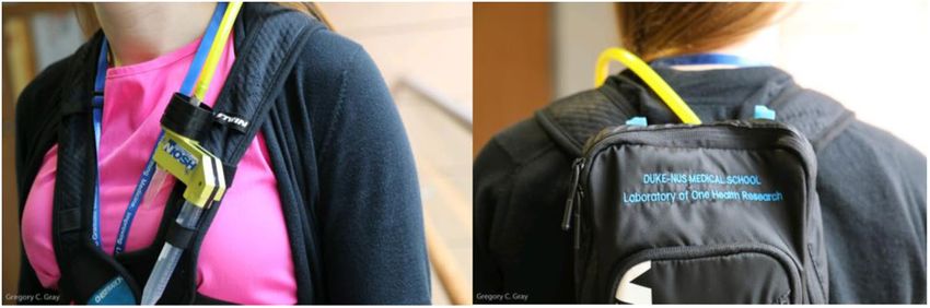
Figure 1. Aerosol sampler attached to a personal backpack carrying an SKC Airchek pump worn by
researchers.
experiences influenza outbreaks year-round, with two peak periods (April–June and September–December).
Although we were able to stratify our pilot study data by month, our results are too limited to properly assess a
correlation between virus-positive aerosols and Singapore’s peak influenza periods. However, it is notable that the
influenza A virus-positive and suspect-positive aerosol samples were both detected during Singapore’s second
peak influenza period in 2017. Additionally, RSV-A-positive aerosols were collected during weeks of increased
polyclinic attendances for acute respiratory infections in Singapore36. Adenovirus-positive aerosols were col-
lected during weeks of increased, average and decreased polyclinic attendances for acute respiratory infections36.
A larger study yielding more aerosol samples over a longer duration would be needed to further characterize the
relationship between bioaerosols in the MRT and community health data in Singapore.
Quickly identifying and robustly tracking respiratory threats are fundamental steps in an early emergency
response to a pandemic. A bioaerosol sampling system is advantageous in this regard as it does not require the
timely acquisition of ethical/IRB approvals and informed consent needed to collect individual samples from
human subjects. If an outbreak is suspected or underway, bioaerosol samplers can be immediately deployed in
high-risk areas, yielding results within a minimum of ~8 hours. Moreover, if bioaerosol samplers are proactively
used to monitor high-risk areas, initial results turnaround times are nearly cut in half, as sample collection time
is ~3–4 hours. Proactively monitoring for respiratory viruses also eliminates the risk of missing the time window
of exposure to pandemic viruses in high-risk areas and would allow a more robust surveillance, which could be
beneficial when the etiologic agent is known but the route of exposure is not fully understood.
Our bioaerosol sampling method also size-fractionates virus-laden particles, which can help measure the pro-
portion of exposure to droplets versus droplet nuclei (i.e., inhalable versus respirable particles) in multiple envi-
ronments and climates, which is important when assessing the type or severity of disease (e.g., an upper versus
lower respiratory tract infection) that might follow. However, the full extent of exposure cannot be determined
without measuring viral load. Our study falls short in that we did not quantify the viral load in our aerosol sam-
ples, making it difficult to compare our results with quantitative aerosol studies. In the future, we will implement
a dilution series of positive controls into our real-time RT-PCR/PCR assay protocols to create a standard curve
that can be used to accurately quantify the viral load in each aerosol sample.
When compared to a mobile/personal bioaerosol sampling method such as the one used in our study, a sta-
tionary sampling method is arguably a more practical approach to conducting disease surveillance.
However, the use/installation of stationary samplers on the MRT would likely require permission from governmen-
tal authorities. Strapping the bioaerosol samplers to our bodies was the least invasive way for our team to recover molec-
ular evidence of aerosolized respiratory viruses on the Singapore MRT. As this type of research is new to Singapore, the
wellbeing of Singapore’s civilians was carefully considered and therefore, researchers wore their Duke-NUS employee
badges while sampling, and our laboratory’s name was stitched onto the sampling backpacks (Fig. 1) for transpar-
ency. Similar to a study measuring the risk of exposure to aerosolized influenza virus among healthcare workers in an
emergency department during influenza season37, strapping the samplers to personal backpacks allowed us to detect
personal exposure to aerosolized viruses and provide insight into the efficacy of potential interventions (e.g., air/surface
decontamination, and wearing face masks when infectious). Moreover, with evidence validating the notion that MRT
riders are at risk of exposure to respiratory viruses, we hope to motivate scientists to conduct similar field studies to
unveil the true risk of exposure while using public transportation, as data on this topic are scarce. Field studies may also
inspire bioengineers and scientific instrument companies to design and test improved bioaerosol sampling such that it
might be employed in a more widespread fashion to surveil for respiratory threats.
In conclusion, our study suggests that when combined with molecular diagnostics, aerosol sampling has
promising potential to work as a noninvasive tool to monitor for respiratory pathogens in public areas. Additional
studies are needed to assess a possible contribution of aerosol sampling to public health surveillance during peri-
ods of increased risk.
Methods
Bioaerosol sampling. From January 2017 through January 2018, National Institute for Occupational Safety
and Health (NIOSH) BC 251 2-stage aerosol samplers were used to collect aerosol samples from the following
Singapore Mass Rapid Transit (MRT) heavy rail lines: East West Line (EWL; green), and North East Line (NEL;
purple). MRT lines were selected for their high capacity (~1,600 passengers per 6-car train), frequent use, and
SCIENtIfIC REPorTS | (2018) 8:17476 | DOI:10.1038/s41598-018-35896-1 4www.nature.com/scientificreports/
connection to high-traffic public areas. The EWL was specifically chosen for its connection to Changi Airport,
and the NEL for its connection to downtown Singapore.
Aerosol samples were collected weekly, with occasional interruptions, during peak ridership (~267 passen-
gers/car or 5 pax/m2) from 7:30–9:00 and 17:30–19:00 on Wednesdays. Sampling days varied occasionally due to
public holidays and the researchers’ availability. Using a 2-foot (61 cm) long piece of ¼” (0.635 cm) Tygon tubing,
®
NIOSH samplers were connected to AirChek TOUCH Sample Pumps (SKC, Eighty Four, Pennsylvania) carried
inside personal backpacks worn by the researchers. Both straps were tightened around the shoulders to secure the
backpack against the researcher’s back. NIOSH samplers were securely fastened to the front of the backpacks (on
or above the researcher’s heart) using Velcro (Fig. 1). For each separate MRT line (i.e., green and purple), the same
assembled NIOSH sampler was used for both AM and PM sampling sessions. To ensure all researchers followed
the same sampling protocol, each researcher sampled one of the middle train-cars and remained standing with
intermittent walking throughout each sampling session. Between AM and PM sampling sessions, sampler inlets
were covered with electrical tape and the assembled samplers were refrigerated. All of the material collected by
one NIOSH sampler during 3 hours of sampling was counted as one aerosol sample.
Before aerosol sampling, air pumps were calibrated to sample at 3.5 L min−1 collecting a total of 630 L of air
per MRT line each week. Calibration was performed by inserting the sampler into a calibration adaptor (designed
for NIOSH samplers) attached to an SKC check-mate calibrator. NIOSH samplers were engineered to separate
collected particles into three size fractions: >4 μm, 1–4 μm, and 4 μm in diameter) collected in stage 1 (15 mL tube).
Cell culture and DNA sequencing. Virus-positive samples were tested for viability using cell culture.
First, 500 μL of each adenovirus-positive aerosol sample was inoculated into A549 cells (ATCC) with Dulbecco’s
Modified Eagle Medium (DMEM) 2% (v/v) Fetal Bovine Serum (FBS) and incubated at 37 °C. MDCK cells were
used to culture the influenza A virus-positive and suspect-positive aerosol samples. Inoculated shell vials were
then observed for cytopathic effect 72 hours after inoculation, and daily for ten days afterwards. Observation
of cytopathic effect was used to score positive or negative cultures. RSV-positive samples were not tested for
viability as RSV assays were not performed until after all of the original aerosol sample material had been used
for DNA/RNA extraction, leaving us with no original sample material to perform cell culture on RSV-positive
SCIENtIfIC REPorTS | (2018) 8:17476 | DOI:10.1038/s41598-018-35896-1 5www.nature.com/scientificreports/
samples. Extracted DNA from adenovirus-positive aerosol samples was typed using conventional PCR targeting
the hexon (predicted amplicon size 764–896 bp) and fiber gene (predicted amplicon size according to species: A
1444–1537 bp; B 670–772 bp; C 1988–2000bp; D 1205–1221 bp; E 967 bp; F 541–586 bp).
Declarations
The findings and conclusions in this report are those of the authors and do not necessarily represent the official
position of the US Centers for Disease Control and Prevention.
Availability of Data and Material
The datasets generated and/or analyzed during this study are available from the corresponding author on reason-
able request.
References
1. Singapore Ministry of Health. Epidemiology and control of SARS in Singapore. Epidemiological news bulletin. 29:7, https://www.
moh.gov.sg/content/dam/moh_web/Statistics/Epidemiological_News_Bulletin/2003/enb07_03w_0.pdf. Accessed 27 April 2018
(2003).
2. Cutter, J. L. et al. Outbreak of pandemic influenza A (H1N1- 2009) in Singapore, May to September 2009. Ann Acad Med Singapore
39, 273–82 (2010).
3. Singapore Ministry of Health. Communicable diseases surveillance in Singapore 2012, https://www.moh.gov.sg/content/moh_web/
home/Publications/Reports/2013/Communicable_Diseases_Surveillance_in_Singapore_2012.html. Accessed 27 April 2018.
4. Chan, K. P. et al. Epidemic Hand, Foot and Mouth Disease caused by human enterovirus 71, Singapore. Emerg Infect Dis. 9(1), 78–85
(2003).
5. Singapore Ministry of Health. Middle East Respiratory Syndrome Coronavirus (MERS-CoV). Health Advisory (January 2018).
https://www.moh.gov.sg/content/moh_web/home/pressRoom/Current_Issues/2014/middle-east-respiratory-syndrome-
coronavirus–mers-cov-.html. Accessed 27April 2018.
6. Azhar, E. I. et al. Detection of the Middle East respiratory syndrome coronavirus genome in an air sample originating from a camel
barn owned by an infected patient. MBio. 5(4), e01450–14 (2014).
7. Wong, T. Y. et al. A cross-sectional study of primary-care physicians in Singapore on their concerns and preparedness for an Avian
Influenza outbreak. Ann Acad Med Singapore. 37, 458–64 (2008).
8. Lee, H. K. et al. Diagnostic testing for pandemic influenza in Singapore. The Journal of Molecular Diagnostics 12(5), 636–43 (2010).
9. World Health Organization. Human infection with avian influenza A(H7N4)virus – China. 2018, http://www.who.int/csr/don/22-
february-2018-ah7n4-china/en/. Accessed 27April 2018.
10. Food and Agriculture Organization of the United States. H7N9 situation update, http://www.fao.org/ag/againfo/programmes/en/
empres/h7n9/situation_update.html. Accessed 27 April 2018 (2018).
11. World Health Organization. Disease Outbreak News, http://www.who.int/csr/don/en/. Accessed 27 April 2018.
12. Roos, R. Study puts global 2009 pandemic H1N1 infection rate at 24%. University of Minnesota CIDRAP News. 2013, http://www.
cidrap.umn.edu/news-perspective/2013/01/study-puts-global-2009-pandemic-h1n1-infection-rate-24. Accessed 20 October 2017.
13. The Straits Times. China’s new H7N9 bird flu strain has pandemic potential: US study, http://www.straitstimes.com/world/united-
states/chinas-new-h7n9-bird-flu-strain-has-pandemic-potential-us-study. Accessed 20 October 2017 (2017).
14. Ke C. et al. Human infection with highly pathogenic avian influenza A(H7N9) virus, China. Emerg Infect Dis. 23(8):1332–1340
https://doi.org/10.3201/eid2308.170600. Accessed 20 October 2017 (2017).
15. Khan, K. et al. Spread of a novel influenza A (H1N1) virus via global airline transportation. N Engl J Med. 361, 212–4 (2009).
16. Imai et al. A highly pathogenic avian H7N9 influenza virus isolated from a human is lethal in some ferrets infected via respiratory
droplets. Cell Host & Microbe. https://doi.org/10.1016/j.chom.2017.09.008. Accessed 20 October 2017 (2017).
17. Afshinnekoo, E. et al. Geospatial resolution of human and bacterial diversity with city-scale metagenomics. Cell Systems. 1(1), 72–87
(2015).
18. The MetaSUB International Consortium. The Metagenomics and Metadesign of the Subways and Urban Biomes (MetaSUB)
International Consortium inaugural meeting report. Microbiome. 4, 24 (2016).
19. Johns Hopkins Center for Health Security. The characteristics of pandemic pathogens. May. Retrieved 2018 June 11 from, http://
www.centerforhealthsecurity.org/about-the-center/pressroom/press_releases/2018-05-10_characteristics-of-pandemic-pathogens.
html (2018).
20. Heo, K. J. & Lee, B. U. Seasonal variation in the concentrations of culturable bacterial and fungal aerosols in underground subway
systems. Journal of Aerosol Science. 92, 122–9 (2016).
21. Heo, K. J., Lim, C. E., Kim, H. B. & Lee, B. U. Effects of human activities on concentrations of culturable bioaerosols in indoor air
environments. Journal of Aerosol Science. 104, 58–65 (2017).
22. Nasir Z. A., Campos L. C., Christie N., Colbeck I. Airborne biological hazards and urban transport infrastructure: current challenges
and future directions. Environmental Science and Pollution Research. Aug 1;23(15):15757–66 (2016).
23. Mohr O. et al. Evidence for airborne infectious disease transmission in public ground transport–a literature review. Eurosurveillance.
Aug 30;17(35):20255 (2012).
24. Browne, A., St-Onge Ahmad, S., Beck, C. R. & Nguyen-Van-Tam, J. S. The roles of transportation and transportation hubs in the
propagation of influenza and coronaviruses: a systematic review. Journal of travel medicine. Jan 18;23(1):tav002 (2016).
25. Triadó‐Margarit, X. et al. Bioaerosols in the Barcelona subway system. Indoor air. May 27(3), 564–75 (2017).
26. Zhu, S., Srebric, J., Spengler, J. D. & Demokritou, P. An advanced numerical model for the assessment of airborne transmission of
influenza in bus microenvironments. Building and Environment. 47, 67–75 (2012).
27. Hertzberg, V. S., Weiss, H., Elon, L., Si, W. & Norris, S. L. FlyHealthy Research Team. Behaviors, movements, and transmission of
droplet-mediated respiratory diseases during transcontinental airline flights. Proceedings of the National Academy of Sciences. Mar
14:201711611 (2018).
28. Mei S. et al. Simulating city-level airborne infectious diseases. Computers, Environment and Urban Systems. May 1;51:97–105 (2015).
29. Noti, J. D. et al. High humidity leads to loss of infectious influenza virus from simulated coughs. PLoS One. 8(2), e57485 (2013).
30. Harper, G. J. Airborne micro-organisms: survival tests with four viruses. J Hyg (Lond). 59, 479–86 (1961).
31. Tang J. W. The effect of environmental parameters on the survival of airborne infectious agents. Journal of the Royal Society Interface.
Dec 6;6(Suppl 6):S737–46 (2009).
32. Kormuth, K. A. et al. Influenza virus infectivity is retained in aerosols and droplets independent of relative humidity. The Journal of
infectious diseases. Jun 7 (2018).
33. Blachere, F. M. et al. Measurement of airborne influenza virus in a hospital emergency department. Clin Infect Dis. 48(4), 438–40
(2009).
34. Cao, G., Noti, J. D., Blachere, F. M., Lindsley, W. G. & Beezhold, D. H. Development of an improved methodology to detect infectious
airborne influenza virus using the NIOSH bioaerosol sampler. Journal of Environmental Monitoring: JEM. Dec 13(12), 3321 (2011).
SCIENtIfIC REPorTS | (2018) 8:17476 | DOI:10.1038/s41598-018-35896-1 6www.nature.com/scientificreports/
35. Lindsley W. G. et al. Distribution of airborne influenza virus and respiratory syncytial virus in an urgent care medical clinic. Clinical
Infectious Diseases. Mar 1;50(5):693–8 (2010).
36. Singapore Ministry of Health. Weekly Infectious Diseases Bulletin, https://www.moh.gov.sg/resources-statistics/infectious-disease-
statistics/2018/weekly-infectious-diseases-bulletin. Accessed2 November 2018.
37. Rule A. M. et al. Healthcare personnel exposure in an emergency department during influenza season. PloS one. Aug 31;13(8):e0203223
(2018).
38. Borkenhagen, L. K. et al. Surveillance for respiratory and diarrheal pathogens at the human-pig interface in Sarawak, Malaysia.
PLOS ONE 13(7), e0201295, https://doi.org/10.1371/journal.pone.0201295 (2018).
39. Van de Pol, A. C. et al. Increased detection of respiratory syncytial virus, influenza viruses, parainfluenza viruses, and adenoviruses
with real-time PCR in samples from patients with respiratory symptoms. Journal of Clinical Microbiology. 1(45(7)), 2260–2 (2007).
40. World Health Organization (WHO). CDC protocol of realtime RT-PCR for influenza H1N1. World Health Organization, Geneva:
Switzerland (2009).
41. Selvaraju, S. B. & Selvarangan, R. Evaluation of three influenza A and B real-time reverse transcription-PCR assays and a new 2009
H1N1 assay for detection of influenza viruses. J Clin Microbiol. 48(11), 3870–5 (2010).
42. Loens K. et al. Performance of different mono-and multiplex nucleic acid amplification tests on a multipathogen external quality
assessment panel. J Clin Microbiol. Mar 1;50(3):977–87 (2012).
43. Bil-Lula, I., De Franceschi, N., Pawlik, K. & WoŸniak, M. Improved real-time PCR assay for detection and quantification of all 54
known types of human adenoviruses in clinical samples. Medical science monitor: international medical journal of experimental and
clinical research. 18(6), BR221 (2012).
Acknowledgements
This study was supported by start-up funding for Dr. Gregory Gray’s Laboratory of One Health Research at Duke-
NUS Medical School, Singapore.
Author Contributions
K.C., T.N., C.H.E., and G.G. designed the study. K.C., T.N., S.Y., and C.H.E. performed aerosol sample collection
and T.N., S.Y., and C.H.E. performed laboratory works. K.C., T.N., S.Y., and C.H.E. analyzed and interpreted the
data. W.L. provided the NIOSH aerosol samplers and K.C. and W.L. guided the researchers in their use. K.C. and
G.G. supervised the study. K.C. drafted the manuscript and W.L. and G.G. contributed to writing and reviewing
the manuscript. All authors read and approved the final manuscript.
Additional Information
Competing Interests: The authors declare no competing interests.
Publisher’s note: Springer Nature remains neutral with regard to jurisdictional claims in published maps and
institutional affiliations.
Open Access This article is licensed under a Creative Commons Attribution 4.0 International
License, which permits use, sharing, adaptation, distribution and reproduction in any medium or
format, as long as you give appropriate credit to the original author(s) and the source, provide a link to the Cre-
ative Commons license, and indicate if changes were made. The images or other third party material in this
article are included in the article’s Creative Commons license, unless indicated otherwise in a credit line to the
material. If material is not included in the article’s Creative Commons license and your intended use is not per-
mitted by statutory regulation or exceeds the permitted use, you will need to obtain permission directly from the
copyright holder. To view a copy of this license, visit http://creativecommons.org/licenses/by/4.0/.
© The Author(s) 2018
SCIENtIfIC REPorTS | (2018) 8:17476 | DOI:10.1038/s41598-018-35896-1 7You can also read