Clinical features and predictors of severity in COVID 19 patients with critical illness in Singapore - Nature
←
→
Page content transcription
If your browser does not render page correctly, please read the page content below
www.nature.com/scientificreports
OPEN Clinical features and predictors
of severity in COVID‑19 patients
with critical illness in Singapore
Ser Hon Puah1, Barnaby Edward Young1,2,9, Po Ying Chia1,2,9, Vui Kian Ho3, Jiashen Loh3,
Roshni Sadashiv Gokhale4, Seow Yen Tan5, Duu Wen Sewa5, Shirin Kalimuddin5,10,
Chee Keat Tan8, Surinder K. M. S. Pada8,11, Matthew Edward Cove6,11, Louis Yi Ann Chai6,11,
Purnima Parthasarathy7, Benjamin Choon Heng Ho1, Jensen Jiansheng Ng1, Li Min Ling1,2,9,
John A. Abisheganaden1,9, Vernon J. M. Lee12,13, Cher Heng Tan1,9, Raymond T. P. Lin2,11,
Yee Sin Leo1,2,9,11,13, David C. Lye1,2,9,11, Tsin Wen Yeo1,2,9,14* & Singapore 2019 novel
coronavirus outbreak research team*
We aim to describe a case series of critically and non-critically ill COVID-19 patients in Singapore.
This was a multicentered prospective study with clinical and laboratory details. Details for fifty
uncomplicated COVID-19 patients and ten who required mechanical ventilation were collected. We
compared clinical features between the groups, assessed predictors of intubation, and described
ventilatory management in ICU patients. Ventilated patients were significantly older, reported
more dyspnea, had elevated C-reactive protein and lactate dehydrogenase. A multivariable logistic
regression model identified respiratory rate (aOR 2.83, 95% CI 1.24–6.47) and neutrophil count (aOR
2.39, 95% CI 1.34–4.26) on admission as independent predictors of intubation with area under receiver
operating characteristic curve of 0.928 (95% CI 0.828–0.979). Median APACHE II score was 19 (IQR
17–22) and PaO2/FiO2 ratio before intubation was 104 (IQR 89–129). Median peak FiO2 was 0.75
(IQR 0.6–1.0), positive end-expiratory pressure 12 (IQR 10–14) and plateau pressure 22 (IQR 18–26) in
the first 24 h of ventilation. Median duration of ventilation was 6.5 days (IQR 5.5–13). There were no
fatalities. Most COVID-19 patients in Singapore who required mechanical ventilation because of ARDS
were extubated with no mortality.
In December 2019, a cluster of severe pneumonia patients linked to an animal wholesale market was reported in
Wuhan, Hubei Province, China1,2. Subsequently the responsible pathogen was identified as the novel zoonotic
severe acute respiratory syndrome coronavirus 2 (SARS-CoV-2)3. Hitherto in this century, two other zoonotic
beta-coronaviruses, SARS-CoV and Middle East Respiratory Syndrome coronavirus (MERS-CoV) have caused
outbreaks of severe respiratory disease with estimated mortality rates of 9% and 30% respectively4.
Since the initial reports, the number of cases diagnosed with coronavirus disease 2019 (COVID-19) has
increased exponentially in China and globally. While the number of reported cases is decreasing in China,
the number of cases in other countries is growing at an alarming rate. While approximately 80% of cases will
experience mild to moderate disease, single-centre studies from hospitals in Wuhan reported that 16–29% of
hospitalized patients developed acute respiratory distress syndrome (ARDS) and 26–32% required intensive care
management1,2,5. Case fatality rates in these reports ranged between 4.3 and 15%1,2,5 A report of 52 critically ill
patients from a tertiary referral hospital in Wuhan described ARDS in 67% of patients with a case fatality rate
of 62.5%6. Currently, there are no reports on clinical details and outcomes of critically ill COVID-19 patients in
1
Tan Tock Seng Hospital, Singapore, Singapore. 2National Centre for Infectious Diseases, Singapore,
Singapore. 3Sengkang General Hospital, Singapore, Singapore. 4Changi General Hospital, Singapore,
Singapore. 5Singapore General Hospital, Singapore, Singapore. 6National University Hospital, Singapore,
Singapore. 7Khoo Teck Puat Hospital, Singapore, Singapore. 8Ng Teng Fong General Hospital, Singapore,
Singapore. 9Lee Kong Chian School of Medicine, Novena Campus Clinical Sciences Building, 11 Mandalay Rd,
Singapore 308232, Singapore. 10Duke-NUS Medical School, Singapore, Singapore. 11Yong Loo Lin School
of Medicine, Singapore, Singapore. 12Ministry of Health, Singapore, Singapore. 13Saw Swee Hock School of
Public Health, Singapore, Singapore. 14Menzies School of Health Research, Charles Darwin University, Darwin,
Australia. *A list of authors and their affiliations appears at the end of the paper. *email: yeotsinwen@ntu.edu.sg
Scientific Reports | (2021) 11:7477 | https://doi.org/10.1038/s41598-021-81377-3 1
Vol.:(0123456789)www.nature.com/scientificreports/
countries outside China, making it difficult to understand the threat COVID-19 poses in different healthcare
settings7.
Singapore is a country in Southeast Asia with high levels of travel and economic connectivity with China.
During the SARS outbreak in 2003, it was one of the worst affected countries, with 238 patients and 33 deaths,
including a significant number of healthcare workers8. Similarly, during the current outbreak, Singapore was one
of the first countries outside China to diagnose a significant number of SAR-CoV-2 infections and document
local transmission7.
In this study, we describe the clinical features and treatment outcomes of the first ten COVID-19 patients in
Singapore who required mechanical ventilation and compared the clinical, laboratory and radiological features
with fifty COVID-19 patients who did not need invasive ventilatory support. We also explored independent pre-
dictors for invasive ventilation in these patients. The findings will not only be important for clinical management
and triage of infected patients, but will have implications for resource planning across the globe, as countries
prepare to deal with the current crisis.
Materials and methods
Study design and participants. This was a multicenter case control study of patients with confirmed
COVID-19 by SARS-CoV-2 real-time polymerase chain reaction (RT-PCR) using a previously described
method7. All COVID-19 patients were admitted for treatment and isolation in government hospitals with nega-
tive pressure facilities. Airborne and contact precautions were observed and staff attending to patients wore
personal protective equipment in accordance with the United States Centers for Disease Control and Prevention
guidelines. Collection of de-identified clinical data from infected individuals was approved by the Ministry of
Health, Singapore under the Infectious Disease Act with waiver of written informed consent. All methods were
carried out in accordance with relevant guidelines and regulations.
Data collection. We recorded demographic, clinical, laboratory and radiological data for COVID-19
patients using a standardized case report form modified from the International Severe Acute Respiratory and
Emerging Infection C onsortium9. Information collected included epidemiological data (age, gender, ethnicity,
exposure to COVID-19 cases, travel history, clinical symptoms and comorbidities), vital signs on admission
and transfer to the intensive care unit (ICU), laboratory values on admission and ICU transfer (hemoglobin;
white blood cell, neutrophil, lymphocyte and platelet counts; lactate dehydrogenase [LDH], C-reactive protein
[CRP], creatinine, arterial blood gas), fraction of inspired oxygen concentration (FiO2), radiological findings
and treatment (oxygen therapy, antibiotics, oseltamivir, interferon beta-1b, lopinavir-ritonavir and inotropes)
for all patients admitted to ICU until 20th February 2020. Data collection was completed on 27th February 2020
for the above, and was censored for patients still in ICU (mortality, days to extubation, and extracorporeal mem-
brane oxygenation) on day of manuscript submission. A non-ICU cohort was selected from the first fifty con-
secutive patients admitted to participating hospitals who recovered without requiring mechanical ventilation.
Two researchers individually reviewed the data forms, and all inconsistent data was clarified with the attending
doctors, patients or their families.
The date of disease onset was defined as the day the symptoms were first noted. We defined ARDS following
recommendations from WHO and the Berlin criteria10. Acute respiratory illness was defined as patients who
developed respiratory failure requiring mechanical ventilation without fulfilling the Berlin criteria. Acute kid-
ney injury was identified according to the Kidney Disease: Improving Global Outcomes d efinition11. For ICU
patients, the Sequential Organ Failure Assessment (SOFA) and Acute Physiology and Chronic Health Evaluation
II (APACHE) scores were recorded on admission.
Clinical management. Respiratory samples were tested for influenza and other respiratory viruses with
a multiplex PCR assay, and urine for pneumococcal and legionella antigen if clinically indicated. Serial naso-
pharyngeal swabs for SARS-CoV-2 RT-PCR were done for all patients. Supportive therapy including supple-
mental oxygen was provided according to the degree of hypoxia and the decision for transfer to ICU was made
by the attending physician and intensivist. Patients clinically suspected of community-acquired or ventilator-
associated pneumonia were administered empiric broad-spectrum antibiotics and oral oseltamivir according to
the treating physician’s discretion. Lopinavir-ritonavir (400 mg/100 mg twice daily orally for up to 14 days) and
interferon beta-1b (8 million units subcutaneously every other day) was prescribed to selected patients, mainly
in the ICU. Corticosteroids were avoided with concern for reported increased mortality in patients with SARS
and MERS12.
Outcomes. We compared the demographic, clinical, laboratory and radiological differences on admission
between patients who required and did not require invasive mechanical ventilation. The incidence of ARDS,
shock, mechanical ventilation, dialysis and mortality were reported.
Statistical analysis. Continuous variables were expressed as mean (95% confidence interval) or median
(interquartile range) depending on distribution, and categorical variables were expressed as frequency and per-
centage. We compared differences for continuous variables using two-sample t test or Wilcoxon rank-sum test
depending on the distribution, and χ2 test or Fisher’s exact test for categorical variables. To assess the predic-
tive utility of continuous variables for invasive ventilation, the area under the receiver operating characteristic
(AUROC) curve and the 95% confidence interval (CI) were calculated.
A multivariable logistic regression model was developed to identify predictors of intubation after exclusion
of individuals who were intubated at presentation. Variables were chosen if complete data was available and
Scientific Reports | (2021) 11:7477 | https://doi.org/10.1038/s41598-021-81377-3 2
Vol:.(1234567890)www.nature.com/scientificreports/
All Non-ICU ICU CI for difference in means p-value*
N 60 50 10
Age, years, mean (95% CI), 44 (41–47) 43 (39–46) 52 (44–59) 0.03
Sex, male, no. (%) 37 (62) 29 (58) 8 (80) 0.29
Chinese ethnicity, no. (%) 52 (87) 44 (88) 8 (80) 0.61
Travel in the 2 weeks before symptoms onset, no. (%) 18 (30) 26 (52) 3 (30) 0.30
Any comorbidity, no. (%) 17 (28) 12 (24) 5 (50) 0.13
Symptoms
Duration of symptoms prior to admission, days, mean (95% CI) 5.1 (3.9–6.2) 4·9 (3.6–6.3) 5.7 (4.1–7.3) 0.63
Fever, no. (%) 47 (78) 37 (74) 10 (100) 0.10
Cough, no. (%) 46 (77) 39 (78) 8 (80) 1.00
Dyspnea, no. (%) 16 (25) 8 (16) 8 (80) < 0.001
Sore throat or coryza, no. (%) 29 (48) 27 (54) 2 (20) 0.08
Diarrhea, no. (%) 10 (17) 9 (18) 1 (10) 1.00
Baseline vital signs (range)
Temperature, °C 37.7 (37.5–38.0) 37.6 (37.3–37.8) 38.7 (38.1–39.2) 0.001
Heart rate, beats per minute 93 (89–97) 89 (85–93) 111 (99–123) < 0.001
Respiratory rate, breaths per minute 18.4 (18.0–18.9) 18.0 (17.6–18.4) 20.5 (18.9–22.1) < 0.001
Systolic blood pressure, mmHg 129 (125–133) 128 (123–132) 136 (121–151) 0.16
Pulse oximeter oxygen saturation, % 97.3 (96.7–98.0) 97.9 (97.5–98.3) 94.4 (91.7–97.1) < 0.001
Baseline blood investigations (range)
White blood cell, × 109/L 4.9 (4.4–5.4) 4.6 (4.2–5.0) 6.3 (4.3–8.2) 0.01
Lymphocyte, × 109/L 1.2 (1.1–1.4) 1.3 (1.2–1.5) 0.6 (0.4–0.8) < 0.001
Neutrophil, × 109/L 3.2 (2.7–3.6) 2.7 (2.4–3.0) 5.3 (3.4–7.2) < 0.001
Neutrophil:lymphocyte ratio 4.0 (2.8–5.3) 2.6 (2.0–3.1) 11.5 (6.4–16.5) < 0.001
Hemoglobin, g/dL 14.0 (13.6–14.4) 14.1 (13.7–14.5) 13.3 (12.4–14.2) 0.10
Platelet, × 109/L 200 (180–219) 201 (178–223) 195 (150–239) 0.84
Creatinine, µmol/L 69 (64–73) 67 (62–72) 77 (64–90) 0.11
ALT U/L (n = 46) 38 (31–44) 32 (27–37) 59 (38–80) 0.002
CRP, mg/L (n = 47) 52 (35–69) 29 (17–41) 137 (91–182) < 0.001
LDH, U/L (n = 51) 562 (485–639) 486 (440–532) 919 (601–1237) < 0.001
LDH:lymphocyte ratio (n = 51) 763 (530–996) 487 (388–585) 2052 (1091–3012) < 0.001
Radiology, no. (%)
Abnormal chest radiograph 30 (50) 21 (42) 9 (90) 0.01
Viral shedding mean (95% CI)
Duration of viral shedding** 15.4 (13.5–17.3), n = 45 15.1 (13.1–17.1), n = 39 17.8 (12.0–23.6), n = 6 0.34
Table 1. Baseline characteristics and laboratory findings of patients infected with SARS-CoV-2 on admission
to hospital and ICU. Only available data were analyzed. N (%) or mean and 95% confidence intervals.
ICU intensive care unit, ALT alanine transaminase, CRP C-reactive protein, LDH lactate dehydrogenase.
*Continuous variables compared with t-test, dichotomous with Fisher’s exact. All continuous variables assessed
to be approximately normal by Mann–Whitney U. **From symptom onset to last detectable PCR.
considered biologically relevant or determined as significantly different between ICU and non-ICU groups on
univariate analysis. All selected variables were included in the model and then removed by backward elimination
if the p-value was < 0.1. The resulting logistic regression equation was used to estimate the logit(probability) for
each individual in the study, and the probability back transformed to generate the receiver operating character-
istic (ROC) and the area under the curve (AUC) with 95% CI of the equation. A simplified model was generated
using only categorical variables.
Tests were two-sided with significance level set at < 0.05. Analyses were performed using MedCalc Statistical
Software version 19.1.7 (MedCalc Software Ltd, Ostend, Belgium) and STATA 13.1 (StataCorp, College Station,
Texas, USA).
Results
Demographic and clinical data. A total of sixty confirmed COVID-19 cases were included in the study
with fifty managed in the general ward and ten who required invasive mechanical ventilation. The mean age was
44 years (95% CI 41–47), 37 (62%) were male and 17 (28%) reported comorbidities (Table 1). The mean symp-
tom duration of symptoms before admission was 5.1 days (95% CI 3.9–6.2) with common complaints being fever
(47 [78%]) and cough (46 [77%]).
Scientific Reports | (2021) 11:7477 | https://doi.org/10.1038/s41598-021-81377-3 3
Vol.:(0123456789)www.nature.com/scientificreports/
Time from
Onset of hospital
Symptoms to admission to Heart Mean arterial White Lactate C-reactive
Patient Age Intubation Intubation Respiratory rate (/ pressure blood cells Neutrophil Lymphocyte dehydrogenase Creatinine protein
number (years) Gender Co-morbidities (days) (days) rate (/min)* min)* (mmHg)* (× 109/L) count (× 109/L) count (× 109/L) (U/L) (µmol/L) (mg/L)
1 47 Female Nil 7 6 22 92 65 6.8 5.84 0.52 650 55 190.5
Diabetes mel-
2 52 Male litus, fatty liver, 9 1 24 92 66 5.1 4.39 0.5 646 81 56
obesity
3 39 Male Nil 6 1 46 102 62 8.2 6.91 0.76 1908 73 202.2
Diabetes
4 71 Male 9 3 45 113 68 6.2 5.23 0.61 632 62 248.7
mellitus
Gastroesopha-
5 62 Male 8 0 32 133 65 9.6 8.70 0.49 1460 93 112.9
geal reflux
6 36 Male Nil 6 0 26 145 69 2.3 2.02 0.25 396 76 291.1
7 39 Female Nil 9 4 34 114 58 5.91 NA 0.34 NA 67 178
Hypertension,
8 54 Male 8 1 33 126 62 6.87 NA 0.55 402 110 115
hyperlipidaemia
9 64 Male Nil 9 3 30 92 62 11.6 10.56 NA 896 78 140
Diabetes
mellitus
10 53 Male without chronic 10 1 33 162 61 10.4 NA 0.4 NA 67 199.6
complications,
obesity
Table 2. Baseline characteristics and laboratory results of mechanically ventilated COVID-19 patients on day
of ICU admission. NA not available. *Highest readings for respiratory rate and heart rate while lowest for mean
arterial pressure in the first 24 h of intensive care unit admission.
Patients requiring mechanical ventilation were significantly older compared to those that did not (mean
52 years, 95% CI 44–59 versus 43 years, 95% CI 39–46). Dyspnea was significantly more common in those who
were intubated (8 [16%] vs. 8 [80%]).
Vital signs and laboratory data. Temperature, heart rate and respiratory rate of ventilated patients on
admission were significantly increased, and oxygen saturation decreased compared with non-ventilated patients
(Table 1). Leukocyte and neutrophil counts were significantly higher and lymphocyte count lower in ICU versus
non-ICU patients resulting in an increased neutrophil/lymphocyte ratio. Alanine aminotransferase, CRP and
LDH were significantly higher in ICU versus non-ICU patients. Significantly more patients requiring mechani-
cal ventilation had abnormal chest X-ray on admission compared with those that did not (9[90%] vs. 21[42%]).
Duration of viral shedding in respiratory samples was similar between the two groups.
Predictors of mechanical ventilation. Two patients were intubated at the emergency department and
excluded from analysis; the remaining eight cases and 50 controls were included. Area under the receiver operat-
ing characteristic (AUROC) curves of baseline neutrophil and lymphocyte counts, neutrophil:lymphocyte ratio,
CRP and LDH indicated that CRP > 68.7 mg/L offered the best discriminatory power for predicting intubation
(AUROC 0.932, 95% CI 0.816–0.986). The neutrophil:lymphocyte ratio AUROC of 0.885 (95% CI 0.774–0.954)
added no additional value to lymphocyte count alone of 0.883 (95% CI 0.771–0.952) (Supplementary Table E1).
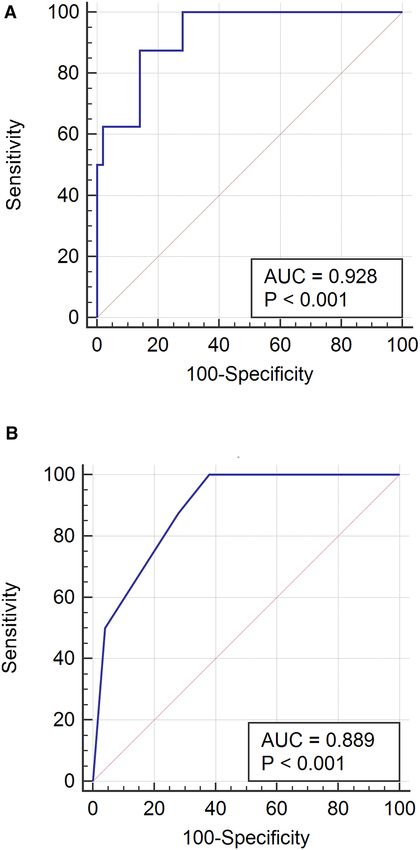
The multivariable logistic regression retained neutrophil count and respiratory rate as predictors of intubation
(Table 2, Fig. 1). A full multivariate model including both as continuous variables generated an AUROC of
0.928 (95% CI 0.828–0.979, p < 0.0001) (Fig. 1A). A simplified model using the categorical variables neutrophil
count > 4 × 10⁹/L and respiratory rate > 18 breaths per minute had an AUROC of 0.889 (95% CI 0.779–0.956,
p = 0.0002) (Fig. 1B). These criteria offered excellent negative predictive value, as neutrophilia and tachypnea
were absent in 31/50 (62%) of patients not requiring intubation while 0/8 (0%) intubated patients met these
criteria. Conversely, 4/8 requiring intubation had neutrophilia and tachypnoea, which were present in only 2/50
(4%) who did not.
Details of mechanically ventilated patients. On 25 February 2020, twelve patients required mechani-
cal ventilation out of a total of 91 COVID-19 patients in Singapore for a risk of 13.1%. Due to a short duration
of follow-up time, two ICU patients were not included in this study.
For the ten included patients, median time from symptom onset to hospital admission was 8.5 days (IQR
7–9) and median time to intubation after admission was 1 day (range, 0.8–3.3), with two intubated in the emer-
gency department (Supplementary Table E2). The median APACHE II score was 19 (IQR 17–22) and SOFA
score 8 (IQR 5–10) (Supplementary Table E3). Nine patients met ARDS criteria on transfer to ICU and one had
acute respiratory injury without other complications. The median PaO2/FiO2 ratio before intubation was 104
(IQR 89–129) increasing to 168 (IQR 134–217) post-intubation. During the first 24 h of ventilatory support,
the median peak FiO2 was 0.75 (IQR 0.6–1.0), positive end-expiratory pressure (PEEP) 12 (IQR 10–14) and
plateau pressure was 22 (IQR 18–26). Five (50%) patients required paralysis, and two (20%) prone positioning
Scientific Reports | (2021) 11:7477 | https://doi.org/10.1038/s41598-021-81377-3 4
Vol:.(1234567890)www.nature.com/scientificreports/
Figure 1. Summary of univariate odds ratios and multivariable logistic regression model. (A) AUC for full
model 0.928 (95% CI 0.828–0.979, p < 0.0001). (B) Simplified model includes the following categorical variables
as risk factors for intubation: neutrophil count > 4 × 10⁹ L and respiratory rate > 18. AUROC for this model is
0.889 (95% CI 0.779–0.956, p = 0.0002). AUROC: Area under receiver operating characteristic.
to maintain oxygenation. No patient required extracorporeal membrane oxygenation. One patient developed
acute kidney injury requiring renal replacement therapy, three patients developed shock requiring inotropes for
a median of 1 days (IQR 1–5), one had cardiac injury with elevated troponin, and four had nosocomial infec-
tions (one with Candida krusei fungemia, three with ventilator-associated pneumonia with endotracheal aspirate
cultures positive for Serratia marcescens, Enterobacter aerogenes, Pseudomonas aeruginosa and Elizabethkingia
species) (Supplementary Table E4). There was also one complication each of upper limb deep vein thrombosis
and gastrointestinal bleeding.
All mechanically ventilated patients received lopinavir-ritonavir and three received interferon beta-1b. Three
patients were treated with lopinavir-ritonavir and interferon beta 1b (median APACHE II 23, IQR 20–25); two
were still intubated for 24 and 30 days. Seven were treated with lopinavir-ritonavir alone (median APACHE II
18, IQR 16–20) with median duration of intubation of 6 days (IQR 5–7). Four patients received at least one dose
of oseltamivir and all were treated with antibiotics for a median of 7 days (IQR 2–11) (Supplementary Table E4).
Scientific Reports | (2021) 11:7477 | https://doi.org/10.1038/s41598-021-81377-3 5
Vol.:(0123456789)www.nature.com/scientificreports/
No patient received corticosteroids. Respiratory viral multiplex PCR, urinary antigen for pneumococcal and
Legionella and other bacterial cultures were negative.
At the time of writing, eight patients were extubated and two remained on ventilatory support with no
fatalities. Two patients were re-intubated, with one able to be extubated on the second attempt while the sec-
ond remained intubated. Of the eight extubated patients, the median duration of intubation was 6.5 days (IQR
5.5–13); four patients were discharged and four remained in the general ward. Of the two patients still intu-
bated, the duration of intubation for the first is currently 30 days. For the remaining patient, he was extubated
after eleven days but was reintubated after 6 days with a new diagnosis of nosocomial pneumonia. There was
no significant difference in age, APACHE II and SOFA scores, and PaO2/FiO2 ratio between patients who were
extubated and those still intubated.
Radiological findings in critically ill patients. Figures 2, 3 and 4 showed the radiographic images of
three patients and their corresponding computed tomography (CT) images as a representation of imaging find-
ings in the cohort patients who were intubated. Generally, all patients had extensive lung involvement with
predominance in the lower zones on radiographs. CT depicted diffuse ground-glass opacities in the affected
regions with varying degrees of organizing and confluent consolidation that appeared to parallel disease severity.
Discussion
In this case control study of 60 COVID-19 patients, fifty were managed in the general ward and ten in intensive
care. The median time of symptom onset to admission was 4 days and to ventilatory support 8.5 days with ARDS
being the major complication in nine of the ten ICU patients. At presentation to hospital, older age, the pres-
ence of dyspnoea, increased temperature, pulse and respiratory rates, higher leukocyte and neutrophil counts,
increased ALT, CRP and LDH, abnormal chest radiograph, decreased oxygen saturation and lymphocyte counts
were associated with an increased risk of invasive ventilation.
In assessing predictors for intubation, elevated CRP and decreased lymphocyte count were found to be reliable
indicators with AUROC of 0.932 and 0.883 respectively. Additionally, we found a combined model of neutrophil
count > 4 × 10⁹/L and respiratory rate > 18 breaths per minute was a reliable predictor for invasive mechanical
ventilation. These predictors performed best at identifying individuals who did not require ventilation and may be
useful as part of the rapid triage of patients who could be managed as outpatient in the event of a large outbreak
if healthcare resources become stretched.
Two single-centre retrospective reports from Wuhan, China described the clinical features and outcomes in
36 and 52 critically ill COVID-19 patients who required ICU c are5,6. The proportion with ARDS was 61% and
67% with case fatality rates of 26·1% and 62·5% respectively. Evidence from Canada, Singapore and Hong Kong
of critically ill SARS patients and a Saudi Arabian study of MERS patients recorded mortality rates of 43%, 38%,
26% and 58% r espectively13–16. Currently, there have been no deaths in the first ten mechanically ventilated
COVID-19 patients in Singapore although two remain intubated. The APACHE II, SOFA score, PaO2/FiO2
ratio and age range in our patients were comparable to the Wuhan ICU reports.
The main complication in our patients was acute hypoxemic respiratory failure with the imaging patterns con-
sistent with a report of severely ill patients in China17. The development of ARDS is consistent with a report on the
pathological findings in a fatal case of COVID-19 with pulmonary c omplications18. Most patients received supple-
mental oxygen via nasal cannula, Venturi masks and masks with reservoir bags prior to intubation although two
patients were intubated on admission. The PaO2/FiO2 ratio improved marginally after intubation and application
of positive pressure. High PEEP and FiO2 were required to maintain adequate oxygenation in all patients dur-
ing the first 24 h. Despite that, plateau and driving pressures for all patients remained low, suggesting the lungs
remained compliant and hypoxia may result from shunt mechanism. The hypoxemia was responsive to PEEP
with no complications even at higher PEEP settings. No patients required extracorporeal membrane oxygenation.
Compared with reports from Wuhan, the proportion of acute kidney injury (AKI) appeared lower in our
patients (10%) while the incidence of cardiac injury and shock (30%) was similar. In Wuhan, AKI was reported as
a complication in 8,·3–30% of patients in two reports while 30.6% had shock and 22% developed cardiac i njury6.
Hitherto, Singapore has reported 16 deaths from COVID-19, out of which 8 demised in the ICU. The propor-
tion of Singapore patients with severe COVID-19 appeared similar to a report using national data from China
at around 13–14%19. However another recent Chinese study found that only 5% required ICU a dmission20.
Compared with case fatality among critically ill COVID-19 patients from Wuhan5,6, our case fatality rate is low.
This may be due to Singapore’s relatively low number of critically ill patients compared with Wuhan, where
healthcare facilities were overwhelmed. The Ministry of Health, Singapore introduced nationwide policies aimed
at containing local spread and preventing an explosive outbreak. Close contacts of confirmed cases were identi-
fied and quarantined, while all confirmed cases isolated in hospitals.
In a mathematical model of COVID-19 transmission, highly effective contact tracing and case isolation
was found to be adequate to control the overall number of cases and a new outbreak within 3 months21. Due to
the stringent screening processes and compulsory admission of confirmed COVID-19 patients, we were able
to monitor for early deterioration and admit to ICU for elective intubation. The association between health-
care resource availability and outcome may account for significant differences in COVID-19 mortality between
Scientific Reports | (2021) 11:7477 | https://doi.org/10.1038/s41598-021-81377-3 6
Vol:.(1234567890)www.nature.com/scientificreports/
Figure 2. (a) Portable supine CXR shows intubated patient with diffuse mixed patchy ground glass opacities
with consolidation predominantly in the peripheries. There is no zonal predilection, hilar adenopathy or
associated pleural effusion. (b) Axial contrast enhanced CT (CECT) image across the lower zones of the lungs
confirms CXR features. There are multifocal areas of consolidation in the peripheries, notably in the dependent
regions of the lower lobes. Ground glass opacities are more diffusely distributed. Air bronchograms are salient
throughout the lower lobes. There is notable absence of pleural effusions or adenopathy in the mediastinum (not
shown).
Scientific Reports | (2021) 11:7477 | https://doi.org/10.1038/s41598-021-81377-3 7
Vol.:(0123456789)www.nature.com/scientificreports/
Figure 3. (a) Portable supine CXR shows intubated patient with dense consolidation in bilateral middle and
lower zones. There is relative sparing of the upper zones and costophrenic recesses. There is no associated
pleural effusion. (b) Axial CECT image across the middle zone of the lungs shows consolidation with air
bronchograms throughout the affected lobes, particularly bilateral lower lobes. The more anterior segments
show some degree of organizing consolidation (arrow). As with other cases in our series, there is absence of
pleural effusions or adenopathy in the mediastinum (not shown). This pattern of confluent consolidation was a
less typical observation.
Scientific Reports | (2021) 11:7477 | https://doi.org/10.1038/s41598-021-81377-3 8
Vol:.(1234567890)www.nature.com/scientificreports/
Figure 4. (a) Portable supine CXR shows intubated patient with mixed ground glass opacification and
consolidation in the peripheries of the middle and lower zones. There is relative sparing of the central and
upper zones. There is no associated pleural effusion. (b) Axial high resolution reconstructed CECT image across
the middle zone of the lungs shows extensive bilateral lung disease with relative sparing of the anterior lobar
segments. Consolidation is most notable in the superior segments of the lower lobes with smaller foci in the
posterior segment of the right upper lobe. There are ground glass opacities in the lateral regions, distinct from
the non-diseased medial portions of the right upper and left lingular lobe.
Scientific Reports | (2021) 11:7477 | https://doi.org/10.1038/s41598-021-81377-3 9
Vol.:(0123456789)www.nature.com/scientificreports/
different geographical regions around the world, but also highlights the importance of containing an outbreak
to ensure healthcare resources can cope e ffectively22.
Our study has several limitations. First, only 60 patients with confirmed COVID-19 infections were included.
While the variables identified as predictors of invasive mechanical ventilation are clinically plausible, the data
is likely to suffer from over-fitting given these small numbers. Validation in a larger cohort will be necessary to
determine the optimal variable selection. Second, we only analysed patients requiring invasive mechanical ven-
tilation and did not include a small number of patients who had high oxygen requirements without ventilatory
support. The strengths of this study are that it was multicenter and the data prospectively collected. Our report
is the first outside of China with detailed description on critically ill COVID-19 patients and the independent
predictors for invasive mechanical ventilation.
In summary, ARDS was the major complication in critically ill COVID-19 patients. Given adequate resources
and manpower, it is possible to intervene in a timely manner to reduce complications and possibly mortality.
Received: 2 June 2020; Accepted: 14 December 2020
References
1. Huang, C. et al. Clinical features of patients infected with 2019 novel coronavirus in Wuhan, China. Lancet 395(10223), 497–506
(2020).
2. Chen, N. et al. Epidemiological and clinical characteristics of 99 cases of 2019 novel coronavirus pneumonia in Wuhan, China: A
descriptive study. Lancet 395(10223), 507–513 (2020).
3. Zhu, N. et al. A novel coronavirus from patients with pneumonia in China, 2019. N. Engl. J. Med. 382(8), 727–733 (2020).
4. de Wit, E. et al. SARS and MERS: Recent insights into emerging coronaviruses. Nat. Rev. Microbiol. 14, 523–534 (2016).
5. Wang, D. et al. Clinical characteristics of 138 hospitalized patients with 2019 novel coronavirus-infected pneumonia in Wuhan,
China. JAMA 323(11), 1061–1069 (2020).
6. Yang, X. et al. Clinical course and outcomes of critically ill patients with SARS-CoV-2 pneumonia in Wuhan, China: A single-
centered, retrospective, observational study. Lancet Respir. Med. https://doi.org/10.1016/S2213-2600(20)30079-5 (2020).
7. Young, B. E. et al. Epidemiologic features and clinical course of patients infected with SARS-CoV-2 in Singapore. JAMA 323(15),
1488–1489 (2020).
8. Wong, J. E. L., Leo, Y. S. & Tan, C. C. COVID-19 in Singapore-current experience: Critical global issues that require attention and
action. JAMA 323(13), 1243–1244 (2020).
9. ISARIC. International Severe Acute Respiratory and emerging Infection Consortium. https://isaric.org.
10. Force, A. D. T. et al. Acute respiratory distress syndrome: The Berlin definition. JAMA 307, 2526–2533 (2012).
11. Kellum, J. A. et al. Kidney disease: Improving global outcomes (KDIGO) acute kidney injury working group. Clinical practice
guideline for acute kidney injury. Kidney Int. 2, 1 (2012).
12. Russell, C. D., Millar, J. E. & Baillie, J. K. Clinical evidence does not support corticosteroid treatment for 2019-nCoV lung injury.
Lancet 395, 473–475 (2020).
13. Fowler, R. A. et al. Critically ill patients with severe acute respiratory syndrome. JAMA 290, 367–373 (2003).
14. Lew, T. W. et al. Acute respiratory distress syndrome in critically ill patients with severe acute respiratory syndrome. JAMA 290,
374–380 (2003).
15. Gomersall, C. D. et al. Short-term outcome of critically ill patients with severe acute respiratory syndrome. Intens. Care Med. 30,
381–387 (2004).
16. Arabi, Y. M. et al. Clinical course and outcomes of critically ill patients with Middle East respiratory syndrome coronavirus infec-
tion. Ann. Intern. Med. 160, 389–397 (2014).
17. Shi, H. et al. Radiological findings from 81 patients with COVID-19 pneumonia in Wuhan, China: A descriptive study. Lancet
Infect. Dis. 20, 425 (2020).
18. Xu, Z. et al. Pathological findings of COVID-19 associated with acute respiratory distress syndrome. Lancet Respir. Med. 8(4),
420–422 (2020).
19. Wu, Z. & McGoogan, J. M. Characteristics of and important lessons from the coronavirus disease 2019 (COVID-19) outbreak in
China: Summary of a report of 72314 cases from the Chinese center for disease control and prevention. JAMA 323(13), 1239–1242
(2020).
20. Guan, W. J. et al. Clinical characteristics of coronavirus disease 2019 in China. N. Engl. J. Med. 382(18), 1708–1720 (2020).
21. Hellewell, J. et al. Feasibility of controlling COVID-19 outbreaks by isolation of cases and contacts. Lancet Glob. Health 8(4),
488–496 (2020).
22. Ji, Y. et al. Potential association between COVID-19 mortality and health-care resource availability. Lancet Glob. Health 8(4), e480
(2020).
Acknowledgements
We thank all clinical and nursing staff who provided care for the patients at National Centre for Infectious
Diseases, Singapore General Hospital, Changi General Hospital, and Sengkang General Hospital; staff at the
Communicable Diseases Division, Ministry of Health, for contributing to outbreak response and contact tracing;
staff at the National Public Health and Epidemiology Unit, National Centre for Infectious Diseases, for assisting
with data analysis; Associate Professor Sanjay Chotirmall for his comments on the manuscript; for staff in the
Singapore Infectious Disease Clinical Research Network and Infectious Disease Research and Training Office,
National Centre for Infectious Diseases, for coordinating patient recruitment, data entry, logistics management
and assistance. None of these individuals received compensation for their role in the study.
Author contributions
S.H.P., B.E.Y., D.C.L. and T.W.Y conceived the study and participated in its design. B.E.Y. and T.W.Y. analysed
and interpreted the data. S.H.P., T.W.Y., B.E.Y. and D.C.L. drafted the initial manuscript and contributed equally.
All authors contributed to the acquisition of data and final edits of the manuscript. All authors approved the
final version of the manuscript.
Scientific Reports | (2021) 11:7477 | https://doi.org/10.1038/s41598-021-81377-3 10
Vol:.(1234567890)www.nature.com/scientificreports/
Competing interests
The authors declare no competing interests.
Additional information
Supplementary Information The online version contains supplementary material available at https://doi.org/
10.1038/s41598-021-81377-3.
Correspondence and requests for materials should be addressed to T.W.Y.
Reprints and permissions information is available at www.nature.com/reprints.
Publisher’s note Springer Nature remains neutral with regard to jurisdictional claims in published maps and
institutional affiliations.
Open Access This article is licensed under a Creative Commons Attribution 4.0 International
License, which permits use, sharing, adaptation, distribution and reproduction in any medium or
format, as long as you give appropriate credit to the original author(s) and the source, provide a link to the
Creative Commons licence, and indicate if changes were made. The images or other third party material in this
article are included in the article’s Creative Commons licence, unless indicated otherwise in a credit line to the
material. If material is not included in the article’s Creative Commons licence and your intended use is not
permitted by statutory regulation or exceeds the permitted use, you will need to obtain permission directly from
the copyright holder. To view a copy of this licence, visit http://creativecommons.org/licenses/by/4.0/.
© The Author(s) 2021
Singapore 2019 novel coronavirus outbreak research team
Poh Lian Lim2, Brenda Sze Peng Ang2, Cheng Chuan Lee2, Lawrence Soon U. Lee2,
Oon Tek Ng2, Monica Chan2, Kalisvar Marimuthu2, Shawn Vasoo2, Chen Seong Wong2,
Tau Hong Lee2, Sapna Pradip Sadarangani2, Ray Junhao Lin2, Mucheli Sharavan Sadasiv2,
Deborah Hee Ling Ng2, Chiaw Yee Choy2, Glorijoy Shi En Tan2, Yu Kit Tan2,
Sean Wei Xiang Ong2, Stephanie Sutjipto2, Pei Hua Lee2, Jun Yang Tay2, Ding Ying2,
Bo Yan Khoo2, Woo Chiao Tay2, Gabrielle Ng2, Yun Yuan Mah2, Wilnard Tan2,
Sennen Jin Wen Lew1, Raymond Kok Choon Fong4, Helen May Lin Oh4,
Jaime Mei Fong Chien4, Humaira Shafi4, Hau Yiang Cheong4, Darren Cheng Han Teo4,
Thuan Tong Tan5, Ban Hock Tan5, Jenny Guek Hong Low5, Limin Wijaya5,
Indumathi Venkatachalam5, Ying Ying Chua5, Benjamin Pei Zhi Cherng5, Yvonne Fu Zi Chan5,
Ghee Chee Phua5, Ken Junyang Goh5, Jade Xiao Jue Soh3, Shuwei Zheng3,
Pushpalatha Bangalore Lingegowda3, Wee Ming Peh3, Yi Lin Lee3, Jun Yang Ho3,
April Yu Jie Chia3, Li Lin8, Say Tat Ooi7, Tambyah Paul Anantharajah6, Jyoti Somani6,
Jolene Ee Ling Oon6 & Gabriel Zherong Yan6
Scientific Reports | (2021) 11:7477 | https://doi.org/10.1038/s41598-021-81377-3 11
Vol.:(0123456789)You can also read