A Straightforward Hypoxic Cell Culture Method Suitable for Standard Incubators - MDPI
←
→
Page content transcription
If your browser does not render page correctly, please read the page content below
Protocol
A Straightforward Hypoxic Cell Culture Method Suitable for
Standard Incubators
Svea Matthiesen † , Rico Jahnke † and Michael R. Knittler *

                                          Institute of Immunology, Friedrich-Loeffler-Institut (FLI), 17493 Greifswald-Insel Riems, Germany;
                                          svea.matthiesen@fli.de (S.M.); rico.jahnke@fli.de (R.J.)
                                          * Correspondence: michael.knittler@fli.de; Tel.: +49-383-5171-170
                                          † These authors contributed equally to the study.

                                          Abstract: We present a new and straightforward method by which standard cell culture plates can be
                                          sealed off from ambient air and be placed under controlled hypoxic cell culture conditions without
                                          costly or highly specialized materials. The method was established on a murine cell culture system
                                          using the dendritic cell line JAWS II but can be readily adapted to other cell cultures. The procedure
                                          was designed to be easy to implement in cell culture laboratories with standard incubators and re-
                                          quires only readily available materials, resources, and consumables, such as six-well plates, degassed
                                          culture medium, CoCl2 , a vacuum sealer, etc., and no further complicated laboratory equipment.
                                          The simple hypoxic cell culture method presented here is technically reliable and experimentally
                                          safe. As it can be performed in any standard incubator, it is suitable for use at both low and higher
                                          biosafety levels.

                                          Keywords: hypoxia; normoxia; cell culture; standard incubator


Citation: Matthiesen, S.; Jahnke, R.;
Knittler, M.R. A Straightforward          1. Introduction
Hypoxic Cell Culture Method                     The oxygen concentration of normal ambient air is 21% (paO2 = 160 mmHg). However,
Suitable for Standard Incubators.         it is well known that the partial pressure of oxygen in the blood decreases significantly
Methods Protoc. 2021, 4, 25. https://
                                          from the arteries to the cells in the tissues [1]. Specifically, while a paO2 of 80–100 mmHg
doi.org/10.3390/mps4020025
                                          is found in the pulmonary vessels, it decreases to 40–60 mmHg in the arterioles and is
                                          only between 1 and 20 mmHg (0.1–3%) in the cells. [1]. O2 serves the mitochondria as a
Academic Editor: Philip Hublitz
                                          terminal electron acceptor for energy production, and one important question in biological
                                          research is how cells adapt to low O2 concentrations (hypoxia), which can develop in
Received: 21 March 2021
Accepted: 6 April 2021
                                          cells/organs during inflammation [2,3] (e.g., after viral or bacterial infections [4,5]), cancer,
Published: 8 April 2021
                                          and metabolic diseases [6,7]. As a specific response to hypoxia, transcriptional upregulation
                                          of specific cellular genes necessary to maintain cellular homeostasis and survival occurs [8].
Publisher’s Note: MDPI stays neutral
                                          Thus, glycolysis is strongly induced in order to facilitate anaerobic ATP synthesis by the
with regard to jurisdictional claims in
                                          cell [9]. Hypoxia-inducible factor-1 (HIF-1: consisting of α and β subunits) plays a crucial
published maps and institutional affil-   role in this process as a master regulator [10]. When cellular hypoxia occurs (≤2%), HIF-1 is
iations.                                  immediately available to the hypoxic cells (within seconds) and enables their survival [11].
                                          However, in the field of infection immunology, research on host–pathogen interaction
                                          in the context of hypoxia is still lacking, but it could be essential for developing future
                                          therapeutic strategies. To study hypoxia in cell culture, sophisticated hypoxia incubators
                                          are usually used to create low-oxygen hypoxic atmospheres; however, this approach is
Copyright: © 2021 by the authors.
Licensee MDPI, Basel, Switzerland.
                                          very investment/cost intensive. For initial pilot experiments and/or research projects
This article is an open access article
                                          that do not focus exclusively on hypoxia, such equipment expenses can be prohibitive.
distributed under the terms and           Moreover, at higher biocontainment levels (BSL3, and 4), a simple technical solution
conditions of the Creative Commons        for hypoxia studies that allows not only safe and easy handling along with appropriate
Attribution (CC BY) license (https://     disposal possibilities represents an important experimental advantage.
creativecommons.org/licenses/by/
4.0/).

Methods Protoc. 2021, 4, 25. https://doi.org/10.3390/mps4020025                                                  https://www.mdpi.com/journal/mps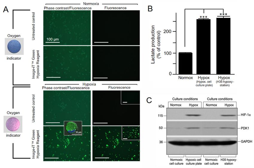
Methods Protoc. 2021, 4, x FOR PEER REVIEW 2 of 11
Methods Protoc. 2021, 4, 25 2 of 11
2. Experimental Design
This article describes a methodical procedure (Figure 1) in which cell culture plates
sealed from ambient
2. Experimental air can be put under hypoxic conditions without significant equip-
Design
ment and This article describesThe
material costs. cell culture
a methodical vessel consists
procedure (Figureof 1)ainstandard
which cell 6-well
culturecellplates
culture
plate,
sealedinfrom
which one of
ambient air the
can besixputavailable wells contains
under hypoxic conditions 0.3 g of oxygen
without significantabsorber
equipment [12] (a
defined mixture of iron powder (≤60% (w/w)), sodium salts (≤20%
and material costs. The cell culture vessel consists of a standard 6-well cell culture plate, (w/w)), and activated
carbon
in which (≤20%
one (w/w)) [13],
of the six which is
available according
wells contains to0.3
manufacturer
g of oxygen able to sustainably
absorber [12] (a definedreduce
Omixture
2 in the entire
of ironvolume
powderof (≤the
60%6-well
(w/w)),plate
sodium(100salts
cm ).(≤Another
3
20% (w/w)),welland
of the plate contains
activated carbon a
(≤20% (w/w))
reversible resazurin/resorufin-based
[13], which is according oxygen indicatorable
to manufacturer [14]to(e.g., AgelessEye),
sustainably reducewhichO2 in themon-
entire 3 ). Another well of the plate contains a reversible
itors thevolume
currentofoxygen
the 6-well plate (in
content (100 cmrange
the ≤ 0.1%–≥0.5%) within the 6-well plate. When
resazurin/resorufin-based
exposed to oxygen, the AgelessEye oxygen indicator
turns from [14]pink
(e.g.,toAgelessEye),
purple to blue whichthen monitors
returnsthe to its
current oxygen content (in the range ≤ 0.1%– ≥ 0.5%) within
original pink color as the oxygen in the cell culture plate is reduced. The remainingthe 6-well plate. When exposed four
to oxygen,
wells of the theculture
AgelessEye
plateturnscanfrom pink to purple
be utilized to blue then
for different cellreturns
cultureto its original pinkDe-
approaches.
color as the oxygen in thecell
gassed/HEPES-buffered cellculture
culturemedium
plate is reduced.
reducesThe the remaining
oxygen tension four wells
beforeof the
the cul-
actual
ture plate can be utilized for different cell culture approaches. Degassed/HEPES-buffered
hypoxia experiment starts. For practical use, the 6-well plate is also evacuated in suitable
cell culture medium reduces the oxygen tension before the actual hypoxia experiment starts.
shrink-wrap by a vacuum sealer machine, and thus a large part of the atmospheric O2 is
For practical use, the 6-well plate is also evacuated in suitable shrink-wrap by a vacuum
already reduced. Sterile “plastic spacers” (placed between the lid and the 6-well plate)
sealer machine, and thus a large part of the atmospheric O2 is already reduced. Sterile
guarantee that the
“plastic spacers” entirebetween
(placed cell culture
the lidvessel
and the with all plate)
6-well six round chambers
guarantee that thehasentire
a uniform
cell
hypoxic atmosphere. To maintain the hypoxic cellular state when
culture vessel with all six round chambers has a uniform hypoxic atmosphere. To maintain opening the 6-well plate
after culturing, 100 µM CoCl 2 is added preventively to the medium
the hypoxic cellular state when opening the 6-well plate after culturing, 100 µM CoCl2 and all other solutions
and buffers.
is added This stabilizes
preventively to thethe cellularand
medium hypoxia
all other regulator
solutions HIF-1α and the
and buffers. cellular
This down-
stabilizes
stream processes
the cellular hypoxia controlled
regulatorbyHIF-1α it [15].andThe thecell cultures’
cellular hypoxic processes
downstream status cancontrolled
be directly
monitored
by it [15]. by TheAgelessEye,
cell cultures’ fluorescent hypoxia
hypoxic status canlive cell dye,monitored
be directly increased by glycolytic
AgelessEye, lactate
fluorescentor
production hypoxia
HIF-1αlive cell dye, increased
stabilization, and pyruvate glycolytic lactate production
dehydrogenase kinase or HIF-1α induc-
1 (PDK1) sta-
bilization, and pyruvate dehydrogenase kinase 1 (PDK1) induction.
tion. The advantages of this new simple method are as follows: fast and stable generation The advantages of
this new simple method are as follows: fast and stable generation
of a hypoxic cell culture environment, availability of required material, simple and safe of a hypoxic cell culture
environment,
disposal of theavailability
device after of the
required
entirematerial,
experiment simple is and safe disposal
finished, as well of asthe device after
cheapness along
the entire experiment is finished, as well as cheapness along
with relatively low time costs for preparation of the sealed hypoxic cell culture with relatively low time costs
plate.
for preparation of the sealed hypoxic cell culture plate.
Figure 1. Sealed hypoxic cell culture plate. For hypoxic cell culture conditions, JAWS II cells are seeded under normoxic
conditions
Figure in degassed,
1. Sealed HEPES-buffered
hypoxic cell culture plate. complete
For hypoxic IMDMcell (selected wells are additionally
culture conditions, JAWS II cells supplemented
are seeded underwith CoCl 2 ).
normoxic
Moreover,inandegassed,
conditions oxygen absorber and an oxygen
HEPES-buffered sensorIMDM
complete (AgelessEye) are wells
(selected addedareto the 6-well platesupplemented
additionally to establish and monitor
with CoCl2).
the low oxygen
Moreover, environment,
an oxygen absorber respectively.
and an oxygen Two sterile(AgelessEye)
sensor 0.75 mm plastic arespacers
added placed on top plate
to the 6-well of theto
6-well plate and
establish allow gas
monitor
theexchange
low oxygen environment,
between respectively.
the different wells of theTwo
cell sterile
culture0.75 mm
plate. plastic
After spacers
the cell placed
culture plateon top of with
is closed the 6-well plate
the lid, it is allow
placedgas
exchange between
in a vacuum bag,the differentfrom
evacuated wells of the cell
ambient culture
air, and plate.
sealed usingAfter the cell sealing
a vacuum culture machine.
plate is closed with
Finally, thethe lid, hypoxic
sealed it is placed
in acell
vacuum evacuated from ambient air, and sealed using◦
culture plate is then placed in a standard incubator at 37 C. When exposed to low oxygen (≤0.5% O2 concentration),cell
bag, a vacuum sealing machine. Finally, the sealed hypoxic
culture plate issensor
the oxygen then placed in a standard
AgelessEye turns fromincubator at 37This
blue to pink. °C. When exposed
color change cantobelow oxygen (≤0.5% O2 concentration),
controlled/monitored throughout thethe
oxygen sensor AgelessEye
cultivation of the cells. turns from blue to pink. This color change can be controlled/monitored throughout the culti-
vation of the cells.Methods Protoc. 2021, 4, 25 3 of 11
2.1. Materials
• 1.5 mL Safe-Lock tubes (Eppendorf, Hamburg, Germany, Cat. No.: 0030120086);
• 15 mL conical tubes (Greiner Bio-One, Frickenhausen, Germany; Cat. No.: 10384601);
• 2-Mercaptoethanol (Roth, Karlsruhe, Germany, Cat. No.: 4227.2);
• 50 mL conical tubes (Greiner Bio-One, Frickenhausen, Germany; Cat. No.: 10711212);
• 6-well plate (Greiner Bio-One, Frickenhausen, Germany; Cat. No.: 657160);
• Acrylamide/bis—Rotiphorese Gel 30 (Roth, Karlsruhe, Germany, Cat. No.: 3029.2);
• AgelessEye (oxygen indicator) (Mitsubishi Gas Chemical Company, Tokyo, Japan;
Distributor: Long Life for Art, Cat. No.: O2INDAEYE);
• Amphotericin B (Thermo Fisher Scientific, Schwerte, Germany, Cat. No.: 15290018);
• Anti-GAPDH antibody (Merk, Darmstadt, Germany, Cat. No.: CB1001-500UG);
• Anti-HIF-1α antibody (Novus Biologicals, Centennial, USA, Distributor: Bio-Techne
GmbH, Wiesbaden, Germany Cat. No.: NB100-479);
• Anti-mouse IgG-HRP (Jackson ImmunoResearch, West Grove, U.S.A., Distributor:
Dianova, Hamburg, Germany, Cat. No.: 115-035-146);
• Anti-rabbit IgG-HRP (Jackson ImmunoResearch, West Grove, U.S.A., Distributor:
Dianova, Hamburg, Germany, Cat. No.: 111-035-144);
• Anti-PDK1 antibody (Novus Biologicals, Centennial, USA, Distributor: Bio-Techne
GmbH, Wiesbaden, Germany, Cat. No.: NB100-2384SS);
• APS (Ammonium peroxodisulfate) (Roth, Karlsruhe, Germany, Cat. No.: 9178.2);
• Blotting filter paper MN 218 B (Macherey-Nagel, Düren, Germany, Cat. No.: 742113);
• Bromophenol blue (Sigma-Aldrich, Munich, Germany, Cat. No.: B0126-25G);
• CoCl2 (cobalt chloride) Merk, Darmstadt, Germany; Cat. No.: 232696-5G);
• FCS (fetal calf serum), 0.2 µm sterile filtered (Pan Biotech, Aidenbach, Germany, Cat.
No.: P30-3302);
• Formaldehyde, 37% (Roth, Karlsruhe, Germany, Cat. No.: 6967.2);
• Glycerin (Roth, Karlsruhe, Germany, Cat. No.: 6967.2);
• Glycine (Roth, Karlsruhe, Germany, Cat. No.: T873.2);
• HCl (hydrogen chloride), 37% (Roth, Karlsruhe, Germany, Cat. No.: 4625.1);
• HEPES (4-(2-hydroxyethyl)-1-piperazineethanesulfonic acid) (Serva, Heidelberg, Ger-
many; Cat. No.: 25245.02);
• Image-iT Green Hypoxia Reagent (Fisher Scientific, Schwerte, Germany, Cat. No.: 15940773);
• IMDM (Iscove’s Modified Dulbecco’s) Medium (Fisher Scientific, Schwerte, Germany;
Cat. No.: GibcoTM21980065);
• JAWS II (American Type Culture Collection (ATCC), Manassas, U.S.A., Cat. No.: CRL-
11904);
• KCl (potassium chloride) (Roth, Karlsruhe, Germany, Cat. No.: 6781.1);
• KH2 PO4 (potassium hydrogen phosphate) (Roth, Karlsruhe, Germany, Cat. No.: 3904.1);
• L-lactate-assay-kit (Sigma-Aldrich, Cat. No.: MAK329-1KT);
• MINI-Vertical Spacer, 0.75 mm (Roth, Karlsruhe, Germany Cat. No.: N624.1);
• Mouse GM-CSF (granulocyte-macrophage colony-stimulating factor), recombinant
protein (Thermo Fisher Scientific, Schwerte, Germany, Cat. No.: PMC2011);
• Na2 HPO4 (sodium hydrogen phosphate) (Roth, Karlsruhe, Germany, Cat. No.: 4984.1);
• NaCl (sodium chloride) (Roth, Karlsruhe, Germany, Cat. No.: 3957.1);
• Nitrocellulose membrane, 0.45µm (Hartenstein, Würzburg, Germany, Cat. No.: 10600002);
• Oxygen absorber (O2frepak, Guangdong, China; Distributor: Amazon, Germany, Cat.
No.: O2frepak 100CC);
• Penicillin–streptomycin (5000 U/mL) (Thermo Fisher Scientific, Schwerte, Germany,
Cat. No.: 15070063);
• Pierce ECL Western Blotting Substrate (Thermo Fisher Scientific, Schwerte, Germany,
Cat. No.: 32209);
• Pipette Tips, 1–200 µL (Thermo Fisher Scientific, Schwerte, Germany, Cat. No.: 11923446);
• Pipette Tips, 200–1000 µL (Greiner Bio-One, Frickenhausen, Germany, Cat. No.: 10557071);Methods Protoc. 2021, 4, 25 4 of 11
• Polyoxyethylene (20)-sorbitan monolaurate (Tween 20) (VWR, Darmstadt, Germany,
Cat. No.: M147-1L);
• Ponceau S (Roth, Karlsruhe, Germany, Cat. No.: 5938.2);
• Prestained protein ladder (Thermo Fisher Scientific, Schwerte, Germany Cat. No.: 26616);
• ProLong™ Gold antifade reagent with DAPI (mounting medium) (Thermo Fisher
Scientific/Invitrogen, Schwerte, Germany, Cat. No.: P36931);
• Protease inhibitor (cOmplete ULTRA Tablets, EDTA-free) (Roche, Basel, Switzerland,
Distributor: Sigma-Aldrich, Munich, Germany, Cat. No.: 5892953001);
• Scalpel (Roth Karlsruhe, Germany, Cat. No.: X004.1);
• SDS (sodium dodecyl sulfate) (Roth, Karlsruhe, Germany, Cat. No.: 2326.2);
• Skim milk powder (Heirler-Cenovis, Radolfzell, Germany, Cat. No.: 4010318030305);
• TEMED (N, N, N0 , N0 - tetramethylethylene-diamine) (Roth, Karlsruhe, Germany, Cat.
No.: 2367.3);
• Transparent foil (Roth Karlsruhe, Germany, Cat. No.: 1255.1);
• Trichloroacetic acid (TCA)-Deproteinization Kit—Deproteinizing Sample Preparation
Kit II (Sigma-Aldrich, Munich, Germany, Cat. No.: MAK342-1KT);
• Tris (Thermo Fisher Scientific, Schwerte, Germany, Cat. No.: 15504020);
• Trypan blue (Roth, Karlsruhe, Germany, Cat. No.: CN76.2);
• Trypsin-EDTA (Ethylenediaminetetraacetic acid) solution (Sigma-Aldrich, Munich,
Germany, Cat. No.: E8008-100ML);
• Urea (Roth Karlsruhe, Germany, Cat. No.: 7638.1);
• Vacuum bags (Shenzhen Green Electrical Appliance Co., Ltd, Guangdong, China;
Distributor: Amazon; Cat. No.: 11 × 16/100);
• X-ray film, UV/blue sensitive Super RX-N (Fujifilm Europe GmbH, Düsseldorf, Ger-
many, Distributor Hartenstein, Würzburg, Germany, Cat. No.: RF11).
2.2. Equipment
• Bio-Rad Trans-Blot Cell (Bio-Rad Laboratories GmbH, Feldkirchen, Germany, Cat.
No.: 1703930);
• Biological safety cabinet—Berner Flow Safe (Berner, Elmshorn, Germany, Cat. No.:
B-[MaxPro]2 -130);
• Centrifuge—Eppendorf 5420 (Eppendorf, Hamburg, Germany, Cat. No.: 5420000318);
• Centrifuge—Hettich Rotina 380R (Hettich, Vlotho, Germany, Cat. No.: 1706);
• Digital camera—Panasonic DMC-TZ18 Lumix (Panasonic Germany, Wiesbaden, Ger-
many, Cat. No.: EAN 5025232608850);
• ELISA Reader—Sunrise Remote (Tecan, Gröding, Austria, Cat. No.: F039300);
• Film developer—Compact 2 (PROTEC GmbH & Co. KG, Oberstenfeld, Germany, Cat.
No.: 11951-1111-5810);
• Fluorescence microscope—Zeiss Axiovert 200 with ApoTome unit (Carl Zeiss Light
Microscopy, Göttingen, Germany, Cat. No.: Axiovert 200 including ApoTome 423660-
0000-00);
• Heating block—Thermomixer comfort 5355 (Eppendorf, Hamburg, Germany Cat. No.:
022670107);
• Hypoxia Cabin—Whitley H35 Hypoxystation (Don Whitley Scientific Pty Ltd, Bingley,
GB, Cat. No.: MEA06060);
• Incubator—Sanyo MCO-19AIC CO2 (Sanyo E & E, Europe B.V., The Netherlands, Cat.
No.: 5519188);
• Inverted microscope—ECLIPSE TS100-F (Nikon, Düsseldorf, Germany, Cat. No.: TS100-F);
• Microscope camera—DFK 21AU04.AS (The Imaging Source Europe GmbH, Bremen,
Germany, Cat. No.: DFK 41AU02);
• Microscope LED illumination source—CoolLED pe-200 (CoolLED, Andover, G.B., Cat.
No.: CoolLED pE-200);
• Neubauer counting chamber (Roth, Karlsruhe, Germany, Cat. No.: T728.1);Methods Protoc. 2021, 4, 25 5 of 11
• pH meter—Hanna Checker (HANNA Instruments, Vöhringen, Germany, Cat. No.:
Z35109);
• Pipette 100–1000 µL (Eppendorf, Hamburg, Germany, Cat. No.: 3123000063);
• Pipette 20–200 µL (Eppendorf, Hamburg, Germany, Cat. No.: 3123000055);
• Pipette 2–20 µL (Eppendorf, Hamburg, Germany, Cat. No.: 3123000098);
• Power supply—Bio-Rad PowerPac Universal Power Supply (Bio-Rad Laboratories
GmbH, Feldkirchen, Germany Cat. No.: 1645070);
• Rocking platform—Heidolph Duomax 1030 (Heidolph Instruments GmbH & CO. KG,
Schwabach, Germany, Cat. No.: 543-32205-00);
• Ultrasound cleaning unit—Elmasonic S40 (SKSONIC, Mörfelden-Walldorf, Germany,
Cat. No.: 1004635);
• Vacuum device—Univapo 150 H & Unijet II Refrigerated Aspirator (Uniequip, Freital,
Germany, Cat. No.: Univapo 150 H & Unijet II 20710);
• Vacuum sealer- Kitchenboss (Shenzhen Green Electrical Appliance Co., Ltd, Guang-
dong, China; Distributor: Amazon, Cat. No.: G200/silver);
• Vertical electrophoresis chamber-Mini-PROTEAN Tetra Cell (Bio-Rad Laboratories
GmbH, Feldkirchen, Germany, Cat. No.: 1658000).
3. Procedure
3.1. Preparation and Performance of Hypoxic Cell Culture Experiment—Time for
Completion: 5–72 h
1. Seed 1 × 10⁵ cells per well in degassed HEPES-buffered complete IMDM (with or
without 100 µM CoCl2 ) in four wells of a 6-well plate. This first step ensures efficient
cell adherence and adaptation. CoCl2 blocks any degradation of the hypoxia factor
HIF-1α under normoxic conditions [15] when the hypoxic cell culture plate is opened
later (see Sections 3.2.2 and 3.2.3).
2. After 1 h of incubation of the cells under normoxic conditions at 37 ◦ C and 7.5% CO2 ,
add 0.3 g of oxygen absorber to the fifth well and a single AgelessEye indicator to the
sixth well of the 6-well plate (Figure 1).
3. To allow gas exchange inside the 6-well plate, place two sterile 0.75 mm plastic spacers
in a parallel alignment across the top and bottom three wells of the open cell culture
plate and close it carefully with the corresponding lid (Figure 1).
4. To microscopically verify the hypoxic conditions and the cultured cells’ hypoxic state,
supplement one of the cell-containing wells with Image-iT Green Hypoxia Reagent
(final concentration of 5 µM). This fluorogenic compound is live cell-permeable and
emits a green fluorescence in hypoxic environments.
5. Put the assembled cell culture plate into a vacuum bag. Insert the bag’s open end into
the vacuum sealing machine and evacuate the air within the experimental unit to a
low vacuum (Figure 1).
6. Place the assembled and evacuated hypoxic cell culture plate into a standard incubator
and cultivate the cells for 5 h (or longer, e.g., 48 or 72 h) at 37 ◦ C. As a control, a 6-
well plate with corresponding cells (in the presence and absence of Image-iT Green
Hypoxia Reagent) can be cultivated with complete IMDM in parallel for the same
time under normoxic conditions. Further, for initial validation of the new hypoxic
cell culture method, we additionally used a Whitley H35 hypoxystation (standard
setting: ≤2% O2 , 7.5% CO2 , 90.5% N2 ) in control experiments.
7. After 30 min of incubation, control the oxygen concentration in the sealed culture
plates visually by the color of the AgelessEye (it should be constant at 0.2–0.3%,
corresponding to the range of oxygen values found in tissues and cells in vivo [1]).Methods Protoc. 2021, 4, 25 6 of 11
3.2. Experimental Verification of Hypoxic Cell Culture Conditions by Immunofluorescence, Lactate
Assay, and Immune Blot Analysis—Time for Completion Depends on Cultivation Period and
Respective Downstream Analyses
3.2.1. Detection of Low Oxygen Concentration in Living Cells by Hypoxia-Sensitive
Fluorescence Dye after 5 h of Hypoxic Cultivation
1. After 5 h of hypoxic cultivation, remove the hypoxic cell culture device from the
incubator, cut the vacuum bag at the sealed end, and remove the 6-well plate. When
exposed to oxygen, the AgelessEye will turn from pink to purple and then blue
(≥0.5%).
2. To distinguish living from dead cells, dispense 500 µL trypsin-EDTA solution into one
of the culture vessels to completely cover the cells and place in the incubator at 37 ◦ C
for up to 5 min. This allows the cells to detach from the culture plate surface. This can
be checked with an inverted microscope. When this is complete, all cells will be in
suspension. Add complete IMDM (Iscove’s Modified Dulbecco’s Medium) containing
FCS (Fetal Calf Serum) to the cell suspension to inhibit further tryptic activity. Mix
one part of 0.4% trypan blue solution with one part cell suspension. Allow mixture to
incubate for 3 min at room temperature and analyze by light microscopy. Do this for
later analysis time points (e.g., for 48 and 72 h) as well.
3. Image the cells that were pretreated with Image-iT Green Hypoxia Reagent in the ves-
sel using the fluorescence microscope with excitation/emission 488/520 nm
(a standard FITC/GFP (Fluorescein Isothiocyanate/Green Fluorescent Protein) excita-
tion/emission filter set is recommended). If necessary, the fluorescent cells can also be
fixed with 2% formaldehyde. Fluorescence of the fixed cells lasts max. 24 h (afterward,
it is hardly detectable). If cells are also grown on coverslips, they can be viewed at
higher magnification with an appropriate fluorescence microscope (e.g., ApoTome
microscope) after respective fixation and embedding in mounting medium.
3.2.2. Measurement of Increased Glycolytic Lactate Production after 48 h of
Hypoxic Cultivation
1. After 48 h of cultivation under norm- and hypoxia, monitor the cells for increased
glycolysis via an L-lactate assay.
2. Remove the medium from the culture vessel by aspiration and wash cells with
cold PBS.
3. Dispense 500 µL trypsin-EDTA (Ethylenediamine Tetraacetic Acid) solution into the
culture vessel and place it in the incubator at 37 ◦ C for up to 5 min to detach the cells
from the culture plate surface. When this is complete, add complete IMDM containing
FCS to the cell suspension to inhibit further tryptic activity.
4. Subsequently, determine the cell number by a Neubauer counting chamber.
5. Wash the detached/harvested cells by centrifugation at 200× g for 5 min and resus-
pend them in PBS at a concentration of 1 × 10⁵ cells/100 µL.
6. After further centrifugation of 300 µL cell suspension at 200× g for 5 min, resuspend
the cell pellet in 200 µL of lactate assay buffer provided with the kit.
7. Homogenize the cells quickly by pipetting up and down ten times.
8. Centrifuge for 5 min at 4 ◦ C and 1000× g in a cold centrifuge to remove insoluble material.
9. Deproteinize the resulting supernatants using a protein precipitating TCA (Tricar-
boxylic acid) kit according to the manufacturer’s protocol.
10. For the further steps of the L-lactate assay, follow the detailed protocol of the man-
ufacturer. The colorimetric response of the assay was measured with an ELISA
(Enzyme-Linked Immunosorbent Assay) reader at a wavelength of 450 nm.
3.2.3. Detection of CoCl2 -Stabilized HIF-1a Levels and PDK1 (Pyruvate Dehydrogenase
Kinase 1) Induction in Cell Extracts after 72 h of Hypoxic Cultivation
1. For the preparation of cell lysates used for SDS-PAGE (Sodium Dodecyl Sulfate-
Polyacrylamide Gel Electrophoresis)/immunoblot analysis, detach the cells with a
trypsin-EDTA solution as described above.Methods Protoc. 2021, 4, 25 7 of 11
2. Collect the detached cells in a centrifuge tube and centrifuge at 400× g for 5 min at 4 ◦ C.
3. Resuspend the resulting cell pellet in 1 mL PBS centrifuge again at 400× g for 5 min at
4 ◦ C. Subsequently, determine the cell number by a Neubauer counting chamber and
resuspend the cell pellet in HIF-lysis buffer at a concentration of 1 × 10⁵ cells/100 µL.
4. Perform the cell lysis on ice for 30 min. Centrifuge the lysed cells at 1000× g and 4 ◦ C
for 30 min. Nuclei and cell debris will form a pellet so that the lysate supernatant can
be transferred to a new reaction tube.
5. For denaturation and complexation with SDS, add one volume of 2× SDS sample
buffer to the cell lysates and boil for 10 min in a heating block at 95 ◦ C.
6. Load the polyacrylamide gel with the respective samples (containing an equal quan-
tity of cell lysates (5 µL)) and a molecular weight marker (prestained protein ladder).
7. Run the gel at 100 V until the dye front migrates from the stacking into the running
gel (15 min) and increase to 200 V until the dye front reaches the bottom of the gel
(45 min).
8. Remove the gel from the apparatus, spacers, and glass plates and equilibrate it by
soaking in transfer buffer for 2 min.
9. Prepare the nitrocellulose membrane by wetting it in transfer buffer for 30 s. Han-
dle the membrane carefully, ideally with rounded tweezers, to avoid scratching or
puncturing the surface.
10. Soak blotting filter papers and sponges in the transfer buffer for 5 min.
11. Starting on the side facing the cathode, sequentially assemble the following compo-
nents: sponge, filter paper, gel, nitrocellulose membrane, filter paper, sponge. Gently
remove any air bubbles with a roller or serological pipette. Bubbles between the gel
and the membrane will inhibit the transfer of proteins to the membrane.
12. Place the completed transfer stack into a transfer cassette and perform wet transfer
according to the manufacturer’s instructions for the blotting apparatus.
13. After transfer, rinse the membrane briefly in distilled water. Gently mark the position
of the molecular weight ladder bands with a pencil for size detection. Using a scalpel,
cut the membrane horizontally at the level of the 40 and 70 KDa markers of the
prestained protein ladder.
14. Stain the membrane with Ponceau S for 30 s and then rinse briefly with distilled water
to visualize protein bands and confirm that the protein transfer was successful. Wash
away Ponceau S with several washes in PBS until the membrane is clear. Incubate
membrane in PBS/0.1% (v/v) Tween 20/5% (w/v) milk powder solution for 1 h at
room temperature with constant rocking.
15. Dilute the primary antibodies to the working concentration (anti-HIF-1α, 1:500; anti-
PDK1, 1:500; anti-GAPDH, 1:1000) in PBS/0.1% Tween 20/10% (v/v) FCS.
16. Incubate the membrane in primary antibody solutions for 2 h at room temperature
with gentle rocking (the upper part of the membrane with anti-HIF-1α, the middle
part with anti-PDK1, and the lower part with anti-GAPDH).
17. Wash the membrane with PBS/0.1% (v/v) Tween 20 solution three times for 10 min
each with gentle rocking.
18. Incubate the membrane with secondary antibody (goat anti-rabbit-HRP, 1:1000; goat
anti-mouse-HRP, 1:1000) in PBS/0.1% (v/v) Tween 20 for 1 h at room temperature
with gentle rocking (the upper/middle parts with anti-rabbit-HRP, and the lower part
with anti-mouse-HRP).
19. Wash the membrane in PBS/0.1% (v/v) Tween 20 three times for 10 min each with
gentle rocking.
20. Prepare the enhanced ECL (chemiluminescence) substrate just before use according
to the manufacturer’s instructions.
21. According to the manufacturer’s suggestions, incubate the membrane in the substrate
(typical incubation times are 10 s to 2 min).
22. Carefully remove the membrane from the detection reagent and sandwich it between
layers of plastic (i.e., a sheet protector or plastic wrap).Methods Protoc. 2021, 4, 25 8 of 11
23. Expose the membrane to autoradiography film in a dark room.
24. After the exposure is complete, place the film into the developer and wait until it is
processed.
25. Once the film has been developed, overlay it back on your blot to mark the position
of the protein ladder with a marker.
4. Expected Results
During incubation of the sealed hypoxic cell culture plate in a standard incubator at
37 ◦ C, the AgelessEye hypoxia sensor was used to visually monitor oxygen levels. Inspec-
tion at the beginning, middle, and end of the cell cultivation revealed that the sensor’s
color remained light pink (0.2–0.3% O2 ) at all times, indicating a constant low oxygen
concentration in the sealed culture plate. Moreover, trypan blue staining showed 91, 89,
and 85% intact live cells after 5, 48, and 72 h of hypoxic incubation, respectively. This
corresponds well to the survival rates obtained for cells cultured in the commercial hypoxys-
tation. Microscopic analysis of the hypoxic cultivation conditions in the sealed hypoxic
cell culture plates was performed for live JAWS II cells using the Image-iT Green Hypoxia
Reagent, which sustains its fluorescence when cells/tissue return to normal oxygen levels.
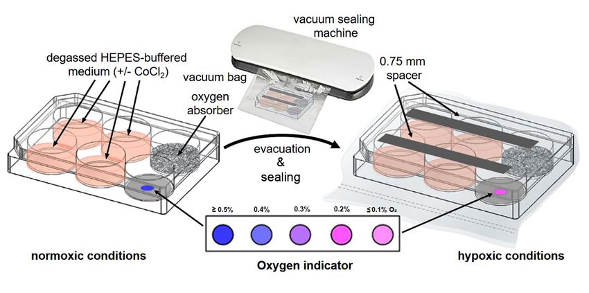
Figure 2A shows JAWS II cultured in the presence or absence of the Image-iT hypoxia
detection reagent under hypoxic and standard normoxic conditions. Only the hypoxic
cell cultures show the appearance of a bright green cellular fluorescence. The low-oxygen
conditions generated in the hypoxic cell culture plate were verified by comparing the ob-
served responses with control experiments using a commercial hypoxystation. As shown
in Figure 2A, the fluorescence staining of cells treated with Image-iT in the hypoxic culture
plate is comparable to those grown in the hypoxystation, suggesting that the two methods
are highly comparable.
Increases in glucose consumption and the glycolytic rate during hypoxia lead to in-
creased conversion of pyruvate to lactate and hence its accumulation in the cytoplasm.
Thus, high lactate levels can be regarded as an indicator of cellular hypoxia. Indeed,
as expected, there were increased levels of lactate in the cells cultured in the sealed hy-
poxic culture plate compared to the cells maintained under standard normoxic conditions
(Figure 2B). Again, no difference was detected between cells cultured in the hypoxic cul-
ture plate or a commercial hypoxystation. Both cases showed a comparable increase in
lactate levels.
Finally, it is well known that CoCl2 enhances the stability of HIF-1α under normoxic
conditions. In our experimental protocol, CoCl2 was added to hypoxic cell cultures to
stabilize HIF-1α (and the cellular processes it controls) after opening the hypoxic cell
culture plate for further downstream work/analyses under normal ambient air conditions.
Immunoblot analyses showed a readily detectable HIF-1α-stabilization for the cells from
the hypoxic culture plate, which was almost the same intensity as that found for hypoxic
cells cultured in the hypoxystation (Figure 2C). No HIF-1a stabilization could be seen
under normoxic cell culture conditions. Moreover, a comparable induction of the second
hypoxia marker (PDK1), which inactivates the mitochondrial TCA cycle enzyme, pyruvate
dehydrogenase (PDH) [16], could also be observed for the JAWS II cells cultured in the
hypoxic cell culture plate and/or the commercial hypoxystation.
Taken together, the sealed hypoxic cell culture plate has proven to be suitable for
performing hypoxic cell culture experiments in standard incubators, and in particular,
the additional use of CoCl2 allows further downstream analyses of HIF-1α related processes.
Previously invented methods and procedures for performing hypoxia experiments in
simple cell culture chambers [17–19] have the additional expense and complexity of gas
mix supply/discharge. This is not necessary for the sealed six-well plate system presented
here and greatly simplifies the entire experimental procedure.Although the presented method has experimental limitations (e.g., a static hypoxic
O2 concentration) and cannot fully replace all the capabilities of a commercial hypoxysta-
tion, it opens new possibilities for the simple and inexpensive experimental application of
hypoxic cell culture conditions in pilot experiments. A further application of this approach
Methods Protoc. 2021, 4, 25
is cell culture work performed under higher biological safety levels (such as BSL3 and 4)
9 of 11
where sophisticated hypoxia chambers are less likely to be available, and where their use
might not be ideal for a variety of practical and safety reasons.
Figure 2.
Figure Cultivation of
2. Cultivation of JAWS
JAWS IIII cells
cells under
under hypoxic
hypoxic cell
cell culture
culture conditions.
conditions. (A)
(A) The
The microscopic
microscopic images
images show
show the
the analysis
analysis
of JAWS
JAWSIIIIcells
cellsafter
after5 5h hofof
cultivation
cultivation under
under hypoxic
hypoxiccellcell
culture conditions
culture in a sealed
conditions hypoxic
in a sealed cell culture
hypoxic plate. plate.
cell culture Untreated
Un-
treated and Image-iT
and Image-iT Green Hypoxia-treated
Green Hypoxia-treated cellsnorm-
cells under under(top)
norm-and(top) and hypoxic
hypoxic (bottom)(bottom)
conditions conditions
are shown areinshown
phase in phase
contrast
contrast
and under and under fluorescence
fluorescence (10× magnification).
(10× magnification). The corresponding
The corresponding color
color of the of the
oxygen oxygen is
indicator indicator
shown on is shown
the left.on the
The left.
lower
The lower left image insert shows a green fluorescent hypoxic cell taken with a Zeiss ApoTome
left image insert shows a green fluorescent hypoxic cell taken with a Zeiss ApoTome fluorescence microscope using a 63× fluorescence microscope
using a 63× oil immersion objective (the cellular nucleus was costained with DAPI, blue). The inserts in the two lower right
oil immersion objective (the cellular nucleus was costained with DAPI, blue). The inserts in the two lower right images
images show corresponding control and Image-iT Green Hypoxia-stained cells cultivated in a commercial hypoxystation.
show corresponding control and Image-iT Green Hypoxia-stained cells cultivated in a commercial hypoxystation. (B) JAWS
(B) JAWS II cells were cultured under norm-, and hypoxic conditions in either the hypoxic cell culture plate or a hypoxys-
II cells as
tation, were culturedfor
indicated, under
48 h norm-, and hypoxic
after which conditions
cellular lactate was in either the
measured hypoxic
with cell culture
an L-lactate assayplate or aphypoxystation,
kit (*** < 0.001 versus
as indicated,
controls; for
n = 3). 48Immunoblot
(C) h after which cellularoflactate
analysis HIF-1αwas measured
stability with induction
and PDK1 an L-lactate assay kit
in JAWS II cells p < 0.001
(*** under norm-versus
andcontrols;
hypoxic
n = 3).
cell (C) Immunoblot
culture conditions after analysis of cultivation
72 h of HIF-1α stability
in theand PDK1
hypoxic induction
cell in JAWS
culture plate II cells
(second lane)under
and norm- and hypoxic
a commercial cell
hypoxys-
culture(fourth
tation conditions
lane).after
GAPDH 72 h of cultivation
was includedin asthe hypoxic
a sample cell culture
loading plate (second lane) and a commercial hypoxystation
control.
(fourth lane). GAPDH was included as a sample loading control.
Although the presented method has experimental limitations (e.g., a static hypoxic O2
concentration) and cannot fully replace all the capabilities of a commercial hypoxystation,
it opens new possibilities for the simple and inexpensive experimental application of
hypoxic cell culture conditions in pilot experiments. A further application of this approach
is cell culture work performed under higher biological safety levels (such as BSL3 and 4)
where sophisticated hypoxia chambers are less likely to be available, and where their use
might not be ideal for a variety of practical and safety reasons.
5. Reagents Setup
Blotting buffer: 25 mM Tris–HCl (pH 7.5), 190 mM glycine. Store at room temperature.
Complete IMDM with 25 mM HEPES and 20% FCS: Add 2.95 g of HEPES and 100 mL
of FCS to 400 mL of IMDM (supplemented with penicillin–streptomycin (100 U/mL)),
amphotericin B (2.5 µg/mL), and GM-CSF (5 ng/mL mouse GM-CSF). Adjust to pH 7.4
with HCl.Methods Protoc. 2021, 4, 25 10 of 11
Electrophoresis buffer: 25 mM Tris–HCl (pH 7.5), 190 mM glycine, 10% (v/v) SDS. Store at
room temperature.
HIF-Lysis buffer: 6.65 M urea, 10 mM Tris (pH 6.8), 1% SDS, 10% glycerin, and one tablet
of cOmplete protease inhibitor per 50 mL of lysis buffer.
Immunoblot blocking buffer: 1× PBS, 0.1% (v/v) Tween 20, 5% (w/v) milk powder. Prepare
freshly (short-term storage on ice).
Immunoblot washing buffer: 1× PBS, 0.3% (v/v) Tween 20. Store at room temperature.
Loading buffer: 10 mM Tris–HCl (pH 6.8); 6.6 M urea; 1% (v/v) SDS; 10% (v/v) glycerin;
0.25% (w/v) bromophenol blue; 7% (v/v) 2-mercaptoethanol, and one tablet of cOmplete
protease inhibitor per 50 mL of loading buffer.
PBS (phosphate-buffered saline): For 1000 mL of 1× PBS, start with 800 mL of distilled
water. Then, add 8 g of NaCl, 0.2 g of KCl, 1.44 g of Na2 HPO4, and 0.24 g of KH2 PO4 .
Adjust the pH to 7.4 with HCl. Add distilled water to a total volume of 1000 mL (final
concentration of 137 mM NaCl, 10 mM phosphate, 2.7 mM KCl). Sterilize the solution
by autoclaving for 30 min and degas the buffer by sonication (in an ultrasonic bath for
45 min at room temperature and by applying a vacuum for 60 min. Store the PBS buffer at
room temperature.
Ponceau S solution: 0.5% (w/v) Ponceau S dissolved in 1% (v/v) acetic acid.
Primary and secondary antibody incubation buffer: 1× PBS, 0.1% (v/v) Tween 20, 10% (v/v)
fetal calf serum (FCS). Prepare fresh (short-term storage on ice until use).
Separating gel (10%): Mix in the following order: H2 O: 4.1 mL; acrylamide/bis (30% 37.5:1):
3.3 mL; Tris–HCl (1.5 M, pH 8.8): 2.5 mL; 10% (v/v) SDS: 100 µL; TEMED: 10 µL; 10% (v/v)
ammonium persulfate (APS): 32 µL.
Stacking gel (4%): Mix in the following order: H2 O: 6.1 mL; acrylamide/bis (30%, 37.5:1):
1.3 l; Tris–HCl (0.5 M, pH 6.8): 2.5 mL; 10% (v/v) SDS: 100 µL; TEMED: 10 µL; 10% (v/v)
APS: 100 µL.
Transfer buffer: 25 mM Tris–HCl (pH 7.5), 190 mM glycine. Store at room temperature.
Trypan Blue Solution: Dissolve 0.4 g of Trypan Blue in 80 mL of 1× PBS and bring to a slow
boil. Cool to room temperature and add PBS to a final volume of 100 mL. Store at room
temperature.
Author Contributions: Conceptualization, M.R.K.; validation, S.M., R.J. and M.R.K.; writing—
original draft preparation, M.R.K.; writing—review and editing, S.M., R.J., and M.R.K.; visualization
S.M., R.J., and M.R.K.; supervision, M.R.K.; methodology, S.M. and M.R.K.; funding acquisition,
M.R.K. All authors have read and agreed to the published version of the manuscript.
Funding: The German Federal Ministry of Education and Research (Bundesministerium für Bil-
dung und Forschung, BMBF) under project number 01KI1726C of “Q-GAPS” as part of the “Re-
search Network Zoonotic Infectious Diseases” is gratefully acknowledged for financial support (S.M.
and M.R.K.).
Institutional Review Board Statement: Not applicable.
Informed Consent Statement: Not applicable.
Data Availability Statement: All data generated or analyzed during this study are included in this
published article.
Acknowledgments: Yolanda Marschner is acknowledged for her technical assistance. We are grateful
to Allison Groseth for her critical reading of the manuscript.
Conflicts of Interest: The authors declare no conflict of interest.
References
1. Wenger, R.H.; Kurtcuoglu, V.; Scholz, C.C.; Marti, H.H.; Hoogewijs, D. Frequently asked questions in hypoxia research. Hypoxia
2015, 3, 35–43. [CrossRef] [PubMed]
2. Palazon, A.; Goldrath, A.W.; Nizet, V.; Johnson, R.S. HIF Transcription factors, inflammation, and immunity. Immunity 2014, 41,
518–528. [CrossRef] [PubMed]Methods Protoc. 2021, 4, 25 11 of 11
3. Taylor, C.T.; Colgan, S.P. Regulation of immunity and inflammation by hypoxia in immunological niches. Nat. Rev. Immunol.
2017, 17, 774–785. [CrossRef] [PubMed]
4. Krzywinska, E.; Stockmann, C. Hypoxia, metabolism and immune cell function. Biomedicines 2018, 6, 56. [CrossRef] [PubMed]
5. Werth, N.; Beerlage, C.; Rosenberger, C.; Yazdi, A.S.; Edelmann, M.; Amr, A.; Bernhardt, W.; von Eiff, C.; Becker, K.;
Schafer, A.; et al. Activation of hypoxia inducible Factor 1 is a general phenomenon in infections with human pathogens.
PLoS ONE 2010, 5, e11576. [CrossRef] [PubMed]
6. Gonzalez, F.J.; Xie, C.; Jiang, C.T. The role of hypoxia-inducible factors in metabolic diseases. Nat. Rev. Endocrinol. 2018, 15, 21–32.
[CrossRef] [PubMed]
7. Muz, B.; de la Puente, P.; Azab, F.; Azab, A.K. The role of hypoxia in cancer progression, angiogenesis, metastasis, and resistance
to therapy. Hypoxia 2015, 3, 83–92. [CrossRef] [PubMed]
8. Wenger, R.H. Cellular adaptation to hypoxia: O2 -sensing protein hydroxylases, hypoxia-inducible transcription factors, and O2 -
regulated gene expression. FASEB J. 2002, 16, 1151–1162. [CrossRef] [PubMed]
9. Kierans, S.J.; Taylor, C.T. Regulation of glycolysis by the hypoxia-inducible factor (HIF): Implications for cellular physiology.
J Physiol. 2021, 599, 23–37. [CrossRef] [PubMed]
10. Epstein, A.C.; Gleadle, J.M.; McNeill, L.A.; Hewitson, K.S.; O’Rourke, J.; Mole, D.R.; Mukherji, M.; Metzen, E.; Wilson, M.I.;
Dhanda, A.; et al. C. elegans EGL-9 and mammalian homologs define a family of dioxygenases that regulate HIF by prolyl
hydroxylation. Cell 2001, 107, 43–54. [CrossRef]
11. Lee, J.W.; Bae, S.H.; Jeong, J.W.; Kim, S.H.; Kim, K.W. Hypoxia-inducible factor (HIF-1)alpha: Its protein stability and biological
functions. Exp. Mol. Med. 2004, 36, 1–12. [CrossRef]
12. Braga, L.R.; Sarantopoulos, C.I.G.L.; Peres, L.; Braga, J.W.B. Evaluation of absorption kinetics of oxygen scavenger sachets using
response surface methodology. Packag. Technol. Sci. 2010, 23, 351–361. [CrossRef]
13. Miltz, J.; Perry, M. Evaluation of the performance of iron-based oxygen scavengers, with comments on their optimal applications.
Packag. Technol. Sci. 2005, 18, 21–27. [CrossRef]
14. Twigg, R.S. Oxidation-reduction aspects of resazurin. Nature 1945, 155, 401–402. [CrossRef]
15. Dai, Z.J.; Gao, J.; Ma, X.B.; Yan, K.; Liu, X.X.; Kang, H.F.; Ji, Z.Z.; Guan, H.T.; Wang, X.J. Up-regulation of hypoxia inducible
factor-1alpha by cobalt chloride correlates with proliferation and apoptosis in PC-2 cells. J. Exp. Clin. Cancer Res. 2012, 31, 28.
[CrossRef]
16. Kim, J.W.; Tchernyshyov, I.; Semenza, G.L.; Dang, C.V. HIF-1-mediated expression of pyruvate dehydrogenase kinase: A metabolic
switch required for cellular adaptation to hypoxia. Cell. Metab. 2006, 3, 177–185. [CrossRef] [PubMed]
17. Bakmiwewa, S.M.; Heng, B.; Guillemin, G.J.; Ball, H.J.; Hunt, N.H. An effective, low-cost method for achieving and maintaining
hypoxia during cell culture studies. Biotechniques 2015, 59, 223–229. [CrossRef]
18. Martinez, C.A.; Cistulli, P.A.; Cook, K.M. A Cell culture model that mimics physiological tissue oxygenation using oxygen-
permeable membranes. Bio Protoc. 2019, 9, e3371. [CrossRef] [PubMed]
19. Wang, R.; Jin, F.; Zhong, H. A novel experimental hypoxia chamber for cell culture. Am. J. Cancer Res. 2014, 4, 53–60. [PubMed]You can also read