Identification and Prevalence of Phascolarctid Gammaherpesvirus Types 1 and 2 in South Australian Koala Populations - MDPI
←
→
Page content transcription
If your browser does not render page correctly, please read the page content below
viruses
Article
Identification and Prevalence of Phascolarctid
Gammaherpesvirus Types 1 and 2
in South Australian Koala Populations
Vasilli Kasimov , Tamsyn Stephenson , Natasha Speight , Anne-Lise Chaber ,
Wayne Boardman , Ruby Easther and Farhid Hemmatzadeh *
 School of Animal and Veterinary Sciences, The University of Adelaide, Roseworthy campus,
 5371 Adelaide, Australia; vasilli.kasimov@hotmail.com (V.K.); tamsyn.stephenson@adelaide.edu.au (T.S.);
 natasha.speight@adelaide.edu.au (N.S.); anne-lise.chaber@adelaide.edu.au (A.-L.C.);
 wayne.boardman@adelaide.edu.au (W.B.); a1685587@student.adelaide.edu.au (R.E.)
 * Correspondence: Farhid.hemmatzadeh@adelaide.edu.au; Tel.: +61-883-137-723

 Received: 20 July 2020; Accepted: 22 August 2020; Published: 27 August 2020

 Abstract: To determine Phascolarctid gammaherpesviruses (PhaHV) infection in South Australian
 koala populations, 80 oropharyngeal swabs from wild-caught and 87 oropharyngeal spleen samples
 and swabs from euthanased koalas were tested using two specific PCR assays developed to detect
 PhaHV-1 and PhaHV-2. In wild-caught koalas, active shedding of PhaHV was determined by positive
 oropharyngeal samples in 72.5% (58/80) of animals, of which 44.8% (26/58) had PhaHV-1, 20.7% (12/58)
 PhaHV-2 and 34.5% (20/58) both viral subtypes. In the euthanased koalas, systemic infection was
 determined by positive PCR in spleen samples and found in 72.4% (63/87) of koalas. Active shedding
 was determined by positive oropharyngeal results and found in 54.0% (47/87) of koalas. Koalas infected
 and actively shedding PhaHV-1 alone, PhaHV-2 alone or shedding both viral subtypes were 48.9%
 (23/47), 14.9% (7/47) and 36.2% (17/47), respectively. Only 45.9% (40/87) were not actively shedding,
 of which 40.0% (16/40) of these had systemic infections. Both wild-caught and euthanased koalas
 actively shedding PhaHV-2 were significantly more likely to be actively shedding both viral subtypes.
 Active shedding of PhaHV-2 had a significant negative correlation with BCS in the euthanased cohort,
 and active shedding of PhaHV-1 had a significant positive relationship with age in both wild-caught
 and euthanased cohorts.

 Keywords: Phascolarctid gammaherpesvirus; koala; South Australia; PhaHV-1; PhaHV-2

1. Introduction
     The herpesviridae family contains widely prevalent double-stranded DNA viruses, classified
into three subfamilies: alphaherpesvirinae, betaherpesvirinae and gammaherpesvirinae. They have been
found to infect many species across the animal kingdom, including all mammalian and avian species
investigated. The persistent and often lifelong infection of herpesviruses has allowed them to co-evolve
with their animal hosts, which may have led to an adaptability advantage over other infectious diseases
and contributed to the survival strategy of the virus [1].
     Several herpesviruses have been characterised in Australian marsupials which include
Macropodid herpesvirus-1 (MaHV-1, alphaherpesvirus) detected from oral and genital mucous
membrane lesions in Parma wallabies (Notamacropus parma) during a mortality event in
1975 [2] Macropodid herpesvirus-2 (MaHV-2, alphaherpesvirinae) isolated from quokka kidney cells
(Setonix brachyurus) [3], Macropodid herpesvirus-3 (MaHV-3, gammaherpesvirus) and Macropodid
herpesvirus-4 (MaHV-4, alphaherpesvirinae) from a variety of tissues including whole blood, mammary

Viruses 2020, 12, 948; doi:10.3390/v12090948                                    www.mdpi.com/journal/viruses
Viruses 2020, 12, 948                                                                                2 of 12

covered gammaherpesviruses, Phascolarctid gammaherpesviruses-1 [4] (PhaHV-1) and PhaHV-2,
detected in the liver, spleen and nasal scrapings from various koalas (Phascolarctos cinereus) [5–7].
Recently, Stalder et al. [8] conducted a surveillance study on a range of Australian marsupials (n = 278)
and detected six additional novel herpesviruses (one alphaherpesvirus and five gammaherpesviruses);
three in common wombats (Vombatus ursinius) (VoHV1–3), one in swamp wallabies (Wallabia bicolor)
(MaHV-5), one in Tasmanian devils (Sarcophilus harrisii) (DaHV-2) and one in Southern brown bandicoots
(Isoodon obesulus) (PeHV-1).
      Gammaherpesviruses such as Epstein Barr virus (EBV) and Kaposi sarcoma-associated herpesvirus
(KHV) in humans, ovine herpesvirus-2 (OVH-2) infections in sheep and cattle, and the recently
discovered novel gammaherpesviruses found in koalas (PhaHV-1 and PhaHV-2) are lymphotropic
by nature, initially infecting epithelial cells and then establishing a latent infection within B-cells
and T-cells which are densely populated within the host spleen and lymph nodes [9]. These viruses
typically lay dormant within host lymphocytes and prevent the cell from dying via the translation of
effector proteins which interfere with natural cell pathways, enabling evasion of the host’s immune
system. Eventually, due to external or environmental stressors, compromised immunity or other factors,
a recrudescence of infection may occur causing the virus to suddenly replicate rapidly. This causes the
cell to lyse, permitting new virions to be actively shed and be transmissible through respiratory and
sexual transmission pathways [10–12].
      Currently, PhaHV infection and prevalence has only been described in Victorian koala populations.
PhaHV-1 and 2 were detected in 10.1% (10/99) and 23.2% (23/99) of surveyed koalas, respectively,
with only one koala (1/99) being co-infected with both viral subtypes. Vaz et al. (2019) [7] also conducted
a survey on 810 koalas from various populations across Victoria, with PhaHV prevalence ranging from
1 to 55%. PhaHV DNA has been detected from conjunctival, nasal, oropharyngeal, cloacal and prepuce
swabs [8].
      The clinical significance of PhaHV is still under scrutiny, with correlations reported between PhaHV
infection and “wet bottom” in koalas, a clinical manifestation of Chlamydia pecorum infection [7,8].
No other direct correlations between disease and PhaHV infection have been reported in koalas.
Vaz et al. [5] describe severe lymphoid depletion in both lymph nodes and spleen of infected koalas.
These koalas also had other comorbidities, such as chronic dermatitis caused by Sarcoptes scabiei,
chronic interstitial nephritis and cystitis, bilateral conjunctivitis, pulmonary congestion, enlarged
nodular spleens and airway haemorrhages [5]. Splenic lymphoid area has been reported to be positively
associated with koala retrovirus (KoRV) viral loads, and disease-free koalas have been shown to
have small numbers or absence of periarteriolar lymphoid sheaths or splenic lymphoid follicles [13].
Cystitis and conjunctivitis are common findings in clinical chlamydiosis [14]. Conditions observed in
PhaHV-positive koalas are similar to those observed during the Macropodid herpesvirus 1 (MaHV-1)
outbreak in 1975 [2], in which infected wallabies displayed signs of conjunctivitis, pneumonia,
splenic and hepatic necrosis [2,3].
      Given the relatively high prevalence of PhaHV in Victoria, we hypothesised that both viral
subtypes occur and are actively being shed within the South Australian koala population. Moreover,
we expected that a significant relationship exists between age and PhaHV infection, due to the lifelong
nature of infection and the increased likelihood of infection with time. We also expected there to be
a relationship between poor body condition of koalas and shedding of the virus, since stress and
being immunocompromised increases the likelihood of recrudescence in other species, resulting in
active shedding, including from the oropharynx [15–17]. Most gammaherpesviruses shed virions
from epithelial cells, therefore DNA detected from swabs of these tissues is most likely to confirm
active shedding [18]. The latent stage of infections occurs within lymphocytes, which are also densely
populated within the spleen [19,20], therefore infection status could be determined through DNA
extracted from spleen samples.
      The primary objectives of this study were to determine if either of the PhaHV viral subtypes were
prevalent in South Australian koala populations, the percentage of koalas actively shedding the virus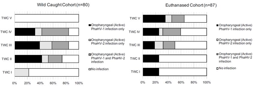
Viruses 2020, 12, 948                                                                                3 of 12

and whether active shedding of either viral subtype has significant correlations with factors such as
age, body condition score or sex.

2. Materials and Methods

2.1. Animal Ethics
     This study was approved by the University of Adelaide Animal Ethics Committee and conducted
in accordance with the guideline set out in the Australian Code for the Care and Use of Animals for
Scientific Purposes 8th Edition (2013) (National Health and Medical Research Council: Canberra, 2013).
Animal ethics approval number for wild-caught koalas: S-2018-022 (granted 6 April 2018) and for the
euthanased examination cohort: S-2016-169 (granted 9 January 2017); DEW scientific permit number:
Y26054-7 (granted 7 September 2017).

2.2. Sample Collection
     Two cohorts of koalas were used for this study: wild-caught and euthanased koalas, both from
the Mount Lofty Ranges koala population in South Australia. The wild-caught cohort was considered
to represent a random sample to investigate active shedding of virus, and the euthanased cohort,
euthanased on welfare grounds, enabled investigations of systemic PhaHV infection status.

2.3. Wild-Caught Koalas
     As part of a larger koala health surveillance project, wild-caught koalas were sourced from three
national parks or reserves in the Mount Lofty Ranges, in Morialta, Cleland and Belair. They were
caught by the flag technique, which utilized a large pole with a flag attached to the far end. The flag end
of the pole was used to direct the koala down the tree. Once the koala was close to the ground,
it was restrained and taken to a field hospital where it was anaesthetised and samples collected,
including oropharyngeal swabs. Oropharyngeal swabs were used for PhaHV testing due to their
increased rate of positivity [8] and suitability in the field. Further demographic data were recorded
and included tag identification, sex, tooth wear class (TWC I-VII) [21] and body condition score
(BCS 1–5) [22]. Global positioning system (GPS) data and tagged trees were recorded so koalas could
be released at their point of capture. Oropharyngeal swabs were placed in sealed plastic bags and kept
on ice before storage at −80 ◦ C within 12 h of sampling. Samples were kept in the −80 ◦ C freezer for up
to 12 months until tested.

2.4. Euthanased Cohort
     For koalas that had been euthanased on welfare grounds, sex, TWC [21] and BCS [22] were
recorded. Oropharyngeal swabs and spleen samples were collected and stored at −20 ◦ C until tested.
Spleen samples were collected to determine systemic infection status due to gammaherpesvirus latency
in immunological cells [11,12,18,23,24].

2.5. DNA Extraction
    DNA was extracted from both oropharyngeal swabs and spleen tissue samples using the QIAamp
DNA Mini Kit (QIAGEN, Hilden, Germany). The concentration of the extracted DNA was measured
using the NanoDrop One Spectrophotometer (Thermo Fisher Scientific Inc, Waltham, MA, USA).
A working solution of 20 ng/µL of DNA from the extracted stock solutions was prepared for PCR tests.

2.6. Quality Control
    The koala beta (β)-actin gene was screened via qPCR, as a quality control, from extracted
oropharyngeal and spleen DNA samples to confirm adequate DNA was extracted, adopting the same
protocol described by Shojima et al. [25]. Any samples negative for β-actin were removed from the
study due to a lack of quality for further testing. DNA samples were run in triplicate in a 5 µL reaction.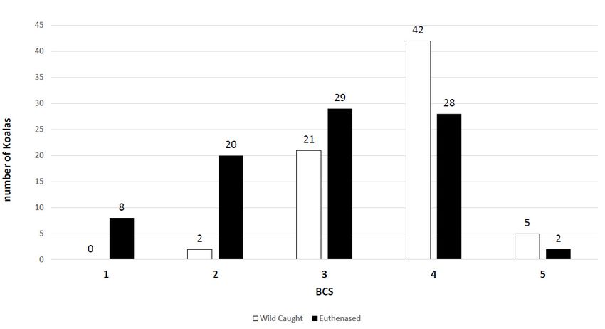
Viruses 2020, 12, 948                                                                                          4 of 12

The DNA copy number was derived from a standard curve from the purified PCR product from
a South Australian koala. Negative control contained no DNA template.

2.7. Molecular Diagnostics (Conventional PCR)
     Specific primers were designed for PhaHV-1 and PhaHV-2 based on the published DPOL gene
(Table 1), due to both viral subtypes only having a 60% nucleotide pairwise identity [6]. Primers were
confirmed to be specific via NCBI Primer Blast, Sanger sequencing and by testing the primer sets on
each of the PhaHV subtypes (Table 2). PCR reactions were run in 20 µL of volume which included
0.5 µM of forward and reverse primer, 5 µL of 4× AllTaq Master Mix solution (QIAGEN, Hilden,
Germany), 5 µL of 20 ng/µL DNA template and 8.5 µL of ultrapure water. PCR conditions were initial
activation and denaturation of 95 ◦ C for 2 min, followed by 34 cycles of denaturation at 95 ◦ C for 5 s,
annealing at 61 ◦ C (PhaHV-1) or 64 ◦ C (PhaHV-2) for 15 s and extension at 72 ◦ C for 10 s. This was
followed by a final extension step of 72 ◦ C for 10 s.

                               Table 1. PCR primers, products and annealing temperatures.

                                                                             Product    Annealing
      Target             Primer Name                Primer Sequence                                   Reference
                                                                               Bp        Temp
                          β-actin-Fwd     50 GAGACCTTCAACACCCCAGC 30                                 Shojima et al.
   β-actin (QC)                                                                111        60 ◦ C
                                                                                                      (2013) [25]
                          β-actin-Rev     50   GTGGGTCACACCATCACCAG     30
                        VK-PhaHV-1-Fwd     50 CGGCATCCTCCCCTGTTTAA 30
     PhaHV-1                                                                   220        61 ◦ C     Current study
                        VK-PhaHV-1-Rev    50 GCCCCTACATTCAACGAACA 30
                        VK-PhaHV-2 Fwd     50 CGCACTCTAAGCTGTCCCTT 30
     PhaHV-2                                                                   330        64 ◦ C     Current study
                        VK-PhaHV-2 Rev     50 TTTCGAGCATCATGCGTCCT 30

      Table 2. Results from AGRF Sanger sequencing, showing primer sets used, sample number, type of
      sample (oropharyngeal or spleen), query and identity to published GenBank sequences (accession
      numbers: JN585829.1, JQ996387.1).

                                           PhaHV-1         PhaHV-2
      Primers              Sample                                        Source        Query Cover     Per Ident
                                         (JN585829.1)    (JQ996387.1)
                           K18-051              +                         Oro             100%            100%
                           K18-051              +                        Spleen           100%            100%
   VK-PhaHV-1
                           K18-064              +                         Oro             100%            100%
                           K18-064              +                        Spleen           100%            100%
                           K18-043                            +           Oro             100%            100%
                           K18-043                            +          Spleen           100%            100%
   VK-PhaHV-2
                           K18-044                            +           Oro             100%            100%
                           K18-044                            +          Spleen           100%            100%

2.8. Statistical Analyses
    Binary Logistic Regression analyses (performed using IBM SPSS Statistics 23) were used to
determine any significant relationships between infection of either PhaHV subtype, coinfections
of PhaHV, BCS, TWC and sex. Compromised BCS was considered as BCS 1 to 3 out of 5 (1–3/5)
(emaciated, poor, or fair muscle condition), and good BCS as 4–5/5. Variables with p-values of ≤0.05
were considered statistically significant.
    A Pearson correlation coefficient (r) analysis was conducted on both wild-caught and euthanased
cohorts (n = 80 and n = 87, respectively) to show correlations between covariates. Covariates
analysed within each cohort were as follows; within the euthanased cohort: infected (PhaHV
DNA detected in the spleen); Oro_Phas1, Oro_Phas2 and Oro_coinfection (PhaHV-1 DNA only,
PhaHV-2 DNA only, or both PhaHV subtypes DNA detected from oropharyngeal swab, respectively);
Spleen_Phas1, Spleen_Phas2 and Spleen_coinfection (PhaHV-1 DNA only, PhaHV-2 DNA only, or
Viruses 2020, 12, 948 5 of 12
both PhaHV subtypes DNA detected from spleen tissue, respectively); TWC (tooth wear class) [21];
BCS (body condition score) [22], BCS_compromised (BCS 1–3/5) and sex. Covariates within the
wild-caught koalas: infected (PhaHV-1 or PhaHV-2 DNA detected from oropharyngeal swab),
Oro_Phas1, Oro_Phas2 and Oro_coinfection, TWC; BCS; BCS_compromised and sex. The results were
Viruses on
visualized 2020,a12, x FOR PEER REVIEW
correlation 5 of 12
heatmap using R version 3.0.1 using ggplot2 and ggcorrplot packages.
swab), Oro_Phas1, Oro_Phas2 and Oro_coinfection, TWC; BCS; BCS_compromised and sex. The
3. Results
results were visualized on a correlation heatmap using R version 3.0.1 using ggplot2 and ggcorrplot
packages.and PhaHV-2 Specific PCR Test
3.1. PhaHV-1
The newly designed primer sets designated, VK-PhaHV-1 and VK-PhaHV-2, were both specific
3. Results
in detecting PhaHV-1 and PhaHV-2, respectively. Samples were confirmed in Sanger Sequencing
3.1. PhaHV-1
(Australian and PhaHV-2
Genome Research Specific PCR Testboth PhaHV-1 and PhaHV-2 showing 100% identity to
Facilities),
GenBank sequence
The newly identifications
designed primer(Table 2) (accession
sets designated, numbers:and
VK-PhaHV-1 JN585829.1, JQ996387.1).
VK-PhaHV-2, were both specific
in detecting PhaHV-1 and PhaHV-2, respectively. Samples were confirmed in Sanger Sequencing
3.2. Wild-Caught Cohort ofResearch
(Australian Genome Koalas Facilities), both PhaHV-1 and PhaHV-2 showing 100% identity to
GenBank sequence identifications (Table 2) (accession numbers: JN585829.1, JQ996387.1).
Approximately three-quarters, 72.5% (58/80), of the wild-caught koalas from the Mount Lofty
Ranges were actively shedding
3.2. Wild-Caught Cohort of KoalasPhaHV. These were shown to be actively shedding just PhaHV-1
(44.8% (26/58)), just PhaHV-2 (20.7% (12/58)) or both viral subtypes (34.5% (20/58)). Only 27.5% (22/80)
Approximately three-quarters, 72.5% (58/80), of the wild-caught koalas from the Mount Lofty
of the wild sampled koalas were not actively shedding either viral subtype (Table 3).
Ranges were actively shedding PhaHV. These were shown to be actively shedding just PhaHV-1
(44.8% (26/58)), just PhaHV-2 (20.7% (12/58)) or both viral subtypes (34.5% (20/58)). Only 27.5%
Table 3. Prevalence of active shedding of Phascolarctid gammaherpesvirus (PhaHV) viral subtypes in
(22/80) of the wild sampled koalas were not actively shedding either viral subtype (Table 3).
the two cohorts, wild-caught and euthanased.
Table 3. Prevalence of active shedding of Phascolarctid gammaherpesvirus
Wild-Caught (PhaHV) viral subtypes
Euthanased
in the two cohorts,Type of Infection
wild-caught and euthanased.
n % n %
Wild-Caught Euthanased
Type of Infection
Active Shedding 58 73% 47 54%
n % n %
Active Shedding Only PhaHV-1 26 33% 23 26%
Active Shedding 58 73% 47 54%
Active Shedding Only PhaHV-2 12 15% 7 8%
Active Shedding Only PhaHV-1 26 33% 23 26%
Coinfected Shedding
Active Shedding Only PhaHV-2
2012 25%15% 17 7
20% 8%
No activeShedding
Coinfected shedding 2220 28%25% 40 17 46% 20%
TOTAL
No active shedding 8022 28% 87 40 46%
TOTAL 80 87
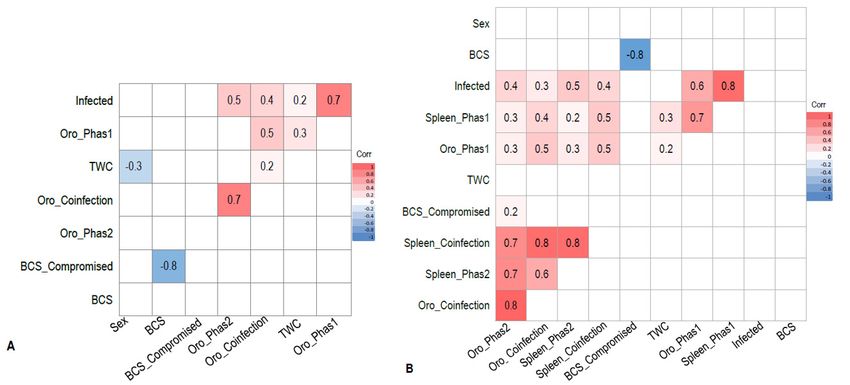
Infection of PhaHV-1 only had a significant positive correlation with TWC (p-value = 0.023; r = 0.2;
Infection of PhaHV-1 only had a significant positive correlation with TWC (p-value = 0.023; r =
n = 80) (Figure 1A). The proportion of koalas in each TWC for each status of infection is shown in
0.2; n = 80) (Figure 1A). The proportion of koalas in each TWC for each status of infection is shown in
Figure 2. There were no significant associations between splenic infection with PhaHV and koalas
Figure 2. There were no significant associations between splenic infection with PhaHV and koalas
with awith
compromised
a compromisedBCSBCS
(p-value = 0.203;
(p-value = 0.203;nn== 80)
80) or withsex
or with sex(p-value
(p-value = 0.776;
= 0.776; n=n = 80).
80).
FigureFigure
1. (A)1.Pearson’s
(A) Pearson’s correlation
correlation matrixdisplaying
matrix displaying interactions
interactions between
between covariates in wild-caught
covariates in wild-caught
koalaskoalas
from from the Mount
the Mount LoftyLofty Ranges,
Ranges, South
South Australia (n(n==80)
Australia 80)portrayed
portrayed on
on aa heat
heatmap.
map.Values
Valuescloser
closer to
to 1.0 indicate a stronger positive correlation between the two variables. Coloured tiles contain a p-
value ≤0.05. White tiles correspond to an insignificant correlation (p-value > 0.05) and are excludedViruses 2020, 12, 948 6 of 12
Viruses1.0
2020, 12, x FOR
indicate a PEER REVIEW
stronger positive correlation between the two variables. Coloured tiles contain6 of 12
a p-value ≤ 0.05. White tiles correspond to an insignificant correlation (p-value > 0.05) and are
from the model. Variable “BCS_compromised” uses compromised koalas (BCS 1–3/5) as the reference
excluded from the model. Variable “BCS_compromised” uses compromised koalas (BCS 1–3/5) as
value.
the (B) Pearson’s
reference correlation
value. (B) Pearson’smatrix displaying
correlation matrixinteractions
displaying between covariates
interactions betweenfrom euthanased
covariates from
koalas sourced from various wildlife hospitals in Adelaide (n = 87), portrayed on a heat
euthanased koalas sourced from various wildlife hospitals in Adelaide (n = 87), portrayed on a heat map. Values
closerValues
map. to 1.0 indicate
closer toa1.0
stronger positive
indicate correlation
a stronger positivebetween the between
correlation two variables.
the twoColoured tiles
variables. contain
Coloured
a p-value ≤0.05. White tiles correspond to an insignificant correlation (p-value > 0.05) and are
tiles contain a p-value ≤0.05. White tiles correspond to an insignificant correlation (p-value > 0.05) and excluded
from
are the model.
excluded fromVariable “BCS_compromised”
the model. uses compromised
Variable “BCS_compromised” koalas (BCS 1–3/5)
uses compromised koalasas(BCS
the reference
1–3/5) as
value. Acronym definitions are provided in Section 2.8 (statistical analysis).
the reference value. Acronym definitions are provided in Section 2.8 (statistical analysis).
2. Histograms
Figure 2. Histogramsdisplaying
displayingthe
thepercentage
percentageofof koalas
koalas actively
actively shedding
shedding PhaHV-1
PhaHV-1 andand PhaHV-2
PhaHV-2 and
and noninfected koalas within each tooth wear classification (TWC) group, for wild-caught and
noninfected koalas within each tooth wear classification (TWC) group, for wild-caught and euthanased
euthanased
cohorts fromcohorts from South Australia.
South Australia.
3.3. Euthanased Cohort
3.3. Euthanased Cohort of
of Koalas
Koalas
It
It was found that
was found that 72.4%
72.4% (63/87)
(63/87) ofof euthanased
euthanased koalas
koalas were
were infected
infected withwith PhaHV.
PhaHV. Active shedding
Active shedding
of the virus occurred in 54.0% (47/87) of koalas in the euthanased cohort, with
of the virus occurred in 54.0% (47/87) of koalas in the euthanased cohort, with active shedding of only active shedding of
only PhaHV-1
PhaHV-1 in 48.9%
in 48.9% (23/47),(23/47), only PhaHV-2
only PhaHV-2 in 14.9%
in 14.9% (7/47),(7/47),
or both orsubtypes
both subtypes simultaneously
simultaneously in
in 36.2%
36.2% (17/47) of koalas. All koalas had matched positive spleen samples
(17/47) of koalas. All koalas had matched positive spleen samples which confirmed infection (Table which confirmed infection
(Table 3). Furthermore,
3). Furthermore, 18.4%18.4%
(16/87)(16/87) of koalas
of koalas were were systemically
systemically infected
infected with
with thevirus
the virusbutbutnot
not actively
actively
shedding
shedding virus.
virus. The
The remaining
remaining27.6% 27.6%(24/87)
(24/87)ofofkoalas
koalaswere
werenot notinfected
infected at at
either
eitherthethe
oropharyngeal
oropharyngeal or
splenic site.
or splenic site.
There
There waswas aa strong
strongcorrelation
correlationbetween
betweensplenic
spleniccoinfection
coinfection with
with both
both PhaHV-1
PhaHV-1 and and PhaHV-2,
PhaHV-2, as
as being co-infected significantly increased the likelihood of actively shedding
being co-infected significantly increased the likelihood of actively shedding (positive oropharyngeal (positive oropharyngeal
sample)
sample) bothboth viral
viral subtypes (p-value <
subtypes (p-value 0.01; rr =
< 0.01; 0.80; n
= 0.80; n= 87) (Figure
= 87) (Figure 1B).
1B). A A significant
significant positive
positive
correlation
correlation exists between active PhaHV-1 and PhaHV-2 infection, as koalas were 3.5 times likely
exists between active PhaHV-1 and PhaHV-2 infection, as koalas were 3.5 times more more
to be actively
likely shedding
to be actively PhaHV-1
shedding if infected
PhaHV-1 with PhaHV-2
if infected with PhaHV-2(p-value = 0.006;= r0.006;
(p-value = 0.3;rExp = 3.538)
(B)Exp
= 0.3; (B) =
(Figure 1B). There
3.538) (Figure 1B). was
There a was
highaprobability of koalas
high probability actively
of koalas shedding
actively PhaHV-1
shedding (Exp (B)
PhaHV-1 (Exp= (B)
28.51) and
= 28.51)
PhaHV-2
and PhaHV-2 (Exp (B)
(Exp= 92.49) when co-infected
(B) = 92.49) with both
when co-infected with viral
bothsubtypes in the spleen
viral subtypes in the(Figure
spleen3).(Figure
Similarly,
3).
to the wild-caught koalas, the euthanased cohort had a significant positive
Similarly, to the wild-caught koalas, the euthanased cohort had a significant positive correlation correlation between TWC
and PhaHV-1
between TWCinfection
and PhaHV-1(Figureinfection (Figure=1A,B)
1A,B) (p-value = 0.2; n ==0.05;
0.05; r(p-value 87). However,
r = 0.2; n = contrary to wild-caught
87). However, contrary
koalas, a significant positive correlation exists between active shedding of PhaHV-2
to wild-caught koalas, a significant positive correlation exists between active shedding of PhaHV-2 and a compromised
BCS
and a(p-value = 0.04; BCS
compromised r = 0.2; n = 97)= (Figure
(p-value 0.04; r =1B),
0.2; with
n = 97)euthanased
(Figure 1B), koalas
withactively
euthanased shedding
koalasPhaHV-2
actively
being 3.5 times more likely to have a compromised BCS (Exp (B) = 3.514).
shedding PhaHV-2 being 3.5 times more likely to have a compromised BCS (Exp (B) = 3.514). No No significant association
between
significant compromised BCS and compromised
association between active shedding BCSof and
PhaHV-1
activeorshedding
sex was found (p-value
of PhaHV-1 = 0.2
or sex wasand 0.16,
found
respectively).
(p-value = 0.2 and 0.16, respectively).Viruses 2020, 12, 948 7 of 12
Viruses 2020, 12, x FOR PEER REVIEW 7 of 12
FigureFigure
3. Histogram of infection
3. Histogram status
of infection within
status withinthe
the euthanased cohort
euthanased cohort (n87),
(n = = 87), displaying
displaying the following
the following
categories: coinfection
categories: with
coinfection withboth viralsubtypes
both viral subtypes (demonstrated
(demonstrated bysplenic
by a dual a dual splenic
infection, andinfection,
actively and
shedding both or singular viral subtypes, or not shedding); infected with just PhaHV-1
actively shedding both or singular viral subtypes, or not shedding); infected with just PhaHV-1 (demonstrated
by a PhaHV-1 splenic infection and shedding or not shedding PhaHV-1); infected with just PhaHV-2
(demonstrated by a PhaHV-1 splenic infection and shedding or not shedding PhaHV-1); infected with
(demonstrated by a PhaHV-2 splenic infection and shedding or not shedding PhaHV-2).
just PhaHV-2 (demonstrated by a PhaHV-2 splenic infection and shedding or not shedding PhaHV-
2).4. Discussion
In this study, oropharyngeal and spleen samples were tested, as previous papers [5,6] succeeded
4. Discussion
in detecting and isolating the same viral subtypes from these sites. In the wild-caught cohort,
Inonly
thisoropharyngeal swabs could be collected, whereas in the euthanased cohort, both oropharyngeal
study, oropharyngeal and spleen samples were tested, as previous papers [5,6] succeeded
and spleen could be collected. As previously mentioned, it has been assumed that the PCR-positive
in detecting and isolating the same viral subtypes from these sites. In the wild-caught cohort, only
oropharyngeal swabs are most likely to confirm active shedding [18], whilst latent infections occur
oropharyngeal swabs could
within lymphocytes whichbeare
collected, whereas within
densely populated in the the
euthanased cohort,
spleen [19,20]. both
In the oropharyngeal
euthanased cohort, and
spleen koalas
couldthat
be were
collected. Asforpreviously
positive mentioned,
PhaHV in both it has
spleen tissue and been assumedswab
oropharyngeal thatsamples
the PCR-positive
were
oropharyngeal
deemed asswabs
infectedare
andmost likely
actively to confirm
shedding active
the virus. shedding
Koalas [18],inwhilst
only positive latent
the spleen wereinfections
classified occur
within as being infected.
lymphocytes which are densely populated within the spleen [19,20]. In the euthanased cohort,
koalas 4.1.
thatActive
wereShedding
positive for PhaHV in both spleen tissue and oropharyngeal swab samples were
deemed as infected and actively shedding the virus. Koalas only positive in the spleen were classified
In this study, the prevalence of PhaHV active shedding in wild-caught koalas was 72.5% (58/80)
as being infected.
and was higher than that of the euthanased koalas at 54% (47/87). The rate of active shedding was
higher than initially expected when compared with other herpesvirus cases found in other koalas in
4.1. Active Shedding
Australia. Stalder et al. [8] surveyed 99 captive and wild koalas in Victoria in 2015 and detected PhaHV-1
Inand
this2 study,
in 10.1%the
(10/99) and 23.2%
prevalence of (23/99)
PhaHV of active
koalas,shedding
respectively.
inAnother study koalas
wild-caught in Victoria
was found the(58/80)
72.5%
prevalence of PhaHV-1 and PhaHV-2 was 17% and 22%, respectively, and an overall prevalence of 33%
and was higher than that of the euthanased koalas at 54% (47/87). The rate of active shedding was
(n = 810), with different populations ranging from 1 to 55% [7]. Mainland populations in Victoria had
higher 22–46%
than initially expected when compared with other herpesvirus cases found in other koalas in
and 31–55% test positive for PhaHV-1 and PhaHV-2 [7], respectively. These numbers are more
Australia. Stalder et al. [8] surveyed 99 captive and wild koalas in Victoria in 2015 and detected
PhaHV-1 and 2 in 10.1% (10/99) and 23.2% (23/99) of koalas, respectively. Another study in Victoria
found the prevalence of PhaHV-1 and PhaHV-2 was 17% and 22%, respectively, and an overall
prevalence of 33% (n = 810), with different populations ranging from 1 to 55% [7]. Mainland
populations in Victoria had 22–46% and 31–55% test positive for PhaHV-1 and PhaHV-2 [7],Viruses 2020, 12, 948 8 of 12
consistent with the prevalence in Mount Lofty Ranges koalas, with active shedding of PhaHV-1 and
PhaHV-2 at 57.5% (46/80) and 40% (32/80), respectively, in the wild-caught koalas and 46% (40/87) and
27.5% (24/87), respectively, in euthanased koalas. These may represent populations with similar high
densities with greater opportunity for infection spread. Differences in prevalence of viral shedding
between PhaHV-1 and PhaHV-2 may show that PhaHV-2 is less established within the SA koala
population than PhaHV-1, or that PhaHV-2 may have a lower tendency to actively shed than PhaHV-1.
This may be due to the genetic differences between the viral subtypes, as PhaHV-2 only shares 60%
pairwise identity with PhaHV-1 [6] and may contribute to higher expression of immune suppressor
genes in PhaHV-2. Latency studies would need to be carried out for this theory to be concluded.
A significant relationship and positive correlation existed between active shedding of PhaHV-1 and
TWC in both the wild-caught cohort (p-value = 0.023, r = 0.3) and the euthanased cohort (p-value = 0.05,
r = 0.2), whereby increasing age increased the likelihood of PhaHV-1 infection. This relationship was
expected: as age increased, the probability of infection increased due to the likelihood of exposure to
the virus and the lifelong nature of the disease. In contrast, there was no correlation between active
shedding of PhaHV-2 and TWC in either cohort. The study by Vaz et al. [7] also showed similar results
regarding PhaHV-1 infection correlation with age and PhaHV-2 spread more evenly across age groups.
This study proposed that PhaHV-1 infection may be more likely due to sexual contact between adult
koalas, whilst PhaHV-2 infection may occur from close contact between mother and joey [7]. Vaz et al.
showed repeatable detection of both PhaHV-1 and PhaHV-2 in koala cloacal regions [7,8], therefore,
viral transmission between mother and joey may not only occur in the pouch, but potentially during
parturition and/or from the consumption of a unique maternal faeces known as “pap”. This faecal
complex contains tannin protein complex-degrading enterobacteria (T-PCDE) essential for digestion
of gum leaves [26] and may also be contaminated with other diseases such as Chlamydia pecorum
and PhaHV-1 and PhaHV-2. Kent et al. [27] conducted a survey on sexually transmitted mustelid
gammaherpesvirus 1 (MusHV-1) in European badgers (Meles meles) and investigated the prevalence
of infection in adults and cubs; the high proportion of infected cubs showed a strong likelihood of
vertical transmission.
Body condition score is a commonly used and effective indicator of the health status of
an animal [18]. In our study, koalas with a BCS of 1–3/5 (emaciated, poor, or fair muscle condition)
were considered compromised, whilst koalas with BCS 4–5/5 were considered in good condition.
It was hypothesised that koalas with a low BCS were more likely to be actively shedding if infected,
due to being unable to keep the herpesvirus in a latent, suppressed state. Active shedding of
PhaHV-2 had a significant correlation with a compromised BCS in the euthanased koala cohort
(p-value = 0.04; r = 0.2; n = 87), but not in the wild-caught cohort. This was likely confounded by
the spread of koalas in each BCS category with a right-skewed bias towards healthy koalas in the
wild-caught cohort and a more normal distribution in the euthanased cohort (Figure 4). Surprisingly,
compromised BCS had no significant correlation with infection or active shedding of PhaHV-1 in
either cohort. The lack of relationship between compromised BCS and active shedding of PhaHV-1
is likely due to apparently healthy animals being able to actively shed herpesviruses without
developing clinical disease [10]. The potential to asymptomatically shed gammaherpesviruses or
have subclinical disease has been demonstrated in other reservoir hosts [11]. In a case study of ovine
herpesvirus-2 (OvHV-2; gammaherpesvirus), Li et al. [10] took a series of nasal swabs and discovered
that healthy lambs between 6 and 9 months of age had an active shedding prevalence of 61% (n = 56).
Gammaherpesviruses can be more likely to cause disease in susceptible, nonreservoir hosts, as seen
with cattle infected with OvHV-2 causing malignant catarrhal fever (MCF) [28].Viruses 2020, 12, 948 9 of 12
Viruses 2020, 12, x FOR PEER REVIEW 9 of 12
Figure4. 4.Histogram
Figure Histogramdisplaying
displayingthe
thedistribution
distributionofofkoalas
koalasinineach
eachcohort
cohortwithin
withintheir
theirassigned
assignedbody
body
condition score (BCS).
condition score (BCS).
4.2. Systemic Infection and Active Shedding
4.2. Systemic Infection and Active Shedding
The majority of infected euthanased koalas were actively shedding the virus (74.6%; 47/63),
The majority of infected euthanased koalas were actively shedding the virus (74.6%; 47/63),
whilst only 25.4% (16/63) of infected koalas were not actively shedding. There was a strong positive
whilst only 25.4% (16/63) of infected koalas were not actively shedding. There was a strong positive
correlation between splenic coinfection of both viral subtypes and active shedding of the virus (0.8)
correlation between splenic coinfection of both viral subtypes and active shedding of the virus (0.8)
(Figure 1B). In the euthanased cohort, koalas infected with PhaHV-2 were 3.5 times more likely to be
(Figure 1B). In the euthanased cohort, koalas infected with PhaHV-2 were 3.5 times more likely to be
coinfected with PhaHV-1 (Exp (B) = 3.538; p-value = 0.006; n = 87) and as a result were 1.5 times more
coinfected with PhaHV-1 (Exp (B) = 3.538; p-value = 0.006; n = 87) and as a result were 1.5 times more
likely to be actively shedding PhaHV-1 with every incremental increase in TWC (Exp (B)) = 1.514;
likely to be actively shedding PhaHV-1 with every incremental increase in TWC (Exp (B)) = 1.514; p-
p-value = 0.05; n = 87). This suggests that viral coinfection highly predisposes koalas to active shedding
value = 0.05; n = 87). This suggests that viral coinfection highly predisposes koalas to active shedding
of the virus. Potentially, increased expression of viral effector proteins from both PhaHV-1 and PhaHV-2
of the virus. Potentially, increased expression of viral effector proteins from both PhaHV-1 and
could lead to greater immunosuppression, initiating recrudescence. A study investigating herpesvirus
PhaHV-2 could lead to greater immunosuppression, initiating recrudescence. A study investigating
coinfection in humans showed that infection of EBV and HHV-7 were also shown to promote HHV-6
herpesvirus coinfection in humans showed that infection of EBV and HHV-7 were also shown to
infection and disease [29–32] however, this may just be a result of severe immunosuppression that
promote HHV-6 infection and disease [29–32] however, this may just be a result of severe
triggers viral coinfection, as stated by Handous et al. [33]. PhaHV-2 had a significant correlation with a
immunosuppression that triggers viral coinfection, as stated by Handous et al. [33]. PhaHV-2 had a
compromised BCS, suggesting that PhaHV-2 may be an indicator of other underlying health conditions
significant correlation with a compromised BCS, suggesting that PhaHV-2 may be an indicator of
such as immunosuppression, stress and coinfection of other diseases including PhaHV-1, Chlamydia
other underlying health conditions such as immunosuppression, stress and coinfection of other
pecorum or koala retrovirus.
diseases including PhaHV-1, Chlamydia pecorum or koala retrovirus.
In our study, there was no significant relationship between sex and infection status.
In our study, there was no significant relationship between sex and infection status. Yet Stalder
Yet Stalder et al. [8] showed a significant relationship between infection and sex, with male koalas
et al. [8] showed a significant relationship between infection and sex, with male koalas more likely to
more
havelikely to have an
an infection. Vazinfection.
et al. [7]Vaz et al. that
showed [7] showed
femalesthat females
without without
young wereyoung
morewere
likelymore
to belikely to
infected
bethan
infected than those with young; they also found a correlation between PhaHV and
those with young; they also found a correlation between PhaHV and “wet bottom”, a clinical“wet bottom”,
a sign
clinical
of sign
overtofchlamydiosis.
overt chlamydiosis. The association
The association of potentially
of potentially infertile
infertile females
females withwith PhaHV
PhaHV maybe
may
beconfounded
confoundedby bychlamydial
chlamydialinfection,
infection,a aknown
knowncause
causeofofreproductive
reproductivepathology
pathologyininkoalas
koalas[34].
[34].
4.3. Sites of Active Viral Shedding
4.3. Sites of Active Viral Shedding
In our study, most oropharyngeal swabs from infected (spleen positive) koalas were positive for
In our study, most oropharyngeal swabs from infected (spleen positive) koalas were positive for
Phascolarctid gammaherpesvirus, suggesting that significant active shedding is from rostral epithelial
Phascolarctid gammaherpesvirus, suggesting that significant active shedding is from rostral
tissues (nasal, oropharyngeal). This finding differs to that of Vaz et al. [7], which found that rostral
epithelial tissues (nasal, oropharyngeal). This finding differs to that of Vaz et al. [7], which found that
(ocular, nasal, oropharyngeal) swabbing was less likely to pick up an infection in comparison with
rostral (ocular, nasal, oropharyngeal) swabbing was less likely to pick up an infection in comparison
caudal (urogenital, cloacal) swabbing. Samples from caudal epithelial tissues were not tested in our
with caudal (urogenital, cloacal) swabbing. Samples from caudal epithelial tissues were not tested in
our study, therefore, shedding from different epithelial sites needs to be investigated further to
provide tissue tropism insights. One concern about the interpretation of active shedding is theViruses 2020, 12, 948 10 of 12
study, therefore, shedding from different epithelial sites needs to be investigated further to provide
tissue tropism insights. One concern about the interpretation of active shedding is the contamination
of the oropharyngeal swabs with cells within which latency has been established, that is, circulating
mononucleocytes. Nevertheless, due to the tissue tropism of herpesviruses, it is less likely to detect
virus in epithelial swabs when the animals are only infected in latent forms [15,35,36]. It is also highly
likely that the detected viruses in epithelial cells are actively shed viruses [37].
4.4. Potential Areas for Further Research
This study focused on the presence and prevalence of Phascolarctid herpesviruses within the
mainland South Australian koala population. Further research is needed to understand the association
of PhaHV with other infections—primarily koala retrovirus (KoRV) and Chlamydial infection—since
PhaHV may be playing a role in the augmentation of clinical disease or be shed secondary to them.
Since PhasHV-2 had an association to koalas with a compromised BCS and increased PhasHV-1
shedding, this could be suggestive of an increase in pathogenicity in this subtype. Investigation into
disease presentations, haematology and immune function markers with and without PhaHV infection
may shed some light on the effect of these viruses. Serological studies could be conducted to investigate
the koalas’ response to PhaHV infection, since the koalas’ immune system has often been perceived as
“immunologically lazy” [38,39]. These studies would help to further determine the clinical significance,
host response to infection and impact of these newly discovered viruses on koala populations.
5. Conclusions
We showed there was a high prevalence of PhaHV infection in koalas in the Mount Lofty Ranges
population, with more than two-thirds of both wild-caught and euthanased cohorts actively shedding
the virus. PhaHV-1 had a greater prevalence within the SA koala populations, with more koalas actively
shedding PhaHV-1 than PhaHV-2.
Despite being less prevalent, koalas coinfected with PhaHV-2 were more likely to also be infected
and actively shedding PhaHV-1. Neither of the viral subtypes were shown to have any significant
relationship with BCS in wild-caught koalas; however, PhaHV-2 infection had a significant correlation
with BCS in euthanased koalas. PhaHV-1 infection was also shown to be positively correlated with
TWC in both cohorts, whilst sex had no significant correlation with either viral subtype in both cohorts.
The clinical significance of these recently discovered Phascolarctid herpesviruses is still unknown,
and additional investigation into the pathogenicity, clinical signs of the virus and coinfection with
other pathogens is important. Uncovering the significance of PhaHV will help determine the health
status and guide the management of koala populations.
Author Contributions: Conceptualization, F.H. and T.S.; methodology, V.K., T.S., N.S.; W.B., R.E.; software,
A.-L.C., V.K.; formal analysis, A.-L.C., V.K.; investigation, F.H., A.-L.C., V.K., N.S.; W.B., and T.S.; resources, F.H.,
N.S.; data curation, A.-L.C.; writing—original draft preparation, V.K.; writing—review and editing, F.H., A.-L.C.,
V.K., T.S., N.S.; W.B.; visualization, F.H., A.-L.C., V.K.; supervision, F.H.; project administration, F.H., N.S.; funding
acquisition, F.H., N.S. All authors have read and agreed to the published version of the manuscript.
Funding: This research was funded by The University of Adelaide HDR support fund grant number 13107182.
Acknowledgments: We thank Kandarp Patel (The University of Adelaide) for advising us on statistical analysis
and modelling, Adelaide Zoo and the Department of Environment and Water, SA for koala capture fieldwork,
Adelaide Koala and Wildlife Hospital, Adelaide and Adelaide Hills based veterinary clinics, SA Fauna Rescue,
Adelaide Koala Rescue, Adelaide and Adelaide Hills Koala Rescue and other fauna rescue groups for their help in
provision of koalas.
Conflicts of Interest: The authors declare no conflict of interest. The funders had no role in the design of the
study; in the collection, analyses, or interpretation of data; in the writing of the manuscript, or in the decision to
publish the results.Viruses 2020, 12, 948 11 of 12
References
1. Chapter 9 - Herpesvirales. In Fenner’s Veterinary Virology (Fifth Edition); MacLachlan, N.J., Dubovi, E.J., Eds.;
Academic Press: Boston, MA, USA, 2017. [CrossRef]
2. Finnie, E.P.; Littlejohns, I.R.; Acland, H.M. Letter: Mortalities in parma wallabies (Macropus parma) associated
with probable herpesvirus. Aust. Vet. J. 1976, 52, 294. [CrossRef] [PubMed]
3. Johnson, M.A.; Whalley, J.M.; Littlejohns, I.R.; Dickson, J.; Smith, V.W.; Wilks, C.R.; Reisner, A.H.
Macropodid herpesviruses 1 and 2: Two herpesviruses from Australian marsupials differentiated by
restriction endonucleases, DNA composition and hybridization. Brief Rep. Arch. Virol. 1985, 85, 313–319.
[CrossRef] [PubMed]
4. Smith, J.A.; Wellehan, J.F., Jr.; Pogranichniy, R.M.; Childress, A.L.; Landolfi, J.A.; Terio, K.A. Identification
and isolation of a novel herpesvirus in a captive mob of eastern grey kangaroos (Macropus giganteus).
Vet. Microbiol. 2008, 129, 236–245. [CrossRef] [PubMed]
5. Vaz, P.; Whiteley, P.L.; Wilks, C.R.; Duignan, P.J.; Ficorilli, N.; Gilkerson, J.R.; Browning, G.F.; Devlin, J.M.
Detection of a novel gammaherpesvirus in koalas (Phascolarctos cinereus). J. Wildl. Dis. 2011, 47, 787–791.
[CrossRef] [PubMed]
6. Vaz, P.; Whiteley, P.L.; Wilks, C.R.; Browning, G.F.; Gilkerson, J.R.; Ficorilli, N.; Devlin, J.M. Detection
of a second novel gammaherpesvirus in a free-ranging koala (Phascolarctos cinereus). Wildl. Dis. 2012,
48, 226–229. [CrossRef]
7. Vaz, P.K.; Legione, A.R.; Hartley, C.A.; Devlin, J.M. Detection and Differentiation of Two Koala
Gammaherpesviruses by Use of High-Resolution Melt (HRM) Analysis Reveals Differences in Viral Prevalence
and Clinical Associations in a Large Study of Free-Ranging Koalas. J. Clin. Microbiol. 2019, 57. [CrossRef]
8. Stalder, K.; Vaz, P.K.; Gilkerson, J.R.; Baker, R.; Whiteley, P.; Ficorilli, N.; Tatarczuch, L.; Portas, T.; Skogvold, K.;
Anderson, G.A.; et al. Prevalence and Clinical Significance of Herpesvirus Infection in Populations of
Australian Marsupials. PLoS ONE 2015, 10, e0133807. [CrossRef]
9. Sugden, B.; Kintner, C.R.; Mark, W. The molecular biology of lymphotropic herpesviruses. Adv. Cancer Res.
1979, 30, 239–278. [CrossRef]
10. Li, H.; Taus, N.S.; Lewis, G.S.; Kim, O.; Traul, D.L.; Crawford, T.B. Shedding of ovine herpesvirus 2 in sheep
nasal secretions: The predominant mode for transmission. J. Clin. Microbiol. 2004, 42, 5558–5564. [CrossRef]
11. Ackermann, M. Pathogenesis of gammaherpesvirus infections. Vet. Microbiol. 2006, 113, 211–222. [CrossRef]
12. Hussy, D.; Janett, F.; Albini, S.; Stauber, N.; Thun, R.; Ackermann, M. Analysis of the pathogenetic basis for
shedding and transmission of ovine gamma herpesvirus 2. Clin. Microbiol. 2002, 40, 4700–4704. [CrossRef]
[PubMed]
13. Fabijan, J.; Sarker, N.; Speight, N.; Owen, H.; Meers, J.; Simmons, G.; Seddon, J.; Emes, R.D.; Tarlinton, R.;
Hemmatzadeh, F.; et al. Pathological Findings in Koala Retrovirus-positive Koalas (Phascolarctos cinereus)
from Northern and Southern Australia. J. Comp. Pathol. 2020, 176, 50–66. [CrossRef]
14. Hemsley, S.; Canfield, P.J. Histopathological and immunohistochemical investigation of naturally occurring
chlamydial conjunctivitis and urogenital inflammation in koalas (Phascolarctos cinereus). J. Comp. Pathol.
1997, 116, 273–290. [CrossRef]
15. Flano, E.; Kim, I.J.; Woodland, D.L.; Blackman, M.A. Gamma-herpesvirus latency is preferentially maintained
in splenic germinal center and memory B cells. J. Exp. Med. 2002, 196, 1363–1372. [CrossRef] [PubMed]
16. Gerow, C.M.; Rapin, N.; Voordouw, M.J.; Elliot, M.; Misra, V.; Subudhi, S. Arousal from hibernation and
reactivation of Eptesicus fuscus gammaherpesvirus (EfHV) in big brown bats. Transbound. Emerg. Dis. 2019,
66, 1054–1062. [CrossRef] [PubMed]
17. Weck, K.E.; Barkon, M.L.; Yoo, L.I.; Speck, S.H. Mature B cells are required for acute splenic infection, but not
for establishment of latency, by murine gammaherpesvirus. Virology 1996. [CrossRef]
18. Feldman, E.R.; Kara, M.; Coleman, C.B.; Grau, K.R.; Oko, L.M.; Krueger, B.J.; Renne, R.; van Dyk, L.F.;
Tibbetts, S.A. Virus-encoded microRNAs facilitate gammaherpesvirus latency and pathogenesis in vivo.
mBio 2014, 5, e00914–e00981. [CrossRef]
19. Sharma, V.; Mobeen, F.; Prakash, T. Comparative Genomics of Herpesviridae Family to Look for Potential
Signatures of Human Infecting Strains. Int. J. Genom. 2016, 2016, 9543274. [CrossRef]
20. van Dyk, L.F.; Virgin, H.W.t.; Speck, S.H. Maintenance of gammaherpesvirus latency requires viral cyclin in
the absence of B lymphocytes. Virology 2003, 77, 5118–5126. [CrossRef]Viruses 2020, 12, 948 12 of 12
21. Martin, R.W. Age-Specific Fertility in Three Populations of the Koala, Phascolarctos cinereus Goldfuss,
in Victoria. Wildl. Res. 1981, 8, 275–283. [CrossRef]
22. Blanshard, W.H.; Bodley, K. Chapter 8: Koalas. In Medicine of Australian Mammals, 1st ed.; Vogelnest, L.,
Woods, R., Eds.; CSIRO Publishing: Collingwood, Clayton, Australia, 2008; pp. 227–328.
23. Lieberman, P.M. Keeping it quiet: Chromatin control of gammaherpesvirus latency. Nat. Rev. Microbiol.
2013, 11, 863–875. [CrossRef] [PubMed]
24. King, A.; Adams, M.J.; Carstens, E.B.; Lefkowitz, E.J. Virus Taxonomy: Ninth Report of the International
Committee on Taxonomy of Viruses. Elsevier 2012, 99–107. [CrossRef]
25. Shojima, T.; Yoshikawa, R.; Hoshino, S.; Shimode, S.; Nakagawa, S.; Ohata, T.; Nakaoka, R.; Miyazawa, T.
Identification of a novel subgroup of Koala retrovirus from Koalas in Japanese zoos. Virology 2013, 87,
9943–9948. [CrossRef] [PubMed]
26. Osawa, R.; Blanshard, W.H.; Ocallaghan, P.G. Microbiological Studies of the Intestinal Microflora of the
Koala, Phascolarctos cinereus. Pap, a Special Maternal Feces Consumed by Juvenile Koalas. Aust. J. Zool. 1993,
41, 527–536. [CrossRef]
27. Kent, A.; Ehlers, B.; Mendum, T.; Newman, C.; Macdonald, D.W.; Chambers, M.; Buesching, C.D. Genital tract
screening finds widespread infection with mustelid gammaherpesvirus 1 in the uropean badger (meles meles).
J. Wildl. Dis. 2018, 133–137. [CrossRef]
28. Meier-Trummer, C.S.; Rehrauer, H.; Franchini, M.; Patrignani, A.; Wagner, U.; Ackermann, M. Malignant
catarrhal fever of cattle is associated with low abundance of IL-2 transcript and a predominantly latent
profile of ovine herpesvirus 2 gene expression. PLoS ONE 2009, 4. [CrossRef]
29. Razonable, R.R.; Brown, R.A.; Humar, A.; Covington, E.; Alecock, E.; Paya, C.V. Herpesvirus infections in
solid organ transplant patients at high risk of primary cytomegalovirus disease. J. Infect. Dis. 2005, 192,
1331–1339. [CrossRef]
30. Blazquez-Navarro, A.; Dang-Heine, C.; Wittenbrink, N.; Bauer, C.; Wolk, K.; Sabat, R.; Westhoff, T.; Sawitzki, B.;
Reinke, P.; Thomusch, O.; et al. BKV, CMV, and EBV Interactions and their Effect on Graft Function One Year
Post-Renal Transplantation: Results from a Large Multi-Centre Study. EBioMedicine 2018, 34, 113–121.
[CrossRef]
31. Aalto, S.M.; Linnavuori, K.; Peltola, H.; Vuori, E.; Weissbrich, B.; Schubert, J.; Hedman, K. Immunoreactivation
of Epstein-Barr virus due to cytomegalovirus primary infection. J. Med. Virol. 1998, 56, 186–191. [CrossRef]
32. Jain, R.; Trehan, A.; Mishra, B.; Singh, R.; Saud, B.; Bansal, D. Cytomegalovirus disease in children with acute
lymphoblastic leukemia. J. Pediatric Hematol./Oncol. 2016, 33, 239–247. [CrossRef]
33. Handous, I.; Achour, B.; Marzouk, M.; Rouis, S.; Hazgui, O.; Brini, I.; Khelif, A.; Hannachi, N.; Boukadida, J.
Co-infections of human herpesviruses (CMV, HHV-6, HHV-7 and EBV) in non-transplant acute leukemia
patients undergoing chemotherapy. Virology 2020, 17, 37. [CrossRef] [PubMed]
34. Robbins, A.; Hanger, J.; Jelocnik, M.; Quigley, B.L.; Timms, P. Longitudinal study of wild koalas (Phascolarctos
cinereus) reveals chlamydial disease progression in two thirds of infected animals. Sci. Rep. 2019, 9, 13194.
[CrossRef] [PubMed]
35. Collins, C.M.; Boss, J.M.; Speck, S.H. Identification of infected B-cell populations by using a recombinant
murine gammaherpesvirus 68 expressing a fluorescent protein. Virology 2009, 83, 6484–6493. [CrossRef]
[PubMed]
36. Rekow, M.M.; Darrah, E.J.; Mboko, W.P.; Lange, P.T.; Tarakanova, V.L. Gammaherpesvirus targets peritoneal
B-1 B cells for long-term latency. Virology 2016, 492, 140–144. [CrossRef] [PubMed]
37. Usherwood, E.J.; Roy, D.J.; Ward, K.; Surman, S.L.; Dutia, B.M.; Blackman, M.A.; Stewart, J.P.; Woodland, D.L.
Control of gammaherpesvirus latency by latent antigen-specific CD8(+) T cells. J. Exp. Med. 2000, 192,
943–952. [CrossRef]
38. Higgins, D.; Lau, Q.; Maher, I. Koala immunology and the koala retrovirus (KoRV). Tech. Rep. Aust. Mus. Online
2014, 35–38. [CrossRef]
39. Wilkinson, R.; Kotlarski, I.; Barton, M. Koala lymphoid cells: Analysis of antigen-specific responses.
Vet. Immunol. Immunopathol. 1992, 33, 237–247. [CrossRef]
© 2020 by the authors. Licensee MDPI, Basel, Switzerland. This article is an open access
article distributed under the terms and conditions of the Creative Commons Attribution
(CC BY) license (http://creativecommons.org/licenses/by/4.0/).You can also read