Inhibition of Sphingosine Phosphate Receptor 1 Signaling Enhances the Efficacy of VEGF Receptor Inhibition
←
→
Page content transcription
If your browser does not render page correctly, please read the page content below
Published OnlineFirst February 20, 2019; DOI: 10.1158/1535-7163.MCT-18-0548

        Cancer Biology and Translational Studies                                                                                         Molecular
                                                                                                                                         Cancer
                                                                                                                                         Therapeutics
     Inhibition of Sphingosine Phosphate Receptor 1
     Signaling Enhances the Efficacy of VEGF Receptor
     Inhibition
     Anthony S. Fischl1, Xiaoen Wang2, Beverly L. Falcon1, Rowena Almonte-Baldonado1,
     Diane Bodenmiller1, Glenn Evans1, Julie Stewart1, Takako Wilson1, Philip Hipskind1,
     Jason Manro1, Mark T. Uhlik1, Sudhakar Chintharlapalli1, Damien Gerald1, David C. Alsop2,
     Laura E. Benjamin1, and Rupal S. Bhatt3

     Abstract
            Inhibition of VEGFR signaling is an effective treatment for                 angiogenic inhibition. Here, we show that inhibition of S1P
         renal cell carcinoma, but resistance continues to be a major                   signaling reduces the endothelial cell barrier and leads to
         problem. Recently, the sphingosine phosphate (S1P) signaling                   excessive angiogenic sprouting. Simultaneous inhibition of
         pathway has been implicated in tumor growth, angiogenesis,                     S1P and VEGF signaling further disrupts the tumor vascular
         and resistance to antiangiogenic therapy. S1P is a bioactive                   beds, decreases tumor volume, and increases tumor cell death
         lipid that serves an essential role in developmental and path-                 compared with monotherapies. These studies suggest that
         ologic angiogenesis via activation of the S1P receptor 1 (S1P1).               inhibition of angiogenesis at two stages of the multistep
         S1P1 signaling counteracts VEGF signaling and is required for                  process may maximize the effects of antiangiogenic therapy.
         vascular stabilization. We used in vivo and in vitro angiogenesis              Together, these data suggest that combination of S1P1 and
         models including a postnatal retinal angiogenesis model and a                  VEGFR-targeted therapy may be a useful therapeutic
         renal cell carcinoma murine tumor model to test whether                        strategy for the treatment of renal cell carcinoma and other
         simultaneous inhibition of S1P1 and VEGF leads to improved                     tumor types.

     Introduction                                                                       urgent need to identify new therapeutic approaches to inhibit
                                                                                        tumor angiogenesis with mechanisms of action that are distinct
        VEGF is the predominant growth factor expressed by tumor
                                                                                        from and/or may complement VEGF/VEGFR modulators. Com-
     cells to drive angiogenesis and solid tumor growth. Antiangio-
                                                                                        binations with other vascular pathway modulators such as sphin-
     genesis therapies targeting VEGF or its receptor VEGF receptor 2
                                                                                        gosine-1-phosphate (S1P1) inhibitors may fill a gap and enable
     (VEGFR) and immune therapies have been clinically demonstrat-
                                                                                        vascular targeting in otherwise VEGF pathway–independent
     ed to be effective in prolonging overall survival and progression-
                                                                                        blood vessels.
     free survival while significantly improving the quality of life for
                                                                                           S1P is a bioactive lipid and important regulator of vascular
     certain cancer patients (1–6). In tumors such as clear cell renal cell
                                                                                        function and immune cell trafficking (7). S1P has also been
     carcinoma (RCC), where VEGF pathway inhibition has demon-
                                                                                        shown to be a potent inducer of many of the hallmarks of cancer
     strated single-agent activity, there are five approved agents that
                                                                                        including tumor angiogenesis, cancer cell growth, immune mod-
     target VEGF signaling. Among these are four VEGFR tyrosine
                                                                                        ulation, migration, and invasion (8, 9). S1P signaling is mediated
     kinase inhibitors (TKI): sunitinib, sorafenib, axitinib, and pazo-
                                                                                        via five G-protein–coupled endothelial differentiation receptors
     panib (4–6). Unfortunately not all patients benefit from these
                                                                                        (S1P1-5 receptors). S1P signaling is diverse and involves many
     VEGF pathway inhibitors. Some patients do not respond to this
                                                                                        signaling pathways known to be important in cancer including
     class of inhibitors, some ultimately develop resistance, and com-
                                                                                        the PI3K, MAPK, and pSTAT3 pathways (8). The S1P receptor 1
     plete responses are extremely rare. For this reason, there is an
                                                                                        (S1P1), in particular, has been shown to play a key role in
                                                                                        angiogenesis, which was first demonstrated by S1P1 genetic
                                                                                        deletion studies in mice (11). Loss of S1P1 function results in
     1
       Eli Lilly and Company, Indianapolis, IN. 2Department of Radiology, Beth Israel   embryonic lethality due to severe hemorrhage likely associated
     Deaconess Medical Center, Harvard Medical School, Boston, Massachusetts.           with defects in pericyte recruitment and vessel maturation. More
     3
       Division of Hematology and Oncology, Beth Israel Deaconess Medical Center,
                                                                                        recent studies evaluating endothelial-specific S1P1 deletion indi-
     Harvard Medical School, Boston, Massachusetts.
                                                                                        cate S1P1 signaling also inhibits angiogenic sprouting in the
     Note: Supplementary data for this article are available at Molecular Cancer        retina of postnatal mice (12–14). S1P signaling via S1P1 appears
     Therapeutics Online (http://mct.aacrjournals.org/).
                                                                                        to be part of a negative feedback mechanism that is required
     Corresponding Author: Rupal S. Bhatt, Beth Israel Deaconess Medical Center,        for maintaining blood vessel integrity by counteracting VEGF
     375 Longwood Avenue, MASCO 426, Boston, MA 02215. Phone: 617-735-2062;             signaling and excessive angiogenic sprouting (13). Our current
     Fax: 617-725-2060; E-mail: rbhatt@bidmc.harvard.edu
                                                                                        understanding of S1P signaling in the vasculature indicates
     doi: 10.1158/1535-7163.MCT-18-0548                                                 that S1P1 plays a critical role in limiting VEGF-dependent angio-
     2019 American Association for Cancer Research.                                    genesis and promoting vascular stability via enhancement of

856 Mol Cancer Ther; 18(4) April 2019

        Downloaded from mct.aacrjournals.org on February 18, 2021. © 2019 American Association for Cancer Research.
Published OnlineFirst February 20, 2019; DOI: 10.1158/1535-7163.MCT-18-0548

                                                                                      S1P1 Inhibition Improves VEGFR-Targeted Therapy

endothelial cell–cell junctions. Loss of S1P1 function has an        minutes. Cells were washed, permeabilized with PBS þ 1%
opposite effect leading to VEGF-dependent hypersprouting angio-      BSA þ 0.5% Triton X100, and stained for VE cadherin using a
genesis, increased vascular permeability, and loss of vascular       goat anti-VE cadherin antibody (BD Biosciences #555661) at
function (12–14). S1P1 inhibition leads to disorganized and          1:50 and with Hoechst 33342 (Invitrogen, 1:1,000) followed by
nonfunctional angiogenesis in nonproliferating tumor vessels         secondary antibodies (goat anti-mouse Alexa Fluor-488; Invitro-
where VEGF inhibition was not previously effective. The blood        gen) at 1:400. Cells were imaged with a Cellinsight NXT imager
vessels resulting from S1P1 antagonism are fragile and effectively   using a 20x objective (Thermo Scientific). For the permeability
eliminated by blockade of VEGF signaling.                            assay, HMVEC monolayers were established on 1 mm pore trans-
   Preclinical studies have shown that modulation of S1P1,           wells (Corning) coated with 5 mg/mL fibronectin (Life Technol-
using several different approaches, will inhibit angiogenesis        ogies) by plating 50,000 cells in 100 mL of EGM2-MV media (21).
and tumor growth. FTY720, a well-characterized agonist that          Media were added to the bottom of the transwell and incubated
activates S1P1, 3, 4, and 5, significantly decreases tumor angio-     for 3 days. The day prior to addition of drugs, fresh media were
genesis as well as vascular permeability and tumor cell viabil-      added to the transwell and receiver plates. HMVEC monolayers
ity (15). The combination of FTY720 with a VEGFR kinase              were treated overnight (18 hours) with a dose response of Ex82.
inhibitor was shown to be additive, suggesting the potential for     The following day 1.8 mg/mL of FITC-dextran (MW 40,000;
improving VEGF pathway–directed therapies. A monoclonal              Sigma) was added to the transwell and incubated for 3 hours.
antibody specific for S1P (S1P mAb) also significantly inhibited       Fluorescence within the receiver plate was measured on a fluo-
tumor angiogenesis and growth in several animal models of            rescent plate reader (excitation 380, emission 505). To ensure that
human cancer (16–18). These effects were associated with             the changes in permeability were due to effects on the barrier
inhibition of S1P-induced cancer cell proliferation and release      function and not loss of cell number or viability, at the end of the
of proangiogenic factors. These inhibitors did not inhibit S1P1      experiment, Presto Blue (Life Technologies) was added to the
specifically. In fact, FTY720 behaves as a functional antagonist      transwell plate for 1 hour and read with a plate reader (excitation
and initially activates the S1P1 receptor followed by the inter-     380, emission 505).
nalization and degradation of the receptor. FTY720 is not
selective for S1P1 and also inhibits S1P3-5 signaling. Thus,         In vitro S1P1 inhibitor assay
selective S1P1 inhibitors may provide more attractive targets           A S1P1 beta-arrestin recruitment assay was used to characterize
due to their specificity. Selective S1P1 inhibitors described in      the in vitro inhibition of S1P1. We used the S1P1-expressing cells
the literature also disrupt the tumor vasculature and inhibit        and PathHunter detection kit (DiscoverRx Corporation) to mea-
tumor growth in preclinical xenograft tumor models but to our        sure inhibition of b-arrestin recruitment to S1P1 by S1P (Avanti
knowledge have never been tested in combination with VEGFR           Polar Lipids). Briefly, cells were plated overnight at 37 C and 5%
inhibition (19, 20). We have previously shown that tumors            CO2 in OPTI-MEM þ 10% FBS (Invitrogen). Appropriate dilu-
pretreated with a VEGFR TKI upregulate many hypoxia-                 tions of inhibitor compounds were added to the cells, incubated
regulated factors including sphingosine kinase 1 (SPHK1;             for 30 minutes at 37 C followed by addition of an EC80 of S1P for
ref. 18). SPHK1 catalyzes the production of S1P, and it is also      another 90 minutes at 37 C. The plate was allowed to equilibrate
expressed in many tumor types including RCC (8). S1P neu-            at room temperature for 30 minutes before adding detection
tralization was able to slow tumor growth in treatment-na€ve as     reagent and incubating 60 minutes at room temperature. Lumi-
well as VEGFR TKI-resistant tumors (18). Together, these stud-       nescence was measured and quantified using an appropriate
ies suggest modulation of vascular VEGF/VEGFR and S1P1               reader.
signaling may provide a novel therapeutic combination
approach for inhibiting tumor angiogenesis and tumor growth.         In vivo target inhibition of murine phosphorylated VEGFR2
Here, we explore the mechanism of S1P1 inhibition. We show              Protocols essentially described by Burkholder and colleagues
inhibition of S1P1 signaling destabilizes endothelial cell junc-     were used to assess VEGFR2 inhibition in vivo (23). Briefly,
tions, delays vessel maturation, and promotes vessel sprouting       female athymic nude mice (22 g) were treated orally with
in response to VEGF. These effects render the tumor vasculature      compounds for 2 hours (Ex82: 30 mpk, sunitinib: 20 mpk)
more sensitive to VEGFR inhibition leading to greater antian-        or 24 hours (DC101: 20 mpk) before VEGFR was stimulated
giogenic and antitumor activities.                                   by tail-vein injection with murine VEGF (500 ng, Peprotech
                                                                     450-30). Lungs were collected 5 minutes after VEGF stimula-
                                                                     tion and homogenized in Tris lysis buffer (MSD R60TX-3)
Materials and Methods                                                containing MSD's protease/phosphatase inhibitor pack
Endothelial cell barrier assays                                      (MSD R70AA-1). Western blot analysis of lung lysates was
   To assess endothelial barrier function, VE cadherin staining of   performed to detect and measure VEGFR activation via phos-
an endothelial cell monolayer and an endothelial cell permeabil-     phorylated VEGFR (pVEGFR). Antibodies from Cell Signaling
ity assay were used. For both assays, adult human dermal micro-      Technology were used: pVEGFR: 2478, B-Actin: 4967, and total
vascular endothelial cells (HMVECs; Lonza) were grown in             VEGFR2:2479. Blots were developed using Pierce's chemilumi-
EGM2-MV on collagen I–coated flasks prior to assay. For               nescent Supersignal West Pico and Femto substrates. Bands
VE cadherin staining, HMVEC monolayers were established in           were visualized using the Fujifilm LAS4000 and quantified
96-well fibronectin-coated plates by plating 42,000 cells in 100 mL   using the software Image J. pVEGFR was normalized using
of EGM2-MV media and incubated for 3 days. Prior to the day of       B-actin, averaged, and compared with the mean of the vehicle
compound addition, fresh media were added to the cells. Cells        group to obtain the fold change. Statistics were performed
were treated overnight (18 hours) with 3 nmol/L Ex82 after          using JMP software. Data are representative of two studies
which the cells were fixed with 3% paraformaldehyde for 10            (n ¼ 8 animals total).

www.aacrjournals.org                                                                                   Mol Cancer Ther; 18(4) April 2019    857

  Downloaded from mct.aacrjournals.org on February 18, 2021. © 2019 American Association for Cancer Research.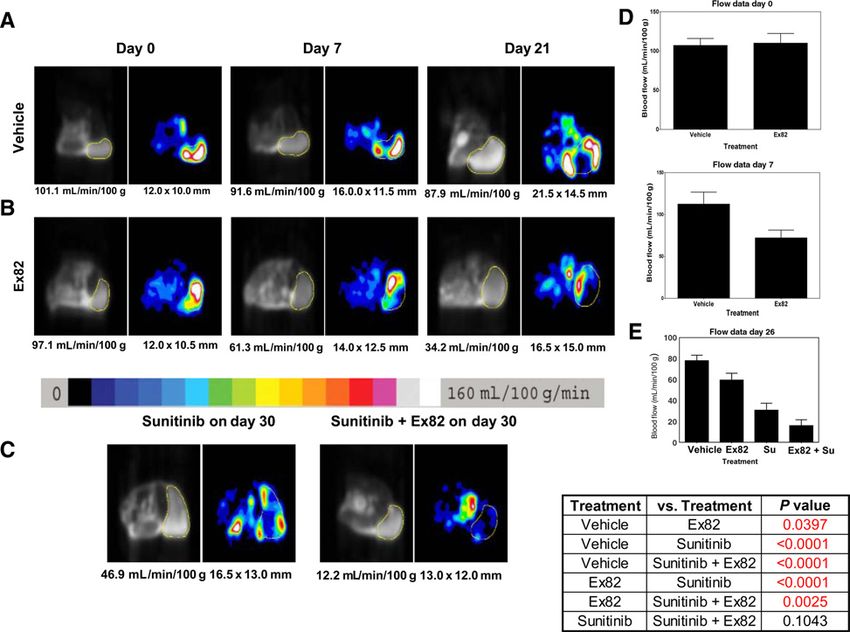
Published OnlineFirst February 20, 2019; DOI: 10.1158/1535-7163.MCT-18-0548

     Fischl et al.

     Multiplexed IHC analysis of tumors                                      gavage), S1P1 antagonist Ex82 (30 mg/kg daily by gavage), or the
        Multiplexed fluorescent immunohistochemistry and high-                combination of sunitinib and S1P1 antagonist Ex82 begun when
     content tissue imaging and quantification were performed as              the tumors reached a diameter of 12 mm as per our previous
     described previously (24). For the angiogenesis panel, blood            reports (25, 26). Tumors were measured daily during therapy to
     vessels were examined with CD34 (Biolegend, 1:100) and                  generate tumor growth curves. The delta t/c or D[T/C] was
     S1P1 (Santa Cruz Biotechnology, 1:100) antibodies multiplexed           calculated using D[T/C] ¼ 100  (Treated Tumor Volume –
     with a myofibroblast/pericyte marker (Cy3-conjugated smooth              Baseline Tumor Volume)/(Control Tumor Volume – Baseline
     muscle actin, SMA, Sigma, 1:400). Secondary antibodies conju-           Tumor Volume). The scale on D[T/C] normally runs between 0
     gated to Alexa Fluor-488 or -647 anti-rat or anti-rabbit were used      and 100. 100 means the treated tumor volume is no difference
     for detection. For the tumor health panel, blood vessels (CD34),        from vehicle. 0 means that the treated tumor volume is the same as
     proliferation (Ki67, NeoMarkers, 1:100), and apoptosis [terminal        baseline or stasis.
     deoxynucleotidyl transferase–mediated dUTP nick end labeling
     (TUNEL), Roche] were examined as described elsewhere (24).              Arterial spin-labeled MRI
     Whole tumor sections were imaged and quantified using the iCys              Imaging of tumor blood flow was performed using arterial
     research imaging cytometer. The percentage of each marker nor-          spin-labeled magnetic resonance imaging (ASL MRI) as previous-
     malized to the total tumor area identified with Hoechst 33342            ly described (25). Briefly mice were anaesthetized and placed in
     (Invitrogen, 1:1,000) was determined. Differences between treat-        the supine position on a 3 cm in diameter custom-built surface
     ment groups were assessed using ANOVA analysis with SAS JMP             coil. Images were acquired using a 3.0 T whole-body clinical MRI
     software.                                                               scanner (3T HD; GE Healthcare Technologies). A single-slice ASL
                                                                             image was obtained with a single-shot fast spin echo sequence
     Retina whole mount assay                                                using a background-suppressed, flow-sensitive alternating inver-
        Following daily i.p. injections of 30 mg/kg Ex82 from postnatal      sion-recovery strategy. The single transverse slice of ASL was
     days 2 to 5, or 20 mg/kg anti-VEGFR (DC101) at day 2 and day 4,         carefully positioned at the center of the tumor, which was marked
     neonatal mice (female and male in C57BL/6 background) were              on the skin with a permanent marker pen for follow-up MRI
     sacrificed at postnatal day 5 and eyes collected into formalin.          studies. To determine tumor blood flow, a region of interest was
     Retinas were dissected and blocked in PBS, 0.2% Triton X-100 and        drawn freehand around the peripheral margin of the tumor by
     10% goat serum overnight at 4 C, and then incubated in blocking        using an electronic cursor on a T2-weighted anatomical reference
     solution successively overnight in isolectin GS-IB4, Alexa Fluor        image that was then copied to the ASL image. The mean blood
     647 (Invitrogen) or primary antibodies (CD31, MEC13.3, BD               flow for the tumor tissue within the region of interest was derived.
     Pharmingen); collagen IV, Abcam; NG2, Millipore; Ter 119, BD            For display, a 16-color table was applied in 10 mL/100 g/min
     Pharmingen, and secondary antibodies (Jackson) each diluted             increments ranging from 0 to 160 mL/100 g/min, with flow values
     1:200 in blocking solution. Retinas were washed (4 to 5 times for       represented as varying shades of black, blue, green, yellow, red,
     1 hour) in PBS, flattened, and photographed using a Nikon Ti             and purple, in order of blood flow. Tumor blood flow was
     microscope. Vascular progression (assessed by measuring the             analyzed with repeated measures ANOVA following the previ-
     distance from the center of the retina to the angiogenic front of       ously described procedure (27).
     the retina), number of tip cells, and vascular density of the
     remodeling plexus were quantified with anti-CD31 staining using          Reagents
     FIJI software. Results were presented as mean  SEM. Statistical          The S1P1 inhibitor tool compound, Ex 82 (28), and DC101
     significance of all data was analyzed using one way ANOVA                were prepared and provided by Eli Lilly and Company. Sunitinib
     (Dunnett test) in GraphPad Prism 6 software. P values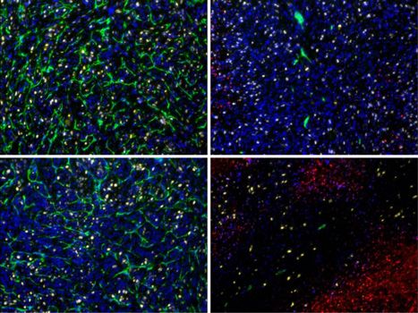
Published OnlineFirst February 20, 2019; DOI: 10.1158/1535-7163.MCT-18-0548

                                                                                                        S1P1 Inhibition Improves VEGFR-Targeted Therapy

Table 1. S1P1 antagonist Ex82 potently and selectively inhibits S1P1 beta-arrestin   staining between cells (Fig. 1B and C, arrows). The loss of barrier
activity                                                                             function with Ex82 was confirmed by using a transwell perme-
Assay                                           S1P1 antagonist IC50 (nmol/L)        ability assay which measures the passage of FITC-labeled dextran
Beta arrestin human S1P1                        5.18  2.3                           across an endothelial monolayer (21). Ex82 increased the per-
Beta arrestin mouse S1P1                        4.0
Beta arrestin human S1P2                       >20,000
                                                                                     meability to FITC-dextran in a dose-dependent manner with an
Beta arrestin human S1P3                       >20,000                               IC50 of 16.0 nmol/L (Fig. 1D). We further characterized the effect
Beta arrestin human S1P4                       >20,000                               of S1P and Ex82 using a transendothelial electrical impedance
Beta arrestin human S1P5                       >20,000                               assay (31, 32). This assay measures changes in electrical imped-
NOTE: Ex82 is a potent antagonist of both human and mouse S1PR1 in a beta-           ance relative to a voltage applied to a monolayer of endothelial
arrestin recruitment assay.                                                          cells (32) and is useful for assessing the modulation of endothelial
                                                                                     barrier function by S1P1 and S1P. S1P treatment (10 nmol/L) of
S1P1 that does not directly activate the receptor or induce receptor                 an endothelial monolayer strongly increases electrical impedance,
internalization and degradation like other reported S1P1 ago-                        whereas Ex82 has the opposite effect and significantly decreases
nists. This allows for the investigation of direct S1P1 inhibition                   electrical impedance (Supplementary Fig. S1). These results are
in vitro and in vivo.                                                                consistent with the known barrier function of S1P1. In addition,
   We next evaluated the effects of S1P1 inhibition by Ex82 on                       pretreatment with Ex82 blocked the S1P-dependent increase in
endothelial function. Because S1P1 has been shown to play an                         electrical impedance (Supplementary Fig. S1). All of these results
essential role in vascular integrity and barrier function (31), we                   demonstrate that Ex82 is a potent and specific S1P1 inhibitor with
determined if inhibition of S1P1 would disrupt endothelial cell                      endothelial barrier disrupting properties consistent with the
junctions and increase permeability of an endothelial monolayer.                     expected effect of S1P1 inhibition in endothelial cells.
Staining of an HMVEC monolayer with the endothelial junction
protein VE cadherin showed that while HMVECs formed tight cell                       S1P1 inhibitor Ex82 modulates circulation of mouse peripheral
junctions with a thick layer of VE cadherin staining (Fig. 1A,                       lymphocytes
arrow), treatment with Ex82 weakened endothelial junctions as                           To determine the potential for using Ex82 in vivo, we
shown by decreased thickness with discontinuous VE cadherin                          assessed the effect of Ex82 on circulating mouse lymphocytes, a

Figure 1.
S1P1 antagonist Ex82 disrupts endothelial barrier function and oral dosing reduces circulating mouse lymphocytes in a dose-dependent manner. Staining of a
HMVEC monolayer with VE cadherin (green) and nuclear Hoechst 33342 (red) is shown after treatment with vehicle (A) or Ex82 (B and C). Junctions between
endothelial cells show a thick area of VE cadherin staining with vehicle and thinning and disruption of junctions (white arrows) after Ex82 exposure. The loss of
barrier function with Ex82 was confirmed by using a transwell assay which measures the permeability of FITC-labeled dextran across an endothelial monolayer.
D, Ex82 increased the permeability to FITC-dextran in a dose-dependent manner with an IC50 ¼ 16.04 nmol/L. Oral dosing of Ex82 in mice led to a dose-
dependent reduction in circulating mouse lymphocytes at 4 hours post dose. Maximal reduction in circulating lymphocytes is shown at a dose of Ex82 at 3 mg/kg
(mpk) or greater (E).

www.aacrjournals.org                                                                                                        Mol Cancer Ther; 18(4) April 2019       859

   Downloaded from mct.aacrjournals.org on February 18, 2021. © 2019 American Association for Cancer Research.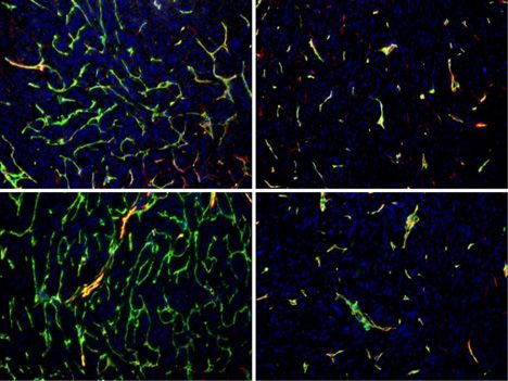
Published OnlineFirst February 20, 2019; DOI: 10.1158/1535-7163.MCT-18-0548

      Fischl et al.

      well-validated assay for characterizing the in vivo effects of S1P1                                studies. Pretreatment with the Anti-VEGFR2 antibody DC101 or
      inhibition (33, 34). Oral dosing of Ex82 induced a rapid and dose-                                 the VEGFR2 kinase inhibitor sunitinib potently inhibited
      dependent reduction in circulating mouse lymphocytes at 4 hours                                    the VEGF-dependent activation of VEGFR2 (Fig. 2). Together,
      post dose (Fig. 1E). At this 4-hour time point, maximal reduction                                  these results demonstrate rationale for coinhibition of S1P1
      in circulating lymphocytes was observed at doses of 3 mg/kg                                        and VEGFR2.
      (mpk) or greater. At 24 hours post dose, 30 mpk of Ex82 reduced
      circulating lymphocytes by greater than 85% compared with                                          Cotargeting S1P1 and VEGFR2 pathways induced vascular
      vehicle control, and there was sufficient plasma exposure of Ex82                                   regression
      to ensure robust S1P1 inhibition based on an IC50 for S1P1                                            To evaluate the antiangiogenic impact of targeting S1P1 and
      inhibition of 4 nmol/L (Table 1). It was for these reasons a 30                                    VEGFR2 pathways in vivo, we used the well-established mouse
      mpk once a day dose of Ex82 was used for all subsequent in vivo                                    retinal angiogenesis model. Previous studies showed that genetic
      mouse studies.                                                                                     ablation of S1P1 receptor in retinal blood endothelial cells
                                                                                                         induced hypersprouting and disorganization of the remodeling
      S1P1 inhibition enhances VEGF activation of VEGFR2                                                 plexus with retained perivascular cell coverage (13). In agreement
         S1P-dependent activation of S1P1 has been shown to inhibit                                      with this study, inhibition of S1P1 by Ex82 increased endothelial
      VEGF activation of VEGFR2 and sprouting angiogenesis (13). For                                     tip cells at the angiogenic front and vascular density of the
      this reason, we hypothesized potent inhibition of S1P1 with Ex82                                   remodeling plexus (Fig. 3A and B). This disorganized angiogenic
      would enhance VEGF-dependent activation of VEGFR2, and this                                        process, however, decreased the progression of blood vessels from
      would have the potential to improve response to VEGFR-targeted                                     the optic disc (OD) into the avascular retinal tissue (Fig. 3A and
      agents. To test this hypothesis, we investigated the effects of S1P1                               B). Despite the presence of pericytes, we also observed hemor-
      inhibition on VEGF activation of VEGFR2 in vivo. Tail vein                                         rhage within the plexus, which is consistent with endothelial
      injection of murine VEGF strongly activated VEGFR2 (Fig. 2).                                       barrier destabilization and increased permeability (Supplemen-
      Pretreatment with Ex82, at a dose that potently inhibits S1P1                                      tary Fig. S2A and S2B).
      in vivo, prior to VEGF tail vein injection resulted in a significant                                   Previous studies have shown that targeting VEGFR2 with a
      increase in pVEGFR2 compared with VEGF alone (Fig. 2). We next                                     selective antibody (DC101) elicits a potent antiangiogenic effect
      use DC101, a monoclonal antibody that blocks murine VEGFR2,                                        on the retinal vasculature (35). To explore the benefit of targeting
      as a tool compound to determine the combined effects of S1P1                                       both VEGFR2 and S1P1, we used 20 mg/kg on days 2 and 4 of
      and VEGFR2 inhibition. Because the standard of care for VEGFR                                      DC101 with 30 mg/kg Ex82 daily. This dose of DC101 is below
      inhibition in metastatic RCC patients is a VEGFR tyrosine kinase                                   the dose needed to see maximal effects in a developing retina
      inhibitor, we also used sunitinib for our in vivo proof-of-concept                                 and is permissive to see additive or combination effects. The

                                                                                                      Ex82                             Ex82
                            Control                                       Ex82             DC101     DC101            Suninib       Suninib
                                                                                                                                                    pVEGFR2
                                                                                                                                                    (Tyr1175)

                                                                                                                                                    β-Acn

                                                                        2.5                   **
                                       Fold change relave to vehicle

                                                                        2.0
                                                       β-acn

                                                                        1.5
                                              pVEGR2/β

                                                                        1.0
                                                                                                     *
                                                                        0.5                                      **
                                                                                                                             **         **

                                                                        0.0
                                                                                 Vehicle     Ex82   DC101      DC101      Suninib   Suninib
                                                                                                                Ex82                   Ex82

      Figure 2.
      Inhibition of S1P1 enhances VEGF activation of endothelial VEGFR2. Mice were orally dosed with compounds for 2 hours (Ex82: 30 mpk, Sunitinib: 20 mpk) or 24
      hours (DC101: 20 mpk) followed by i.v. injection of murine VEGF to activate VEGFR. Lungs were collected 5 minutes after VEGF stimulation, and Western blot
      analysis of lung lysates was performed to detect and measure VEGFR activation. The S1P1 inhibitor Ex82 increases VEGFR activation 1.8 fold (P value < 0.0001),
      whereas the VEGFR inhibitor DC101 decreases VEGFR activation by 53% (P value < 0.0004). Sunitinib was used as a control, and 95% target inhibition was
      achieved (P value < 0.0001).

860 Mol Cancer Ther; 18(4) April 2019                                                                                                        Molecular Cancer Therapeutics

         Downloaded from mct.aacrjournals.org on February 18, 2021. © 2019 American Association for Cancer Research.
Published OnlineFirst February 20, 2019; DOI: 10.1158/1535-7163.MCT-18-0548
S1P1 Inhibition Improves VEGFR-Targeted Therapy
Figure 3.
Cotargeting S1P1 and VEGFR2 pathways induces vascular regression. Whole-mount staining of blood vessels by anti-CD31 (endothelial cell membrane) and
anti-collagen IV (extracellular basement membrane) in mouse retinas of young pups (postnatal day 5) treated with vehicle or anti-VEGFR2 (DC101, 20 mpk) and
S1P1 antagonist (Ex82, 30 mpk) or the combination of DC101 and Ex82 (A). Bottom plots represent a higher magnification of the retinal remodeling plexus (white
boxes). Vessel regression was identified by collagen IV–positive and CD31-negative structures (sleeves of former blood vessel basement membranes, white
arrowheads). Quantification of vascular progression from the retinal center (OD) to the angiogenic front, endothelial tip cells at the angiogenic front, and
vascular density of the remodeling plexus (B). Results are pooled from 3 independent experiments (n 6 animals per group per experiment, mean SEM,
one-way ANOVA Dunnett test). Scale bars: top plots, 200 mm; bottom plots, 50 mm.
www.aacrjournals.org Mol Cancer Ther; 18(4) April 2019 861
Downloaded from mct.aacrjournals.org on February 18, 2021. © 2019 American Association for Cancer Research.Published OnlineFirst February 20, 2019; DOI: 10.1158/1535-7163.MCT-18-0548
Fischl et al.
combination of VEGFR2 and S1P1 inhibition in the retina trig- the mechanism of the combined effect of sunitinib and Ex82. The
gered a significant regression of CD31-positive endothelial cells in percent area of tumor vessels (labeled with CD34) tended to
the remodeling plexus, leaving behind basement membrane increase with S1P1 inhibition (20.4% for vehicle vs. 30.5% with
sleeves of collagen IV (Fig. 3A, white arrowheads). This resulted Ex82; Fig. 6Aiii and Av), consistent with the observed increased EC
in a lower vascular density in the combination treatment com- sprouting seen in the mouse retinal assay (Fig. 3). As expected,
pared with the vehicle-treated mice. The combination also sunitinib significantly reduced the percent area of vessels (6Aii),
decreased the vascular progression and increased the number of but combination of Ex82 with sunitinib did not lead to significant
endothelial tip cells compared with VEGF inhibition alone further reduction in tumor vessels (7.2% for sunitinib and 4.8%
(Fig. 3A and B). With this combination, the areas of hemorrhage for the combination; Fig. 6Aiv and Av). S1P1 expression increased
were reduced within the vascular plexus and restricted only to the with Ex82 (P ¼ 0.0073), and this was significantly reduced with
angiogenic front indicating that the remaining vessels in the sunitinib and the combination treatment (P ¼ 0.0027 Ex82 vs.
remodeled area are less permeable (Supplementary Fig. S2). These sunitinib and P ¼ 0.0052 Ex82 vs. combination; Fig. 6Aiii and
results suggest increased sensitivity of remodeling blood vessels to Avi). Examination of the effects of treatment on tumor cell
anti-VEGFR2 therapies when the vessels are destabilized by S1P1 proliferation showed little effect of any of the treatments (percent
inhibition. These data, along with our mouse lung data, support area of Ki67; Fig. Bv). Sunitinib treatment alone led to a nonsig-
the idea of combining VEGF and S1P1 pathway inhibition as a nificant increase in the percent area of TUNEL-positive cells
novel antiangiogenic therapeutic regimen that may improve upon compared with vehicle (Fig. 6Bii and Bvi and 6C), whereas Ex82
VEGF pathway blockade alone. single treatment had no effect on tumor cell apoptosis (Fig. 6Biii
and Bvi). The combination treatment, however, significantly
Combination of S1P1 and VEGF pathway inhibition decreased increased TUNEL staining more than the vehicle or either of
RCC tumor growth and blood flow the single agents. The percent area of TUNEL was 25.3% for the
RCC is a vascular tumor that is highly dependent on VEGF likely combination, compared with 4.9% for the vehicle (P ¼ 0.0003),
due to the Von Hippel-Lindau (VHL) loss seen in most RCC. In 10.9% for sunitinib (P ¼ 0.0065), and 3.06% for Ex82
RCC, VEGF pathway inhibition has shown clinical effects, and we (P < 0.0001; Fig. 6Bvi). Because S1P1 was not expressed on tumor
have previously shown that VEGFR TKI therapy leads to induction cells (Supplementary Fig. S3), these effects on tumor cell death are
of the S1P pathway (25). Because inhibition of S1P1 signaling by likely to be attributed to secondary effects due to the direct effects
Ex82 destabilizes endothelial cell junctions, delays vessel matu- on the functional tumor vascular network, despite a modest
ration, and promotes vessel sprouting in response to VEGF reduction in vessel density.
(ref. 13; Figs. 1 and 3), we hypothesized that these vascular
features following S1P1 inhibition would render the tumor
vasculature more sensitive to VEGF pathway blockade. To test Discussion
this hypothesis, we evaluated the effect of S1P1 inhibition alone Resistance to antiangiogenic therapy is a major obstacle in the
and in combination with sunitinib in the 786-O VHL-deficient management of metastatic RCC as well as other tumor types.
RCC murine xenograft model compared with a vehicle control Tumor angiogenesis is initiated and largely driven by VEGF,
group (n ¼ 6–8 per group). Treatment with either sunitinib or especially in RCC in which the VHL deficiency (including muta-
Ex82 led to slowed tumor growth as single agents compared with tion, deletion, or LOH) renders the tumors highly dependent on
vehicle control (Fig. 4; P ¼ 0.0082 for Ex82 vs. vehicle and VEGF (25, 38). However, other angiogenic pathways are alsoPublished OnlineFirst February 20, 2019; DOI: 10.1158/1535-7163.MCT-18-0548
S1P1 Inhibition Improves VEGFR-Targeted Therapy
Figure 4.
Combination of S1P1 and VEGF pathway inhibition reduces RCC tumor growth. Tumor growth curves from the 786-O RCC tumor xenograft model are shown for
the four treatment arms: vehicle, S1P1 antagonist (Ex82), sunitinib, or the combination. The table shows that tumors from mice treated with sunitinib and the Ex82
grow more slowly than the vehicle-treated tumors and that the addition of Ex82 to sunitinib adds to the tumor growth control of sunitinib (P ¼ 0.008).
result in enhanced inhibition of angiogenesis compared with there was a significant reduction in vascular progression and
either agent alone. vascular density. Importantly, we saw evidence of empty base-
We show that inhibition of S1P1 alone destabilized the ment membrane sleeves with the combination treatment,
retinal vasculature resulting in hypersprouting blood vessels. which indicates vascular regression (43). These results uncover
The hypersprouting was accompanied by vascular hemorrhage the dynamic sensitivity of remodeling blood vessels, which
as seen with TER 119 staining of red blood cells. Our results have been destabilized by S1P1 antagonism, to anti-VEGFR2
obtained with Ex82 phenocopied the results obtained by therapies. This indicates mechanistically that S1P1 inhibition
genetic knockout of endothelial S1P1, suggesting Ex82 mod- makes the vessels more sensitive to VEGFR2 inhibition by
ulates S1P1-dependent vascular biology in vivo (11–14). When making the vessels more dependent on VEGF signaling leading
VEGF pathway inhibition was combined with S1P1 inhibition, to reduced tumor growth and tumor cell apoptosis. These data
www.aacrjournals.org Mol Cancer Ther; 18(4) April 2019 863
Downloaded from mct.aacrjournals.org on February 18, 2021. © 2019 American Association for Cancer Research.Published OnlineFirst February 20, 2019; DOI: 10.1158/1535-7163.MCT-18-0548
Fischl et al.
Figure 5.
Combination of S1P1 and VEGF pathway inhibition lowers tumor blood flow. ASL MRI blood flow images are shown for tumors from mice treated serially with
vehicle (A) or Ex82 (B) at day 0, day 7, and day 21. For each image pair, the black and white image is the MRI anatomic image, and the corresponding colored
image is the ASL image. The tumor is circled with a yellow line, and the area in yellow is the region of interest for which the blood flow is measured. The color
scale corresponds to tumor blood flow values. Below the color scale are two representative images of tumors from mice treated with sunitinib or sunitinib þ Ex82
and imaged on day 26 (4 days depending on availability of MR scanner; C). Statistical analysis is shown in the accompanying graphs (D and E) and Table (E),
which shows the P values for differences in tumor blood flow at day 26.
support the development of this combination to enhance the apoptosis demonstrating the potential increase in clinical
sensitivity to VEGFR targeting. response that this combination could have. The specific stresses
We next evaluated the benefit of combined S1P1 and VEGFR2 placed upon the tumor cells with combination treatment are not
therapy in a 786-O VHL-deficient mouse xenograft model of RCC. fully known but may be in part due to increased tumor hypoxia
We have previously shown that this model is dependent on and nutrient deprivation. We believe the significant effects on
SPHK1/S1P signaling when tumors progress on anti-VEGFR2 tumor apoptosis are secondary to the effects on the vasculature
therapy (18). In addition, the 786-O model exclusively expresses as S1P1 expression was not detected on tumor cells. S1P1 is
S1P1 on the endothelial cells of the tumor-associated vasculature. well expressed in the tumor-associated blood vessels in 786-O
Serial ASL MRI perfusion imaging studies showed inhibition of xenografts and in all other tumor xenograft models we have
S1P1 reduced tumor blood flow but not to the extent of sunitinib. characterized. To date, there is only one known model
The combination of S1P1 and VEGFR inhibition reduced blood (SK-Hep-1) which shows both tumor and tumor-associated
flow even further. The magnitude of reduction in flow induced by blood vessel S1P1 expression (Supplementary Fig. S3).
sunitinib alone may mask the additional effects of S1P1 inhibi- It is also likely that the S1P pathway modulates the tumor
tion, although a trend for decreased blood flow was seen with the immune microenvironment, but the specifics of these effects are
combination. Differences observed in tumor blood flow after not yet fully understood and should be explored in an immune
S1P1 inhibition versus VEGFR inhibition further support the competent model. One known class effect of S1P inhibitors is
distinct effects of VEGFR2 and S1P1 on the tumor vasculature. their ability to modulate circulating lymphocytes. S1P has potent
Interestingly, treatment with the combination but not either roles in limiting T-cell egress from tissues into circulation, and we
single agent alone led to a dramatic induction of tumor cell also demonstrate that S1P1 inhibition with Ex82 reduces
864 Mol Cancer Ther; 18(4) April 2019 Molecular Cancer Therapeutics
Downloaded from mct.aacrjournals.org on February 18, 2021. © 2019 American Association for Cancer Research.Published OnlineFirst February 20, 2019; DOI: 10.1158/1535-7163.MCT-18-0548
S1P1 Inhibition Improves VEGFR-Targeted Therapy
A i ii v 40
% CD34 area
30
20
*† *†
10
0
Vehicle Su Vehicle S1P1
Ex82 Sutent
Su Su + Ex82
Combo
iii iv vi 15
% S1P1 area
10
5 † †
0
Ex82 SU + Ex82 Vehicle
Vehicle S1P1
Ex82 Sutent
Su Su Combo
+ Ex82
CD34 SMA S1P1 Hoechst
B
i ii v 25
20
% Ki67 area
15
10
5
0
Vehicle Su Vehicle Ex82 Sutent
Vehicle S1P1 Su Combo
Su + Ex82
iii iv vi 40
*†‡
% TUNEL area
30
20
10
0
Vehicle Ex82
Vehicle S1P1 Sutent
Su Combo
Su + Ex82
Ex82 Su + Ex82
CD34 TUNEL Ki67 Hoechst
C
TUNEL
Vehicle Ex82 Su Su + Ex82
Figure 6.
The combination of VEGFR and S1P1 inhibition induces tumor cell apoptosis. Multiplexed panels to assay tumor angiogenesis are shown. The percent area of
tumor vessels (labeled with CD34; green) increased with Ex82 (Aiii and Av) and sunitinib (Su) significantly reduced the percent area of vessels (Aii and Av), but
further reduction in tumor vessels was not detected with the combination of Ex82 and sunitinib (Av). Pericyte staining was assessed by SMA (red), Hoechst
staining is shown in blue, and S1P1 is shown in yellow. S1P1 expression tended to increase with Ex82 (Aiii and vi) and was significantly less with sunitinib and the
combination treatment. Examination of the effects of treatment on tumor cells showed that sunitinib treatment tended to increase apoptosis (TUNEL stain
shown in red), and the combination treatment significantly increased apoptosis more than the vehicle or either of the single agents (Biv and vi). CD34 staining is
shown in green, and Ki67 is shown in yellow. Hoechst staining is shown in blue. Whole tumor cross-sections are shown in C stained for TUNEL (gray). Bars
represent mean SEM. , P < 0.05 vs. vehicle; †, P < 0.05 vs. Ex82; and z, P < 0.05 vs. Su.
circulating lymphocytes. S1P1 signaling has been shown to drive effects of S1P inhibition in combination with VEGF and PD1
Treg cell accumulation in tumors limiting CD8þ T-cell recruit- pathway inhibition.
ment and activation thus promoting tumor growth (44). Testing Inhibitors of the S1P pathways are currently being developed in
of these agents in immune competent models may help elucidate the clinical setting. Inhibition of the S1P pathway has been
the role of S1P inhibition in enhancing of immune-mediated achieved by two main strategies. S1P receptor modulators such
antitumor responses. Moreover, it will be important to assess the as FTY720 mimic S1P and have been shown to have activity in
www.aacrjournals.org Mol Cancer Ther; 18(4) April 2019 865
Downloaded from mct.aacrjournals.org on February 18, 2021. © 2019 American Association for Cancer Research.Published OnlineFirst February 20, 2019; DOI: 10.1158/1535-7163.MCT-18-0548
Fischl et al.
A B C D
Sproung front Figure 7.
Conceptual role of S1P/S1P1 signaling in tumor
angiogenesis. This model depicts our hypothesis
about the effects of S1P1 and VEGFR inhibition. A
shows an abundant tumor vascular bed. VEGF
Maturing
network pathway inhibition leads to decreased sprouting and a
defect in the development of the vascular bed (B). S1P
induces vascular sprouting. Thus, S1P1 inhibition leads
to hypersprouting resulting in nonfunctional
angiogenesis (C). Combination therapy leads to loss of
No inhibitor VEGF inhibion S1P1 inhibion Combinaon S1P-dependent vessels likely induced by VEGFR
Normal vessel Sproung Excessive sproung Limited sproung inhibition and the VEGF-dependent hypersprouting
network inhibited Lack of maturaon Lack of maturaon induced by the S1P1 inhibitor (D).
Vessel regression
multiple sclerosis, allograft rejection, and inflammatory bowel Authors' Contributions
disease (45). An antibody against S1P (Sphingomab) has also Conception and design: A.S. Fischl, D. Bodenmiller, P. Hipskind,
been shown to have antitumor and antiangiogenic effects in S. Chintharlapalli, L.E. Benjamin, R.S. Bhatt
preclinical models but did not meet its primary endpoints in a Development of methodology: A.S. Fischl, X. Wang, D. Bodenmiller, G. Evans,
P. Hipskind, S. Chintharlapalli, D.C. Alsop, R.S. Bhatt
phase II clinical trial (18, 46). Acquisition of data (provided animals, acquired and managed patients,
In summary, using a potent and selective antagonist tool provided facilities, etc.): A.S. Fischl, X. Wang, B.L. Falcon, R. Almonte-
compound against endothelial protein S1P1 (Ex82), alone and Baldonado, D. Bodenmiller, G. Evans, J. Stewart, T. Wilson, M.T. Uhlik,
in combination with VEGFR-targeted agents, we show S1P1 D.C. Alsop, R.S. Bhatt
inhibition destabilizes endothelial junctions in vitro and in vivo Analysis and interpretation of data (e.g., statistical analysis, biostatistics,
during the early and remodeling/maturation phases of retinal and computational analysis): A.S. Fischl, X. Wang, B.L. Falcon, R. Almonte-
Baldonado, D. Bodenmiller, J. Stewart, P. Hipskind, J. Manro, M.T. Uhlik,
tumor angiogenesis which leads to vascular beds that are vulner- D. Gerald, R.S. Bhatt
able to VEGF pathway inhibition (Fig. 7). S1P1 signaling is Writing, review, and/or revision of the manuscript: A.S. Fischl, X. Wang,
distinct yet complementary from the initiation phase of angio- B.L. Falcon, D. Bodenmiller, P. Hipskind, J. Manro, S. Chintharlapalli,
genesis where VEGFA/VEGFR2 signaling is dominant. Targeting D. Gerald, D.C. Alsop, L.E. Benjamin
S1P1 and VEGFR2 simultaneously provides a novel therapeutic Administrative, technical, or material support (i.e., reporting or organizing
approach by inhibiting two mechanisms required for functional data, constructing databases): A.S. Fischl, D. Bodenmiller, P. Hipskind,
R.S. Bhatt
vasculature. Combined inhibition has the potential to enhance Study supervision: P. Hipskind, S. Chintharlapalli, R.S. Bhatt
response rates compared with currently approved antiangiogenic
agents, and this combination has the potential to overcome S1P- Acknowledgments
dependent resistance to anti-VEGF pathway therapies. R.S. Bhatt, X. Wang, and D.C. Alsop were supported by NIH R01 CA196996
and NIH P50 CA101942-12.
Disclosure of Potential Conflicts of Interest
B.L. Falcon and J. Manro have an ownership interest (including stock, The costs of publication of this article were defrayed in part by the
patents, etc.) in Eli Lilly and Co. P. Hipskind has an ownership interest payment of page charges. This article must therefore be hereby marked
(including stock, patents, etc.) in LLY. M.T. Uhlik is Vice President, Translational advertisement in accordance with 18 U.S.C. Section 1734 solely to indicate
Oncology, at Biothera Pharmaceuticals, Inc., and has an ownership interest this fact.
(including stock, patents, etc.) in Biothera Pharmaceuticals, Inc. and Eli Lilly and
Company. L.E. Benjamin has an ownership interest (including stock, patents,
etc.) in Oncologie Ltd. and Eli Lilly and Company. No potential conflicts of Received May 25, 2018; revised October 4, 2018; accepted February 4, 2019;
interest were disclosed by the other authors. published first February 20, 2019.
References
1. Casak SJ, Fashoyin-Aje I, Lemery SJ, Zhang L, Jin R, Li H, et al. FDA Approval 6. Rini BI, Escudier B, Tomczak P, Kaprin A, Szczylik C, Hutson TE, et al.
Summary: ramucirumab for gastric cancer. Clin Cancer Res 2015;21: Comparative effectiveness of axitinib versus sorafenib in advanced renal cell
3372–6. carcinoma (AXIS): a randomised phase 3 trial. Lancet 2011;378:1931–9.
2. Gerald D, Chintharlapalli S, Augustin HG, Benjamin LE. Angiopoietin-2: 7. Yanagida K, Hla T. Vascular and immunobiology of the circulatory sphin-
an attractive target for improved antiangiogenic tumor therapy. Cancer Res gosine 1-phosphate gradient. Annu Rev Physiol 2017;79:67–91.
2013;73:1649–57. 8. Pyne NJ, Pyne S. Sphingosine 1-phosphate and cancer. Nat Rev Cancer
3. Zhao Y, Adjei AA. Targeting angiogenesis in cancer therapy: moving beyond 2010;10:489–503.
vascular endothelial growth factor. Oncologist 2015;20:660–73. 9. Kunkel GT, Maceyka M, Milstien S, Spiegel S. Targeting the sphingosine-1-
4. Escudier B, Eisen T, Stadler WM, Szczylik C, Oudard S, Staehler M, et al. phosphate axis in cancer, inflammation and beyond. Nat Rev Drug Discov
Sorafenib for treatment of renal cell carcinoma: final efficacy and safety 2013;12:688–702.
results of the phase III treatment approaches in renal cancer global 10. van der Weyden L, Arends MJ, Campbell AD, Bald T, Wardle-Jones H,
evaluation trial. J Clin Oncol 2009;27:3312–8. Griggs N, et al. Genome-wide in vivo screen identifies novel host regulators
5. Motzer RJ, Hutson TE, Tomczak P, Michaelson MD, Bukowski RM, Oudard of metastatic colonization. Nature 2017;541:233–6.
S, et al. Overall survival and updated results for sunitinib compared with 11. Liu Y, Wada R, Yamashita T, Mi Y, Deng CX, Hobson JP, et al. Edg-1, the G
interferon alfa in patients with metastatic renal cell carcinoma. J Clin Oncol protein-coupled receptor for sphingosine-1-phosphate, is essential for
2009;27:3584–90. vascular maturation. J Clin Invest 2000;106:951–61.
866 Mol Cancer Ther; 18(4) April 2019 Molecular Cancer Therapeutics
Downloaded from mct.aacrjournals.org on February 18, 2021. © 2019 American Association for Cancer Research.Published OnlineFirst February 20, 2019; DOI: 10.1158/1535-7163.MCT-18-0548
S1P1 Inhibition Improves VEGFR-Targeted Therapy
12. Ben Shoham A, Malkinson G, Krief S, Shwartz Y, Ely Y, Ferrara N, et al. S1P1 28. Angst D, Bollbuck B, Janser P, Quancard J. Biaryl benzylamine derivatives.
inhibits sprouting angiogenesis during vascular development. Develop- World Intellectual Property Organization 2010: Patent Number WO
ment 2012;139:3859–69. 2010072712 A1.
13. Gaengel K, Niaudet C, Hagikura K, Lavina B, Muhl L, Hofmann JJ, et al. The 29. Bigaud M, Guerini D, Billich A, Bassilana F, Brinkmann V. Second gener-
sphingosine-1-phosphate receptor S1PR1 restricts sprouting angiogenesis ation S1P pathway modulators: research strategies and clinical develop-
by regulating the interplay between VE-cadherin and VEGFR2. Dev Cell ments. Biochim Biophys Acta 2014;1841:745–58.
2012;23:587–99. 30. Quancard J, Bollbuck B, Janser P, Angst D, Berst F, Buehlmayer P, et al. A
14. Jung B, Obinata H, Galvani S, Mendelson K, Ding BS, Skoura A, et al. Flow- potent and selective S1P(1) antagonist with efficacy in experimental
regulated endothelial S1P receptor-1 signaling sustains vascular develop- autoimmune encephalomyelitis. Chem Biol 2012;19:1142–51.
ment. Dev Cell 2012;23:600–10. 31. Garcia JG, Liu F, Verin AD, Birukova A, Dechert MA, Gerthoffer WT, et al.
15. LaMontagne K, Littlewood-Evans A, Schnell C, O'Reilly T, Wyder L, Sphingosine 1-phosphate promotes endothelial cell barrier integrity by
Sanchez T, et al. Antagonism of sphingosine-1-phosphate receptors by Edg-dependent cytoskeletal rearrangement. J Clin Invest 2001;108:
FTY720 inhibits angiogenesis and tumor vascularization. Cancer Res 2006; 689–701.
66:221–31. 32. Garbison KE, Heinz BA, Lajiness ME, Weidner JR, Sittampalam GS.
16. Ader I, Gstalder C, Bouquerel P, Golzio M, Andrieu G, Zalvidea S, et al. Impedance-based technologies. In: Sittampalam GS, Gal-Edd N, Arkin
Neutralizing S1P inhibits intratumoral hypoxia, induces vascular remo- M, Auld D, Austin C, Bejcek B, et al., editors. Assay guidance manual.
delling and sensitizes to chemotherapy in prostate cancer. Oncotarget Bethesda, MD; 2004.
2015;6:13803–21. 33. Sanna MG, Wang SK, Gonzalez-Cabrera PJ, Don A, Marsolais D, Matheu
17. Visentin B, Vekich JA, Sibbald BJ, Cavalli AL, Moreno KM, Matteo RG, et al. MP, et al. Enhancement of capillary leakage and restoration of lymphocyte
Validation of an anti-sphingosine-1-phosphate antibody as a potential egress by a chiral S1P1 antagonist in vivo. Nat Chem Biol 2006;2:434–41.
therapeutic in reducing growth, invasion, and angiogenesis in multiple 34. Cyster JG, Schwab SR. Sphingosine-1-phosphate and lymphocyte egress
tumor lineages. Cancer Cell 2006;9:225–38. from lymphoid organs. Annu Rev Immunol 2012;30:69–94.
18. Zhang L, Wang X, Bullock AJ, Callea M, Shah H, Song J, et al. Anti-S1P 35. Benedito R, Rocha SF, Woeste M, Zamykal M, Radtke F, Casanovas O, et al.
antibody as a novel therapeutic strategy for VEGFR TKI-resistant renal Notch-dependent VEGFR3 upregulation allows angiogenesis without
cancer. Clin Cancer Res 2015;21:1925–34. VEGF-VEGFR2 signalling. Nature 2012;484:110–4.
19. Sarkisyan G, Gay LJ, Nguyen N, Felding BH, Rosen H. Host endothelial 36. Wang X, Bullock AJ, Zhang L, Wei L, Yu D, Mahagaokar K, et al. The role of
S1PR1 regulation of vascular permeability modulates tumor growth. Am J angiopoietins as potential therapeutic targets in renal cell carcinoma.
Physiol Cell Physiol 2014;307:C14–24. Transl Oncol 2014;7:188–95.
20. Ibrahim MA, Johnson HW, Jeong JW, Lewis GL, Shi X, Noguchi RT, 37. Wang X, Zhang L, O'Neill A, Bahamon B, Alsop DC, Mier JW, et al. Cox-2
et al. Discovery of a novel class of potent and orally bioavailable inhibition enhances the activity of sunitinib in human renal cell carcinoma
sphingosine 1-phosphate receptor 1 antagonists. J Med Chem 2012;55: xenografts. Br J Cancer 2013;108:319–26.
1368–81. 38. Bergers G, Benjamin LE. Tumorigenesis and the angiogenic switch. Nat Rev
21. Hoang MV, Nagy JA, Senger DR. Active Rac1 improves pathologic VEGF Cancer 2003;3:401–10.
neovessel architecture and reduces vascular leak: mechanistic similarities 39. Bhatt RS, Atkins MB. Molecular pathways: can activin-like kinase pathway
with angiopoietin-1. Blood 2011;117:1751–60. inhibition enhance the limited efficacy of VEGF inhibitors? Clin Cancer Res
22. Mandala S, Hajdu R, Bergstrom J, Quackenbush E, Xie J, Milligan J, et al. 2014;20:2838–45.
Alteration of lymphocyte trafficking by sphingosine-1-phosphate receptor 40. Chae SS, Paik JH, Furneaux H, Hla T. Requirement for sphingosine
agonists. Science 2002;296:346–9. 1-phosphate receptor-1 in tumor angiogenesis demonstrated by in vivo
23. Burkholder TP, Clayton JR, Rempala ME, Henry JR, Knobeloch JM, RNA interference. J Clin Invest 2004;114:1082–9.
Mendel D, et al. Discovery of LY2457546: a multi-targeted anti- 41. Anelli V, Gault CR, Snider AJ, Obeid LM. Role of sphingosine kinase-1 in
angiogenic kinase inhibitor with a novel spectrum of activity and paracrine/transcellular angiogenesis and lymphangiogenesis in vitro.
exquisite potency in the acute myelogenous leukemia-Flt-3-internal FASEB J 2010;24:2727–38.
tandem duplication mutant human tumor xenograft model. Invest 42. Chae SS, Paik JH, Allende ML, Proia RL, Hla T. Regulation of limb
New Drugs 2012;30:936–49. development by the sphingosine 1-phosphate receptor S1p1/EDG-1
24. Falcon BL, Stewart J, Ezell S, Hanson J, Wijsman J, Ye X, et al. High-content occurs via the hypoxia/VEGF axis. Dev Biol 2004;268:441–7.
multiplexed tissue imaging and quantification for cancer drug discovery. 43. Inai T, Mancuso M, Hashizume H, Baffert F, Haskell A, Baluk P, et al.
Drug Discov Today 2013;18:510–22. Inhibition of vascular endothelial growth factor (VEGF) signaling in cancer
25. Bhatt RS, Wang X, Zhang L, Collins MP, Signoretti S, Alsop DC, et al. causes loss of endothelial fenestrations, regression of tumor vessels, and
Renal cancer resistance to antiangiogenic therapy is delayed by appearance of basement membrane ghosts. Am J Pathol 2004;165:35–52.
restoration of angiostatic signaling. Mol Cancer Ther 2010;9:2793– 44. Priceman SJ, Shen S, Wang L, Deng J, Yue C, Kujawski M, et al. S1PR1 is
802. crucial for accumulation of regulatory T cells in tumors via STAT3. Cell Rep
26. Zhang L, Bhasin M, Schor-Bardach R, Wang X, Collins MP, Panka D, et al. 2014;6:992–9.
Resistance of renal cell carcinoma to sorafenib is mediated by potentially 45. Brinkmann V, Billich A, Baumruker T, Heining P, Schmouder R, Francis G,
reversible gene expression. PLoS One 2011;6:e19144. et al. Fingolimod (FTY720): discovery and development of an oral drug to
27. Yan SB, Peek VL, Ajamie R, Buchanan SG, Graff JR, Heidler SA, et al. treat multiple sclerosis. Nat Rev Drug Discov 2010;9:883–97.
LY2801653 is an orally bioavailable multi-kinase inhibitor with potent 46. Pal SK, Drabkin HA, Reeves JA, Hainsworth JD, Hazel SE, Paggiarino DA,
activity against MET, MST1R, and other oncoproteins, and displays anti- et al. A phase 2 study of the sphingosine-1-phosphate antibody sonepci-
tumor activities in mouse xenograft models. Invest New Drugs 2013;31: zumab in patients with metastatic renal cell carcinoma. Cancer 2017;123:
833–44. 576–82.
www.aacrjournals.org Mol Cancer Ther; 18(4) April 2019 867
Downloaded from mct.aacrjournals.org on February 18, 2021. © 2019 American Association for Cancer Research.Published OnlineFirst February 20, 2019; DOI: 10.1158/1535-7163.MCT-18-0548
Inhibition of Sphingosine Phosphate Receptor 1 Signaling
Enhances the Efficacy of VEGF Receptor Inhibition
Anthony S. Fischl, Xiaoen Wang, Beverly L. Falcon, et al.
Mol Cancer Ther 2019;18:856-867. Published OnlineFirst February 20, 2019.
Updated version Access the most recent version of this article at:
doi:10.1158/1535-7163.MCT-18-0548
Supplementary Access the most recent supplemental material at:
Material http://mct.aacrjournals.org/content/suppl/2019/02/20/1535-7163.MCT-18-0548.DC1
Cited articles This article cites 44 articles, 12 of which you can access for free at:
http://mct.aacrjournals.org/content/18/4/856.full#ref-list-1
Citing articles This article has been cited by 3 HighWire-hosted articles. Access the articles at:
http://mct.aacrjournals.org/content/18/4/856.full#related-urls
E-mail alerts Sign up to receive free email-alerts related to this article or journal.
Reprints and To order reprints of this article or to subscribe to the journal, contact the AACR Publications Department at
Subscriptions pubs@aacr.org.
Permissions To request permission to re-use all or part of this article, use this link
http://mct.aacrjournals.org/content/18/4/856.
Click on "Request Permissions" which will take you to the Copyright Clearance Center's (CCC)
Rightslink site.
Downloaded from mct.aacrjournals.org on February 18, 2021. © 2019 American Association for Cancer Research.You can also read