Elimination of Aspergillus flavus from Pistachio Nuts with Dielectric Barrier Discharge (DBD) Cold Plasma and Its Impacts on Biochemical Indices
←
→
Page content transcription
If your browser does not render page correctly, please read the page content below

Hindawi
Journal of Food Quality
Volume 2021, Article ID 9968711, 12 pages
https://doi.org/10.1155/2021/9968711

Research Article
Elimination of Aspergillus flavus from Pistachio Nuts with
Dielectric Barrier Discharge (DBD) Cold Plasma and Its
Impacts on Biochemical Indices

 Mina Makari,1 Mohammad Hojjati ,1 Samira Shahbazi,2 and Hamed Askari 2

 1
 Department of Food Science and Technology, Agricultural Sciences and Natural Resources University of Khuzestan, Ahvaz, Iran
 2
 Nuclear Agriculture School, Nuclear Science and Technology Research Institute (NSTRI),
 Atomic Energy Organization of Iran (AEOI), Karaj, Iran

 Correspondence should be addressed to Mohammad Hojjati; hojjati@asnrukh.ac.ir

 Received 28 March 2021; Accepted 31 May 2021; Published 7 June 2021

 Academic Editor: Chunpeng Wan

 Copyright © 2021 Mina Makari et al. This is an open access article distributed under the Creative Commons Attribution License,
 which permits unrestricted use, distribution, and reproduction in any medium, provided the original work is properly cited.
 In the present research, the effects of different durations (0, 15, 30, 60, 90, 120, 150, and 180 sec) of dielectric barrier discharge
 (DBD) cold plasma on decontaminating Aspergillus flavus, detoxifying pure aflatoxin B1 (AFB1), and the quality attributes of
 pistachio nuts (total phenolic content, antioxidant activity, chlorophylls, total carotenoids, instrumental color, total soluble
 protein, and malondialdehyde determination) were studied. The results showed that the viable spore population reduced with the
 increase of plasma treatment duration, so that after 180 s of the treatment, a decrease by 4 logs was observed in the spore
 population. Chlorophyll a and b, as well as total carotenoid levels and color parameters, decreased, which led to darker pistachio
 samples and intensity reduction in soluble protein content and protein bands. Plasma treatment did not alter the total phenolic
 content but slightly increased the antioxidant activity of pistachio nuts samples. The malondialdehyde values significantly in-
 creased all the plasma treatment durations. The maximum reduction of AFB1 was observed after 180 s of the treatment, which was
 64.63% and 52.42% for glass slides and pistachio nut samples, respectively. The present findings demonstrated that cold plasma
 could be used as an efficient decontamination method of food products without inducing undesirable quality changes in nuts.

1. Introduction half a billion dollars in that year [1]. This valuable com-
 modity is highly susceptible to contamination by one of the
Pistachio nut (Pistacia vera L.) is a member of Anacardiaceae most prevalent aflatoxin producing fungi Aspergillus flavus
family and is the most famous tree nuts in the world. Iran, during preharvest and postharvest stages, which leads to
USA, Turkey, China, Syria, Greece, and Italy are the main reduction in quality and nutritional value; finally, it causes
pistachio-growing countries [1, 2]. Pistachio kernels have economic losses to the producing countries [4, 5].
been recognized as health promoting food because of their Aflatoxins are extremely toxic, teratogenic, and carci-
high content of unsaturated fatty acids, proteins, minerals, nogenic secondary metabolites that are synthesized by the
vitamins, phytosterols, and polyphenols and have been polyketide pathway [6]. Aflatoxins B1 (AFB1) and B2 (AFB2)
consumed as a salted and roasted snack. They also can be are two popular toxins made by A. flavus [7]. AFB1 has a
used as the main ingredient of many food products such as high potential for the liver cancer and is one of the most
confections, biscuits, ice cream, and sausage [2]. Pistachio is common toxins in pistachios [8, 9]. The strict international
the most important nonpetroleum export commodity that laws have been enacted to monitor the level of aflatoxin
plays a vital role in the economy of Iran [3]. Iran is the first contamination in pistachios, and the acceptable maximum
and biggest producer and exporter of pistachio nuts in the levels for pistachio nuts are 8 μg/kg for AFB1 and 10 μg/kg
world. It produced 337,815 tons and exported 78,547 tons in for the total aflatoxins (TotAFs) according to the Iran and
2019. The total export revenue from pistachio nuts was about European commission regulations [10, 11].
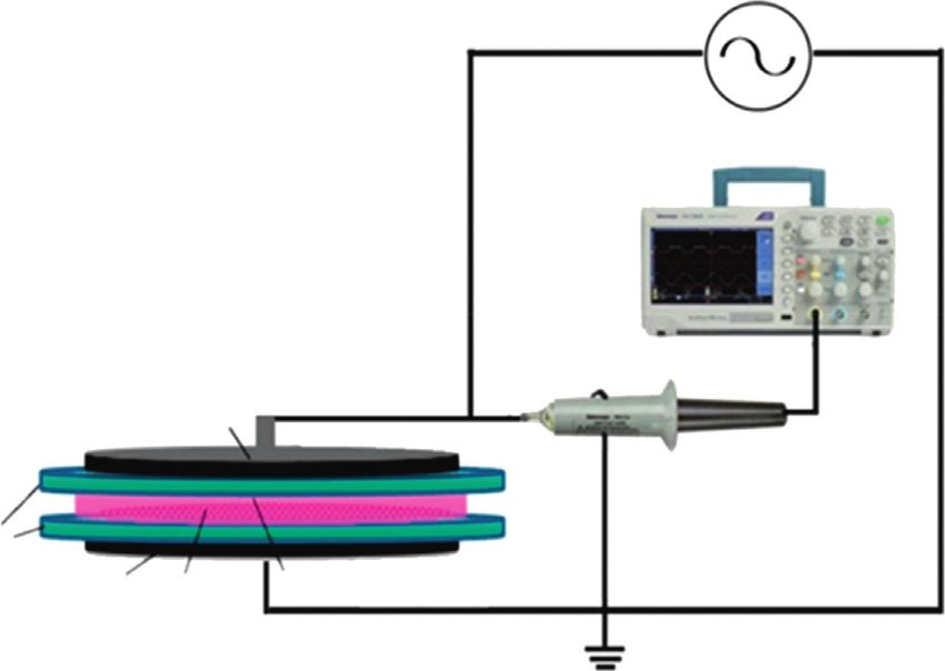
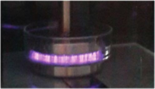
2 Journal of Food Quality

 Various physical, thermal, and chemical methods have spores in pistachio powder was set to (6.18 ± 1.18)
been studied to prevent the mold growth and mycotoxin ×105 spores/g pistachio power. The contaminated pistachio
decontamination; but using these methods is limited due to powders were packed in sterile Petri dishes and kept at
potential risks to the human health ,as well as reduced 4 ± 1°C [9].
quality, sensory properties (flavor, color, and texture), and
nutritional value of the foodstuffs [12, 13]. Hence, choosing
 2.3. Cold Plasma Treatment of Pistachio Powder and Pure
reliable alternative decontamination methods to maintain-
 Aflatoxin B1. The nonthermal atmospheric pressure di-
ing the quality and safety of food products is of great im-
 electric barrier discharge (DBD) air plasma system consisted
portance. Cold plasma is a futuristic nonthermal technique,
 of two parallel metallic electrodes separated by a 5 mm gap
which is considered as the fourth state of matter including
 (Figure 1). The upper electrode had the diameter of 75 mm,
radicals, electrons, charged ions, neutral and/or excited
 was made of stainless steel 316, and was covered from the
particles, molecules, atoms, and UV photons with inacti-
 bottom by the dielectric pyrex glass with the thickness of
vating properties of microorganisms [4, 14]. Several factors
 3 mm and diameter of 80 mm. The plasma treatment was
such as type of feeding gas, electric and magnetic field, gas
 operated in ambient air as working gas at 15, 30, 60, 90, 120,
flow rate and pressure, order and shape of electrodes, relative
 150, and 180 s time intervals. The process was done at 130 W
humidity, treatment time, and type of cold plasma system
 power, 20 kHz frequency, and 15 kV voltage, and the dis-
can affect the composition of plasma reactive species [4, 8].
 tance between the electrodes and sample was 3 mm.
Several studies have been done on applying cold plasma
 Treatments were done in triplicate. About 4 g of pistachio
technology in food processing such as modifying wheat flour
 nut samples contaminated with A. flavus active spores and/
functionality [15], degrading pesticides residue in vegetables
 or AFB1 with the thickness of 3 mm was placed between two
and fruits [16], reducing the cooking time of rice [17],
 electrodes and treated using a DBD plasma device at dif-
inactivating enzymes [18], and inactivating microbes in
 ferent time intervals. AFB1-contaminated glass slides with
various food products [17, 19, 20]. Recently, promising
 the size of 24 × 50 were used alongside with pistachio nut
results of A. flavus decontamination and aflatoxin detoxi-
 samples for quantifying the AFB1 residual.
fication using cold plasma in nuts have been reported
[4–9, 12–14, 21].
 The purposes of this research are the possibility of using 2.4. Determining A. flavus Total Count. After plasma treat-
cold plasma in decreasing the A. flavus spore population and ment of pistachio samples, each sample was suspended with
reducing the amount of AFB1 in pistachio powder along sterile saline solution (8.5 g NaCl in 1000 ml distilled water)
with the plasma effect on some quality characteristics of by vortex; after serial dilution preparation, 100 μl of each
pistachios. dilution was inoculated into two Petri dishes comprising
 Potato dextrose agar (Merck, Germany) with 50 ppm
2. Materials and Methods chloramphenicol and 0.033 g·L−1 of Rose-Bengal by surface
 plating and, then, incubated at 28°C for 3–5 days. Fungal
All the chemical materials and solvents were prepared colonies were counted visually after incubation, and the
mainly from Sigma-Aldrich (St. Louis, MO, US) and Merck results were means of triplicate reported as log colony
(Darmstadt, Germany) companies. Pure AFB1 from forming unit per gram (log cfu/g) [23].
A. flavus was purchased from Sigma-Aldrich (Darmstadt,
Germany) Company.
 2.5. Total Phenolic Content (TPC) and Antioxidant Activity.
 Samples were analyzed for TPC and antioxidant activity
2.1. Pistachio and Sample Preparation. Pistachio nuts using Folin-Ciocalteu method and DPPH free radical
(P. vera L.), Akbari cultivar, were prepared from Iranian scavenging activity, respectively, according to the procedure
Pistachio Research Centre located in Kerman Province in used by Hojjati et al. [2]. About 1 gr of each ground pistachio
Autumn 2019. The peel of all the pistachios was removed; the samples was extracted for 15 min with 80% methanol so-
kernels were milled and stored properly in dry conditions in lution (10 ml) and sonicated (Sonorex Digitech DT 1028 H,
sterile Petri dishes at 4 ± 1°C and protected from direct Bandelin, Germany) two times for 15 min. The mixture was
sunlight until further experiments. kept at the room temperature for a day and, then, centri-
 fuged (5000 ×g at 4°C for 4 min) (Httich Refrigerated
 Centrifuge Universal 320R, Germany). The collected
2.2. Inoculation of Pistachio with Aspergillus flavus and Af-
 supernatants were used for both experiments.
latoxin B1. The aflatoxigenic fungus A. flavus R5 [22] was
grown on potato dextrose agar (PDA) at 28°C for 7 days.
Some pistachio kernels were inoculated with pathogen 2.6. Malondialdehyde Determination. Lipid oxidation was
fungal spores or Aflatoxin B1 (final concertation about assessed based on thiobarbituric acid reactive substances
400 ppb) and milled with other pistachio kernels. Concen- (TBARS) analysis according to Papastergiadis et al. [24]. The
tration of conidial in contaminated pistachio powder was sample extracts were obtained by mixing about 7 gr of
determined by hemocytometer counts using suspended 0.5 g ground pistachio nuts with 15 mL of 7.5% trichloroacetic
of inoculated pistachio powder in 4.5 ml of sterile saline acid (TCA) (w/v) with 0.1% of ethylenediaminetetraacetic
dilute solution containing 0.5% Tween-80. The population of acid (EDTA) (w/v) and 0.1% of propyl gallate (w/v),
Journal of Food Quality 3

 7

 6

 1

 2
 3 4 5

 (a) (b)

Figure 1: Photograph (a) and schematics (b) of an atmospheric pressure diffuse plasma generated by DBD: (1) discharge electrode, (2) pyrex
glass dielectric barrier, (3) ground electrode, (4) pistachio nut sample, (5) cold plasma, (6) oscilloscope, and (7) power supply. The discharge
in the photo (a) was driven by kHz sinusoidal high voltage, and the working gas was air. Photo was taken at the author’s laboratory.

homogenized at 18,000 rpm for 1 min, and adjusted to 30 mL describing the accurate location of a color within a three
with TCA. After adding 2.5 mL of the filtrated extract (with dimensional space, in which parameter L∗ indicates the
150 mm filter paper) and 2.5 mL of thiobarbituric acid re- brightness of the color and is between zero (black) and 100
agent (46 mM) in glacial acetic acid (99%) to a 15 mL tube, (white), a∗ parameter shows the characteristic of red and
the tubes were placed in boiling water bath for 35 min and green color and has negative (green) and positive (red)
cooled to the room temperature. The collected supernatant values, parameter b∗ represents yellowness and blueness,
absorbance was read at 532 nm using a spectrophotometer. and the negative and positive values indicate the colors blue
The blank sample was TBARS reagent in distilled water, and and yellow, respectively; finally, C∗ is chroma, and the value
the malondialdehyde (MDA) standard curve was plotted of C∗ is 0 at the center of a color sphere and increases with
using different MDA standard solutions in TCA solution distance from the center. For determining the color values, a
(7.5%). The results were reported as nanomolar malon- watch glass was placed on the samples, and the L∗ , a∗ , and b∗
dialdehyde per gram pistachio powder. parameters were calculated using a colorimeter. Chroma was
 determined using [19]
2.7. Chlorophylls and Total Carotenoids. Total carotenoids, as ��������
well as chlorophyll a and b, were measured by spectro- C∗ � a∗2 + b∗2 . (2)
photometric method according to Roueita et al. [25]. The
ground pistachio nut samples (0.2 g) were mixed with 80% Color analyses of the samples were done in five
(v/v) acetone (5 ml). The mixtures were left in darkness for replicates.
15 min and, then, centrifuged (1500 ×g for 10 min) and
filtrated with no. 2 Whatman filter paper. The absorbance of
the filtrated extracts was determined at 470 (A470), 663 2.9. Total Soluble Protein. Total soluble protein was mea-
(A663), and 645 (A645), nm. The contents of carotenoids sured using Bradford method [26]. The pistachio nut powder
(μg·g−1) and chlorophyll a and b (μg·g−1) were calculated was mixed well with 10% sodium phosphate buffer solution
according to the following formulae: (pH � 6.8, W/V) using a mortar and pestle; then, it was
 stirred for 30 min. The mixture was poured into a falcon tube
 Ca � 12.21 A663 − 2.81 A645 , and centrifuged at 5000 ×g for 5 min. The collected super-
 natant was kept at −20°C for later examination. Also, 5 ml of
 Cb � 21.13 A645 − 5.03 A663 , Bradford reagent was added to 100 μl of the collected ex-
 (1)
 tracts, and, after 30 min, the absorbance of the extracts was
 1000 A470 − 3.27Ca − 104Cb calculated using the bovine serum albumin (BSA) standard
 Ct � .
 229 curve at 595 nm. The results were reported as mg of protein
 per g of pistachio nuts (mg/g).

2.8. Instrumental Color. The color parameters of pistachio
nuts’ powder samples were examined by means of a Minolta 2.10. Electrophoretic Pattern of Proteins. 250 μ of protein
Colorimeter CR-400 (Konica Minolta, Inc., Osaka, Japan) extracts was precipitated with cold acetone and centrifuged
with a D65 illuminant as a light source and a 10 standard (5000 ×g for 4 min at 4°C). The supernatant was gathered
observer at 25°C. This colorimeter is based on International and placed in the room to remove the remaining acetone.
Commission on Illumination (CIE) L∗ , a∗ and b∗ values for Then, a certain amount of the samples was prepared in
4 Journal of Food Quality

distilled water and the sample buffer (nonreducing buffer contaminated pistachio nuts. The results showed that the
condition; 65 mM Tris, pH 6.8, 10% glycine, 2 SDS, 0.2% population of the viable spores significantly decreased with
Bromophenol blue). On the other hand, the same samples the increase of plasma exposure time (p < 0.05). The total
were mixed with 5% 2-mercaptoethanol and heated in count of the plasma-treated pistachio nut samples was in-
boiling water bath for reducing buffer. All the samples were crementally reduced by increasing the treatment duration in
tested for electrophoretic pattern of proteins by SDS-PAGE comparison to the control (Figure 2(a)). As shown in
using a Mini Protean ΙΙ Cell system (Bio-Rad, Hercules, Figure 2(b), the rate of spore reduction was slow at the
USA) by adopting Laemmli method [27]. Also, 50 μg of the treatment duration of 15 s and 30 s. However, the rate of
samples was transferred onto the gel consisting of 12.5% spore reduction gradually increased with increasing plasma
acrylamide separating gel and 5% acrylamide stacking gel. treatment up to 180 s duration. After 120 s and 150 s of the
The gels were stained with Coomassie Brilliant Blue R-250 treatment, a reduction of 2 and 3 log (CFU/g) in A. flavus
and destained in a methanol-acetic acid-water (1 : 1 : 8 v/v) spore population was achieved, respectively. As the plasma
mixture. The Quantity One 1-D Analysis software and Gel treatment duration raised to 180 s, a 4 log (CFU/g) reduction
Doc XR + (Bio-Rad, Hercules, USA) were used for capturing in spore population was observed, and no detectable spore
gels images, and the relative density, molecular weight, and was found after plating the plasma-treated sample at the
bands intensity were analyzed by Gel-Pro Analyzer (ver.6.0). treatment duration of 180 s (Figures 2(a) and 2(b)).
 The present results were in agreement with the findings
 by Sohbatzadeh et al. [5] and Ghorashi et al. [8] who have
2.11. Aflatoxin Extraction and HPLC Analysis of Aflatoxin B1. investigated the effect of cold plasma treatment on A. flavus
In order to extract AFB1 for HPLC analysis, the sample (1 g population in pistachio nut samples. Dasan et al. [12] also
pistachio nut powder) was mixed with methanol 80% v/v observed the 4 log reduction of A. flavus after 5 min of
and shaken well at 150 rpm for 24 h. The mixture was plasma treatment on hazelnut samples. Mošovská et al. [21]
centrifuged at 10000 ×g for 5 min, and the supernatant was studied the cold plasma using different gases (O2, N2,
purified using an ASPEC 401 immunoaffinity purification ambient air, synthetic air with or without vapor) on hazelnut
column. The immunoaffinity column was washed with PBS samples and observed that the use of synthetic air (with
(10 ml) to remove impurities; then, it was loaded with the vapor) as plasma working gas reduced the A. flavus spore
sample and washed again with water (10 ml). Finally, the population below the detection limit (4 log) after 180 s of the
column-bounded AFB1 was released by the elution with treatment. Sen et al. [4] also reported that population of
acetonitrile (1.5 ml). One aliquot of eluate (0.5 ml) was A. flavus and A. parasiticus in hazelnut samples decreased by
retained in a glass vial, and the remainder was diluted with about 4 log after low pressure (LP) plasma treatment (100 W
water (2 ml) before HPLC analysis. 400 ml of the diluted for 30 min). Bagheri et al. [28] observed that A. flavus spores
eluate was injected into the HPLC system equipped with a reduced after cold atmospheric plasma (15 V, 10 min, and
Spherisorb Excel ODS1 (250 × 4.6 mm; 5 μm) C18 reversed 3 cm) treatment on contaminated military ration snack
column with a guard column (25 × 4.6 mm i.d.). Perkin samples. Šimončicová et al. [29] stated that cold plasma
Elmer LC420 fluorescence detector with excitation at treatment duration of 180 s showed a remarkable reduction
364 nm and emission at 440 nm were applied to detection. in A. flavus growth on agar medium and also observed
Water: methanol: acetonitrile (56 : 14 : 30, v/v/v) at the flow changes in hyphae structure after plasma treatment dura-
rate of 0.86 ml/min was used as the mobile phase. Post- tions of 60 s and 180 s. Suhem et al. [30] investigated the
column derivatization was achieved with a zero dead volume inactivation effect of cold atmospheric plasma on A. flavus
T-piece and 30 cm × 0.3 mm i.d. PTFE reaction tube. Pyri- spores in both agar medium and brown rice cereal bars and
dine hydrobromide perbromide reagent was added at 0.3 ml/ found that plasma treatment (40 W for 25 min) significantly
min. The retention time for AFB1 was approximately suppressed the fungi growth on both samples. They observed
13.55 min [13]. that cold plasma treatment effectively damaged the fungi
 structure and broke the conidiophores and vesicles. Several
2.12. Statistical Analyses. Data analyses were performed inactivation mechanisms have been suggested for plasma
using SPSS Software v.26.0 (SPSS Inc., Chicago, IL, USA) treatment by many authors. The air plasma is composed of
using one-way analysis of variance (ANOVA). Differences oxygen reactive species (ROS), nitrogen reactive species
were significant at 95% confidence level (statistical signifi- (RNS), charged particles, atoms, excited molecules, and UV
cance at p < 0.05) using Duncan’s multiple comparison test. radiation, which can lead to spore inactivation by creating a
All the experiments were administrated in a completely harsh oxidative environment in collision with sample sur-
randomized design, and data presented in this study were face and cause denaturation of the proteins of spore coat and
the mean ± standard deviation (SD) of the triplicate loss of spore coat integrity, exposing the center of the spore
experiments. to plasma reactive species and reducing the cell viability
 [5, 12, 14]. In addition, plasma reactive species can result in
3. Results and Discussion rupturing the fungal cell wall via affecting the lipid bilayers
 of cell wall and/or accumulating charged particles on the
3.1. Decontamination Effect of Plasma Treatment. external surface of the cell membrane, which forms elec-
Figure 2 shows the effect of different durations of cold trostatic force between the reactive particles and the cell
plasma on the reduction of A. flavus spore population in membrane and leads to fungal deactivation [4, 6]. On the
Journal of Food Quality 5

 (a)

 Control 15 sec 30 sec 60 sec 90 sec 120 sec 150 sec 180 sec
 5
 (b)

 a
 4 b
 c
 (CFUs/g pistachio nuts)
 Log10 A. flavus

 3
 d

 e
 2
 f

 1 g

 h
 0
 0 15 30 60 90 120 150 180
 Time of plasma treatment (sec)
Figure 2: Decontamination of A. flavus by different durations of cold plasma on contaminated pistachio nut powders. (a) Cultured Petri
dishes in similar dilution. (b) Enumeration of viable spores in untreated (0 sec) and plasma-treated (15–180 s) pistachio nut powders with
PDA media after 72 h of incubation at 28°C. Data are means of three replicates. The same letter does not differ statistically according to
Duncan’s test (p ≤ 0.05).

other hand, UV photons generated by plasma treatment can this initial decrement of phenolic compounds as the result of
damage cell wall and inhibit cell replicate by damaging DNA interaction between plasma reactive oxygen species (peroxyl
strand and result in cell inactivation [6]. However, the exact radicals, hydroxyl radicals, singlet oxygen, and atomic ox-
antimicrobial mechanism of action of cold plasma is still ygen) and phenolic compounds known for their free radical
unknown and requires further investigation. scavenging ability. As shown in Table 1, some of the plasma-
 treated samples were slightly enhanced in antioxidant ac-
 tivity. This increment can be due to the chemical reactions of
3.2. Total Phenolic Content (TPC) and Antioxidant Activity. plasma reactive species (ions, chemically reactive species,
The results obtained from the effect of different durations of and UV irradiation) that lead to cell membrane breakdown
cold plasma on TPC and antioxidant activity of samples are and, consequently, collapse the cell membrane-bound
summarized in Table 1. The results indicated that the TPC phenolic compounds of food product, promoting poly-
and antioxidant values of pistachio nut samples were sig- phenols concentration and antioxidant activity after plasma
nificant at some of the plasma treatment durations. The treatment because of their role as antioxidant agents [20, 31].
lowest amount of the TPC of plasma-treated samples was Bagheri et al. [28] reported that no remarkable differences
observed after treatment durations of 30 s, 60 s, and 180 s, were found in TPC of military ration snacks after exposure
while the maximum amount of TPC was shown in the of atmospheric cold plasma treatment. Ramazzina et al. [32]
samples treated for 120 s and 150 s. No significant difference reported that DBD plasma had no effect on TPC and an-
was found between TPC values at other plasma treatment tioxidant activity of kiwifruit. In another study, Ramazzina
durations in comparison with the control; plasma treatment et al. [33] indicated that TPC and antioxidant activity of
had no effect on the amount of TPC of pistachio nut samples. apples were not affected by DBD plasma exposure in less
The lowest amount of antioxidant activity of control and than 30 minutes.
treated samples was observed after treatment duration of
15 s. Other plasma-treated samples displayed slight changes
in antioxidant activity, and plasma treatment slightly en- 3.3. Malondialdehyde Determination. The results showed
hanced the antioxidant activity after longer plasma treat- that different plasma durations significantly affected the
ment durations. Nevertheless, there was no significant MDA content of the plasma-treated pistachio samples
difference between treated samples after treatment durations (Table 1), and an increase of plasma duration increased the
of longer than 60 s to 180 s. A similar trend of reduction in MDA content of all the samples. The lowest and highest
TPC was observed in the initial part of the plasma treatment MDA values were observed at the treatment durations of
(30 s and 60 s) on orange juice, tomato juice, apple juice, and 30 s, and 15 s (3%), and 180 s (27.88%), respectively. On the
sour cherry juice by Dasan and Boyaci [31] who described other hand, MDA content of plasma-treated pistachio
6 Journal of Food Quality

Table 1: Effect of different durations of cold plasma on the values of the total phenolic content (TPC), antioxidant activity, malondialdehyde
(MDA), pigments, total soluble protein (TSP), and instrumental color parameters of pistachio nuts.
 Time of plasma treatments (sec)
Properties
 0 15 30 60 90 120 150 180
TPC
 1.81ab ± 0.05 1.78a–c ± 0.08 1.67c ± 0.08 1.71bc ± 0.08 1.76a–c ± 0.03 1.84a ± 0.05 1.85a ± 0.08 1.79a–c ± 0.01
(mg GAE/g)
Inhibitory of
DPPH activity 86.85c ± 0.58 86.43c ± 0.60 87.01bc ± 0.40 87.82ab ± 0.35 87.90a ± 0.29 87.12a–c ± 0.42 87.82ab ± 0.42 87.94a ± 0.42
(%)
MDA
 24.96d ± 1.34 25.77cd ± 0.39 25.71cd ± 1.14 28.31bc ± 1.66 29.04b ± 1.66 28.82b ± 1.14 29.49ab ± 1.46 31.92a ± 2.61
(n mol. g−1)
TSP (mg/g) 3.56b ± 0.22 3.40b ± 0.23 4.10a ± 0.10 3.61b ± 0.10 4.30a ± 0.20 3.68b ± 0.30 2.86c ± 0.08 2.50d ± 0.09
 −1
Pigments (μg·g )
Chlorophyll a 25.91a ± 0.27 22.89b ± 0.39 21.83c ± 0.51 18.65e ± 0.76 18.50e ± 0.53 20.16d ± 0.62 20.65d ± 0.36 20.13d ± 0.60
Chlorophyll b 26.44a ± 0.21 23.86c ± 0.43 24.63bc ± 0.56 24.63d ± 0.76 22.17d ± 0.55 24.15c ± 1.06 26.28a ± 0.61 25.83ab ± 1.02
Total
 52.35a ± 0.43 46.75b ± 0.79 46.46bc ± 1.05 46.46d ± 1.39 40.67d ± 1.07 44.31b ± 1.69 46.93b ± 0.97 45.96bc ± 1.60
chlorophyll
Total
 24.23a ± 0.10 21.63d ± 0.28 22.42b ± 0.13 18.08g ± 0.16 17.51h ± 0.08 18.77f ± 0.09 19.10e ± 0.07 21.90c ± 0.04
carotenoid
Instrumental color
Lightness (L) 62.07a ± 0.38 61.03b ± 0.60 60.82bc ± 0.40 61.20ab ± 0.56 61.25ab ± 0.24 59.85d ± 0.47 59.97cd ± 0.31 61.45ab ± 0.80
Redness (a∗ ) −5.51f ± 0.30 −4.74e ± 0.16 −4.13d ± 0.43 −3.83cd ± 0.17 −3.54bc ± 0.15 −3.37bc ± 0.38 −3.04b ± 0.47 −0.38a ± 0.06
Yellowness
 32.04a ± 0.25 30.13b ± 0.33 30.48b ± 0.18 30.77b ± 0.50 30.04bc ± 0.58 30.39b ± 0.34 29.26cd ± 0.65 28.49d ± 0.57
(b∗ )
Chroma (C∗ ) 32.51a ± 0.29 30.50b ± 0.35 30.76b ± 0.23 31.00b ± 0.52 30.24b ± 0.56 30.57b ± 0.35 29.42c ± 0.70 28.49d ± 0.57
Values (mean ± SD) in a row without the same superscript letter differ statistically (p < 0.05).

samples had no statistically significant differences at the 180 s) showed lower amount of chlorophyll a than the
treatment durations of 90 s, 120 s, and 150 s; however, they control sample, but there was no significant difference be-
had greater MDA content than plasma-treated samples at tween the plasma-treated samples. Chlorophyll b showed
shorter treatment durations. There are a few reports that lower values than the control at the treatment duration of
have investigated the effect of cold plasma on lipid oxidation 120 s, but no significant difference was observed after the
in nuts and nut products. Bagheri et al. [28] reported an treatment durations of 150 s and 180 s compared to the
increment of peroxide values of military ration snacks by control sample. Also, the total chlorophyll content was re-
increase in the voltage of generating the plasma device. duced by the duration of plasma exposure. Total chlorophyll
Thirumdas et al. [34] indicated that plasma treatment of content significantly reduced with the increase of plasma
walnuts and peanuts at different powers and time intervals treatment duration of 90 s. Total chlorophyll reduction after
led to increase in the peroxide value (20%) of the samples. treatment durations of 60 s and 90 s was 24% compared to
Ahangari et al. [19] found that peroxide value of plasma- the control. Treatment durations of 120 s showed lower
treated walnuts (50 W for 15 and 20 min) slightly increased, amount of total chlorophyll than the control, but there was
but no differences were observed between the plasma- no remarkable difference between the treated and control
treated and control samples. This increase in MDA content samples at the treatment times of 150 s and 180 s. In general,
can be because cold plasma reactive species can initiate lipid increasing the plasma treatment durations to at least 90 s
oxidation by attacking various compounds including un- resulted in significant reduction in total chlorophyll. From
saturated fatty acids and increasing the MDA content of data in Table 1, it is apparent that total carotenoids content
plasma-treated food products [35]. of plasma-treated pistachio nuts reduced significantly as the
 plasma treatment increased to 90 s (p < 0.05), and the lowest
 amount of total carotenoids was observed after the treatment
3.4. Chlorophylls and Carotenoids. The pigment content data durations of 60 s and 90 s. Generally, the duration of plasma
of pistachio nuts submitted to different durations of cold treatment to 90 s led to a reduction in total content of ca-
plasma are set out in Table 1. The results suggested that the rotenoids. The reduction in total carotenoids after these
amount of different pigments in pistachio samples decreased treatment durations was about 25% compared to the control.
with the duration of the plasma treatment. The results Ramazzina et al. [32] reported a 15% decrement in chlo-
revealed that chlorophyll a and b significantly reduced after rophyll a right after applying an air DBD plasma treatment
plasma exposure (p > 0.05). However, chlorophyll a re- to the kiwifruit samples. In this study, chlorophyll and
duction was greater than chlorophyll b. The lowest amount carotenoid content showed a significant decrease after
of chlorophyll a (28%) and chlorophyll b (17.6% and 16.1%, storage time. Beyer et al. [20] observed that air plasma
respectively) was observed after 60 s and 90 s of the process, treatment reduced the chlorophyll a concentration of
respectively. Longer treatment durations (120, 150, and spirulina algae powder. It has been suggested that
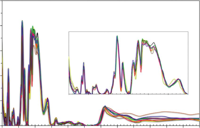
Journal of Food Quality 7

chlorophyll degradation upon plasma treatment can be study, the authors explained that atmospheric pressure
related to pigment oxidation by plasma reactive species and/ plasma jet (APPJ) treatment can expose the hydrophobic
or denaturation of enzymes responsible for chlorophyll groups to the protein surface by unfolding both secondary
catabolism during the plasma treatment [32]. and tertiary structures of the protein molecules. Interaction
 of this hydrophobic groups led to protein agglomeration and
 induced a reduction in protein solubility [39].
3.5. Impact of Cold Plasma on Instrumental Color. The results
of different plasma treatment durations on instrumental of
pistachio nuts color are presented in Table 1, showing that 3.7. Electrophoretic Pattern of Proteins. Impact of different
the L∗ value decreased with an increase in plasma treatment durations of plasma treatment on soluble protein patterns of
times. This parameter significantly reduced after 120 s and pistachio nuts is provided in Figures 3 and 4. In accordance
150 s of plasma exposure (p < 0.05), but it remained un- with Figure 3, a variety of protein bands were identified in a
changed at other exposure durations. The b∗ index slightly molecular weight range from 5 to 204 kDa and 6 to 236 kDa
decreased during plasma treatment compared to the control in reduced and nonreduced gels for all the samples, re-
sample, while a∗ values increased considerably after treat- spectively. According to the results and presence of low
ment durations of longer than 120 s. Parameter C∗ or molecular weight peptides in the reducing condition, the
chroma also decreased with the increase of the treatment presence of β-mercaptoethanol in reducing conditions led to
duration, but this reduction was not significant between the the breakdown of the polypeptides disulfide bonds and
samples treated with treatment durations of less than 120 s. converted them into smaller subunits. Nonreducing con-
This parameter remarkably decreased after the treatment dition (without β-mercaptoethanol) did not change the
durations of 150 s and 180 s. This overall color changes proteins’ disulfide bonds (Figure 3). Comparing polyacryl-
during plasma treatment resulted in darker pistachio nuts amide gel in both nonreducing and reducing conditions
color at longer cold plasma treatment durations due to showed slight differences in the pattern of protein band due
Maillard reactions and/or chlorophyll and carotenoids to plasma treatment. According to the densitometry analyses
degradation. The breakdown of glycosidic and peptidic of protein bands, with the increase of exposure time, the
bonds during radiation led to the formation of breakdown intensity of protein bands in nonreducing gel decreased in
products such as carbonyl and amino compounds, which comparison with the control sample (Figure 4). Neverthe-
induced the Maillard reactions and, finally, formed colored less, these decreasing changes were not the same in the
compounds [3]. In a similar study, a decrease in L∗ and b∗ of intensity of all protein bands, and the intensity of some of the
walnut samples after plasma treatment (20 min and 40 W) protein bands increased as a result of plasma treatment. For
was observed [19]. Hertwig et al. [36] found a decrement in example, protein bands in the molecular weight of 7 kDa
L∗ , b∗ , and chroma parameters after cold plasma treatment increased at all the treatment durations. The plasma treat-
resulted in darker almond samples. ment of 30 s showed the lowest intensity of pistachio protein
 bands in pistachio samples (Figure 4). These results can be
 related to the results of protein solubility (Table 1), because
3.6. Total Soluble Protein. The effect of different plasma
 plasma reactive species may lead to the alteration of protein
durations on total soluble protein of pistachio nuts is il-
 structure and/or creation of new proteins through cross-
lustrated in Table 1. All the data were significantly different
 linkage of free amino acids to the protein and protein-
(p < 05.0). Total soluble protein showed an increment with
 protein aggregation and reduce the solubility [40]. Results of
the increase of the plasma exposure time to 90 s, and the
 a similar study conducted by Alinezhad et al. [3] demon-
highest amount of total soluble protein was observed after
 strated that gamma irradiation (1–6 kGy) altered the pattern
30 s and 90 s. No differences were shown between the protein
 and intensity of pistachio protein; the intensity of protein
solubility of control and plasma-treated samples after the
 bonds was declined in all pistachio nut samples, while
treatment durations of 15 s, 60 s, and 120 s. Plasma treatment
 gamma irradiation at dose of 4 kGy showed an increase in
durations of longer than 90 s resulted in a decrease in protein
 the intensity of pistachio protein bands. Similar reports were
solubility, so that the lowest protein solubility was observed
 stated by Meinlschmidt et al. [40] who indicated that CAPP
after 180 s and 150 s compared to the control sample.
 (1, 2.5, 5, 7.5, and 10 min) reduced the intensity of soy bean
Nonthermal plasma contains a wide range of reactive species
 protein bonds corresponding to Gly m5 allergen. On the
(ROS, RNS, and UV photons) that can attack the protein
 contrary, Ekezie et al. [39] reported that ATPJ (2–10 min)
backbone and lead to alteration and unfolding of the sec-
 had no impact on the intensity of king prawn protein bands
ondary and tertiary structures of the protein. Consequently,
 in both reducing and nonreducing conditions.
the interaction between hydrophobic groups of unfolded
proteins resulted in aggregation, followed by coagulation
and precipitation inducing changes in chemical properties of 3.8. HPLC Analysis of Aflatoxin B1. The cold plasma effect on
the proteins such as decrease in protein solubility [37, 38]. AFB1 concentration is shown in Table 2. AFB1 was extracted
Bubler et al. [38] observed that the protein solubility of with 80% methanol solution from glass slides contaminated
Tenebrio flour declined after cold atmospheric pressure with the same concentration of pure toxin and was used to
plasma (CAPP) treatment. Similar results were reported by determine the AFB1 residual by means of a HPLC method.
Ekezie et al. [39]: protein solubility of king prawn samples The chromatograms of pure AFB1 (left) and contaminated
decreased with increasing the plasma treatment time. In this pistachio nuts (right) treated at different durations (0, 60,
8 Journal of Food Quality

 Non-reducing condition Reducing condition
 kDa (1) (2) (3) (4) (5) (6) (7) (8) M (1) (2) (3) (4) (5) (6) (7) (8)
 245
 180
 135
 100
 75
 63

 45

 35

 25
 20

 17

 11

Figure 3: Profiles of proteins from nontreated (lane 1) and treated pistachios with different durations of cold plasma (lane numbers of 2, 3, 4,
5, 6, 7, and 8 for 15, 30, 60, 90, 120, 150, and 180 sec, respectively) under reducing and nonreducing conditions. “M” shows a molecular
weight marker (SinaClon BioScience, prestained protein ladder, PR901641).

 1

 0.8
 0.9
 0.8
 0.7
 Optical density

 0.6 0.6
 0.5
 0.4
 0.3
 0.4
 0.2
 0.1
 0
 8 18 28 38 48 58 68
 0.2

 0
 8 28 48 68 88 108 128 148 168 188 208 228 248
 Molecular weight (kDa)
 Control 60 150
 15 90 180
 30 120
 (a)
 Figure 4: Continued.
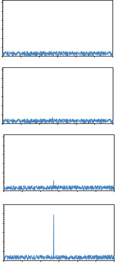
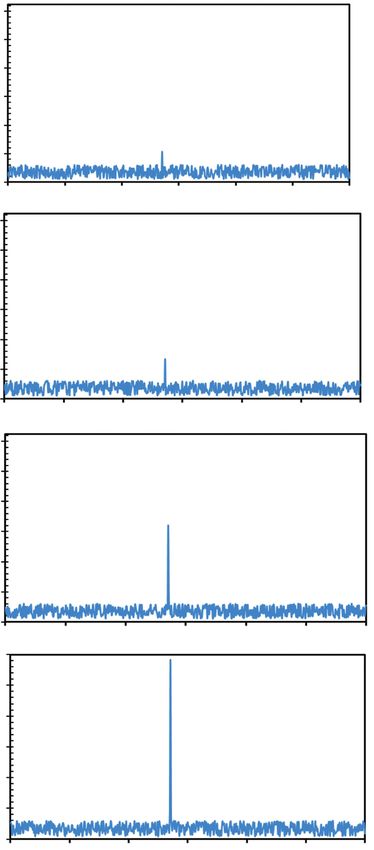
Journal of Food Quality 9

 1

 0.8
 0.9

 0.75
 Optical density
 0.6 0.6

 0.45

 0.3
 0.4
 0.15

 0
 0.2 8 13 18 23 28 33 38

 0
 8 28 48 68 88 108 128 148
 Molecular weight (kDa)
 Control 60 150
 15 90 180
 30 120
 (b)

Figure 4: The densitometric analysis of the SDS-PAGE profile of soluble proteins extracted from untreated (0.0 sec) and treated (15–180 sec)
pistachios nuts with different durations of cold plasma in nonreducing (a) and reducing conditions (b).

Table 2: Effect of cold plasma (0–180 sec) on aflatoxin B1 concentration (ppb) in pure toxin and contaminated pistachio nut powder.
 Time of plasma treatments (sec)
Aflatoxin B1 concentration (ppb)
 0 60 120 180
Pure toxin 710.38a ± 0.93 458.89b ± 0.88 277.27c ± 1.06 251.32d ± 0.88
Pistachio nut powder 383.19a ± 0.38 259.39b ± 1.11 211.64c ± 1.70 182.32d ± 1.78
Values (mean ± SD) in a row without the same superscript letter differ statistically (p < 0.05).

120, and 180 s) are illustrated in Figure 5. The increment of plasma treatment of contaminated hazelnuts (400 W and
the plasma exposure time significantly decreased the AFB1 4 min) was reported [7]. Devi et al. [6] observed 65% and
concentration in plasma-treated samples, as shown in Ta- 95% reduction in AFB1 on air plasma-treated groundnuts
ble 2. The concentration of AFB1 remarkably decreased after with the power of 60 W and 12 min and 40 W and 15 min,
60 s and 120 s of the plasma treatment. A reduction of respectively. Plasma generated reactive species (ROS, RNS,
35.41%, 60.97%, and 64.63% was observed in contaminated free radicals, excited particles, and UV radiation) can ef-
glass slides after 60 s, 120 s, and 180 s of the treatment, re- fectively degrade mycotoxins including aflatoxins [9]. There
spectively. This rate of reduction in AFB1-contaminated are some factors that affect mycotoxin degradation upon
pistachio powder samples was 32.31%, 44.77%, and 52.42%, cold plasma treatment such as molecular structure of toxins,
respectively. According to the results, the rate of AFB1 re- plasma chemistry, and interaction between plasma reactive
duction in pistachio nut samples was less than the AFB1- species and toxin molecules [28]. It has been stated that
contaminated glass slides. This could be the result of plasma treatment is capable of AFB1 degradation by the
complex matrix of pistachio nuts compared to the glass ozonolysis of the AFB1, and it is due to the opening of
slides, which limited the AFB1 decontamination process by terminal furan ring by the reaction of the ozone with C8�C9
cold plasma treatment. Basaran et al. [13] investigated the double band. Also, modifying the lactone ring structure with
effect of LP plasma on the reduction of Aflatoxins on three the opening of lactone ring (disappearance of the C8�C9
different nuts (hazelnut, pistachio nut, and peanut). The double band) and blocking AFB1-exo-8,9-epoxide formation
results of the investigation represented that the AFB1 con- led to complete or partial degradation of the toxin to
centration in hazelnut samples decreased from 15.75 to nontoxic or lower toxic materials [7]. However, in this study,
1.74 ng/g (88.95%) after 20 min of air plasma treatment. Sen the breakdown products of AFB1 after DBD plasma treat-
et al. [9] reported that AP and LP plasma treatments on ment were not specified. The results of the present work
contaminated hazelnuts reduced the concentration of AFB1 showed that although cold plasma treatment may not be able
by about 72% and 73%, respectively. In another report, to induce complete elimination of AFB1, it can effectively
83.2% of decrement in AFB1 after atmospheric nitrogen reduce the AFB1 concentration in pistachio samples.
10 Journal of Food Quality

 120 120
 180 sec 180 sec
 100 100

 80 80

 60 60

 40 40

 20 20

 0 0
 0 5 10 15 20 25 30 0 5 10 15 20 25 30

 120 120
 120 sec 120 sec
 100 100

 80 80

 60 60

 40 40

 20 20

 0 0
 0 5 10 15 20 25 30 0 5 10 15 20 25 30

 120 120
 60 sec 60 sec
 100 100

 80 80

 60 60

 40 40

 20
 20
 0
 0
 0 5 10 15 20 25 30
 0 5 10 15 20 25 30
 120 120

 100 Control Control
 100

 80 80
 mVolt

 mVolt

 60 60

 40 40

 20 20

 0 0
 0 5 10 15 20 25 30 0 5 10 15 20 25 30
 Time (min) Time (min)

 (a) (b)

Figure 5: Chromatograms: effect of cold plasma (0–180 sec) on detoxification of pure aflatoxin B 1 (a) and pistachio nut powder
(b) contaminated with aflatoxin B 1 .

4. Conclusion plasma exposure, the spores were not detectable (4 log). The
 TPC of pistachios were not affected by the plasma. The
Investigating the effect of different durations of DBD cold antioxidant activity was marginally promoted, but there was
plasma treatment (15, 30, 60, 90, 120, 150, and 180 s) on the no significant difference between plasma-treated samples at
inactivation of A. flavus, reduction in AFB1 mycotoxin, and the treatment durations of longer than 60 s. The MDA values
quality attributes of pistachio nuts demonstrated that cold raised with the increase of the treatment duration and
plasma significantly decreased the viable spore population of showed the highest increment after the treatment duration
A. flavus by increase in the treatment duration; after 180 s of of 180 s. Plasma exposure resulted in lower levels of
Journal of Food Quality 11

chlorophyll a, b, and total carotenoids content. Color pa- [4] Y. Sen, B. Onal-Ulusoy, and M. Mutlu, “Aspergillus decon-
rameters were affected by the plasma treatment, and plasma tamination in hazelnuts: evaluation of atmospheric and low-
treatment led to darker pistachio nuts. Plasma treatment pressure plasma technology,” Innovative Food Science &
reduced protein solubility and altered the pattern and in- Emerging Technologies, vol. 54, pp. 235–242, 2019.
tensity of proteins in the pistachio nuts. The maximum [5] F. Sohbatzadeh, S. Mirzanejhad, H. Shokri, and M. Nikpour,
reduction in AFB1 concentration was observed after the “Inactivation of Aspergillus flavus spores in a sealed package
 by cold plasma streamers,” Journal of Theoretical and Applied
treatment duration of 180 s; the reduction rate of this toxin
 Physics, vol. 10, no. 2, pp. 99–106, 2016.
at this time interval for AFB1-contaminated glass slides and
 [6] Y. Devi, R. Thirumdas, C. Sarangapani, R. R. Deshmukh, and
pistachio nut samples was 64.63% and 52.42%, respectively. U. S. Annapure, “Influence of cold plasma on fungal growth
Plasma treatment had several positive and negative effects on and aflatoxins production on groundnuts,” Food Control,
quality characteristics of plasma-treated pistachio nuts. vol. 77, pp. 187–191, 2017.
However, plasma treatment induced a smaller amount of [7] I. Siciliano, D. Spadaro, A. Prelle et al., “Use of cold atmo-
quality changes than other decontamination traditional spheric plasma to detoxify hazelnuts from aflatoxins,” Toxins,
technologies. The results of this experiment showed that cold vol. 8, no. 5, p. 125, 2016.
plasma treatment can be used efficiently for decontamina- [8] A. H. Ghorashi, M. A. R. Tasouji, and A. Kargarian, “Opti-
tion of nuts. Further work is recommended to investigate the mum cold plasma generating device for treatment of As-
impact of various plasma generating systems with different pergillus flavus from nuts surface,” Journal of Food Science and
input power, voltage, frequency, working gas, and flow rate Technology, vol. 57, no. 11, pp. 3988–3994, 2020.
alongside with exposure duration on the decontamination of [9] Y. Sen, B. Onal-Ulusoy, and M. Mutlu, “Detoxification of
A. flavus or other toxigenic fungus and their toxins, and/or hazelnuts by different cold plasmas and gamma irradiation
quality attributes of pistachio nuts or other food products. treatments,” Innovative Food Science & Emerging Technolo-
 gies, vol. 54, pp. 252–259, 2019.
 [10] ISIRI, Food and Feed-Mycotoxins–Maximum Tolerated Level,
Data Availability Institute of Standard and Industrial Research of Iran, Tehran,
 Iran, 2020.
The data used to support the findings of this study are
 [11] European Commission, “Setting maximum levels for certain
available from the corresponding author upon request. contamination in foodstuffs as regards aflatoxins,” Official
 Journal of the European Union, vol. L50, pp. 8–12, 2010.
Conflicts of Interest [12] B. G. Dasan, M. Mutlu, and I. H. Boyaci, “Decontamination of
 Aspergillus flavus and Aspergillus parasiticus spores on
The authors notify that they have no conflicts of interest. hazelnuts via atmospheric pressure fluidized bed plasma re-
 actor,” International Journal of Food Microbiology, vol. 216,
Authors’ Contributions pp. 50–59, 2016.
 [13] P. Basaran, N. Basaran-Akgul, and L. Oksuz, “Elimination of
M. Makari carried out all the experimental tasks and wrote Aspergillus parasiticus from nut surface with low pressure cold
the first draft of the manuscript. M. Hojjati conceptualized plasma (LPCP) treatment,” Food Microbiology, vol. 25, no. 4,
the study, developed the methodology, performed formal pp. 626–632, 2008.
analysis and investigation, edited the manuscript, and su- [14] B. G. Dasan, I. H. Boyaci, and M. Mutlu, “Nonthermal plasma
pervised all the analyses. S. Shahbazi conceived the research treatment of Aspergillus spp. spores on hazelnuts in an at-
and contributed to the design and coordination. H. Askari mospheric pressure fluidized bed plasma system: impact of
carried out all the experimental tasks and contributed to process parameters and surveillance of the residual viability of
designing the research methodology and data analysis. spores,” Journal of Food Engineering, vol. 196, pp. 139–149,
 2017.
Acknowledgments [15] N. Bahrami, D. Bayliss, G. Chope, S. Penson, T. Perehinec,
 and I. D. Fisk, “Cold plasma: a new technology to modify
This investigation has been supported by Agricultural Sci- wheat flour functionality,” Food Chemistry, vol. 202,
ences and Natural Resources University of Khuzestan. pp. 247–253, 2016.
 [16] C. Sarangapani, G. O’Toole, P. J. Cullen, and P. Bourke,
 “Atmospheric cold plasma dissipation efficiency of agro-
References chemicals on blueberries,” Innovative Food Science &
 [1] FAOSTATE, FAOSTATE Database Results, 2021, http:// Emerging Technologies, vol. 44, pp. 235–241, 2017.
 www.fao.org/faostat/en/#data. [17] K. H. Lee, H.-J. Kim, K. S. Woo et al., “Evaluation of cold
 [2] M. Hojjati, L. Noguera-Artiaga, A. Wojdyło, and plasma treatments for improved microbial and physico-
 Á. A. Carbonell-Barrachina, “Effects of microwave roasting chemical qualities of brown rice,” LWT, vol. 73, pp. 442–447,
 on physicochemical properties of pistachios (Pistacia vera 2016.
 L.),” Food Science and Biotechnology, vol. 24, no. 6, [18] S. K. Pankaj, N. N. Misra, and P. J. Cullen, “Kinetics of tomato
 pp. 1995–2001, 2015. peroxidase inactivation by atmospheric pressure cold plasma
 [3] M. Alinezhad, M. Hojjati, H. Barzegar, S. Shahbazi, and based on dielectric barrier discharge,” Innovative Food Science
 H. Askari, “Effect of gamma irradiation on the physico- & Emerging Technologies, vol. 19, pp. 153–157, 2013.
 chemical properties of pistachio (Pistacia vera L.) nuts,” [19] M. Ahangari, Y. Ramezan, and M. R. Khani, “Effect of low
 Journal of Food Measurement and Characterization, vol. 15, pressure cold plasma treatment on microbial decontamina-
 no. 1, pp. 199–209, 2021. tion and physicochemical properties of dried walnut kernels
12 Journal of Food Quality

 (Juglans regia L.),” Journal of Food Process Engineering, vol. 44, [34] R. Thirumdas, C. Sarangapani, and U. S. Annapure, “Cold
 Article ID e13593, 2021. plasma: a novel non-thermal technology for food processing,”
[20] M. Beyrer, M. C. Pina-Perez, D. Martinet, and W. Andlauer, Food Biophysics, vol. 10, no. 1, pp. 1–11, 2015.
 “Cold plasma processing of powdered Spirulina algae for [35] M. Gavahian, Y.-H. Chu, A. Mousavi Khaneghah, F. J. Barba,
 spore inactivation and preservation of bioactive compounds,” and N. N. Misra, “A critical analysis of the cold plasma in-
 Food Control, vol. 118, Article ID 107378, 2020. duced lipid oxidation in foods,” Trends in Food Science &
[21] S. Mošovská, V. Medvecká, M. Gregová et al., “Plasma in- Technology, vol. 77, pp. 32–41, 2018.
 activation of Aspergillus flavus on hazelnut surface in a diffuse [36] C. Hertwig, A. Leslie, N. Meneses, K. Reineke, C. Rauh, and
 barrier discharge using different working gases,” Food Con- O. Schlüter, “Inactivation of Salmonella enteritidis PT30 on
 trol, vol. 104, pp. 256–261, 2019. the surface of unpeeled almonds by cold plasma,” Innovative
[22] A. Gorran, M. Farzaneh, M. Shivazad, M. Rezaeian, and Food Science & Emerging Technologies, vol. 44, pp. 242–248,
 A. Ghassempour, “Aflatoxin B1-reduction of Aspergillus 2017.
 flavus by three medicinal plants (Lamiaceae),” Food Control, [37] W. Zhang, S. Xiao, and D. U. Ahn, “Protein oxidation: basic
 vol. 31, no. 1, pp. 218–223, 2013. principles and implications for meat quality,” Critical Reviews
[23] S. Khodavaisy, A. Maleki, B. Hossainzade et al., “Occurrence in Food Science and Nutrition, vol. 53, no. 11, pp. 1191–1201,
 of fungal contamination in pistachio and peanut samples from 2013.
 retail shops in Sanandaj province, Iran,” African Journal of [38] S. Bubler, B. A. Rumpold, A. Fröhling, E. Jander, H. M. Rawel,
 Microbiology Research, vol. 6, pp. 6781–6784, 2012. and O. K. Schlüter, “Cold atmospheric pressure plasma
[24] A. Papastergiadis, E. Mubiru, H. Van Langenhove, and processing of insect flour from Tenebrio molitor: impact on
 B. de Meulenaer, “Malondialdehyde measurement in oxidized microbial load and quality attributes in comparison to dry
 foods: evaluation of the spectrophotometric thiobarbituric heat treatment,” Innovative Food Science & Emerging Tech-
 acid reactive substances (TBARS) test in various foods,” nologies, vol. 36, pp. 277–286, 2016.
 Journal of Agricultural and Food Chemistry, vol. 60, no. 38, [39] F.-G. C. Ekezie, J.-H. Cheng, and D.-W. Sun, “Effects of at-
 pp. 9589–9594, 2012. mospheric pressure plasma jet on the conformation and
[25] G. Roueita, M. Hojjati, and M. Noshad, “Study of physico- physicochemical properties of myofibrillar proteins from king
 chemical properties of dried kiwifruits using the natural prawn (Litopenaeus vannamei),” Food Chemistry, vol. 276,
 hypertonic solution in ultrasound-assisted osmotic dehy- pp. 147–156, 2019.
 dration as pretreatment,” International Journal of Fruit Sci- [40] P. Meinlschmidt, E. Ueberham, J. Lehmann et al., “The effects
 ence, vol. 20, no. 2, pp. S491–S507, 2020. of pulsed ultraviolet light, cold atmospheric pressure plasma,
[26] M. M. Bradford, “A rapid and sensitive method for the and gamma-irradiation on the immunoreactivity of soy
 quantitation of microgram quantities of protein utilizing the protein isolate,” Innovative Food Science & Emerging Tech-
 principle of protein-dye binding,” Analytical Biochemistry, nologies, vol. 38, pp. 374–383, 2016.
 vol. 72, no. 1-2, pp. 248–254, 1976.
[27] U. K. Laemmli, “Cleavage of structural proteins during the
 assembly of the head of bacteriophage T4,” Nature, vol. 227,
 no. 5259, pp. 680–685, 1970.
[28] H. Bagheri, S. Abbaszadeh, and A. Salari, “Optimization of
 decontamination conditions for Aspergillus flavus inoculated
 to military rations snack and physicochemical properties with
 atmospheric cold plasma,” Journal of Food Safety, vol. 40,
 Article ID e12850, 2020.
[29] J. Šimončicová, B. Kaliňáková, D. Kováčik et al., “Cold plasma
 treatment triggers antioxidative defense system and induces
 changes in hyphal surface and subcellular structures of As-
 pergillus flavus,” Applied Microbiology and Biotechnology,
 vol. 102, pp. 6647–6658, 2018.
[30] K. Suhem, N. Matan, M. Nisoa, and N. Matan, “Inhibition of
 Aspergillus flavus on agar media and brown rice cereal bars
 using cold atmospheric plasma treatment,” International
 Journal of Food Microbiology, vol. 161, no. 2, pp. 107–111,
 2013.
[31] B. G. Dasan and I. H. Boyaci, “Effect of cold atmospheric
 plasma on inactivation of Escherichia coli and physico-
 chemical properties of apple, orange, tomato juices, and sour
 cherry nectar,” Food and Bioprocess Technology, vol. 11, no. 2,
 pp. 334–343, 2018.
[32] I. Ramazzina, A. Berardinelli, F. Rizzi et al., “Effect of cold
 plasma treatment on physico-chemical parameters and an-
 tioxidant activity of minimally processed kiwifruit,” Post-
 harvest Biology and Technology, vol. 107, pp. 55–65, 2015.
[33] I. Ramazzina, S. Tappi, P. Rocculi et al., “Effect of cold plasma
 treatment on the functional properties of fresh-cut apples,”
 Journal of Agricultural and Food Chemistry, vol. 64, no. 42,
 pp. 8010–8018, 2016.
You can also read