Mandibular Bone Loss after Masticatory Muscles Intervention with Botulinum Toxin: An Approach from Basic Research to Clinical Findings - MDPI
←
→
Page content transcription
If your browser does not render page correctly, please read the page content below
toxins
Review
Mandibular Bone Loss after Masticatory Muscles
Intervention with Botulinum Toxin: An Approach
from Basic Research to Clinical Findings
Julián Balanta-Melo 1,2,3 , Viviana Toro-Ibacache 1,4,5 , Kornelius Kupczik 3,4
and Sonja Buvinic 1,6, *
 1    Institute for Research in Dental Sciences, Faculty of Dentistry, Universidad de Chile, Santiago 8380492, Chile;
      julian.balanta@correounivalle.edu.co (J.B.-M.); mtoroibacache@odontologia.uchile.cl (V.T.-I.)
 2    School of Dentistry, Universidad del Valle, Cali 760043, Colombia
 3    Max Planck Weizmann Center for Integrative Archaeology and Anthropology, Max Planck Institute for
      Evolutionary Anthropology, 04103 Leipzig, Germany; kornelius_kupczik@eva.mpg.de
 4    Center for Quantitative Analysis in Dental Anthropology, Faculty of Dentistry, Universidad de Chile,
      Santiago 8380492, Chile
 5    Department of Human Evolution, Max Planck Institute for Evolutionary Anthropology, 04103 Leipzig,
      Germany
 6    Center for Exercise, Metabolism and Cancer Studies CEMC2016, Faculty of Medicine, Universidad de Chile,
      Independencia 8380453, Chile
 *    Correspondence: sbuvinic@u.uchile.cl

 Received: 30 December 2018; Accepted: 28 January 2019; Published: 1 February 2019

 Abstract: The injection of botulinum toxin type A (BoNT/A) in the masticatory muscles, to cause
 its temporary paralysis, is a widely used intervention for clinical disorders such as oromandibular
 dystonia, sleep bruxism, and aesthetics (i.e., masseteric hypertrophy). Considering that muscle
 contraction is required for mechano-transduction to maintain bone homeostasis, it is relevant to
 address the bone adverse effects associated with muscle condition after this intervention. Our
 aim is to condense the current and relevant literature about mandibular bone loss in fully mature
 mammals after BoNT/A intervention in the masticatory muscles. Here, we compile evidence from
 animal models (mice, rats, and rabbits) to clinical studies, demonstrating that BoNT/A-induced
 masticatory muscle atrophy promotes mandibular bone loss. Mandibular bone-related adverse effects
 involve cellular and metabolic changes, microstructure degradation, and morphological alterations.
 While bone loss has been detected at the mandibular condyle or alveolar bone, cellular and molecular
 mechanisms involved in this process must still be elucidated. Further basic research could provide
 evidence for designing strategies to control the undesired effects on bone during the therapeutic use
 of BoNT/A. However, in the meantime, we consider it essential that patients treated with BoNT/A
 in the masticatory muscles be warned about a putative collateral mandibular bone damage.

 Keywords: botulinum toxin type A; bone quality; muscle atrophy; temporomandibular joint;
 mandibular condyle; alveolar process; alveolar bone loss

 Key Contribution: After botulinum toxin type A injection in the masticatory muscles the mandible
 exhibits bone loss as an adverse effect. Since this procedure is a widely used approach for several
 movement disorders in the clinical dentistry, the potential damage of mandibular bone should be
 considered and inform the patients.

Toxins 2019, 11, 84; doi:10.3390/toxins11020084                                       www.mdpi.com/journal/toxins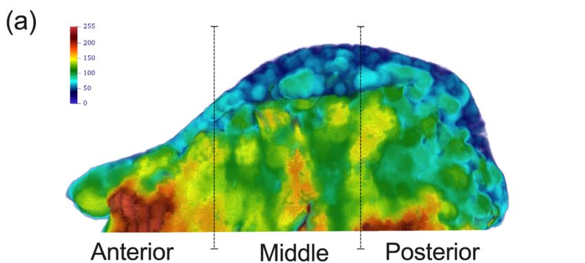
Toxins 2019, 11, 84                                                                                  2 of 16

1. Introduction
      The intervention of the masticatory muscles with botulinum toxin is increasing among general and
specialized dentists, aiming to reduce the activity of muscles such as the masseter during parafunctions
like sleep bruxism and oral movement disorders (i.e., oromandibular dystonia) [1–3]. In addition,
the transitory paralysis caused by this neurotoxin results in masseter atrophy, which is intended for
aesthetic alterations like benign masseteric hypertrophy [1,4]. However, the lack of evidence regarding
its effectiveness and the potential adverse effects in associated musculoskeletal structures have raised
interest in scientifically assessing the latter, something that has not been considered by many clinical
practitioners until now [2,3,5–8].
      The mammalian masticatory apparatus is a complex and specialized anatomical system necessary
for functions such as mastication, self-defense, and social relationships among others. Its main
components are the teeth, the maxillary and mandibular bones, the temporomandibular joint (TMJ),
and the masticatory muscles, with a common origin in the first and second pharyngeal arches [9–11].
The masticatory muscles (medial and lateral pterygoids, temporalis, and masseter) work synchronously
to provide the mandibular movements, with the TMJ as a fulcrum. The TMJ articulates the mandibular
condyle with the mandibular fossa in the skull base. The mandibular condyle exhibits an articular
surface covered with fibrocartilage and a subjacent subchondral bone [12–14]. It is also a growth center
that maintains its activity in adult individuals, which allows it to adapt to external stimuli changes [15].
      During ontogeny, the structure and function of masticatory muscles are necessary for the
proper development of the mammalian TMJ, as well as mastication performance [13,16]. Moreover,
the biomechanical input from the masticatory muscles is required during adulthood to maintain the
homeostasis of the joint [17,18]. The functional and/or structural alterations in one or more of the
components of the TMJ are recognized as temporomandibular disorders (TMDs), grouped by muscular,
articular or developmental conditions [19,20]. The TMDs include several pathological conditions
that severely impair the quality of life, with a high cost in both diagnosis and management [19].
In addition, TMDs-related alterations affect, worldwide, the human daily activities such as talking,
eating, and sleeping, with a higher prevalence in adult women between 20 and 40 years [21,22].
      The mandible is an irregular bone that provides support and protection of the soft craniofacial
tissues [23]; its alveolar bone houses the mandibular teeth [24], it allows for the insertion of masticatory
muscles [10,25], while keeping general properties of bone as mineral reservoir [26] and primary center
for hematopoiesis [27]. Since muscle function and bone homeostasis are related at biomechanical and
biochemical levels [28], it remains necessary to understand the effect of masticatory muscle alterations,
such as paralysis induced by botulinum toxin type A (BoNT/A) for therapeutic purposes [1,3,4,29],
on the bone remodeling process in the mammalian mandible.

1.1. Bone Remodeling as an Integration Mechanism in the Masticatory Apparatus
     In general, bone tissue is highly organized at several levels (from nano to macrostructure). It is
composed of cortical bone with high mineral content and concentric laminae, and less mineralized
and irregular trabecular bone [30–32]. Additionally, bone is highly innervated and vascularized,
and presents a specialized cover called periosteum on the outside surface [33], with an internal analog
called endosteum, in direct contact with the bone marrow [27,34].
     Bone remodeling is the process of bone turnover after maturation, and the first process is the
degradation of damaged tissue (bone resorption) [35,36]. The osteoclasts are a specialized cell line
that performs the bone resorption process [37,38]. These cells result from the fusion of monocytes,
recruited by specific molecular signals like the receptor activator of nuclear factor κ B ligand (RANKL),
necessary for osteoclastogenesis [39,40]. After bone resorption, bone apposition is performed by
the osteoblasts [41]. Once bone apposition is complete, a portion of the osteoblast population is
covered by mineralized bone tissue and become differentiated into osteocytes [42,43]. The osteocytes
represent almost 95% of the bone cells and they exhibit extended longevity, are sensitive to mechanical
stimuli, and are the main source of RANKL in the trabecular bone [44]. The balance between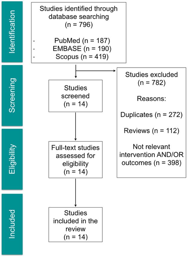
Toxins 2019, 11, 84                                                                                3 of 16

RANKL and its soluble antagonist, Osteoprotegerin (OPG), determines the bone homeostasis and bone
remodeling processes [45,46]. The reduction of bone mineral density (BMD), which reflects the bone’s
mineral content, is called osteopenia. It precedes osteoporosis, which compromises trabecular bone
microstructure and quality under different conditions such as immobilization, lack of gravity, hormonal
alterations, among others [45]. Since most of the mandible is formed by bone, the adaptive capacity of
this mineralized tissue is necessary for the integrity of the mammalian masticatory apparatus [47,48].

1.2. Mechanism of Action and Treatment of Oral Muscular Disorders: The Use of Botulinum Toxin Type A
in Dentistry
      Botulinum toxin is a neurotoxin produced by the anaerobic bacteria Clostridium botulinum.
The most potent serotype is the A (BoNT/A) and it is composed of two chains; a heavy chain of
100 kDa and a light chain of 50 kDa [1,49,50]. The former acts as a specific ligand for presynaptic
membrane receptors in cholinergic nerve endings (such as NMJ) and the latter, once inside the motor
neuron, cleaves the SNAP25 protein through zinc-dependent proteolytic activity; SNAP25 is part of the
SNARE complex, which is necessary for exocytosis [49]. Therefore, acetylcholine release to the NMJ
cleft is blocked, and skeletal muscle is temporally paralyzed and subsequently atrophied [49–51]. This
is an expected outcome in dentistry for oral muscular disorders such as sleep bruxism [3] or aesthetic
conditions like masseteric hypertrophy [1,4]. In addition, the block of neurotransmitter release (like
Substance P) also seems to be helpful for the treatment of myofascial pain [1,29]. However, there is
a lack of high-quality evidence that supports the effectiveness of this intervention for such mentioned
disorders. Moreover, this neurotoxin is not approved for interventions in the masticatory apparatus,
according to the US Food and Drug Administration (FDA) [52,53]; as such, its clinical use is off-label.
Despite this, BoNT/A has been considered as an option for some clinical trials [54] and is commonly
used by dental practitioners and aesthetic therapists. This raises the concern about the safety of this
procedure [5,6,8].

1.3. BoNT/A and Bone Loss at the Masticatory Apparatus
      Considering that muscle contraction drives the mechano-transduction and molecular signaling
required for bone homeostasis, what happens to mandibular bone when masticatory muscles are
paralyzed? Pre-clinical evidence showed that masticatory muscle atrophy induced by BoNT/A
impairs craniofacial bone development by reducing the size of particular regions of mandible (such
as mandibular condyle) and altering its morphology, when compared with normally developed
individuals [55–61]. However, the adverse effects of this intervention in adult individuals remains
poorly understood. Therefore, the aim of this work is to condense the current and relevant
literature about the mandibular bone loss in fully mature mammals after BoNT/A intervention
in the masticatory muscles.

2. Methods
     An electronic literature search of the databases PubMed, EMBASE and Scopus, was conducted in
January 2019 by two independent reviewers with the aim to identify the relevant literature regarding
the effect of masticatory muscle atrophy induced by botulinum toxin type A (BoNT/A) on the
mandibular bone structure of adult individuals. No time range was considered, and the search
was restricted to English language. The following search strategy combining Medical Subject Headings
(MeSH), search terms, and truncated terms was used:

      ((((((((((("Mandible"[Mesh]) OR Mandib*[Title/Abstract]) OR "Temporomandibular Joint"[Mesh])
      OR "Mandibular Condyle"[Mesh]) OR Mandibular head*[Title/Abstract]) OR Mandibular
      condyle*[Title/Abstract]) OR Subchondral bone*[Title/Abstract]) OR "Alveolar Bone Loss"[Mesh])
      OR "Alveolar Process"[Mesh]) OR Alveolar bone*[Title/Abstract])) AND ((("Botulinum Toxins,
      Type A"[Mesh]) OR "Botulinum Toxins"[Mesh]) OR Botulinum toxin*[Title/Abstract])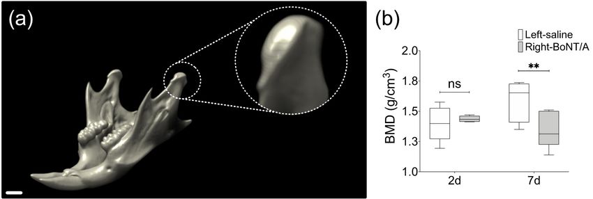
Toxins 2019,11,
Toxins2019,  11,84
                 x FOR PEER REVIEW                                                                          44 of
                                                                                                               of 16
                                                                                                                  16

     Additionally, a Google Scholar search was performed to identify any other relevant studies. The
Additionally, a Google Scholar search was performed to identify any other relevant studies. The search
search terms were screened initially in the title and the abstract before selection for full-text review.
terms were screened initially in the title and the abstract before selection for full-text review. Only
Only publications that report mandibular bone effects after BoNT/A intervention in the masticatory
publications that report mandibular bone effects after BoNT/A intervention in the masticatory muscles
muscles without any other conditions (e.g., induced osteoporosis) were considered. Previously
without any other conditions (e.g., induced osteoporosis) were considered. Previously published
published narrative or systematic reviews and conference abstracts were considered as exclusion
narrative or systematic reviews and conference abstracts were considered as exclusion criteria. Bone
criteria. Bone parameters such as histomorphometry and microtomography were considered as
parameters such as histomorphometry and microtomography were considered as primary outcomes;
primary outcomes; time of evaluation after intervention, molecular expression (gene and/or protein),
time of evaluation after intervention, molecular expression (gene and/or protein), and Bone Mineral
and Bone Mineral Density (BMD) from affected bones were considered as secondary outcomes.
Density (BMD) from affected bones were considered as secondary outcomes.
3. Results
3. Results
     The search
     The  search strategy
                 strategy retrieved
                           retrieved aa total
                                         total of
                                               of 796
                                                  796 articles.
                                                       articles. After
                                                                 After title
                                                                        title and
                                                                              and abstract
                                                                                   abstract evaluation,
                                                                                            evaluation, 14
                                                                                                        14 articles
                                                                                                           articles
were included
were  included for
                for full-text
                     full-text review.
                               review. From
                                         From thethe selected
                                                      selected articles,
                                                                 articles, 10
                                                                           10 were
                                                                               were experimental
                                                                                     experimental studies
                                                                                                    studies using
                                                                                                              using
animal   models  (mice,  rats and   rabbits)  and   4  were   reports  from    human   studies: one  clinical
animal models (mice, rats and rabbits) and 4 were reports from human studies: one clinical case-report,       case-
two retrospective studies and one clinical trial. The selection process is shown in Figure 1, and and
report, two retrospective   studies  and  one  clinical  trial. The selection   process is shown  in Figure  1, the
the descriptive summary     of  the articles that  met  the  inclusion   criteria
descriptive summary of the articles that met the inclusion criteria in Table 1.   in Table  1.

                          Figure 1. Flow chart of selection process for relevant studies.
                          Figure 1. Flow chart of selection process for relevant studies.
Toxins 2019, 11, 84 5 of 16
Table 1. Summary of selected articles.
Author Individuals Intervention Time after Intervention and Bone Evaluation Methods
Experimental group: 0.2 U BoNT/A in the right masseter and 2 weeks; 3D bone parameters from mandibular condyle
Adult male BALB/c mice (8–9
Balanta-Melo et al. 2018 [62] saline solution in the left masseter and alveolar process, and shape analysis of mandibular
weeks-old)
Control group: without intervention condyle using microCT
Experimental group: 0.3 U BoNT/A in the right masseter and
Young adult female C57BL/6J mice (6 4 weeks; 3D bone parameters using microCT, BMD and
Dutra et al. 2018 [63] no intervention in the left masseter
weeks-old) histomorphometry from mandibular condyle
Control group: without intervention
Experimental group: 2 U BoNT/A bilateral in both masseter
Young adult female Sprague-Dawley rats 4 weeks; 3D bone parameters using microCT and
Shi et al. 2018 [64] muscles
(5 weeks-old) histomorphometry from mandibular condyle
Control group: without intervention
Experimental group: 0.2 U BoNT/A in the right masseter and 2 weeks; bone histomorphometry and mRNA
Balanta-Melo et al. 2018 [51] Adult male BALB/c mice (8 weeks-old)
saline solution in the left masseter quantification from mandibular condyle
Morphology of the mandibular condyle (qualitative
Aziz et al. 2017 [5] Adult woman (55 years-old) 140 U BoNT/A quarterly in the left masseter description) using diagnostic imaging (Dynamic
Magnetic Resonance Imaging)
Experimental group I: 25 U BoNT/A bilaterally in the
masseter muscles
6 months after first intervention; evaluation of bone
Lee et al. 2017 [8] Adult men and women (28–48 years-old) Experimental group II: 25 U BoNT/A bilaterally in the
volume in the mandibular angle using CBCT
masseter muscles; repetition 4 months after the first
intervention
Experimental group: 1 U BoNT/A unilaterally in the
Adult male Sprague–Dawley rats (18 masseter and temporalis muscles 4 weeks; 3D bone parameters using microCT of
Kün-Darbois et al. 2017 [65]
weeks-old) Control group: unilateral injection of saline solution in the mandibular condyles
masseter and temporalis muscles
Young adult female transgenic mice
Experimental group: 0.3 U BoNT/A in the right masseter and 4 weeks; 3D bone parameters using microCT, BMD and
Dutra et al. 2016 [66] (Col10a1) on a CD-1 background (5
no intervention in the left masseter histomorphometry of mandibular condyles
weeks-old)
Experimental group: 10 U BoNT/A unilateral in the masseter
Adult New Zeland white female rabbits muscle 4 weeks and 12 weeks; bone histomorphometry of
Matthys et al. 2015 [67]
(5 months-old) Control group: unilateral injection of saline solution in the mandibular condyles
masseter muscle
Experimental group: 1 U BoNT/A unilateral in the masseter
Adult male Sprague–Dawley rats (18 and the temporalis muscles 4 weeks; 2D analysis of microCT slices from mandibular
Kün-Darbois et al. 2015 [68]
weeks-old) Control group: unilateral injection of saline solution in the condyles and alveolar bone
masseter and the temporalis muscles
Exposed group: Adult women with myofascial pain exposed
to BoNT/A for treatment. No dose of BoNT/A reported. CBCT 6-10 weeks after exposure to BoNT/A
Raphael et al. 2014 [69] Adult women (Mean age 45 years-old)
Unexposed group: Adult women with myofascial pain with intervention
no previous exposure to BoNT/AToxins 2019, 11, 84 6 of 16
Table 1. Cont.
Author Individuals Intervention Time after Intervention and Bone Evaluation Methods
Experimental group: 10 U BoNT/A unilateral in the masseter
Adult New Zeland white female rabbits muscle 4 weeks and 12 weeks; 2D and 3D evaluation using
Rafferty et al. 2012 [70]
(5 months-old) Control group: unilateral injection of saline solution in the microCT of mandibular condyles and alveolar bone
masseter muscle
3 months; 3D analysis of cortical thickness of the
Chang et al. 2011 [71] Adult women Bilateral injection of 120 U BoNT/A in both masseter muscles
mandibular ramus using CT
3 months; Linear measurements and BMD of mandibles
Adult male Sprague-Dawley rats (8 Experimental group: 7.5 U BoNT/A in the left masseter and
Tsai et al. 2010 [72] 2D histomorphometry of slices at first molar and
weeks-old) saline solution in the right masseter
coronoid levels
BoNT/A, Botulinum toxin type A; BMD, Bone Mineral Density; U, Mouse Units; CT, computerized tomography; CBCT, cone-beam computerized tomography; 2D, two dimensional; and
3D, three dimensional.Toxins2019,
Toxins 2019,11,
11,84
x FOR PEER REVIEW 77 of
of 16
16
Toxins 2019, 11, x FOR PEER REVIEW 7 of 16
Our literature search retrieved preclinical and clinical studies related to mandibular bone
Our literature
Our
changes after
literature search
BoNT/Asearch retrieved
intervention
retrieved preclinical
in andandclinical
the masticatory
preclinical studies
muscles.
clinical related
Taken
studies to together,
relatedmandibular bone
it is
to mandibular changes
possible
bone to
after
changesBoNT/A
describe after intervention
the main results
BoNT/A in the masticatory
based on three
intervention muscles.
different
in the Taken
levels for
masticatory together,
bone quality
muscles. it is possible
Takenevaluation
together, it to
[73]: describe
is cellular the
possibleandto
main results
metabolic
describe basedresults
thechanges,
main on three different
microstructural
based levels
changes
on three for
andbone
different quality
morphological
levels for boneevaluation
changes. [73]: cellular
quality evaluation and
[73]: metabolic
cellular and
changes, microstructural changes and morphological
metabolic changes, microstructural changes and morphological changes. changes.
3.1. Cellular and Metabolic Changes
3.1. Cellular and Metabolic Changes
3.1. Cellular and Metabolic Changes
Tsai et al. (2010) found no differences in BMD from the whole mandible samples in a rat model
Tsai et al. (2010) found no differences in BMD from the whole mandible samples in a rat model of
of unilateral
Tsai et al. injection
(2010) foundof BoNT/A in theinmasseter
no differences BMD from muscle, threemandible
the whole months samples
after intervention [72].
in a rat model
unilateral injection of BoNT/A in the masseter muscle, three months after intervention [72]. However,
However,
of unilaterala 3–4% reduction
injection in BMD was
of BoNT/A described
in the in mandibular
masseter muscle, threecondyles of unilateral
months BoNT/A-injected
after intervention [72].
a 3–4% reduction in BMD was described in mandibular condyles of unilateral BoNT/A-injected
young female
However, a 3–4%mice four weeks
reduction in BMDafterwas
intervention,
describedcomparing
in mandibular the injected
condylesside with the contralateral
of unilateral BoNT/A-injected non-
young female mice four weeks after intervention, comparing the injected side with the contralateral
injected
young side or
female with
mice samples
four weeksfrom afteranimals without
intervention, intervention
comparing [63,66]. side
the injected Unpublished results fromnon-
with the contralateral our
non-injected side or with samples from animals without intervention [63,66]. Unpublished results
mouse model
injected side or (Figure 2) show
with samples even
from earlierwithout
animals changesintervention
in BMD [74] (Figure
[63,66]. 3). In adult mice,
Unpublished resultsseven
fromdays
our
from our mouse model (Figure 2) show even earlier changes in BMD [74] (Figure 3). In adult mice,
after unilateral
mouse BoNT/A
model (Figure intervention
2) show even earlierin the masseter
changes muscle,
in BMD [74]a(Figure
significant
3). Inreduction
adult mice, in the
sevenBMDdaysof
seven days after unilateral BoNT/A intervention in the masseter muscle, a significant reduction in the
the mandibular
after condyles
unilateral BoNT/A from the BoNT/A-injected
intervention in the masseter side,muscle,compared to thereduction
a significant saline-injected
in the side
BMDwas of
BMD of the mandibular condyles from the BoNT/A-injected side, compared to the saline-injected side
found
the (Figure 3).condyles from the BoNT/A-injected side, compared to the saline-injected side was
mandibular
was found (Figure 3).
found (Figure 3).
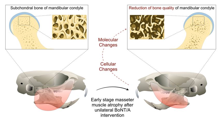
Figure 2.2. Mouse
Figure Mouse model
model of
of mandibular
mandibular condyle
condyle degradation
degradation during
during the
the early
early stage
stage (2
(2 weeks)
weeks) of
of
BoNT/A-induced
Figure
BoNT/A-induced masseter
2. Mouse model of muscle
masseter atrophy
mandibular
muscle in
in adult
condyle
atrophy animals.
degradation
adult animals. during the early stage (2 weeks) of
BoNT/A-induced masseter muscle atrophy in adult animals.
Figure 3.3.Bone
Figure BoneMineral
MineralDensity
Density(BMD)
(BMD) from
from mandibular
mandibular condyles
condyles of adult
of adult malemale
micemice
2 days2 days
and 7anddays7
days unilateral
after
Figure after
3. Boneunilateral
BoNT/A
Mineral BoNT/A
Density intervention
intervention
(BMD)infrom in the
the right right condyles
masseter
mandibular masseter
muscle.of(a)muscle.
3D view
adult (a) of
male 3D view
mouse
mice of mouse
mandible
2 days and 7
performed
mandible
days with
after performedDataViewer
unilateral with
BoNT/A (v1.5.6.2,
DataViewer Bruker
intervention microCT).
(v1.5.6.2,
in Bruker
the rightThe scan was
microCT).
masseter The carried
scan was
muscle. outcarried
(a) under
3D the
outof
view following
under
mouse the
parameters:
mandible SkyScan with
followingperformed
parameters: 1278 (Bruker),
SkyScan 1278Voltage
DataViewer (Bruker),65 Voltage
(v1.5.6.2,kV, Current
Bruker 65 692Current
kV,
microCT).µA,TheAluminum
692
scanµA, filter
wasAluminum 1 mm,
carried voxel
outfilter
under size
1 mm,
the
51.48
voxelµmsizeand
following reconstruction
51.48 µm and
parameters: program
reconstruction
SkyScan NRecon
program
1278 (Bruker), (v1.7.4.2,
NRecon
Voltage Bruker
65 kV, microCT).
(v1.7.4.2,
Current Bruker
692 µA, Dotted
microCT).
Aluminumcircle:Dotted
Close1circle:
filter up
mm, of
volume
voxel upofof
Closesize interest,
51.48 µmthe
volume of mandibular
and interest, the condyle.
mandibular
reconstruction programScale bar: 1 mm.
condyle.
NRecon Scale (b)
bar:Measurement
(v1.7.4.2, 1Bruker
mm. (b) of BMD
Measurement
microCT). in samples
Dotted ofcircle:
BMD
from
Close both
up ofsides
in samples fromofboth
volume experimental individuals
sides of experimental
of interest, the mandibular2 individuals
days and 7 Scale
condyle. days
2 daysafter
bar:and1BoNT/A
7 days
mm. (b)injection,
after obtained
BoNT/A
Measurement with
injection,
of BMD
the
in CT
obtainedAnalyzer
sampleswith from (v1.18.4.0,
theboth
CT sides Bruker
Analyzer microCT). Min
(v1.18.4.0, Bruker
of experimental to Max;
microCT).
individuals n = 5 per
Minand
2 days day;
to Max; paired
7 days t-test
n =after between
5 perBoNT/A
day; pairedsamples
t-test
injection,
from the same
between
obtained samples
with individual;
the from theShapiro
CT Analyzer Wilk test:
same(v1.18.4.0,
individual; p > 0.05;
Shapiro
Bruker ** p to0.05;
Max; **pn 0.05; **p < 0.01; ns: non-significant.Toxins 2019, 11, 84 8 of 16
A significant reduction in bone remodeling processes of the samples from the experimental side
was detected in young adult female mice as a decrease in both osteoclast activity (measured with
TRAP staining) [63,66] and bone mineralization (assessed through alkaline phosphatase staining and
fluorescent dyes for mineralized bone) after four weeks [63]. In contrast to these findings, Shi et al.
reported, in adult female rats, a significant increase of the TRAP staining in the subchondral bone of
mandibular condyles four weeks after bilateral injection of BoNT/A in the masseter muscles, when
compared with samples from animals bilaterally injected with saline solution [64]. However, different
techniques were implemented to assess TRAP staining: In the samples from mice [63,66], a fluorescent
approach was employed using TRAP-positive pixels for quantification, whereas the study with rats
used an immunohistochemical procedure with multinucleated positive TRAP cells quantitation [64].
In addition, in a pilot study with adult male mice, we demonstrated a significant increase in mRNA
levels of the bone resorption marker RANKL in extracts from mandibular condyles just two days after
unilateral BoNT/A injection in the masseter muscle [51].
Another result that involves a cellular change in the bone tissue was reported by Dutra et al. [66].
They founded an increased number of apoptotic cells (visualized by TUNEL reaction) in the
subchondral bone of the mandibular condyle from the BoNT/A-injected side in young adult female
mice, four weeks after intervention [66]. However, there was no identification of the cell type labeled
for apoptosis.
3.2. Microstructural Changes
In a pilot study, we implemented a mouse model to determine the effect of unilateral
injection of BoNT/A in the masseter muscle on mandibular condyle microstructure in adult male
mice [51]. We found a significant reduction in bone per tissue area (B.Ar/T.Ar; 30%) and trabecular
thickness (Tb.Th; 55%) of the subchondral bone in the treated side, assessed by histomorphometry
of representative slices from the middle portion of the mandibular condyles two weeks after
intervention [51]. In adult male rats, the same intervention showed a significant decrease in the
cortical thickness and the trabecular bone in coronal slices at coronoid and molar levels, three months
after following intervention [72]. Additionally, using 2D bone histomorphometry in adult female
rabbits, a similar significant reduction in the subchondral bone (20%) was found four weeks after
unilateral intervention in the masseter muscle, with a statistically non-significant recovery at 12 weeks,
when compared with the control side injected with saline solution [67]. Moreover, a microCT evaluation
of 2D representative slices from the middle portion of the mandibular condyle in rabbits showed
a significant reduction of B.Ar/T.Ar four weeks after intervention (compared with the control side);
the difference in this bone parameter was still statistically significant at 12 weeks [70]. In addition,
a loss of alveolar bone (at molar level) was detected in the experimental side, but no longer detected
12 weeks after BoNT/A intervention [70]. In adult male rats, the unilateral injection of BoNT/A in the
masseter and the temporalis muscles resulted in a significant reduction of B.Ar/T.Ar in the alveolar
bone and the mandibular condyle (20 and 35%, respectively) when compared with the control side
four weeks after, using microCT imaging [68].
In comparison with 2D evaluations, the 3D (volumetric) analyses of mandibles using microCT
technology offers a more complete picture of how bone loss presents following BoNT/A intervention.
It has been demonstrated for example that the extent of bone loss is less than expected from 2D
analyses, albeit still significant. An advanced 3D analysis using high resolution microCT showed
a reduction of 10–11% in bone volume fraction (BV/TV) in the mandibular condyle of young adult
female mice [63] and adult male mice [62] four weeks and two weeks after, respectively. A higher
value was reported in another study (loss of 21% in BV/TV), but these mice were younger (five
weeks old) [66]. In mandibular condyles of adult male mice, Tb.Th was also significantly reduced,
while trabecular number (Tb.N) and trabecular density (Conn.D) were significantly increased at two
weeks. However, no statistically difference was detected for these bone parameters in the alveolar
bone at the first molar level [62]. On the other hand, in the mandibular condyles of young adultToxins 2019, 11, 84 9 of 16
female mice, Tb.Th was significant reduced (17%) and trabecular separation (Tb.Sp) was increased
by 18% at four weeks, compared with control side [66]. Moreover, unpublished results from our lab
using a high resolution microCT approach previously described [62] has confirmed that the significant
reduction of 2019,
Toxins the 11,
bone
x FORmicrostructure
PEER REVIEW parameters such as BV/TV and Tb.Th is more pronounced 9 of 16 in
the middle portion of the mandibular condyle [74] (Figure 4). This may explain why findings of
resolution microCT approach previously described [62] has confirmed that the significant reduction
studies that only assessed the middle portion of the mandibular condyle using a 2D approach reported
of the bone microstructure parameters such as BV/TV and Tb.Th is more pronounced in the middle
higher bone
portionloss than
of the those using
mandibular 3D [74]
condyle evaluation.
(Figure 4). Interestingly,
This may explainbilateral injection
why findings of BoNT/A
of studies that only in the
masseterassessed
muscles themay have
middle a greater
portion of theadverse effect
mandibular on mandibular
condyle condyle microstructure.
using a 2D approach reported higher bone As shown
by Shi etloss
al. than
in adult
thosefemale
using 3Drats,evaluation.
at 4 weeks, BV/TV and
Interestingly, Tb.Thinjection
bilateral of the mandibular
of BoNT/A incondyles
the masseterwere both
decreasedmuscles
by a may50%have anda the
greater adverse
Tb.Sp waseffect on mandibular
significantly condylewhen
increased microstructure.
compared Aswith
shownsamples
by Shi from
et al. in adult female rats, at 4 weeks, BV/TV and Tb.Th of the mandibular condyles were both
control individuals [64].
decreased by a 50% and the Tb.Sp was significantly increased when compared with samples from
In humans, a pilot study in adult women suggested a potential damage of the mandibular condyle
control individuals [64].
after BoNT/A intervention
In humans, a pilot in study
the masticatory
in adult womenmuscles [69]. Patients
suggested were
a potential administered
damage between 2 and
of the mandibular
7 BoNT/A injections
condyle with an
after BoNT/A average in
intervention time
the of three months
masticatory musclesbetween sessions.
[69]. Patients Imaging analysis
were administered
from two between 2 and 7 BoNT/A
independent injections
radiologists withCone
using an average
Beam time of three monthsTomography
Computerized between sessions. Imaging
(CBCT) detected
analysis from two independent radiologists using Cone Beam Computerized
significant reduction of trabecular bone density and cortical thickness, when compared with a similar Tomography (CBCT)
detected significant reduction of trabecular bone density and cortical thickness, when compared with
cohort of unexposed individuals [69]. With the same CBCT approach, a clinical trial in patients with
a similar cohort of unexposed individuals [69]. With the same CBCT approach, a clinical trial in
masseteric hypertrophy
patients (adult
with masseteric men and (adult
hypertrophy women) menshowed
and women)a significant reductionreduction
showed a significant of the boneof thevolume
in the mandibular
bone volumeangle in the after two different
mandibular angle aftersessions of bilateral
two different sessions BoNT/A
of bilateral injections in the masseter
BoNT/A injections in
muscles,thewith a time
masseter of four
muscles, withmonths
a time ofbetween
four monthseach one, each
between andone,
assessment six months
and assessment six months after
afterthe first
BoNT/Athe first BoNT/A[8].
intervention intervention [8]. a
In contrast, Inretrospective
contrast, a retrospective study in
study in adult adult women
women with squared-as chief
with squared-face
face as chief complaint, found no significant difference in the whole
complaint, found no significant difference in the whole mandible volume and the cortical mandible volume and the cortical
thickness of
thickness of the mandibular ramus three months after of bilateral BoNT/A injections in the masseter
the mandibular ramus three months after of bilateral BoNT/A injections in the masseter muscles [71].
muscles [71].
Figure 4.Figure 4. MicroCT-based
MicroCT-based histomorphometry ofofthethe
histomorphometry mandibular
mandibularcondyle in adultinmale
condyle mice.
adult (a) mice.
male
Parasagittal view of 3D Trabecular Thickness (Tb.Th) depiction of a representative
(a) Parasagittal view of 3D Trabecular Thickness (Tb.Th) depiction of a representative mandibular mandibular
condyle from BoNT/A-injected side in adult male mice, 2 weeks after (performed with Paraview,
condyle from BoNT/A-injected side in adult male mice, 2 weeks after (performed with Paraview,
v5.4.1). Samples are from three locations and contain the same number of microCT slices. Color scale
v5.4.1). Samples are from three locations and contain the same number of microCT slices. Color
in gray values. (b) A significant reduction of Bone Volume Fraction (BV/TV) was detected only for
scale in gray values.
the middle (b) Aofsignificant
portion reduction
the mandibular offrom
condyles BoneBoNT/A-injected
Volume Fraction (BV/TV)
sides, was detected
when compared with only
for the middle portion of the mandibular condyles from BoNT/A-injected sides,
saline-injected control. A higher reduction was found when compared with the whole volume when compared
with saline-injected control. A higher reduction was found when compared with the whole volume
assessment of the mandibular condyle (16 vs 11%). (c) The same result was found for the Tb.Th, with
a significant
assessment difference in the
of the mandibular middle(16
condyle portion of the mandibular
vs 11%). (c) The same condyle,
resultand a higher
was founddifference
for the Tb.Th,
between sides (26%) (Min to Max; n = 7; One-way ANOVA, p-values after Bonferroni´s multiple
with a significant difference in the middle portion of the mandibular condyle, and a higher difference
comparisons test; ***p < 0.001; ns: non-significant).
between sides (26%) (Min to Max; n = 7; One-way ANOVA, p-values after Bonferroni´s multiple
comparisons test; *** p < 0.001; ns: non-significant).Toxins 2019, 11, 84 10 of 16
3.3. Morphological Changes
Tsai et al. reported several mandibular changes three months after unilateral BoNT/A intervention
in the masseter muscle of adult male rats [72]. Linear measurements demonstrated a significant
reduction of the mandibular ramus, and a significant increase in the length of the mandible,
measured between the mandibular condyle and the tip of the mandibular incisor [72]. Additionally,
they described qualitative morphological alterations in the insertion of the masticatory muscles. This
is consistent with our findings in the insertion site of the BoNT/A-injected masseter muscle on the
vestibular side of the mandible, close to the first molar zone [62]. However, assessment of bone
microstructure in this specific portion of the alveolar process revealed no significant differences when
compared with saline-injected control side, 2 weeks after [62]. The 3D assessment of condyle shape
using geometric morphometrics showed that 2 weeks after unilateral BoNT/A intervention in the
masseter muscle, mouse mandibular condyles of injected side were more extended anteroposteriorly,
with a decreased width, and exhibited a concave anterior-superior surface, when compared with
samples from control side [62]. These findings contrasted with a study using young adult female mice,
where a reduction in the anterior-posterior dimension of the mandibular condyle was found four
weeks after unilateral BoNT/A intervention but without any alteration of mandibular length (between
condyle and incisor) [66]. In humans, a clinical case-report of an adult woman that received repetitive
BoNT/A injections in the masseter muscle every three months for the treatment of oromandibular
dystonia showed a condylar bone resorption only in the BoNT/A-injected side, a morphological
change detected with Dynamic Magnetic Resonance Imaging [5].
4. Discussion
The BoNT/A intervention is a promising tool in dentistry for the management of several clinical
conditions, including those related to myofascial pain [1,7,29]. Moreover, for dentists, injecting
BoNT/A in the facial region is not technically difficult, making this procedure highly available for
patients in dentistry [1]. However, concerns about its effectiveness are based on clinical studies
with a poor design and high risk of bias [2,3,7]. In addition, there are no indications for its use in
the masticatory apparatus, and the adverse effects are rarely or never reported in the clinical trials,
assuming the absence of effects beyond the injected muscles. Most of the relevant literature found
here about mandibular damage after BoNT/A intervention in the masticatory apparatus comes from
pre-clinical studies. However, heterogeneity in the design (i.e., animals used, brand of BoNT/A, dose
equivalence, dose assessment methods, among others) does not make it possible to properly compare
the results. Interestingly, the use of 3D technologies such as microCT adds significant improvements in
the evaluation of bone effects in irregular structures like the mandibular condyle.
In clinical dentistry, the BoNT/A intervention in the masticatory muscles has been used for
the treatment of several oral movement disorders such as oromandibular dystonia [75] and sleep
bruxism [3] and aesthetics conditions like masseteric hypertrophy [1]. However, there is no official
approval by the FDA for this therapeutic strategy [53]. In addition, safety considerations regarding the
adverse effects of BoNT/A-induced masticatory muscle atrophy on mandibular bone from pre-clinical
studies are relevant in order to avoid unnecessary risks in human trials. Thus, bone mandibular
assessment during clinical trials should be considered as an important outcome after BoNT/A
intervention in the masticatory muscles.
The muscle-bone crosstalk in the masticatory apparatus is still poorly understood, and results
from animal experiments could be useful to study the cellular and molecular dynamics behind soft
and hard tissue homeostasis. The establishment of a mouse model (Figure 2) allows us to design better
and controlled experiments with less resources (compared with the use of larger animals), less time for
intervention/response and easier genetic manipulation. However, it is necessary to consider the genetic
and physiological differences between mice and humans before suggesting potential similarities in
the bone tissue response during altered masticatory function induced by BoNT/A [76–79]. The bone
remodeling process is quite a lot faster in mice (two weeks) when compared with humans (up toToxins 2019, 11, 84 11 of 16
nine months) [76]. In addition, the bone loss after BoNT/A intervention in the skeletal muscle is
strain-dependent in mice, which suggests that careful attention must be dedicated during experimental
design and comparison between studies [80]. Interestingly, the molecular response during bone loss
such as the increase of RANKL has been explored using genetic modified mice and represents some
common features of the bone biology between these species with humans [81]. Therefore, although the
mastication biomechanics in mice and animals is different, it is possible that they share cellular and
molecular mechanisms related to bone remodeling processes. Without doubt it would be relevant to be
able to evaluate these processes in humans. However, due to the bone damage observed by BoNT/A
intervention of masseter muscles in the animal model, we do not consider it pertinent or ethical to
conduct a clinical study.
Most of the studies that use rodents as models to evaluate the effects of BoNT/A-induced
masticatory imbalance are highly focused on the mandibular condylar cartilage (MCC). This is
important to highlight, because early effects of masseter muscle atrophy after BoNT/A intervention
seem to be related to cellular/molecular and microstructural changes in the subchondral bone,
preceding detectable damage of the MCC. Therefore, mouse models are important to understand the
potential adverse effects of BoNT/A in the homeostasis of mandibular, as well as to unveil the cellular
and molecular mechanisms responsible for these outcomes. Taken together, results from pre-clinical
models using BoNT/A intervention could also shed light on understanding pathologies such as
temporomandibular joint osteoarthritis, where changes in the remodeling process of subchondral bone
have been suggested as an early manifestation of the disease [82].
Gathering evidence, we have found that the best way to calculate the dose of BoNT/A to be
injected into the masseter muscle of different preclinical models is the relationship between the toxin
units and the muscle mass. In this way, we have found that Botox®injection of 1.2–3.3 U/g masseter
muscle is safe for interventions in mouse, rat and rabbit models (Table 2). This is an important
parameter to consider when starting any pilot study, as it will reduce the loss of animals used in
the calibration of the procedure. In addition, it is relevant to note that units of different brands
of commercialized BoNT/A are not equivalent [52]; thus, it is highly necessary to consider the
BoNT/A source when comparing evidence. The onabotulinumtoxin A molecule is the most reported
BoNT/A in experimental studies in animals and it was reported in one clinical trial [8], whereas the
abobotulinumtoxin A was used in another study in humans [71] (Table 2). However, all preclinical
studies with bone-related outcomes used BoNT/A from the same company (Allergan, Inc.) [51,62–68,
70,72], while both clinical studies used BoNT/A from Medytox, Inc. [8] and Ipsen Biopharm Ltd [71]
(Table 2). Therefore, the brand and dosage of BoNT/A should be carefully considered when reviewing
literature and evaluating the effectiveness or adverse effects of the procedures with BoNT/A.Toxins 2019, 11, 84 12 of 16
Table 2. Summary of the characteristics of BoNT/A interventions in the included literature.
Average BoNT/A
Average Masseter Dose/Volume
Individual Generic Name/Brand Dose Per Masseter
Mass (g) (U/ml)
Mass (U/g)
Onabotulinumtoxin A; Botox®,
Allergan Chile, Santiago, Chile
Mouse [51,62]
0.075 [51] 0.2–0.3/0.01–0.03 3.3
[51,62,63,66] Onabotulinumtoxin A; Botox®,
Allergan, Plc, Parsippany-Troy
Hills, NJ, USA [63,66]
Onabotulinumtoxin A; Botox®,
Allergan Inc., Irvine, CA, USA
[64,65,68]
Rat [64,65,68,72] 1.1 [64] 1–7.5/0.1–0.3 4.3
Onabotulinumtoxin A; Botox®,
Allergan Pharmaceuticals,
Dublin, Ireland [72]
Onabotulinumtoxin A; Botox®,
Rabbit [67,70] 7.9 [70] Allergan Inc., Irvine, CA, USA 10/0.25 1.2
[67,70]
Onabotulinumtoxin A;
Neuronox®, Medytox Inc.,
Seoul, Korea [8] 25/0.5 [8]
Human [8,71] 20.14 [83] 3.6
Abobotulinum A; Dysport®, 120/0.6 [71]
Ipsen Biopharm Ltd, Wrexham,
UK [71]
5. Conclusions
The findings from pre-clinical studies reviewed here suggest that intervention with BoNT/A
in the masticatory muscles presents adverse effects related to bone loss in the mandible at specific
and time-dependent regions, such as the mandibular condyle and the alveolar process. However,
the cellular and molecular mechanisms behind these phenomena remain to be fully understood. It will
be relevant to address in the future if muscle atrophy/paralysis leads to osteopenia events through
mechanical unloading, a deregulation of biochemical factors normally secreted by muscles to maintain
bone homeostasis, or both. Further basic research may also provide tools for new indications and to
control undesired effects. On the other hand, studies in humans are scarce and reported contrasting
designs and results regarding mandibular bone loss effects. However, the only clinical trial reviewed
here demonstrated mandibular angle bone loss after repetitive injections of BoNT/A in the masseter
muscle. Therefore, the current reviewed evidence warnings about potential mandibular bone loss that
may affect the temporomandibular joint and the alveolar bone around teeth, and this statement should
be communicated to the patients before BoNT/A intervention in the masticatory muscles.
Author Contributions: J.B.-M. and S.B. performed the conceptualization and methodology of the review. J.B.-M.,
S.B., V.T.-I. and K.K. wrote, reviewed and edited the manuscript. All authors approved the final manuscript.
Funding: This research was funded by the FONDECYT-Chile Grants N◦ 1151353 (SB) and N◦ 11150175 (VT-I), the
Max Planck Society (K.K.), the CONICYT-Chile Scholarship N◦ 21170015 (J.B.-M.) and the Professor Scholarship
Semillero Docente 2014 of the Universidad del Valle (J.B.-M.).
Acknowledgments: We are grateful to Jean-Jacques Hublin (Max Planck Institute for Evolutionary Anthropology)
for providing access to the microCT scanning facilities. We thank David Plotzki and Zewdi Tsegai, from the
Max Planck Institute for Evolutionary Anthropology, for their assistance during microCT scanning and help
with the 3D reconstructions with Paraview, respectively. Also, we thank Daniela Poblete, from the Plataforma
Experimental Bio-CT (Faculty of Dentistry, Universidad de Chile, FONDEQUIP EQM150010), for her assistance
during microCT scan and BMD analysis.
Conflicts of Interest: The authors declare no conflict of interest.Toxins 2019, 11, 84 13 of 16
References
1. Miller, J.; Clarkson, E. Botulinum Toxin Type A: Review and Its Role in the Dental Office. Dent. Clin.
North. Am. 2016, 60, 509–521. [CrossRef] [PubMed]
2. Comella, C.L. Systematic review of botulinum toxin treatment for oromandibular dystonia. Toxicon 2018,
147, 96–99. [CrossRef] [PubMed]
3. De la Torre Canales, G.; Camara-Souza, M.B.; do Amaral, C.F.; Garcia, R.C.; Manfredini, D. Is there enough
evidence to use botulinum toxin injections for bruxism management? A systematic literature review.
Clin. Oral Investig. 2017, 21, 727–734. [CrossRef] [PubMed]
4. Fedorowicz, Z.; van Zuuren, E.J.; Schoones, J. Botulinum toxin for masseter hypertrophy. Cochrane Database
Syst. Rev. 2013. [CrossRef] [PubMed]
5. Aziz, J.; Awal, D.; Ayliffe, P. Resorption of the mandibular condyle after injections of botulinum toxin A.
Br. J. Oral Maxillofac. Surg. 2017, 55, 987–988. [CrossRef] [PubMed]
6. Balanta-Melo, J.; Buvinic, S. Mandibular bone loss: A hidden side effect of botulinum toxin type A injection
in masticatory muscles. J. Oral Res. 2018, 7, 44–47. [CrossRef]
7. Laskin, D.M. The Use of Botulinum Toxin for the Treatment of Myofascial Pain in the Masticatory Muscles.
Oral Maxillofac. Surg. Clin. North. Am. 2018, 30, 287–289. [CrossRef] [PubMed]
8. Lee, H.J.; Kim, S.J.; Lee, K.J.; Yu, H.S.; Baik, H.S. Repeated injections of botulinum toxin into the masseter
muscle induce bony changes in human adults: A longitudinal study. Korean J. Orthod. 2017, 47, 222–228.
[CrossRef] [PubMed]
9. Santagati, F.; Rijli, F.M. Cranial neural crest and the building of the vertebrate head. Nat. Rev. Neurosci. 2003,
4, 806–818. [CrossRef] [PubMed]
10. Baverstock, H.; Jeffery, N.S.; Cobb, S.N. The morphology of the mouse masticatory musculature. J. Anat.
2013, 223, 46–60. [CrossRef]
11. Tzahor, E. Head muscle development. Results Probl. Cell. Differ. 2015, 56, 123–142. [CrossRef] [PubMed]
12. Orset, E.; Chaffanjon, P.; Bettega, G. Temporomandibular joint model: anatomic and radiologic comparison
between rat and human. Surg. Radiol. Anat. 2014, 36, 163–166. [CrossRef] [PubMed]
13. Liang, W.; Li, X.; Gao, B.; Gan, H.; Lin, X.; Liao, L.; Li, C. Observing the development of the
temporomandibular joint in embryonic and post-natal mice using various staining methods. Exp. Ther. Med.
2016, 11, 481–489. [CrossRef] [PubMed]
14. Shibata, S.; Sato, R.; Murakami, G.; Fukuoka, H.; Francisco Rodríguez-Vázquez, J. Origin of mandibular
condylar cartilage in mice, rats, and humans: Periosteum or separate blastema? J. Oral Biosci. 2013,
55, 208–216. [CrossRef]
15. Mizoguchi, I.; Toriya, N.; Nakao, Y. Growth of the mandible and biological characteristics of the mandibular
condylar cartilage. Jpn. Dent. Sci. Rev. 2013, 49, 139–150. [CrossRef]
16. Dickinson, E.; Fitton, L.C.; Kupczik, K. Ontogenetic changes to muscle architectural properties within the
jaw-adductor musculature of Macaca fascicularis. Am. J. Phys. Anthropol. 2018, 167, 291–310. [CrossRef]
[PubMed]
17. de Jong, W.C.; Korfage, J.A.; Langenbach, G.E. The role of masticatory muscles in the continuous loading of
the mandible. J. Anat. 2011, 218, 625–636. [CrossRef] [PubMed]
18. Tsouknidas, A.; Jimenez-Rojo, L.; Karatsis, E.; Michailidis, N.; Mitsiadis, T.A. A Bio-Realistic Finite Element
Model to Evaluate the Effect of Masticatory Loadings on Mouse Mandible-Related Tissues. Front. Physiol.
2017, 8, 273. [CrossRef] [PubMed]
19. Ahmad, M.; Schiffman, E.L. Temporomandibular Joint Disorders and Orofacial Pain. Dent. Clin. North. Am.
2016, 60, 105–124. [CrossRef]
20. Okeson, J.P.; de Leeuw, R. Differential diagnosis of temporomandibular disorders and other orofacial pain
disorders. Dent. Clin. North. Am. 2011, 55, 105–120. [CrossRef]
21. List, T.; Jensen, R.H. Temporomandibular disorders: Old ideas and new concepts. Cephalalgia 2017,
37, 692–704. [CrossRef] [PubMed]
22. Maixner, W.; Diatchenko, L.; Dubner, R.; Fillingim, R.B.; Greenspan, J.D.; Knott, C.; Ohrbach, R.; Weir, B.;
Slade, G.D. Orofacial pain prospective evaluation and risk assessment study-the OPPERA study. J. Pain 2011,
12, T4–T11. [CrossRef] [PubMed]Toxins 2019, 11, 84 14 of 16
23. Yang, H.M.; Won, S.Y.; Kim, H.J.; Hu, K.S. Neurovascular structures of the mandibular angle and condyle:
A comprehensive anatomical review. Surg. Radiol. Anat. 2015, 37, 1109–1118. [CrossRef] [PubMed]
24. Bagi, C.M.; Berryman, E.; Moalli, M.R. Comparative bone anatomy of commonly used laboratory animals:
Implications for drug discovery. Comp. Med. 2011, 61, 76–85. [PubMed]
25. Chong, D.A.; Evans, C.A. Histologic study of the attachment of muscles to the rat mandible. Arch. Oral. Biol.
1982, 27, 519–527. [CrossRef]
26. Kwan, P. Osteoporosis: From osteoscience to neuroscience and beyond. Mech. Ageing Dev. 2015, 145, 26–38.
[CrossRef] [PubMed]
27. Morrison, S.J.; Scadden, D.T. The bone marrow niche for haematopoietic stem cells. Nature 2014, 505, 327–334.
[CrossRef] [PubMed]
28. Laurent, M.R.; Dubois, V.; Claessens, F.; Verschueren, S.M.; Vanderschueren, D.; Gielen, E.; Jardi, F.
Muscle-bone interactions: From experimental models to the clinic? A critical update. Mol. Cell. Endocrinol.
2016, 432, 14–36. [CrossRef] [PubMed]
29. Pihut, M.; Ferendiuk, E.; Szewczyk, M.; Kasprzyk, K.; Wieckiewicz, M. The efficiency of botulinum toxin
type A for the treatment of masseter muscle pain in patients with temporomandibular joint dysfunction and
tension-type headache. J. Headache Pain 2016, 17, 29. [CrossRef]
30. Oftadeh, R.; Entezari, V.; Sporri, G.; Villa-Camacho, J.C.; Krigbaum, H.; Strawich, E.; Graham, L.; Rey, C.;
Chiu, H.; Muller, R.; et al. Hierarchical analysis and multi-scale modelling of rat cortical and trabecular bone.
J. R. Soc. Interface 2015, 12. [CrossRef]
31. Burr, D.B.; Akkus, O. Bone Morphology and Organization. In Basic and Applied Bone Biology; Academic Press:
San Diego, CA, USA, 2014.
32. Martin, R.B.; Burr, D.B.; Sharkey, N.A.; Fyhrie, D.P. Skeletal Biology. In Skeletal Tissue Mechanics; Martin, R.B.,
Burr, D.B., Sharkey, N.A., Fyhrie, D.P., Eds.; Springer: New York, NY, USA, 2015.
33. Lai, X.; Price, C.; Lu, X.L.; Wang, L. Imaging and quantifying solute transport across periosteum: Implications
for muscle-bone crosstalk. Bone 2014, 66, 82–89. [CrossRef] [PubMed]
34. Bellido, T.; Plotkin, L.I.; Bruzzaniti, A. Bone Cells A2. In Basic and Applied Bone Biology; Allen, M.R., Burr, D.B.,
Eds.; Academic Press: San Diego, CA, USA, 2014.
35. Sims, N.A.; Martin, T.J. Coupling the activities of bone formation and resorption: A multitude of signals
within the basic multicellular unit. Bonekey Rep. 2014, 3, 481. [CrossRef] [PubMed]
36. Xiao, W.; Wang, Y.; Pacios, S.; Li, S.; Graves, D.T. Cellular and Molecular Aspects of Bone Remodeling.
Front Oral. Biol. 2016, 18, 9–16. [CrossRef]
37. Cappariello, A.; Maurizi, A.; Veeriah, V.; Teti, A. Reprint of: The Great Beauty of the osteoclast.
Arch. Biochem. Biophys. 2014, 561, 13–21. [CrossRef] [PubMed]
38. Boyle, W.J.; Simonet, W.S.; Lacey, D.L. Osteoclast differentiation and activation. Nature 2003, 423, 337–342.
[CrossRef] [PubMed]
39. Takahashi, N.; Udagawa, N.; Suda, T. A new member of tumor necrosis factor ligand family,
ODF/OPGL/TRANCE/RANKL, regulates osteoclast differentiation and function. Biochem. Biophys.
Res. Commun. 1999, 256, 449–455. [CrossRef] [PubMed]
40. Kim, J.H.; Kim, N. Signaling Pathways in Osteoclast Differentiation. Chonnam Med. J. 2016, 52, 12–17.
[CrossRef]
41. Long, F. Building strong bones: Molecular regulation of the osteoblast lineage. Nat. Rev. Mol. Cell. Biol. 2011,
13, 27–38. [CrossRef]
42. Plotkin, L.I.; Bellido, T. Osteocytic signalling pathways as therapeutic targets for bone fragility.
Nat. Rev. Endocrinol. 2016, 12, 593–605. [CrossRef]
43. Bonewald, L.F. The amazing osteocyte. J. Bone Miner. Res. 2011, 26, 229–238. [CrossRef]
44. Xiong, J.; Piemontese, M.; Onal, M.; Campbell, J.; Goellner, J.J.; Dusevich, V.; Bonewald, L.; Manolagas, S.C.;
O’Brien, C.A. Osteocytes, not Osteoblasts or Lining Cells, are the Main Source of the RANKL Required for
Osteoclast Formation in Remodeling Bone. PLoS ONE 2015, 10, e0138189. [CrossRef] [PubMed]
45. Xiao, W.; Li, S.; Pacios, S.; Wang, Y.; Graves, D.T. Bone Remodeling Under Pathological Conditions.
Front. Oral Biol. 2016, 18, 17–27. [CrossRef]
46. Walsh, M.C.; Choi, Y. Biology of the RANKL-RANK-OPG System in Immunity, Bone, and Beyond.
Front. Immunol. 2014, 5, 511. [CrossRef] [PubMed]Toxins 2019, 11, 84 15 of 16
47. Anderson, P.S.; Renaud, S.; Rayfield, E.J. Adaptive plasticity in the mouse mandible. BMC Evol. Biol. 2014,
14, 85. [CrossRef] [PubMed]
48. Klingenberg, C.P.; Navarro, N. Development of the mouse mandible. In Evolution of the House Mouse;
Macholán, M., Baird, S.J.E., Munclinger, P., Piálek, J., Eds.; Cambridge University Press: Cambridge, UK, 2012.
49. Rossetto, O.; Pirazzini, M.; Montecucco, C. Botulinum neurotoxins: Genetic, structural and mechanistic
insights. Nat. Rev. Microbiol. 2014, 12, 535–549. [CrossRef]
50. Pirazzini, M.; Rossetto, O.; Eleopra, R.; Montecucco, C. Botulinum Neurotoxins: Biology, Pharmacology, and
Toxicology. Pharmacol. Rev. 2017, 69, 200–235. [CrossRef] [PubMed]
51. Balanta-Melo, J.; Toro-Ibacache, V.; Torres-Quintana, M.A.; Kupczik, K.; Vega, C.; Morales, C.;
Hernandez-Moya, N.; Arias-Calderon, M.; Beato, C.; Buvinic, S. Early molecular response and
microanatomical changes in the masseter muscle and mandibular head after botulinum toxin intervention in
adult mice. Ann. Anat. 2018, 216, 112–119. [CrossRef]
52. Brin, M.F.; James, C.; Maltman, J. Botulinum toxin type A products are not interchangeable: A review of the
evidence. Biologics 2014, 8, 227–241. [CrossRef]
53. Kane, C.D.; Nuss, J.E.; Bavari, S. Novel therapeutic uses and formulations of botulinum neurotoxins: A patent
review (2012–2014). Expert Opin. Ther. Pat. 2015, 25, 675–690. [CrossRef]
54. Jankovic, J. An update on new and unique uses of botulinum toxin in movement disorders. Toxicon 2018,
147, 84–88. [CrossRef]
55. Chen, Z.; Chen, Z.; Zhao, N.; Shen, G. An Animal Model for Inducing Deviation of the Mandible. J. Oral
Maxillofac. Surg. 2015, 73, 2207–2218. [CrossRef] [PubMed]
56. Seok, H.; Kim, S.G.; Kim, M.K.; Jang, I.; Ahn, J. Effect of the masseter muscle injection of botulinum toxin
A on the mandibular bone growth of developmental rats. Maxillofac. Plast. Reconstr. Surg. 2018, 40, 5.
[CrossRef] [PubMed]
57. Park, C.; Park, K.; Kim, J. Growth effects of botulinum toxin type A injected unilaterally into the masseter
muscle of developing rats. J. Zhejiang Univ. Sci. B 2015, 16, 46–51. [CrossRef] [PubMed]
58. Tsai, C.Y.; Yang, L.Y.; Chen, K.T.; Chiu, W.C. The influence of masticatory hypofunction on developing rat
craniofacial structure. Int. J. Oral Maxillofac. Surg. 2010, 39, 593–598. [CrossRef] [PubMed]
59. Kim, J.Y.; Kim, S.T.; Cho, S.W.; Jung, H.S.; Park, K.T.; Son, H.K. Growth effects of botulinum toxin type
A injected into masseter muscle on a developing rat mandible. Oral Dis. 2008, 14, 626–632. [CrossRef]
[PubMed]
60. Kwon, T.G.; Park, H.S.; Lee, S.H.; Park, I.S.; An, C.H. Influence of unilateral masseter muscle atrophy
on craniofacial morphology in growing rabbits. J. Oral Maxillofac. Surg. 2007, 65, 1530–1537. [CrossRef]
[PubMed]
61. Tsai, C.Y.; Chiu, W.C.; Liao, Y.H.; Tsai, C.M. Effects on craniofacial growth and development of unilateral
botulinum neurotoxin injection into the masseter muscle. Am. J. Orthod. Dentofacial Orthop. 2009,
135, 142.e1–142.e6. [CrossRef] [PubMed]
62. Balanta-Melo, J.; Torres-Quintana, M.A.; Bemmann, M.; Vega, C.; Gonzalez, C.; Kupczik, K.; Toro-Ibacache, V.;
Buvinic, S. Masseter muscle atrophy impairs bone quality of the mandibular condyle but not the alveolar
process early after induction. J. Oral. Rehabil. 2018. [CrossRef]
63. Dutra, E.H.; O’Brien, M.H.; Logan, C.; Tadinada, A.; Nanda, R.; Yadav, S. Loading of the Condylar Cartilage
Can Rescue the Effects of Botox on TMJ. Calcif. Tissue Int. 2018, 103, 71–79. [CrossRef]
64. Shi, Z.; Lv, J.; Xiaoyu, L.; Zheng, L.W.; Yang, X.W. Condylar Degradation from Decreased Occlusal Loading
following Masticatory Muscle Atrophy. Biomed. Res. Int. 2018, 2018, 1–11. [CrossRef]
65. Kun-Darbois, J.D.; Manero, F.; Rony, L.; Chappard, D. Contrast enhancement with uranyl acetate allows
quantitative analysis of the articular cartilage by microCT: Application to mandibular condyles in the BTX
rat model of disuse. Micron 2017, 97, 35–40. [CrossRef] [PubMed]
66. Dutra, E.H.; MH, O.B.; Lima, A.; Kalajzic, Z.; Tadinada, A.; Nanda, R.; Yadav, S. Cellular and Matrix Response
of the Mandibular Condylar Cartilage to Botulinum Toxin. PLoS ONE 2016, 11, e0164599. [CrossRef]
67. Matthys, T.; Ho Dang, H.A.; Rafferty, K.L.; Herring, S.W. Bone and cartilage changes in rabbit mandibular
condyles after 1 injection of botulinum toxin. Am. J. Orthod. Dentofacial Orthop. 2015, 148, 999–1009.
[CrossRef] [PubMed]You can also read