Doxycycline Hyclate Modulates Antioxidant Defenses, Matrix Metalloproteinases, and COX-2 Activity Accelerating Skin Wound Healing by Secondary ...
←
→
Page content transcription
If your browser does not render page correctly, please read the page content below
Hindawi
Oxidative Medicine and Cellular Longevity
Volume 2021, Article ID 4681041, 16 pages
https://doi.org/10.1155/2021/4681041

Research Article
Doxycycline Hyclate Modulates Antioxidant Defenses, Matrix
Metalloproteinases, and COX-2 Activity Accelerating Skin Wound
Healing by Secondary Intention in Rats

 Luciana S. Altoé ,1 Raul S. Alves ,1 Lyvia L. Miranda ,1 Mariáurea M. Sarandy ,1
 Daniel S. S. Bastos ,1 Elda Gonçalves-Santos ,2 Rômulo D. Novaes ,2
 and Reggiani V. Gonçalves 3
 1
 Departament of General Biology, Federal University of Viçosa, Viçosa, Minas Gerais 36570-900, Brazil
 2
 Departament of Structural Biology, Federal University of Alfenas, Alfenas, Minas Gerais 37130-001, Brazil
 3
 Departament of Animal Biology, Federal University of Viçosa, Viçosa, Minas Gerais 36570-900, Brazil

 Correspondence should be addressed to Reggiani V. Gonçalves; reggysvilela@yahoo.com.br

 Received 3 August 2020; Revised 16 November 2020; Accepted 31 March 2021; Published 19 April 2021

 Academic Editor: Alexandros Georgakilas

 Copyright © 2021 Luciana S. Altoé et al. This is an open access article distributed under the Creative Commons Attribution License,
 which permits unrestricted use, distribution, and reproduction in any medium, provided the original work is properly cited.

 The main objective of this study was to investigate the action of doxycycline hyclate (Dx) in the skin wound healing process in Wistar
 rats. We investigated the effect of Dx on inflammatory cell recruitment and production of inflammatory mediators via in vitro and
 in vivo analysis. In addition, we analyzed neovascularization, extracellular matrix deposition, and antioxidant potential of Dx on
 cutaneous repair in Wistar rats. Male animals (n = 15) were divided into three groups with five animals each (protocol: 72/2017),
 and three skin wounds (12 mm diameter) were created on the back of the animals. The groups were as follows: C, received distilled
 water (control); Dx1, doxycycline hyclate (10 mg/kg/day); and Dx2, doxycycline hyclate (30 mg/kg/day). The applications were
 carried out daily for up to 21 days, and tissues from different wounds were removed every 7 days. Our in vitro analysis
 demonstrated that Dx led to macrophage proliferation and increased N-acetyl-β-D-glucosaminidase (NAG) production, besides
 decreased cyclooxygenase-2 (COX-2), prostaglandin E2 (PGE2), and metalloproteinases (MMP), which indicates that macrophage
 activation and COX-2 inhibition are possibly regulated by independent mechanisms. In vivo, our findings presented increased
 cellularity, blood vessels, and the number of mast cells. However, downregulation was observed in the COX-2 and PGE2
 expression, which was limited to epidermal cells. Our results also showed that the downregulation of this pathway benefits the
 oxidative balance by reducing protein carbonyls, malondialdehyde, nitric oxide, and hydrogen peroxide (H2O2). In addition, there
 was an increase in the antioxidant enzymes (catalase and superoxide dismutase) after Dx exposure, which demonstrates its
 antioxidant potential. Finally, Dx increased the number of types I collagen and elastic fibers and reduced the levels of MMP, thus
 accelerating the closure of skin wounds. Our findings indicated that both doses of Dx can modulate the skin repair process, but the
 best effects were observed after exposure to the highest dose.

1. Introduction which provides a humid and nutritious environment for
 microbial proliferation and colonization [2]. An infected
The skin is a complex organ that serves as a barrier to protect cutaneous wound increases the risks of chronification,
the body from the external environment [1]. However, differ- reduces the quality of life, and causes a high mortality rate
ent aggressive agents, such as trauma and microorganisms, of patients [3]. Skin wounds represent a serious health prob-
can affect the structure and functions of this organ. In the lem worldwide and are frequently associated with high costs
case of a lesion, there is an exposure of subcutaneous tissue, and inefficient treatments with limited efficiency [4]. The
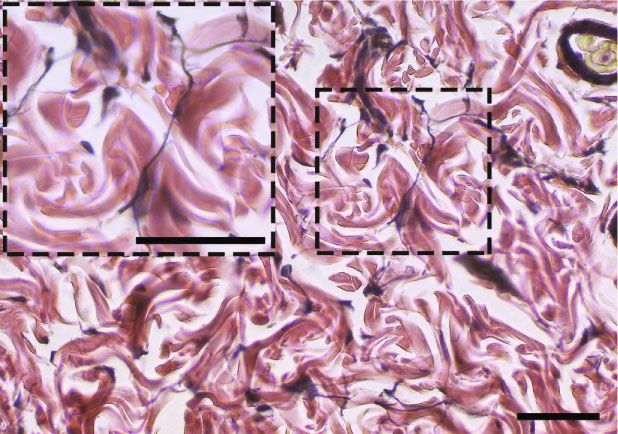
2 Oxidative Medicine and Cellular Longevity

therapies available today are aimed at improving the healing may affect the cutaneous healing process. Although Dx is
of wounds by promoting their rapid closure. However, the effective in treating various disorders [23–25], little is known
control of infections is generally neglected [5]. As a result, about the role of Dx in skin tissue repair. Therefore, this
it is desirable to develop therapeutic interventions that con- study evaluated the effect of Dx on the viability of macro-
trol the infection and increase cutaneous repair. phages, monitored the inflammatory changes in vitro, and
 The repairing of cutaneous wounds is a process that used an experimental model to understand the effect of Dx
involves a complex interaction between cells, extracellular on inflammation, oxidative status, angiogenesis, and fibro-
matrix, blood vessels, and tissue growth factors. Further- genic responses during wound healing in rats.
more, the process is separated into the phases of inflamma-
tion, proliferation (granulation), and tissue remodeling [6]. 2. Materials and Methods
During the inflammation phase, there is a migration of leuco-
cytes to the injured site, with the release of cell mediators. 2.1. In Vitro Assays
During the proliferative phase, there is a multiplication of
keratinocytes, fibroblasts, and endothelial cells, resulting in 2.1.1. Cell Viability. Cell viability for RAW264.7 macro-
the formation of granulation tissue, which is also rich in ves- phages was evaluated by 3-[4,5-dimethylthiazol-2yl]-2,5-
sels and collagen type III [7]. The next phase is characterized diphenyl tetrazolium bromide (MTT) assay as previously
by tissue remodeling and maturation, in which collagen III is described [26, 27]. Cells were cultured in DMEM supple-
replaced by collagen I, thus making the scar stronger and mented with 10% fetal bovine serum and 100 U/mL of peni-
more resistant to mechanical forces [8, 9]. cillin/streptomycin in a humidified 5% CO2 37°C incubator.
 The skin healing process is known as acute or chronic, To evaluate the effect of Dx on cell viability, RAW264.7 mac-
according to its duration and nature [7]. During a chronifica- rophages were seeded into 96-well plates at a density of 1 ×
tion process, there is persistent activation of COX way and 105 cells/well in 200 μL medium. After 24 h, the various con-
neutrophils and macrophages release cytokines and chemo- centrations of Dx (10, 30, 100, and 300 μg/mL) were added to
kines, which attract more cells to the location of the inflam- media, and the incubation continued for the next 24 h at 37°C
mation and promote oxidative stress in the repairing tissue and 5% CO2. The control (100% of growth) was carried out
[10, 11]. The excess of proinflammatory mediators increases with cells cultured in medium only. The MTT solution was
the peroxide of hydrogen (H2O2) and oxide nitric content, added to each well, and the cells were further incubated for
which accelerate the peroxidation of lipids and proteins 2 h, at 37°C. The MTT formazan generated during incubation
[12]. The prooxidant mediators cause damage to the cutane- was dissolved in DMSO, and the absorbance was measured at
ous tissue and delay the wound healing process. Therefore, a 570 nm. For each sample, the result was expressed as the per-
controlled inflammation process is necessary to avoid persis- centage absorbance in relation to the control group.
tent tissue damage through the continued action of free rad-
icals and reactive oxygen species (ROS) [13]. Associated with 2.1.2. Macrophage Challenge with LPS. RAW264.7 macro-
this, we can highlight that the skin healing environment is phages cultured under the same conditions were seeded in
usually prooxidant and generally presents decreased synthe- 24-well polystyrene plates, at 2:5 × 10⁵ cells, and 1 mL of cul-
sis and expression of the antioxidant enzymes, such as super- ture medium per well. After 24 h, the culture medium was
oxide, glutathione, and catalase, which impair the healing replaced, and the RAW264.7 cells were incubated for 24 h with
environment [14]. a fresh medium containing 10% FBS, without or with
 In general, a desirable repair process results from a bal- 100 ng/mL LPS (Sigma-Aldrich, St. Louis, Missouri, USA)
anced process of synthesis and degradation of inflammatory and different Dx concentrations (10, 30, 100, and 300 μg/mL).
mediators and pro- and antioxidant compounds conse- The control cells were treated with a fresh culture medium.
quently in the extracellular matrix components, especially After 24 h of incubation, the macrophages were harvested;
collagen [15]. Matrix metalloproteinases (MMP) play an the cell number was quantified and adjusted by cell counting
essential role in the turnover of the extracellular matrix in a Neubauer chamber. The cells were lysed with 100 mM
(ECM) remodeling, by degrading collagen and noncollagen- NaCl/50 mM Tris-HCl buffer and centrifuged (1000g for
ous elements, such as glycosaminoglycans, proteoglycans, 15 min at 4°C). The culture supernatant was collected to mea-
cytokines, growth factors, and their receptors [16]. However, sure the prostaglandin production enzymatic analysis of
the overexpression of MMP may lead to uncontrolled degra- MMP, cyclooxygenase-2, and N-acetylglucosaminidase.
dation of ECM and delayed the wound healing process [17–
19]. Thus, MMP modulatory drugs may cause an important (1) Metalloprotease Activity. The enzymatic activity of matrix
impact on cutaneous tissue repair, with a particular effect metalloproteases in macrophage homogenate was measured
on tissue inflammation and maturation. In this context, using a fluorometric enzymatic kit, according to the manu-
doxycycline (Dx) has already been described as an inhibitor facturer’s instructions (ABCAM, Cambridge, MA, USA).
of MMP activity [20], and its usage has already been proven The MMP activity was measured at 490 nm/525 nm (excita-
in the modulation of tissue levels of collagen in cutaneous tion/emission), as previously reported [28].
repair [21], which stimulates collagen deposition. In addi-
tion, Dx proved to be an important tool to inhibit the release (2) Cyclooxygenase-2 Activity. An aliquot (100 μL) of the
of proinflammatory cytokines, such as tumor necrosis factor- macrophage homogenate was applied to measure the
alpha (TNF-α), IL-6, and IL-8 [22]. Thus, we believed that Dx cyclooxygenase-2 (COX-2) activity, which was analyzed
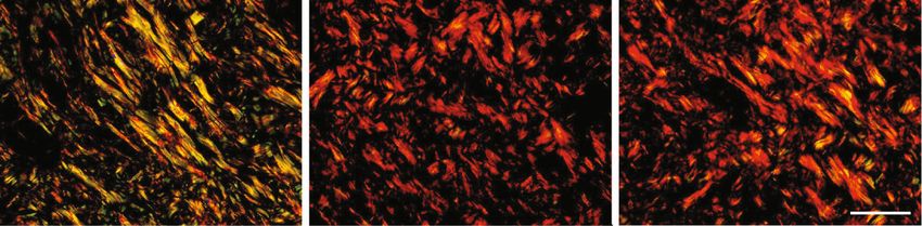
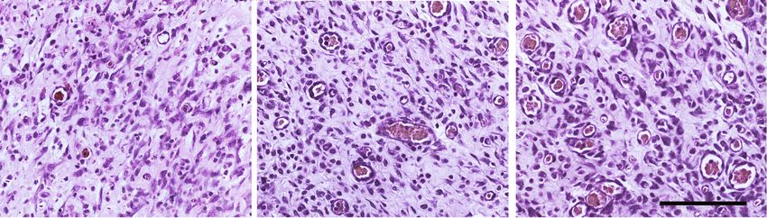
Oxidative Medicine and Cellular Longevity 3

 Realization of
 Acclimation
 the wound
 Dorsal region

 W1 W2 W3

 7 days

 Day 0 Day 7 Day 14 Day 21
 12 mm2

Figure 1: Representation of the experimental model of wound healing by secondary intention and time-dependent evolution of wound
closure. The top image shows the distribution of the three excisional wounds in the back of the animal. The general appearance of wound
closure from the initial wound (day 0) is represented by photographs. W1 (day 7), W2 (day 14), and W3 (day 21); macroscopic aspect of
the wounds observed every 7 days. The wound areas were calculated on days 0, 7, 14, and 21 (mean ± SD), based on the digitized images.

using a biochemical colorimetric kit, following the manufac- were randomly allocated in individual cages, which were
turer’s instructions (Cayman Chemical, Ann Arbor, MI, cleaned daily and maintained under controlled environmen-
USA). The enzymatic assay was based on the peroxidase tal conditions (temperature: 22 ± 2° C, humidity: 60–70%,
component of cyclooxygenases, in which the peroxidase and light/dark cycle: 12/12 h). Commercial food and water
activity was spectrophotometrically measured by monitoring were provided ad libitum. All the experiments were approved
the production of oxidized N,N,N ′ ,N ′ -tetramethyl-p-pheny- by the Animal Ethics Committee of the Federal University of
lenediamine, at 590 nm. Viçosa (registration no. 72/2017).

(3) Prostaglandin Production. The prostaglandin E2 (PGE2) 2.2.2. The Procedure of Surgical Wounds. The rats were anes-
levels in the macrophage homogenate were quantified by thetized with an intraperitoneal injection of sodium pento-
the specific enzyme-linked immunosorbent assay (ELISA) barbital (70 mg/kg). After anesthesia, the dorsolateral
kit, according to the manufacturer’s instructions (Cayman shaving of the animals was performed, and the area was
Chemical, Ann Arbor, MI, USA). Briefly, 10 μL homogenates cleaned with 70% alcohol. Three circular skin wounds of
were added to 96-well microplates previously sensitized with 12 mm diameter were created in the dorsolateral region of
specific antibodies against PGE2. Prostaglandin levels were each rat by secondary intention, with surgical excision of
determined by spectrophotometry at 412 nm [29]. the skin and subcutaneous cellular tissue using surgical scis-
 sors. The area of the wound was marked with violet crystal
(4) N-Acetylglucosaminidase Activity. The activation of the and measured with an analog caliper (Mitutoyo Sul Ameri-
RAW264.7 cells was measured based on the quantification cana Ltda®, São Paulo, Brazil) [31]. No analgesia was admin-
of N-acetyl-β-D-glucosaminidase (NAG) activity, which is istered after the surgical procedure since the application of
a lysosomal enzyme intensely produced by activated mono- drugs can alter cell migration and proliferation and compro-
cytes/macrophages [30]. The N-acetyl-β-D-glucosaminidase mise the skin repairing process. Tissue samples were
activity was measured in skin homogenate by using a com- obtained from different wounds at 7, 14, and 21 days, for his-
mercial biochemical colorimetric kit, according to the manu- tological and biochemical analyses, as presented in Figure 1.
facturer’s instructions (Abcam, Cambridge, UK). This assay
uses a synthetic p-nitrophenol derivative (R-pNP) as a 2.2.3. Experimental Design. The animals were randomized
NAG substrate and releases pNP, which is measured by spec- into three groups, with five animals in each group: C (dis-
trophotometry, at 400 nm. tilled water, control); Dx1 (doxycycline 10 mg/kg/day), and
 Dx2 (doxycycline 30 mg/kg/day). The treatments were
2.2. In Vivo Assays administered by gavage for 21 days. After this period, the ani-
 mals were euthanized by cardiac puncture and exsanguina-
2.2.1. Animals and Ethics. Fifteen healthy three-month-old tion, after an anesthetic procedure. The doses were
male Wistar rats (Rattus norvegicus) (339:16 ± 16:25 g) were provided according to studies that used oral Dx for corneal
obtained from the Central House of the health and Biological reepithelialization in the rabbit model [32], and Dx was given
Sciences Center, Federal University of Viçosa. These animals by gavage to inhibit MMP-mediated vascular changes in
4 Oxidative Medicine and Cellular Longevity

hypertension in rats [33]. They found that many animals milk prepared at pH 7.6 TBST (1X Tris-buffered saline with
died after 100 mg/kg daily, thus suggesting that the therapeu- 0.05% Tween 20). Then, the sections were incubated for
tic window for doxycycline may be rather narrow. Therefore, 12 h, at 4°C, with a primary rabbit anti-COX-2 antibody
we decided to study the effects associated with the 10 and (ab15191, Abcam, Cambridge, UK), at 1 : 1000 dilution. The
30 mg/kg dose, because studies have not reported harmful slides were washed with TBST and incubated for 2 h at room
effects on animals. temperature, with a ready-to-use secondary goat anti-rabbit
 IgG antibody conjugated with horseradish peroxidase (Dako
2.2.4. Calculation of the Area and the Rate of Wound EnVision™+ Dual Link System-HRP, Agilent, Santa Clara,
Contraction. The area and rate of contraction of the third CA, USA). The slides were washed with TBST, and the
wound were evaluated every 7 days, using images scanned COX-2 marking was revealed with 0.5% 3,3 ′ -diaminobenzi-
with 320 × 240 pixels (24 bits/pixel) obtained by an Asus Zen- dine for 5 min. Finally, the slides were released in ethanol,
fone 2 ZE551ML smartphone (ASUS, Taipei, Taiwan). The treated with xylene, and mounted with coverslips.
wound area was calculated by the formula A = π × ðrÞ2 , where
r is the radius. The wound contraction index (WCI) was calcu- 2.5. Biochemical Assays. Tissue fragments were collected
lated by the ratio: initial wound area ðAo Þ − the area on a from each wound, immediately frozen in liquid nitrogen
given day ðAi Þ/initial wound area ðAo Þ × 100 [34, 35]. (-196°C), and stored in a freezer at −80°C. The samples
 (200 mg) were homogenized in 2 mL phosphate-buffered
2.3. Histological and Stereological Analysis. The samples col- saline (PBS) and centrifuged for 5 minutes at 10,000g, under
lected from the wounds, with tissue from the center of the refrigeration, at 5°C [14]. The supernatant and tissue pellets
lesion, were immersed in histological fixative for 24 h, dehy- were separately collected and used in all biochemical analyses
drated in ethanol, diaphanized in xylene, and immersed in described below.
paraffin. Histological sections (4 μm thick) were obtained
on a rotary microtome (Leica Multicut 2045, Reichert-Jung
 2.5.1. Cyclooxygenase-2, N-Acetylglucosaminidase Activity,
Products, Germany). We used 1 of every 20 sections to avoid
 and Prostaglandin Production. Cyclooxygenase-2, N-
repeating the analysis of the same histological area. The sec-
 acetylglucosaminidase activity, and prostaglandin E2 pro-
tions were stained with hematoxylin and eosin (HE) for the
 duction in the scar tissue were measured with the aid of the
analysis of the fibroblasts and blood vessels [31]. Further-
 same commercial kits used to quantify these parameters in
more, the samples were stained with Sirius red for the analy-
 the in vitro model. All measures were performed from intact
sis of collagen fiber types I and III [36]. Toluidine blue was
 skin (day 0) and scar tissue collected at days 7, 14, and 21 of
used to identify mast cells [37], and Verhoeff was used to dif-
 wound healing. The enzymatic activity and prostaglandin
ferentiate elastic fibers [14]. The slides were visualized and
 levels were measured in tissue homogenate supernatant.
captured in a BX601 light microscope (Olympus, São Paulo,
Brazil) coupled with a QColor-31 digital camera (Olympus,
São Paulo, Brazil). Five images were selected at random using 2.5.2. Hydrogen Peroxide and Nitric Oxide Production. The
a 20x objective lens. For this analysis, a grid containing 256 hydrogen peroxide (H2O2) production was measured in
points within a standard test area (AT) of 73 × 103 μm2 was supernatants of tissue homogenates. 50 μL of supernatants
superimposed over each image. The stereological parameters were incubated with 50 μL of α-Phenylenediamine dihy-
of volumetric density (V v ) were calculated by counting the drochloride (OPD) and an equal volume of peroxidase type
points that occurred over cells, blood vessels, types I and III II 15 mmol/L. The conversion of absorbance into micromolar
collagen, and elastic fibers, using the ratio: V v = PP/PT, concentrations of H2O2 was calculated based on a standard
where PP is the number of points occurring over the struc- curve, using a known concentration of H2O2. The results
tures of interest and PT is the total number of points on the were expressed as μmol/L [41].
test system [31, 38]. Collagen fibers were analyzed according Nitric oxide (NO) was indirectly quantified through the
to the different properties of birefringence, as thick collagen detection of nitrite/nitrate (NO2-/NO3-) levels by the stan-
fibers (type I) appear in shades of bright colors ranging from dard Griess reaction [42]. 50 μL of supernatants were incu-
red to yellow, whereas thin reticular fibers (collagen type III) bated with an equal volume of Griess reagent (1%
appear bright green under polarization [31, 39]. The mast sulfanilamide, 0,1% N-(1-Naftil) ethylenediamine, and 2.5%
cells were analyzed using a 40x objective lens. Ten micro- H3PO4) and kept at room temperature for 10 minutes. The
scopical fields were randomly analyzed in each histological conversion of absorbance into micromolar concentrations
section to obtain a total area (TA) of 1.96 mm2. The number of NO was obtained from a sodium nitrite standard curve
of mast cells per unit of histological area was calculated (0–125 μM) and expressed as NO concentrations
according to the relation QA = Σ mast cells/TA [40]. (μmol × L−1 ).

2.4. COX-2 Immunohistochemistry. The histological sections 2.5.3. Determination of Lipid and Protein Oxidation. Lipid
(4 μm thick) were dewaxed with xylene and hydrated in dis- peroxidation (LPO) was estimated according to the total mal-
tilled water. Antigen recovery was performed with citrate ondialdehyde levels (MDA) [43]. The MDA concentration
buffer (pH 6) in a pressure cooker for 4 min. The sections was determined by using the standard curve of known con-
were incubated for 10 min in 3% hydrogen peroxide to block centrations of 1, 1, 3, 3-tetramethoxypropane (TMPO). The
endogenous peroxidase, followed by 15 min in 5% nonfat results were expressed as μmol × L−1 per mg of protein.
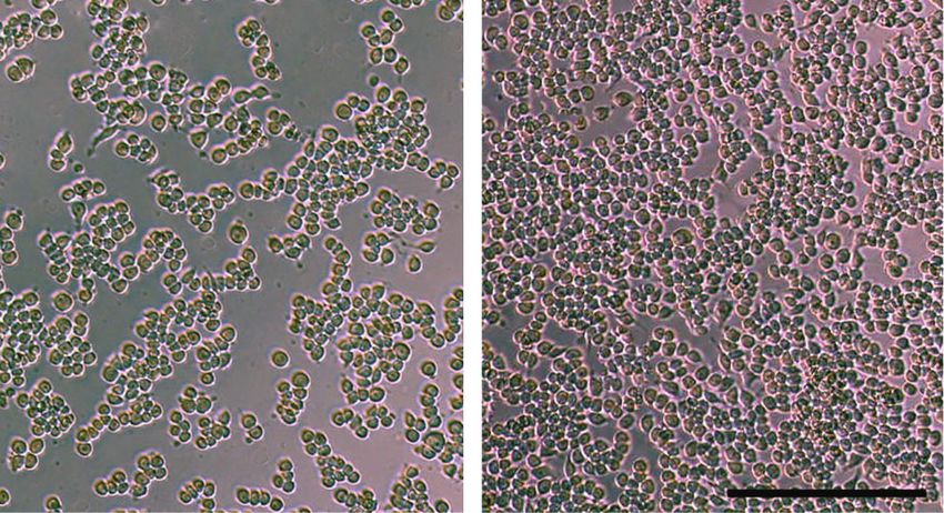
Oxidative Medicine and Cellular Longevity 5

 Control 300 g/ml
 300 ⁎
 Read % value versus control

 200 ⁎

 100

 0
 10 g/ml

 30 g/ml

 100 g/ml

 300 g/ml
 (a) (b)

Figure 2: Effects of doxycycline hyclate (Dx) on cell viability. (a) RAW264.7 macrophages were treated with various doses of Dx (10, 30, 100,
and 300 μg/mL) for 24 h. (b) Representative photomicrographs showing cells in cultured medium (control group) and 300 μg/mL Dx added to
the medium (phase-contrast microscopy, bar = 200 μm). The results are presented as the percentage absorbance of the control group. The
data are expressed in the graphics as a mean and standard deviation. ∗ Statistical difference compared to control cells (Student-Newman-
Keuls test, p < 0:05).

 Protein oxidation was estimated from protein carbonyl ELISA commercial immunoenzymatic kit was used accord-
content, which was measured using 2,4-dinitrophenylhydra- ing to the manufacturer’s instructions (Sigma-Aldrich;
zine (DNPH) [44], based on the carbonyl group reaction with Merck KGaA, Darmstadt, Germany).
DNPH. The pellets resulting from the tissue homogenates
from previous extractions were used for quantification. The 2.6. Statistical Analysis. The statistical analysis was carried
results were expressed as nmol per mL of protein. out using the GraphPad Prism 7 software system (GraphPad
 Software Inc., San Diego, Calif., USA). The results were
2.5.4. Superoxide Dismutase Activity. The activity of superox- expressed as mean and standard deviation (mean ± SD).
ide dismutase (SOD) was determined by the superoxide (O2-) The parametric data were compared using one-way ANOVA
and hydrogen peroxide reduction method, thereby decreas- variance analysis, followed by the Student-Newman-Keuls
ing the autooxidation of pyrogallol [45]. SOD activity was post hoc test. The nonparametric data were compared using
calculated as units per milligram of protein, with one unit the Kruskal–Wallis test. Statistical significance was estab-
(U) of SOD defined as the amount that inhibited the rate of lished at p ≤ 0:05.
pyrogallol autoxidation by 50%.
2.5.5. Catalase Activity. The catalase (CAT) activity was eval- 3. Results
uated according to the method described by Aebi [46], by
 3.1. Impact of Dx on Macrophage Viability, Prostaglandin
measuring the rate of decomposition of hydrogen peroxide.
 Production, and COX-2, MMP, and NAG Activity. The effect
One unit of CAT activity was calculated using the number
 of Dx on macrophage viability is presented in Figure 2. No
of enzymes that decompose 1 mmol H2O2 for 1 min. The
 cytotoxicity was observed after exposing the macrophages
results were expressed as units of catalase/milligram of
 to Dx. Furthermore, the highest cell proliferation was
protein.
 observed after macrophage incubation with 100 μg/mL
2.5.6. Glutathione S-Transferase Activity. The glutathione S- (182:03 ± 6:57) and 300 μg/mL (253:05 ± 30:92) of Dx,
transferase (GST) activity was measured using the method which indicates a clear dose-dependent effect (Figures 2(a)
of Habig et al. [47]. Glutathione S-transferase activity was and 2(b)).
analyzed according to the formation of glutathione- In the in vitro analysis, Dx also reduced glycoprotein
conjugated 2,4-dinitrochlorobenzene (CDNB). One unit of cyclooxygenase-2 (COX-2) in the Dx100 group when com-
GST activity was defined as the amount of enzyme that cata- pared to CM, Dx10, and Dx30. The Dx300 group presented
lyzed the formation of one μmol of product/min/mL. GST reduced COX2, in relation to NC, CM, Dx10, Dx30, and
activity was expressed as U per milligram of protein. Dx100. The prostaglandin E2 levels were higher in CM and
 Dx (10, 30, and 100), when compared to the NC group.
2.5.7. Evaluation of MMP-10 Cutaneous Activity. For the Dx100 presented a decrease in relation to CM, Dx10, and
evaluation of MMP-10 activity, 200 mg samples of the skin Dx30. Dx300 presented a decrease when compared to NC,
were homogenized in 1 mL of 5 mM Tris-HCl (pH 7.4) buffer CM, and Dx (10, 30, and 100). Metalloproteinases (MMP)
containing 0.15 M NaCl, 10 mM CaCl2, and 0.02% NaN3. presented a decrease in the CM, Dx10, Dx30, and Dx100
After centrifugation at 10,000g for 30 min, the supernatant groups when compared to the NC group. The Dx100 group
was collected for analysis of MMP activity. For such, an values were lower, compared to the CM, Dx10, and Dx30
6 Oxidative Medicine and Cellular Longevity

 40 700 ⁎ ⁎ ⁎

 COX-2 (nmol/min/ml)
 500 ⁎&
 30

 PGE2 (pg/mL)
 300 #
 & 100
 20
 # 10

 10 5
 < 13
 0 0
 NC CM 10 30 100 300 NC CM 10 30 100 300
 Dx ( M) Dx ( M)
 10 0.8
 ⁎ ⁎ ⁎
 107)

 ⁎ ⁎

 NAG (U/mg tissue)
 8 0.6 ⁎ ⁎
 MMP (RFU

 6 ⁎
 0.4
 4 ⁎&
 # 0.2
 2

 0 0.0
 NC CM 10 30 100 300 NC CM 10 30 100 300
 Dx ( M) Dx ( M)

Figure 3: Effects of doxycycline hyclate (Dx) on cyclooxygenase-2 (COX-2), prostaglandin E2 production, and metalloproteinases (MMP)
and N-acetyl-β-D-glucosaminidase (NAG) activity in LPS-stimulated RAW264.7 macrophages treated with various doses of Dx for 24 h.
NC: not stimulated cells (not treated with LPS or Dx), CM: cells treated with culture medium containing LPS, and Dx: cells treated with
culture medium containing LPS and Dx at 10, 30, 100, and 300 μM. The data are expressed as mean and standard deviation. Statistical
difference (Student-Newman-Keuls test, p < 0:05), compared to ∗ NC, &CM, Dx10, and Dx30, #NC, CM, and Dx (10, 30, and 100), and +
CM and Dx10.

 15 100 ⁎ ⁎
 80
 Wound area (mm2)

 contraction (%)
 Rate of wound

 10 ⁎
 60
 ⁎
 ⁎ 40
 5

 ⁎ 20

 0 0
 C Dx1 Dx2 C Dx1 Dx2 C Dx1 Dx2 C Dx1 Dx2 C Dx1 Dx2 C Dx1 Dx2 C Dx1 Dx2
 Day 0 Day 7 Day 14 Day 21 Day 7 Day 14 Day 21
 (a) (b)

Figure 4: (a) Area (mm ) and (b) rate of wound contraction (%) in rats treated with doxycycline hyclate (Dx) after 7, 14, and 21 days of
 2

treatment. Dx1: doxycycline hyclate (10 mg/kg), Dx2: doxycycline hyclate (30 mg/kg). The data are expressed as mean and standard
deviation. Statistical difference (Kruskal–Wallis test, p < 0:05) compared to the ∗ control group.

groups. The Dx300 group presented lower values when com- 3.3. Histopathological Results. On day 7, the proportion of
pared to NC, CM, and Dx (10, 30, and 100). The NAG values total cells in the tissue was higher in the Dx2 group, com-
were increased in all groups (CM, Dx10, Dx30, Dx100, and pared to other groups. On the same day, Dx1 showed a
Dx300) when compared to the NC group (Figure 3). higher proportion of cells, compared to the control group.
 On day 14, Dx2 presented a higher proportion of cells in rela-
3.2. Wound Area and Contraction Index. The wound area tion to the control group (Figure 5(a)). In relation to blood
was smaller on days 7, 14, and 21 in the Dx1 group, com- vessels, Dx1 and Dx2 groups showed an increased proportion
pared to the control animals, as well as on day 14, in the of vessels when compared to the control group, on day 7.
Dx2 group, in relation to the control group. The rate of However, on day 14, the proportion of vessels was reduced
wound contraction was higher in the Dx1 and Dx2 groups, in the Dx1 group, compared to the control animals. On day
compared to the control group, on day 21 (Figure 4). 21, Dx1 presented reduced blood vessels compared to the
Oxidative Medicine and Cellular Longevity 7

 8 5
 ⁎ ⁎
 ⁎&
 ⁎ 4
 6 ⁎

 Blood vessels (%)
 Cellularity (%)

 3
 4 ⁎
 2
 ⁎#
 2
 1

 0 0
 C Dx1Dx2 C Dx1Dx2 C Dx1Dx2 C Dx1Dx2 C Dx1Dx2 C Dx1Dx2 C Dx1Dx2 C Dx1Dx2
 Day 0 Day 7 Day 14 Day 21 Day 0 Day 7 Day 14 Day 21
 (a)

 C Dx1 Dx2

 (b)

Figure 5: The proportion of cell nucleus and blood vessels (a) and representative photomicrographs showing cells and blood vessel
distribution (b) in the scar tissue of rats untreated and treated with doxycycline hyclate (Dx), on day 7 (H&E staining, bar = 100 μm). C:
control, Dx1 = 10 mg/kg Dx, and Dx2 = 30 mg/kg Dx. In the graphics, the data are represented as mean and standard deviation. The
statistical difference compared to the groups ∗ C, &Dx1, and #Dx2 (Student-Newman-Keuls test, p < 0:05).

control and Dx2 groups (Figure 5(a)). The distribution of when compared to control and Dx1, on day 7. These results
cells and blood vessels in the scar tissue of the different were confirmed by the analysis of the photomicrographs,
groups is shown in Figure 5(b). showing decreased COX-2 expression in Dx2, mainly in epi-
 The number of mast cells, on day 7, was higher in the Dx2 thelial cells. In relation to PGE2, the Dx2 group showed lower
group, compared to the other groups. In addition, the Dx1 values compared to the other groups, on day 7 (Figure 8(c)).
group presented a higher number of cells compared to the The macrophage accumulation/activation was proven by
control group (Figure 6(a)). The distribution of mast cells increased NAG values in the Dx1 and Dx2 groups when
in the scar tissue in the Dx2 group, on day 7, is shown in compared to the control (Figure 8(d)).
Figure 6(b).
 A higher proportion of type I collagen fibers were 3.4.2. Oxidative Stress Markers. On day 7, H2O2 levels were
observed in the groups treated with Dx1 and Dx2, on day higher in the Dx1 and Dx2 groups in relation to the control
21, when compared to the control group. The type III colla- group. On day 14, Dx2 showed lower H2O2 levels in relation
gen fibers were reduced in Dx1 and Dx2, on day 21, in com- to the control and Dx1 groups. On day 21, Dx2 showed lower
parison to the control group (Figure 7(a)). The distribution levels of H2O2, when compared to the control group
of type I and type III fibers in the scar tissue of the different (Figure 9(a)). Nitrite and nitrate levels were lower in the
groups and the predominance of type I collagen fibers after Dx1 and Dx2 groups, compared to the control group, on
treatment with Dx are shown in Figure 7(b). On days 14 day 14 (Figure 9(b)). Concerning malondialdehyde, the levels
and 21, the number of elastic fibers was higher in the Dx2 were lower in the Dx1 and Dx2 groups, on day 7, when com-
group, compared to the other groups (Figure 7(c)). The rep- pared to the control (Figure 9(c)). On the other hand,
resentative distribution of elastic fibers in the scar tissue in reduced carbonyl protein levels were identified in the group
the Dx2 group, on day 21, is shown in Figure 7(d). Dx1, compared to the control group, on day 21 (Figure 9(d)).

3.4. Biochemical Results 3.4.3. Antioxidant Enzymes and Metalloproteinase-10. Super-
 oxide dismutase activity was higher in the Dx1 group, on day
3.4.1. Immunohistochemistry and COX-2, PGE2, and NAG 21, when compared to the Dx2 group (Figure 10(a)). Catalase
Activity. Figures 8(a) and 8(b) show the results of the COX- activity was higher on days 7 and 14, in the Dx1 and Dx2
2 analysis, which presented lower values in the Dx2 group groups when compared to the control. On day 21, CAT
8 Oxidative Medicine and Cellular Longevity

 80

 ⁎&

 Mast cells/1.96 mm2
 60

 40 ⁎

 20

 0
 C Dx1Dx2 C Dx1Dx2 C Dx1Dx2 C Dx1Dx2
 Day 0 Day 7 Day 14 Day 21
 (a)

 (b)

Figure 6: The number of mast cells (a) and representative photomicrograph showing mast cell distribution (b) in the scar tissue from a rat
treated with doxycycline hyclate (Dx, group 2) on day seven of wound healing (Toluidine blue staining, bar = 50 μm). C: control, Dx1 = 10
 mg/kg Dx, and Dx2 = 30 mg/kg Dx. Data represented as mean and standard deviation. In the graphics, the data are represented as mean
and standard deviation. The statistical difference compared to the groups ∗ C and &Dx1 (Student-Newman-Keuls test, p < 0:05).

activity was lower in Dx2, concerning the Dx1 group mainly when related to its antioxidant capacity. In a recent
(Figure 10(b)). The glutathione S-transferase values were systematic review, we found positive results of antibiotic
not significantly different over the trial period therapy for the treatment of cutaneous wounds. However,
(Figure 10(c)). On day 14, MMP-10 levels were lower in the there is limited evidence that Dx can exert beneficial effects
group Dx2 compared to Dx1. On day 21, both groups treated on the treatment of skin lesions in vivo [52]. In this review,
with Dx presented lower levels of MMP-10 when compared sulfonamides were the antibiotic most commonly used, and
to the control group (Figure 10(d)). doxycycline was tested in only one study [52]. As the main
 mechanisms involved in the cutaneous repair after Dx expo-
4. Discussion sure remain unknown, studies using immunological, bio-
 chemical, and oxidative markers are required.
Doxycycline constitutes a large group of broad-spectrum Due to its potent bacteriostatic properties, Dx is an effec-
antibiotics derived from tetracycline [48]. Studies have sug- tive antibiotic to treat diseases, such as syphilis [23], peri-
gested that Dx presents therapeutic activities unrelated to odontal diseases [24], pneumonia [53], and cholera [54].
its antimicrobial activity [49, 50]. The present study used Although the effect of Dx on cutaneous repair is still poorly
an integrated cellular and tissue analysis to evaluate the effect explored, this drug increases collagen deposition [55], stimu-
Dx when administrated orally in Wistar rats, to repair their lates tissue reepithelization [20] and favors the elimination of
skin. Therefore, we observed that Dx was effective for com- reactive oxygen species, thereby preventing or reducing path-
plete wound closure and increased the rate of wound con- ological tissue destruction [56]. In addition, Dx changes the
traction. This effect is possibly associated with Dx proliferation of inflammatory cells [57]. In this sense, we
antimicrobial and anti-inflammatory capacity [49, 51]. These observed that Dx increased macrophages proliferation
results showed that Dx has beneficial action for the treatment in vitro, exerting a potential dose-dependent immunoregula-
of skin lesions. However, studies investigating the action of tory activity. This effect was aligned with increased macro-
this drug on cutaneous repair are still scarce and limited, phage activation, which was indicated by the upregulation
Oxidative Medicine and Cellular Longevity 9

 100 100
 ⁎ ⁎

 Type III collagen (%)
 80 80
 Type I collagen (%)

 60 60

 40 40

 20 20 ⁎ ⁎

 0 0
 C Dx1Dx2 C Dx1Dx2 C Dx1Dx2 C Dx1Dx2 C Dx1Dx2 C Dx1Dx2 C Dx1Dx2 C Dx1Dx2
 Day 0 Day 7 Day 14 Day 21 Day 0 Day 7 Day 14 Day 21
 (a)

 C Dx1 Dx2

 (b)
 4 ⁎&

 3 ⁎&
 Elastic fiber (%)

 2 ⁎

 1

 0
 C Dx1Dx2 C Dx1Dx2 C Dx1Dx2 C Dx1Dx2
 Day 0 Day 7 Day 14 Day 21
 (c) (d)

Figure 7: The proportion of type I and III collagen fibers (a), representative photomicrographs showing collagen fiber distribution, on day 21
((b) Sirius red staining under polarized light, bar = 100 μm), the proportion of elastic fibers (c), and representative photomicrograph showing
elastic fiber distribution in group Dx2, on day 21 ((d) Verhoeff’s elastic stain, bar = 50 μm) in the scar tissue of rats untreated and treated with
doxycycline hyclate (Dx). C: control, Dx = 10 mg/kg Dx, and Dx2 = 30 mg/kg Dx. In the graphics, the data are represented as mean and
standard deviation. The statistical difference compared to the groups ∗ C and &Dx1 (Student-Newman-Keuls test, p < 0:05).

of NAG activity induced by all doses of Dx. Conversely, Dx- In addition, our results also revealed an increased overall
treated macrophages exhibited a dose-dependent decrease in amount of scar tissue cells, highlighted by a high amount of
COX-2 and PGE2 levels, which indicates that macrophage mast cells. This was mainly observed when we used higher
activation and COX-2 inhibition are regulated by Dx through doses of Dx and in the early stages of the healing process.
potentially independent mechanisms, which requires further Mast cells are important for the inflammation phase of the
investigation. However, there is evidence that different drugs repair process, as they promote the activation of macro-
with antimicrobial and anti-inflammatory properties such as phages and the formation of granulation tissue, cell migra-
nimesulide, ibuprofen, and amoxicillin can differentially reg- tion, and maturation of collagen fibers [61]. Therefore, we
ulate macrophages activity (i.e., endocytosis and nitric oxide believe that Dx can stimulate the process of cell migration
production) and cytokine and prostaglandin secretion [58– and speed up the closure of the wound and consequently
60], which corroborates relative independence of these meta- decrease the risk of infection. On the other hand, we observed
bolic processes. Thus, this specific effect of Dx on COX-2 and that Dx stimulates an increase in the proportion of type I col-
macrophage activity is potentially relevant in wound healing, lagen, associated with a low proportion of type III collagen,
since it can modulate the intensity of the inflammatory after 21 days of treatment. These characteristics are very
response without inhibiting cell activation in response to important since a high proportion of type I collagen fibers
antigenic stimulation, which exerts an essential protective increases tissue resistance and force [62]. These collagen
role against infections. fibers exhibit covalent bonds and are more common in intact
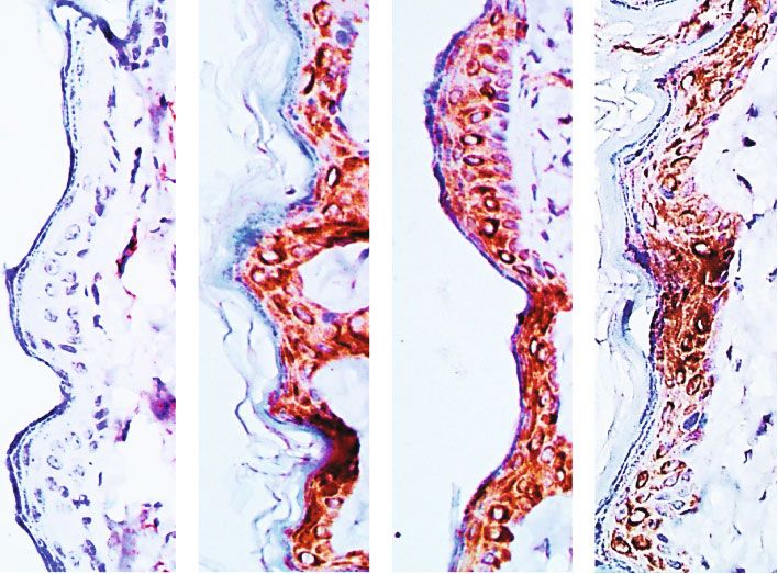
10 Oxidative Medicine and Cellular Longevity

 CIm C Dx1 Dx2
 40

 COX-2 (nmol/min/ml)
 30

 20 ⁎

 10
 < 13
 0
 C Dx1Dx2 C Dx1Dx2 C Dx1Dx2 C Dx1Dx2
 Day 0 Day 7 Day 14 Day 21
 (a) (b)
 30
 ⁎ 0.8
 PGE2 (pg/mg protein)

 ⁎
 ⁎

 NAG (U/mg tissue)
 20 ⁎ 0.6

 0.4
 10
 0.2

 0 0.0
 C Dx1Dx2 C Dx1Dx2 C Dx1Dx2 C Dx1Dx2 C Dx1Dx2 C Dx1Dx2 C Dx1Dx2 C Dx1Dx2
 Day 0 Day 7 Day 14 Day 21 Day 0 Day 7 Day 14 Day 21
 (c) (d)

Figure 8: Immunohistochemical detection of cyclooxygenase-2 (COX-2) in the epithelial cells (a), COX-2 activity (b), prostaglandin E2
(PGE2) levels (c), and N-acetyl-β-D-glucosaminidase (NAG) activity (d) in the scar tissue from rats untreated and treated with
doxycycline hyclate (Dx). Cim: control of the immunohistochemical method (the primary antibody was omitted in the technique), C:
control, Dx = 10 mg/kg Dx, and Dx2 = 30 mg/kg Dx. In the graphics, data are represented as mean and standard deviation. ∗ Statistical
difference compared to group C (Student-Newman-Keuls test, p < 0:05).

skin [63]. In general, at the start of the wound healing pro- cess and reduce the levels of vessels at the end of the
cess, it is observed high deposition of type III collagen, but process. For Altoé et al. [52], the tetracycline class repre-
as the process progresses, the type III collagen fibers are sented by doxycycline increases collagen organization and,
replaced by type I collagen fibers [14], which makes the tissue consequently, the rupture force of the wound. Corroborating
more resistant to traction. About elastic fibers, after treat- the current evidence, our findings indicate that Dx stimu-
ment with Dx, there was an increase in this fiber in the extra- lated the increase of new vessels at the start of the wound pro-
cellular matrix in the scar tissue, especially in the final phase cess (day 7) and the number of cells, possibly due to the
of tissue remodeling. Similarly, the role of Dx on elastic fibers intense migration and activation of the inflammatory cells
was analyzed by Chung et al. [64], in the Marfan syndrome, proven by the increased NAG, at the same time. On the other
in which Dx preserved the elastic fibers in the thoracic aorta, hand, reduced vessels and cells were observed, while fibers
probably by inhibiting the action of metalloproteinases. increased in more advanced phases of the wound repair.
Thus, our findings show the induction of the proliferative These changes are very important for the evolution of wound
capacity of cells and, consequently, the synthesis of the extra- healing and provide resistance and force for the new tissue.
cellular matrix after the application of Dx. Interestingly, Dx was also effective in attenuating COX-2
 Cellularity and neovascularization are essential to the activity and PGE2 only in the initial stage of wound healing
process of wound healing, especially in the inflammatory (day 7). As this inhibition occurred simultaneously to
phase [6]. A good vascularization can provide oxygen and increased cellularity and NAG activity in the scar tissue, the
nutrients for the cells, which are important for the recovery main source of COX-2 activity and PGE2 seems to be unre-
of injured tissue [65]. Besides, the formation of new vessels lated to the increased recruitment of immune cells, including
is directly linked to the formation of a new matrix, called macrophages. Corroborating this proposition, we identified
granulation tissue, rich in vessels and cells. When the process that animals treated with the higher dose of Dx presented a
advances, the number of vessels and cells decreases, but the reduced COX-2 expression in the scar tissue, which was lim-
number of fibers increases, which characterizes mature scar ited to epidermal cells. Thus, our findings indicated that ker-
tissue [66]. Therefore, new effective treatments should pro- atinocytes are the primary targets of Dx-induced COX-2
mote intense neoangiogenesis at the start of the repair pro- downregulation, which represents an effect potentially
Oxidative Medicine and Cellular Longevity 11

 0.8 60

 0.6 ⁎
 H2O2 ( mol/L)

 NO ( mol/L)
 ⁎ 40
 ⁎
 ⁎
 0.4 ⁎&
 20
 0.2 ⁎ ⁎

 0.0 0
 C Dx1Dx2 C Dx1Dx2 C Dx1Dx2 C Dx1Dx2 C Dx1Dx2 C Dx1Dx2 C Dx1Dx2 C Dx1Dx2
 Day 0 Day 7 Day 14 Day 21 Day 0 Day 7 Day 14 Day 21

 (a) (b)
 4 18
 MDA ( mol/L/mg protein)

 3

 CP (nmole/mL)
 12
 ⁎
 2
 ⁎
 6
 1
 ⁎
 0 0
 C Dx1Dx2 C Dx1Dx2 C Dx1Dx2 C Dx1Dx2 C Dx1Dx2 C Dx1Dx2 C Dx1Dx2 C Dx1Dx2
 Day 0 Day 7 Day 14 Day 21 Day 0 Day 7 Day 14 Day 21
 (c) (d)

Figure 9: Levels of oxidative stress markers in the tissue: (a) hydrogen peroxide (H2O2), (b) nitrite and nitrate (NO2-/NO3-), (c)
malondialdehyde (MDA), and (d) carbonyl proteins (CP) in the scar tissue of rats untreated and treated with doxycycline hyclate
(Dx). C: control, Dx1 = 10 mg/kg Dx, and Dx2 = 30 mg/kg Dx. Data are represented as mean and standard deviation. The statistical
difference compared to the group ∗ C and &Dx1 (Student-Newman-Keuls test, p < 0:05).

limited to the initial stages of wound healing. From COX-2 study, as expected, the levels of H2O2 increased on the 7th
activity, intense PG levels are detected after skin injuries, day in the groups treated with Dx, corresponding to the
which are potent proinflammatory effectors that attract inflammatory phase, but decreased in the later phases, on
immune cells, stimulate fibroblast, and endothelial cell prolif- days 14 and 21, in the Dx2 group. In this case, the excess
eration and metabolism in the wound area [11]. COX-2 of H2O2 probably occurred in the inflammatory phase,
played a central and regulatory role in the arachidonic acid due to cell migration, of neutrophils and macrophages,
pathway, by regulating hemostasis and inflammation [67] which release mediators and reactive oxygen species during
and directly impacting the development of the subsequent phagocytosis [73]. Also, we believe that, in this phase, the
phases of wound healing [68]. Thus, controlling the inflam- activation of nicotinamide adenine dinucleotide phosphate
mation from COX-2 downregulation may be a relevant strat- (NADPH) takes place, which generates H2O2 and activates
egy by which Dx accelerates the onset of the proliferative and cell proliferation and apoptosis. In our study, we believed
remodeling phases, stimulating the resolution of the healing that an increased number of macrophages, proven by
process. Since PG release mediated by COX-2 activation trig- in vitro and in vivo analysis of NAG, could justify the
gers prooxidant mechanisms, Dx potentially attenuates the increased level of H2O2 in the early repair process. On the
oxidative stress as well, which can prolong the inflammatory other hand, H2O2 and nitric oxide (NO) showed decreased
phase due to secondary reactive molecular damage [49]. levels during day 14, which indicates that Dx was efficient
 The production of intense free radicals is usually to inhibit lipid peroxidation and protein oxidation in the
observed in the initial stages of the healing process. In skin later phase of the process. However, the excess of NO may
lesions, phagocytic immune cells produce free radicals in a cause irreversible damage in the cells, impaired homeostasis,
process known as respiratory burst [69]. Free radicals, reac- and the activation of various signaling cascades, such as
tive species of oxygen (ROS), and reactive species of nitro- mitogen-activated protein (MAP) or c-Jun N terminal
gen (RNS) promote cellular oxidative stress, damaging kinase (JNK), thus causing cell degeneration and death
membranes, proteins, and genetic material [70, 71]. In [74]. In general, it is difficult to measure NO due to the
respect to ROS, it is important to consider the presence of short half-life in tissues. NO, nitrite (NO2), and nitrate
peroxide hydrogen (H2O2), known as an important marker (NO3) are the markers usually employed [75, 76]. As a
of redox metabolism and more commonly found during the result of this, in our study, we measured the content of
inflammatory phase of the healing process [72]. In our nitrite and nitrate in the scar tissue of Wistar rats.
12 Oxidative Medicine and Cellular Longevity

 120 8
 SOD (U/mg protein)

 ⁎&

 CAT (U/mg protein)
 90 6
 ⁎ ⁎
 60 4 &
 ⁎ ⁎

 30 2

 0 0
 C Dx1Dx2 C Dx1Dx2 C Dx1Dx2 C Dx1Dx2 C Dx1Dx2 C Dx1Dx2 C Dx1Dx2 C Dx1Dx2
 Day 0 Day 7 Day 14 Day 21 Day 0 Day 7 Day 14 Day 21
 (a) (b)
 0.8 40
 GST (U/mg protein)

 MMP-10 (pg/mL)
 0.6 30

 0.4 20

 & ⁎ ⁎
 0.2 10

 0
 0.0
 C Dx1Dx2 C Dx1Dx2 C Dx1Dx2 C Dx1Dx2
 C Dx1Dx2 C Dx1Dx2 C Dx1Dx2 C Dx1Dx2
 Day 0 Day 7 Day 14 Day 21
 Day 0 Day 7 Day 14 Day 21
 (c) (d)

Figure 10: Levels of (a) superoxide dismutase (SOD), (b) catalase (CAT), (c) glutathione S-transferase (GST), and (d) metalloproteinase-10
(MMP-10) in the scar tissue of rats untreated and treated with doxycycline hyclate (Dx). C: control, Dx1 = 10 mg/kg Dx, and Dx2 = 30 mg/
kg Dx. The data are represented as mean and standard deviation. The statistical difference compared to the group ∗ C, &Dx1, and Dx2
(Student-Newman-Keuls test, p < 0:05).

 Markers of oxidative stress are important tools to evalu- to cellular components [83]. In this sense, in addition to act-
ate the redox balance of the healthy and damaged tissues ing as an antimicrobial, Dx also presents antioxidant second-
[77]. MDA is a compound of three carbons synthesized from ary action, which protects the recovering tissue.
the peroxidation of polyunsaturated fatty acids, generally To maintain redox homeostasis in an injured tissue the
formed during the oxidation of the cell membrane lipids action of the antioxidant defense system is necessary, which
[78]. During cutaneous repair, MDA usually increases, exhibits components, such as superoxide dismutase (SOD),
mainly in the inflammatory phase, which indicates damaged catalase (CAT), and glutathione S- transferase (GST) [84,
tissue and slow wound closure [79]. In our study, we 85]. SOD removes superoxide radical, which is highly dan-
observed reduced levels of MDA on day 7, after treatment gerous and destructive for the cells [86]. CAT enzyme accel-
with Dx1 and Dx2, which indicates that doxycycline can help erates the passage of electron and reduces the residence of
to positively modulate the redox status of tissue in recovery. H2O2 inside the cells, consequently avoiding the harmful
Similar results were found by Nogueira et al. [80], after the effect of this compound on the tissues [87]. This means that,
induction of convulsive lesions, following the application of for a drug to be considered effective to treat lesions caused by
pilocarpine, a parasympathomimetic alkaloid extracted from free radicals, it is highly desirable that it stimulates the tran-
jaborandi leaves. These authors showed that Dx reduced lipid scription and translation of these antioxidant enzymes. Our
peroxidation in the brain, thus protecting the tissue from the findings demonstrated that Dx increases SOD and CAT
action of free radicals. Another important marker of the activity in the tissue. Increased CAT was mainly found in
action of ROS and RNS in the tissue is the high content of the inflammatory phase, which corroborates our findings in
carbonylated protein, which indicates protein oxidation [66, relation to the levels of H2O2. We believed that increased
81]. In the present study, after treatment with Dx, only using SOD and CAT reduced superoxide ion (O2-) and H2O2, thus
the lower dose, reduced levels of protein carbonylation were protecting the tissue.
observed, suggesting the antioxidant secondary action of this The imbalance between collagen synthesis and degrada-
drug. Serra et al. [82] evaluated the antioxidant action of tion is a common feature in cutaneous lesions, mainly in
doxycycline and demonstrated that this drug, as well as other infected wounds [88]. A common consequence of this imbal-
tetracyclines, is similar to vitamin E, mainly due to the pres- ance is the formation of hypertrophic scars and keloid, which
ence of a phenolic ring with multiple substitutions. This phe- results in fibrosis and loss of tissue function [8]. MMPs are
nolic ring reacts with the free radical and generates a enzymes found in acute or chronic skin wounds, which reg-
phenolic radical, which is relatively stable and not reactive ulate the deposition of collagen and degrade the extracellular
Oxidative Medicine and Cellular Longevity 13

matrix, which is essential for wound reepithelization [19]. Acknowledgments
The excess of these enzymes may cause disorganization in
the epithelium, cell-cell contact changes, and increased apo- The authors are grateful to the support provided by the Fun-
ptosis of fibroblasts and keratinocytes [89]. In our study, we dação do Amparo à Pesquisa do Estado de Minas Gerais
observed decreased levels of MMP in in vivo analysis, after (FAPEMIG, processes APQ-01895-16, PPM-00687-17, and
treatment with Dx1 and Dx2, which corroborates our find- PPM-00077-18), Conselho Nacional de Desenvolvimento
ings for decreased wound area and increased rate of wound Científico e Tecnológico (CNPq, processes 303972/2017-3,
contraction in these groups. For the healing process to occur 423594/2018-4, 305093/2017-7, and MCTIC 408503/2018-
smoothly and efficiently, a balance between the synthesis and 1), and Coordenação de Aperfeiçoamento de Pessoal de Nível
degradation of collagen is required. If there is a predomi- Superior, Brazil (CAPES, finance code 001).
nance of MMP in the tissue, an exacerbated degradation of
the extracellular matrix may occur, which compromises References
wound closure [19]. Other studies demonstrate that the
administration of Dx speeds up the closure of cutaneous [1] L. Marrot, “Pollution and sun exposure: a deleterious synergy.
wounds by inhibiting MMP-9 [82] and accelerates the recov- mechanisms and opportunities for skin protection,” Current
ery of gastric wounds by inhibiting MMP-2 and H2O2 [90]. Medicinal Chemistry, vol. 25, no. 40, pp. 5469–5486, 2019.
The balance in the synthesis of this enzyme is required for [2] T. Dai, Y. Huang, S. K. Sharma, J. T. Hashmi, D. B. Kurup, and
efficient cutaneous repair and can facilitate cellular migration M. R. Hamblin, “Topical antimicrobials for burn wound infec-
and accelerate the recovery of the tissue. tions,” Recent Patents on Anti-Infective Drug Discovery, vol. 5,
 no. 2, pp. 124–151, 2010.
 [3] J. M. Badia, A. L. Casey, N. Petrosillo, P. M. Hudson, S. A.
5. Conclusions Mitchell, and C. Crosby, “Impact of surgical site infection on
 healthcare costs and patient outcomes: a systematic review in
Taken together, our findings indicate that both doses of Dx six European countries,” Journal of Hospital Infection,
can modulate the repair of skin wounds in rats. However, vol. 96, no. 1, pp. 1–15, 2017.
in general, the best results in cellularity, mast cells, elastic [4] C. K. Sen, “Human wounds and its burden: an updated com-
fibers, hydrogen peroxide levels, and metalloproteinase-10 pendium of estimates,” Advances in Wound Care, vol. 8,
were observed after exposure to the highest dose of doxycy- no. 2, pp. 39–48, 2019.
cline hyclate (30 mg/kg). In vitro, our findings showed that [5] S. Sarabahi, “Recent advances in topical wound care,” Indian
 Journal of Plastic Surgery, vol. 45, no. 2, pp. 379–387, 2012.
Dx increased macrophage proliferation but decreased the
COX-2 and PGE-2 levels. This indicates that macrophage [6] J. Larouche, S. Sheoran, K. Maruyama, and M. M. Martino,
 “Immune regulation of skin wound healing: mechanisms and
activation and COX-2 inhibition are possibly regulated by
 novel therapeutic targets,” Advances in Wound Care, vol. 7,
independent mechanisms. Our findings showed that, no. 7, pp. 209–231, 2018.
in vitro, despite the increased in the number of cells in the
 [7] R. G. Frykberg and J. Banks, “Challenges in the treatment of
initial phase of wound healing, there was a reduction in the chronic wounds,” Advances in Wound Care, vol. 4, no. 9,
expression of COX-2, which was limited to epidermal cells. pp. 560–582, 2015.
Therefore, our results indicated that keratinocytes are the [8] M. Xue and C. J. Jackson, “Extracellular matrix reorganization
primary targets of Dx-induced COX-2 downregulation, and during wound healing and its impact on abnormal scarring,”
this capacity of modulating the intensity of the inflammatory Advances in Wound Care, vol. 4, no. 3, pp. 119–136, 2015.
response without inhibiting cell activation can play an [9] R. V. Gonçalves, R. D. Novaes, M. M. Sarandy et al., “5α-Dihy-
important role in the favorable evolution of the healing pro- drotestosterone enhances wound healing in diabetic rats,” Life
cess. In addition, since the COX-2 is involved in prooxidant Sciences, vol. 152, pp. 67–75, 2016.
mechanisms, the regulation of this pathway by Dx also [10] S. A. Eming, P. Martin, and M. Tomic-Canic, “Wound repair
favored the oxidative balance into the cell and protected the and regeneration: mechanisms, signaling, and translation,”
molecules against the action of free radicals, showing an anti- Science Translational Medicine, vol. 6, no. 265, p. 265sr6, 2014.
oxidant potential in cutaneous wound repair. This informa- [11] S. A. Abd el-Aleem, S. Abdelwahab, H. AM-Sherief, and
tion may be relevant for the selection of the treatment, A. Sayed, “Cellular and physiological upregulation of inducible
mainly in the acute phase of cutaneous wound healing. nitric oxide synthase, arginase, and inducible cyclooxygenase
 in wound healing,” Journal of Cellular Physiology, vol. 234,
 no. 12, pp. 23618–23632, 2019.
Data Availability [12] I. Dalle-Donne, A. Scaloni, D. Giustarini et al., “Proteins as
 biomarkers of oxidative/nitrosative stress in diseases: the con-
The data used to support the findings of this study are avail- tribution of redox proteomics,” Mass Spectrometry Reviews,
able from the corresponding author upon request. vol. 24, no. 1, pp. 55–99, 2005.
 [13] M. Mittal, M. R. Siddiqui, K. Tran, S. P. Reddy, and A. B.
 Malik, “Reactive oxygen species in inflammation and tissue
Conflicts of Interest injury,” Antioxidants & Redox Signaling, vol. 20, no. 7,
 pp. 1126–1167, 2014.
The authors declare that there are no conflicts of interest [14] M. M. Sarandy, L. L. Miranda, L. S. Altoé et al., “Strychnos
regarding the publication of this manuscript. pseudoquina modulates the morphological reorganization of
14 Oxidative Medicine and Cellular Longevity

 the scar tissue of second intention cutaneous wounds in rats,” a mouse subcutaneous sponge model,” Microvascular
 PLoS One, vol. 13, no. 4, article e0195786, 2018. Research, vol. 97, pp. 130–136, 2015.
[15] C. Bonnans, J. Chou, and Z. Werb, “Remodelling the extracel- [31] R. V. Gonçalves, R. D. Novaes, S. L. P. Matta, G. P. Benevides,
 lular matrix in development and disease,” Nature Reviews F. R. Faria, and M. V. M. Pinto, “Comparative study of the
 Molecular Cell Biology, vol. 15, no. 12, pp. 786–801, 2014. effects of gallium-aluminum-arsenide laser photobiomodula-
[16] B. Yue, “Biology of the extracellular matrix,” Journal of Glau- tion and healing oil on skin wounds in Wistar rats: a histomor-
 coma, vol. 23, 8 Suppl 1, pp. S20–S23, 2014. phometric study,” Photomedicine and Laser Surgery, vol. 28,
[17] P. Lu, K. Takai, V. M. Weaver, and Z. Werb, “Extracellular no. 5, pp. 597–602, 2010.
 matrix degradation and remodeling in development and dis- [32] H. D. Perry, L. W. Hodes, J. A. Seedor, E. D. Donnenfeld, T. F.
 ease,” Cold Spring Harbor Perspectives in Biology, vol. 3, McNamara, and L. M. Golub, “Effect of doxycycline hyclate on
 no. 12, pp. 1–24, 2011. corneal epithelial wound healing in the rabbit alkali-burn
[18] P. Rousselle, F. Braye, and G. Dayan, “Re-epithelialization of model,” Cornea, vol. 12, no. 5, pp. 379–382, 1993.
 adult skin wounds: Cellular mechanisms and therapeutic strat- [33] D. A. Guimaraes, E. Rizzi, C. S. Ceron et al., “Doxycycline
 egies,” Advanced Drug Delivery Reviews, vol. 146, pp. 344–365, dose-dependently inhibits MMP-2-mediated vascular changes
 2019. in 2K1C hypertension,” Basic & Clinical Pharmacology & Tox-
[19] M. P. Caley, V. L. C. Martins, and E. A. O'Toole, “Metallopro- icology, vol. 108, no. 5, pp. 318–325, 2011.
 teinases and wound healing,” Advances in Wound Care, vol. 4, [34] M. S. Ågren, P. M. Mertza, and L. Franzén, “A comparative
 no. 4, pp. 225–234, 2015. study of three occlusive dressings in the treatment of full-
[20] G. Simona, B. Buchanan, A. Kundu et al., “Stability, activity, thickness wounds in pigs,” Journal of the American Academy
 and application of topical doxycycline formulations in a dia- of Dermatology, vol. 36, no. 1, pp. 53–58, 1997.
 betic wound case study,” Wounds : a compendium of clinical [35] N. Lemo, G. Marignac, E. Reyes-Gomez, T. Lilin, O. Crosaz,
 research and practice, vol. 31, pp. 49–54, 2019. and D. M. D. Ehrenfest, “Cutaneous reepithelialization and
[21] J. Stechmiller, L. Cowan, and G. Schultz, “The role of doxycy- wound contraction after skin biopsies in rabbits: a mathemat-
 cline as a matrix metalloproteinase inhibitor for the treatment ical model for healing and remodelling index,” Veterinarski
 of chronic wounds,” Biological Research For Nursing, vol. 11, Arhiv, vol. 80, pp. 637–652, 2010.
 no. 4, pp. 336–344, 2010. [36] P. C. Dolber and M. S. Spach, “Conventional and confocal
[22] R. Di Caprio, S. Lembo, L. Di Costanzo, A. Balato, and fluorescence microscopy of collagen fibers in the heart,” Jour-
 G. Monfrecola, “Anti-inflammatory properties of low and high nal of Histochemistry and Cytochemistry, vol. 41, no. 3,
 doxycycline doses: an in vitro study,” Mediators of Inflamma- pp. 465–469, 1993.
 tion, vol. 2015, 10 pages, 2015. [37] C. J. Churukian and E. A. Schenk, “A toluidine blue method
[23] T. Dai, R. Qu, J. Liu, P. Zhou, and Q. Wang, “Efficacy of doxy- for demonstrating mast cells,” Journal of Histotechnology,
 cycline in the treatment of syphilis,” Antimicrobial Agents and vol. 4, no. 2, pp. 85-86, 1981.
 Chemotherapy, vol. 61, no. 1, 2017. [38] R. D. Novaes, R. V. Gonçalves, M. C. Cupertino et al., “The
[24] S. Spasovski, Z. Belazelkoska, M. Popovska et al., “Clinical energy density of laser light differentially modulates the skin
 therapeutic effects of the application of doxycycline in the morphological reorganization in a murine model of healing
 treatment of periodontal disease,” Open Access Macedonian by secondary intention,” International Journal of Experimental
 Journal of Medical Sciences, vol. 4, no. 1, pp. 152–157, 2016. Pathology, vol. 95, no. 2, pp. 138–146, 2014.
[25] D. A. Barbie and B. K. Kennedy, “Doxycycline: new tricks for [39] M. C. Cupertino, K. L. C. Costa, D. C. M. Santos et al., “Long-
 an old drug,” Oncotarget, vol. 6, no. 23, pp. 19336-19337, 2015. lasting morphofunctional remodelling of liver parenchyma
[26] Y. Liu, W. Su, S. Wang, and P. Li, “Naringin inhibits chemo- and stroma after a single exposure to low and moderate doses
 kine production in an LPS-induced RAW 264.7 macrophage of cadmium in rats,” International Journal of Experimental
 cell line,” Molecular Medicine Reports, vol. 6, no. 6, pp. 1343– Pathology, vol. 94, no. 5, pp. 343–351, 2013.
 1350, 2012. [40] C. A. Mandarim-de-Lacerda, “Stereological tools in biomedi-
[27] M. Tyszka-Czochara, P. Paśko, W. Reczyński, M. Szlósarczyk, cal research,” Anais da Academia Brasileira de Ciencias,
 B. Bystrowska, and W. Opoka, “Zinc and propolis reduces vol. 75, no. 4, pp. 469–486, 2003.
 cytotoxicity and proliferation in skin fibroblast cell culture: [41] J. M. Oliveira, N. F. Losano, S. S. Condessa et al., “Exposure to
 total polyphenol content and antioxidant capacity of propolis,” deltamethrin induces oxidative stress and decreases of energy
 Biological Trace Element Research, vol. 160, no. 1, pp. 123–131, reserve in tissues of the Neotropical fruit-eating bat Artibeus
 2014. lituratus,” Ecotoxicology and Environmental Safety, vol. 148,
[28] M. V. Dias, A. P. Castro, C. C. Campos et al., “Doxycycline pp. 684–692, 2018.
 hyclate: A schistosomicidal agent in vitro with immunomodu- [42] D. Tsikas, “Analysis of nitrite and nitrate in biological fluids
 latory potential on granulomatous inflammation in vivo,” by assays based on the Griess reaction: appraisal of the
 International Immunopharmacology, vol. 70, pp. 324–337, Griess reaction in the l-arginine/nitric oxide area of
 2019. research,” Journal of Chromatography B, vol. 851, no. 1-2,
[29] E. C. Santos, R. D. Novaes, D. S. S. Bastos et al., “Modulation of pp. 51–70, 2007.
 oxidative and inflammatory cardiac response by nonselective [43] J. A. Buege and S. D. Aust, “[30] Microsomal lipid peroxida-
 1- and 2-cyclooxygenase inhibitor and benznidazole in mice,” tion,” Methods in Enzymology, vol. 52, pp. 302–310, 1978.
 The Journal of Pharmacy and Pharmacology, vol. 67, no. 11, [44] R. L. Levine, J. A. Williams, E. R. Stadtman, and E. Shacter,
 pp. 1556–1566, 2015. “[37] Carbonyl assays for determination of oxidatively modi-
[30] F. H. Guedes-da-Silva, D. Shrestha, B. C. Salles et al., “Trypa- fied proteins,” Methods in Enzymology, vol. 233, pp. 346–357,
 nosoma cruzi antigens induce inflammatory angiogenesis in 1994.
You can also read