Neuromodulation of Visual Cortex Reduces the Intensity of Intrusive Memories
←
→
Page content transcription
If your browser does not render page correctly, please read the page content below
Cerebral Cortex, 2021;00: 1–10

                                                                              https://doi.org/10.1093/cercor/bhab217
                                                                              Original Article

                                                                                                                                                         Downloaded from https://academic.oup.com/cercor/advance-article/doi/10.1093/cercor/bhab217/6322323 by guest on 27 August 2021
ORIGINAL ARTICLE

Neuromodulation of Visual Cortex Reduces
the Intensity of Intrusive Memories
Noa Herz 1,2 , Yair Bar-Haim1,2 , Ido Tavor2,3 , Niv Tik2,3 , Haggai Sharon3,4 ,
Emily A. Holmes5,6 and Nitzan Censor1,2
1 School of Psychological Sciences, Tel Aviv University, Tel Aviv 69978, Israel, 2 Sagol School of Neuroscience, Tel
Aviv University, Tel Aviv 69978, Israel, 3 Sackler Faculty of Medicine, Tel-Aviv University, Tel Aviv 6997801, Israel,
4 Tel Aviv Sourasky Medical Center, Tel Aviv 6423906, Israel, 5 Department of Psychology, Uppsala University,

Uppsala 75142, Sweden and 6 Department of Clinical Neuroscience, Karolinska Institutet, Solna 17177, Sweden
Address correspondence to Noa Herz, School of Psychological Sciences and Sagol School of Neuroscience, Tel Aviv University, Sharet Building,
Tel Aviv 69978, Israel. Email: herz.noa@gmail.com

Abstract
Aversive events can be reexperienced as involuntary and spontaneous mental images of the event. Given that the vividness
of retrieved mental images is coupled with elevated visual activation, we tested whether neuromodulation of the visual
cortex would reduce the frequency and negative emotional intensity of intrusive memories. Intrusive memories of a viewed
trauma film and their accompanied emotional intensity were recorded throughout 5 days. Functional connectivity,
measured with resting-state functional magnetic resonance imaging prior to film viewing, was used as predictive marker
for intrusions-related negative emotional intensity. Results indicated that an interaction between the visual network and
emotion processing areas predicted intrusions’ emotional intensity. To test the causal inf luence of early visual cortex
activity on intrusions’ emotional intensity, participants’ memory of the film was reactivated by brief reminders 1 day
following film viewing, followed by inhibitory 1 Hz repetitive transcranial magnetic stimulation (rTMS) over early visual
cortex. Results showed that visual cortex inhibitory stimulation reduced the emotional intensity of later intrusions, while
leaving intrusion frequency and explicit visual memory intact. Current findings suggest that early visual areas constitute a
central node inf luencing the emotional intensity of intrusive memories for negative events. Potential neuroscience-driven
intervention targets designed to downregulate the emotional intensity of intrusive memories are discussed.

Key words: intrusive memory, memory, reactivation, TMS, visual cortex

                                                                              1999; Cattaneo et al. 2012). Here, we found that functional
Introduction                                                                  connectivity of the visual network with emotion processing
Following traumatic and aversive events, people commonly                      and memory-related brain regions prior to exposure to an
experience spontaneous intrusive recollections of the distress-               aversive event predicts the negative emotional intensity of to-be
ing incident (intrusive memories) (Iyadurai et al. 2019). These               intruding memory images. In addition, we found that inhibiting
memory intrusions are predominantly visual, experienced in                    early visual activity using noninvasive neuromodulation can
the form of mental imagery of discrete moments from within the                reduce the emotional intensity of such memory intrusions.
distressing event (Ehlers et al. 2004; Holmes et al. 2004, 2005). The             Intrusive memories constitute a basic mode of remembering
early visual cortex has been implicated in such visual mental                 (Berntsen 2010). While the functional role of memory intrusions
imagery (Pearson et al. 2015; Pearson 2019), and it has a causal              is still not clear, there are indications that memory intrusions
role in the ability to hold vivid mental images in mind (Kosslyn              function to maintain the visual memory of negative events

© The Author(s) 2021. Published by Oxford University Press.
This is an Open Access article distributed under the terms of the Creative Commons Attribution Non-Commercial License (http://creativecommons.org/
licenses/by-nc/4.0/), which permits non-commercial re-use, distribution, and reproduction in any medium, provided the original work is properly cited.
For commercial re-use, please contact journals.permissions@oup.com
2     Cerebral Cortex, 2021, Vol. 00, No. 00

across time (Herz et al. 2020). While memory persistence for        visual working memory resources necessary for the reformation
stressful situations can adaptively guide behavior in similar       of perceptual memory intrusions (Kessler et al. 2018, 2020).
future circumstances, pathological persistence can lead to post-    Given the relevance of sensory-visual elements to the intrusion
traumatic stress disorder (PTSD), where intrusive memories          formation, direct inhibition of visual brain regions may function
from the trauma lead to extreme distress and functional             as a mechanistically specific target for intervention. Indeed,
impairment (Bryant et al. 2011; Bar-Haim et al. 2021).              inhibitory 1 Hz repetitive transcranial magnetic stimulation
    According to dual representation theory (Brewin et al.          (rTMS) over the early visual cortex has been shown to
2010), intrusive memories arise from high sensory-bound             effectively interfere with a previously consolidated perceptual
representations that lack sufficient contextual representation      memory (Shmuel et al. 2021), suggesting that this protocol may

                                                                                                                                         Downloaded from https://academic.oup.com/cercor/advance-article/doi/10.1093/cercor/bhab217/6322323 by guest on 27 August 2021
during encoding of the traumatic event. Emergence of these          also prove effective for interfering with perceptual memory
sensory-bound memories, which are usually accompanied by            intrusions.
a strong emotional reaction, involve projections of the dorsal          Here, using an experimental analog of trauma, we used rTMS
visual stream to superior parietal areas as well as to the          to determine whether inhibitory stimulation of early visual
amygdala and insula (Brewin et al. 2010). Exposing patients with    cortex following memory reactivation can reduce the frequency
PTSD to reminders of their trauma while undergoing functional       and negative emotional intensity of intrusive memories of trau-
magnetic resonance imaging (fMRI) revealed that relative to         matic material. In addition, we investigated whether baseline
ordinary episodic memories of the trauma, intrusive memories        functional connectivity, measured by resting-state fMRI, can
involved increased activation of the sensory and motor areas,       predict the frequency and emotional intensity of intrusive mem-
corresponding with the affective and visual components of           ories. Previous studies indicated that baseline brain activity can
the sensory-bound representations predicted by the dual             predict participants’ behavioral responses to external stimuli
representation theory (Whalley et al. 2013).                        (Fox and Raichle 2007) and that resting-state functional con-
    Studies have used the trauma film paradigm to study intru-      nectivity of the visual network predicts evoked blood oxygen
sive memories in a more controlled laboratory environment           level–dependent (BOLD) responses to visual stimuli (Liu et al.
(James et al. 2016). Using fMRI during encoding of aversive         2011). High variability in intracranial electroencephalography
material, it was found that subsequently intrusive scenes,          (iEEG) fluctuations within visual areas during resting state also
relative to neutral scenes, involved elevated activation of the     predicts performance in a visual memory task, even when the
amygdala and the ventral occipital cortex. Relative to aversive     resting state data are acquired 1 day or more apart from the
material that did not intrude, an elevated activation of the        behavioral task (Grossman et al. 2019). We therefore expected
inferior frontal gyrus and bilateral middle temporal gyrus          that interindividual variability in the frequency and emotional
was observed for the material that did intrude (Bourne et al.       intensity of intrusions would be predicted from the baseline
2013). A subsequent study found a similar pattern of results        functional connectivity of the visual network with memory- and
and also used machine learning on fMRI data acquired during         emotion-related brain regions, such as the hippocampus and the
encoding to predict scenes that would subsequently become           amygdala.
intrusive. The classifier indicated a number of brain networks
predictive of intrusions, including areas involved in emotional
processing and a perception-vision-shape network involving
visual brain regions (Clark et al. 2014, 2016). These results       Materials and Methods
are consistent with the highly emotional and visual nature of
                                                                    Participants
intrusive memories. Together with enhanced visual priming for
subsequently intrusive material (Ehlers et al. 2006), findings      Participants were 44 individuals (24 females, Mage = 24.8 years,
appear to generally support the involvement of emotion-             standard deviation [SD] = 2.6, range = 20–30) recruited through
related and visual brain areas in the formation of intrusive        online and printed advertisements. Exclusion criteria were
memories.                                                           current self-reported neurological, physical, or mental disorders,
    Various ways have been considered to modify negative intru-     or the use of any medication that can affect the central
sive memories. Key approaches have drawn on reconsolidation         nervous system. Individuals with a score ≥ 33 (the suggested
theory (Haubrich and Nader 2016) and relied on reminder             clinical cutoff) in the PTSD checklist for DSM-5 were excluded
stimuli, intended to reactivate the memory trace, to enable         (PCL-5; MPCL = 7.0, SD = 5.9, range = 2–15) (Blevins et al. 2015).
its modification and downregulation (Kindt and van Emmerik          Presence of a metal object within or near the head that
2016; Monfils and Holmes 2018). For example, administration         precluded magnetic resonance imaging (MRI) or transcra-
of Propranolol in proximity to memory reactivation stimuli          nial magnetic stimulation (TMS) also served as exclusion
was found to reduce behavioral expressions of fear (Kindt           criteria. All participants had normal or corrected-to-normal
and van Emmerik 2016) and subjective feelings of anxiety            vision.
(Soeter and Kindt 2012). In a similar vein, trauma-focused              Due to technical failures, four participants did not receive
cognitive behavior therapy relies on the systematic recall          the rTMS intervention. The remaining 40 participants were
of trauma memories and their elaboration in the safe and            allocated to a visual-rTMS (n = 19, 11 females) or a vertex-rTMS
supportive context of psychotherapy (McLean and Foa 2011).          (n = 21, 11 females) as a control stimulation site (Jung et al.
Such interventions aim to reduce the involuntary nature             2016). Allocation to groups was made on day 2 of the study
of memory retrieval and the emotional intensity associated          immediately prior to the rTMS procedure, alternately matching
with it, while leaving voluntary memory of the trauma intact        participants between groups on the sum of intrusions on day 1
(Visser et al. 2018; Lau-Zhu et al. 2019). Visuospatial cognitive   of the study. Participants provided their written and informed
tasks delivered following a memory reminder procedure were          consent prior to testing and were compensated $125 for their
shown to reduce the amount of intrusions experienced in             participation. The Tel Aviv Sourasky Medical Center (reference
the week following an aversive event, presumably by taxing          number: 0561-14) and the Tel-Aviv University Institutional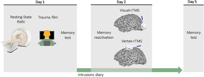
Neuromodulation of Visual Cortex Reduces Intrusive Memories Intensity Herz et al.                3

Review Boards (reference number: 0000912-1) approved the              with 3-s interstimulus interval (ISI). The same eight reactivation
study.                                                                images were presented to all participants, which were different
                                                                      from the images used in the recognition memory tests.
Tasks and Measures                                                       Subsequent to the memory reactivation procedure, partici-
                                                                      pants were provided with a 10-min standardized filler task that
MRI Data Acquisition                                                  consisted of pleasantness ratings of 15 classical music pieces
Participants underwent an MRI session which included anatom-          (James et al. 2015).
ical sequences and resting-state fMRI. Scans were acquired
using a 3 Tesla scanner (Siemens) with a 64-channel head
                                                                      Digitized Intrusive Memories Diary
coil at the Alfredo Federico Strauss Center for Computa-

                                                                                                                                           Downloaded from https://academic.oup.com/cercor/advance-article/doi/10.1093/cercor/bhab217/6322323 by guest on 27 August 2021
                                                                      Participants recorded their intrusive memories about the film
tional Neuro-Imaging, Tel Aviv University. For structural
                                                                      in a digitized online diary that was accessible through their
data, T1w high-resolution (1 mm3 ) whole brain images were
                                                                      mobile phones (Qualtrics—version November 2017–July 2018,
acquired with an MPRAGE sequence (time repetition/time
                                                                      Provo, UT), which was developed in a previous study (Herz
echo [TR/TE] = 1750/2.62 ms; flip angle = 8◦ ; in-plane acquisition
                                                                      et al. 2020). It was explained that intrusive memories come to
matrix [AM] = 224 × 224; field of view [FOV] = 256 × 256 mm;
                                                                      mind unbidden (rather than deliberately recalled) in the form
slice thickness = 1 mm; 176 axial slices). Additional anatomical
                                                                      of sensory mental images. A distinction was made between
sequences (T2w and fluid-attenuated inversion recovery
                                                                      intrusive memories versus pure verbal thoughts, which were
[FLAIR]) were acquired for radiological screening. Functional
                                                                      defined as words/verbal language relating to the film, without
imaging data were acquired with a CMRR multiband accelerated
                                                                      remembering any specific perceptual details from it. Partici-
echo planar imaging pulse sequence of functional T2∗-weighted
                                                                      pants were requested to record any involuntary memories of the
images (TR/TE = 2000/30 ms; flip angle = 82◦ ; AM = 104 × 104;
                                                                      film immediately following its occurrence, to indicate its type
FOV = 208 × 208 mm; slice thickness = 2 mm; 66 interleaved axial
                                                                      (mental image, verbal thought, or both) and describe its content
slices per volume). All images were acquired at a 30◦ angle off the
                                                                      in a way that would allow identifying the intrusion as related
anterior–posterior commissures (AC–PC) line. The resting-state
                                                                      to a specific scene of the film. Verbal thoughts, if they occurred
functional scans comprised a total of 240 volumes (acquisition
                                                                      alone, were not included in the intrusion analysis.
duration = 8 min). The first five volumes were discarded to
                                                                         The emotional intensity of each of the recorded intrusive
account for T1-equilibrium effects.
                                                                      memories was rated as the amount of distress elicited by the
                                                                      recorded intrusion on a scale ranging from 0 (not at all) to 10
Trauma Film
                                                                      (extremely). Ratings were completed in the diary immediately
The trauma film consisted of eight different scenes displaying
                                                                      after recording the occurrence of a given intrusion.
distressing content, including: self-injury, eye surgery, animal
                                                                         Separate diary entries were requested for each intrusive
cruelty, car accident, knee surgery, a scene of humans attacked
                                                                      memory even if the same memory intruded more than once
by an animal, tooth extraction, and a man stabbed with a knife.
                                                                      or if several memories intruded in succession. Participants
These scenes were taken from a previous study (Herz et al. 2020).
                                                                      were asked to provide diary entries for at least three times
Average scene duration was 80.12 s (SD = 25.35). The presenta-
                                                                      per day (morning, afternoon, and evening) even if they did not
tion of each scene was followed by a 2-min break during which
                                                                      experience any intrusive memories in which case a “no intrusive
participants were requested to remain seated with their eyes
                                                                      memory” option could be selected. An experimenter checked
closed until a sound signaled the beginning of the next scene.
                                                                      whether each diary entry was matched to one of the eight scenes
Author N.H. received training from author E.A.H. on administra-
                                                                      viewed and that participants completed the diary for each of
tion of the trauma film paradigm, including a lab exchange visit
                                                                      the study days. Detailed description of data extracted from the
to learn and practice methods.
                                                                      digital diary can be found in the Supplementary Material.

Trauma Film Mood Manipulation Check
Before and after the trauma film, participants rated how sad,         Visual Recognition Memory Test
scared, angry, and happy they felt “at this very moment” on an        The visual recognition memory test was a two-alternative
analog scale ranging from (0) “not at all” to (10) “extremely.”       forced-choice test. The test consisted of three trials per scene,
A composite mood score (ranging 0–40) was computed by                 yielding a total of 24 trials. On each trial, two images were
summing all scale scores after inverting the happy scale score        presented serially: One image was taken from the original
(Holmes et al. 2010). Further manipulation checks, conducted          viewed film, and the other depicted a similar content taken
immediately following film watching, included ratings of              from another unviewed film. Each image was presented for
attention paid to the film and distress experienced in response       1000 ms with 1000 ms ISI. The order of presentation of the two
to the film ranging from not at all (0) to extremely (10).            pictures was random, as well as the order of the trials. After
                                                                      watching the two images, participants had to indicate as fast
Memory Reactivation Procedure                                         and accurate as possible which of the two images was taken
A procedure intended to facilitate memory reactivation was            from the film. Pictures were presented on a 19.6-inch screen,
applied 1 day following film viewing and consisted of presen-         with 1600 × 1200 resolution. Viewing distance was 80 cm. No
tation of one neutral still image from each of the eight scenes       time limit was imposed on responses. The next trial began
of the trauma film. The images served as reminder cues for            following participants’ response.
scenes from the trauma film without reexposing participants to
its aversive content (James et al. 2015). The cues were presented     Noninvasive Brain Stimulation
on the same computer in the same room as used for the trauma          rTMS was delivered using a cooled figure-of-eight coil. Stimula-
film viewing. Following the participants’ key-press, images were      tion was given at 115% of resting motor threshold, which was
presented one at a time against a black background for 3 s each,      measured over the contralateral hemisphere of the participant’s
4 Cerebral Cortex, 2021, Vol. 00, No. 00
Downloaded from https://academic.oup.com/cercor/advance-article/doi/10.1093/cercor/bhab217/6322323 by guest on 27 August 2021
Figure 1. Overview of study design. On day 1, participants underwent anatomical MRI and a resting-state fMRI scan. They then viewed the trauma film, completed a
visual recognition memory test, and were asked to record any intrusions about the film in a digitized diary every day until day 5. On the following day (day 2), brief
reminder cues were used to reactivate participants’ memory of the film, and then rTMS was delivered either over the early visual cortex (visual rTMS) or a control site
(vertex rTMS). On day 5, participants attended a final meeting in which they were retested using the visual recognition memory test.
dominant hand. Resting motor threshold was determined as the was followed by a 8-min resting-state fMRI during which they
minimal stimulation intensity over the primary motor cortex were asked to remain awake with their eyes closed. Participants
(M1) inducing 5 out of 10 motor-evoked potentials that were then completed a demographic questionnaire and the visual
greater than 0.05 mV in the left first dorsal interosseous (FDI) analog mood scales. Participants were next given specific view-
muscle (Rossini et al. 1994). rTMS was delivered over either ing instructions for the trauma film. They watched the film
the early visual cortex or the vertex as a control site by using alone, in a darkened room, using earphones that were set to a
the same stimulation parameters (Jung et al. 2016). rTMS was predefined volume intensity. Following film watching, the visual
delivered at 1-Hz stimulation frequency for 15-min duration, analog mood scales were completed again, as well as the ratings
which is a common protocol that is expected to decrease cortical of attention paid to the film and the day 1 visual recognition
excitability (Chen et al. 1997). memory test. To complete the diary, participants were then
Early visual cortex was localized at the tip of the calcarine given detailed explanation about intrusive memories and were
fissure (Brodmann’s area 17) (Kosslyn 1999) based on each partic- shown how to record intrusions of the film using a practice
ipant’s anatomical MRI acquired on the previous day. A neuron- diary entry on their private mobile phones. A checklist ensured
avigation system (Brainsight) enabled identification of the stim- understanding of diary completion.
ulation target by marking the stimulation site on each partici- On day 2, participants completed the memory reactivation
pant’s anatomical MRI and coregistering the participant’s head procedure in the same context as the film-viewing in day 1.
to his or her MRI using four anatomical landmarks (nasion, tip of Following a 10-min standardized filler task, rTMS was applied
the nose, and the left and right crus of helix). During stimulation, over either the early visual cortex, or over the vertex. Participants
the coil was held with the handle pointing directly upward. The did not receive information about the purpose or the location of
target of stimulation was maintained online using the neuron- the stimulation. At the end of day 2, participants were reminded
avigation system. Mean Montreal Neurological Institute (MNI) to continue filling in the digital diary until day 5.
coordinates of early visual cortex stimulation across partici- On day 5 (laboratory session 3), participants arrived for a final
pants were x = 1.4, y = −84.3, and z = −6.8. Mean MNI coordinates meeting and completed the visual recognition memory test for
of vertex stimulation across participants were x = 0, y = −12.5, the second time. They also rated how accurately they thought
and z = 84.1. they had completed the diary (diary compliance) from 1 (“not
accurate at all”) to 10 (“extremely accurate”). They were then
thanked, debriefed, and compensated for their participation.
Procedure
As shown in Figure 1, the study consisted of three laboratory
fMRI Analysis
sessions.
On day 1, a detailed explanation about the study was given. MRI Preprocessing
Participants were informed that the study consisted of three The anatomical data were skull-stripped using the FSL Brain
laboratory sessions that included MRI and TMS and that they Extraction Tool (BET) (Smith 2002). Preprocessing of the resting-
would be required to watch a film containing distressing content state functional imaging data was performed using FMRIB soft-
and were to fill a diary in between sessions. Participants were ware library (FSL version 6.0.0) (Smith et al. 2004), and it included
required to sleep at least 6 h before each of the experimental high-pass filtering at 0.01 Hz, correction for motion artifacts,
sessions and were informed that they could stop participa- linear registration to the T1w anatomical scan, nonlinear regis-
tion at any time. After providing their written and informed tration to 152 MNI space, and smoothing with 5-mm Gaussian
consents, participants underwent an anatomical MRI scan that kernel. Residual noise was cleaned using FMRIB’s ICA-basedNeuromodulation of Visual Cortex Reduces Intrusive Memories Intensity Herz et al. 5
Downloaded from https://academic.oup.com/cercor/advance-article/doi/10.1093/cercor/bhab217/6322323 by guest on 27 August 2021
Figure 2. Predictive functional connectivity edges of the emotional intensity of intrusive memories. (A) Overview of the fMRI analysis on a single subject’s data.
Each individual resting-state fMRI data were parcellated to seven known resting-state networks based on Thomas Yeo et al. (2011) as well as to the amygdala and
hippocampus. Functional connectivity between all networks was computed for each individual. The resulting functional correlation matrix was used to predict
participants’ emotional intensity at baseline (day 1). Vis, visual; SomMot, somatomotor; Cont, frontoparietal control; DorsAttn, dorsal attention; SalVenAtt, saliency
ventral attention. (B) Variable reduction was used to select predictive networks for the emotional intensity of intrusive memories across participants. In a stepwise
regression model, two variables were selected as predictive features of intrusions’ intensity: the visual network–amygdala connectivity, and the somatomotor–ventral
attention connectivity. (C) The graph shows the correlation between real intrusions’ emotional intensity values and the predicted emotional intensity values based on
the two functional connectivity edges in (B) (R2 = 0.364, P = 0.0001).
X-noiseifier (FIX) (Griffanti et al. 2014), which is a semiauto- correlations, 43 connections were computed for each partic-
matic ICA-based method to identify and remove artefactual ipant. The correlation values were normalized using Fisher’s
components from the data. z-transformation. These 43 features were then correlated with
the intrusions’ emotional intensity and frequency, which were
measured on day 1 of the study (prior to the rTMS intervention)
Resting-State fMRI Analysis
(Fig. 2A).
To compute functional connectivity at rest, we first parcellated
To avoid overfitting due to high number of predictors (43)
the cortex into 400 regions based on Schaefer and colleagues
relative to the number of observations (N = 44), a stepwise regres-
(Schaefer et al. 2018), where each parcel is assigned to one of
sion was applied with all functional connectivity edges as pre-
seven brain networks. In addition to these seven cortical net-
dictors of intrusions’ emotional intensity at baseline (day 1). To
works, we considered two subcortical limbic regions—the amyg-
ensure the robustness of the features selected by the stepwise
dala and hippocampus—due to their central role in memory and
regression, we also computed a least absolute shrinkage and
emotion preprocessing. The amygdala and hippocampus were
selection operator (Lasso) (Tibshirani 1996) as a secondary anal-
extracted based on the Harvard-Oxford subcortical structural
ysis. Lasso was computed using 10-fold cross-validation, and
atlas (Frazier et al. 2005). Preprocessed resting-state fMRI time
the mean squared error (MSE) of the model was inspected to
courses were averaged within each of 400 cortical parcels and
determine a regularization parameter (λ) for which the MSE is
4 subcortical regions (right/left amygdala and right/left hip-
the smallest. Corresponding to standard recommendations, the
pocampus), resulting in a 404 × 240 matrix (regions × mea-
regularization parameter λ of the model (controlling the degree
surements) of resting-state time courses for each participant.
of variables shrinkage) was selected to be the largest value of λ,
Pearson correlations between all regions pairs were then cal-
that is, within one standard error of MSE value (Waldmann et al.
culated, yielding a 404 × 404 Pearson’s correlation matrix per
2013).
participant.
The Pearson’s correlation matrix was then averaged within
each region (seven cortical + two subcortical), resulting in a
Behavioral Analysis
functional connectivity matrix of 9∗ (9 − 1)/2= 36 unique edges.
We also calculated within-network connectivity by averaging Age, attention paid to the film, postfilm distress, and diary
the pairwise correlations between all nodes within each of the compliance records were compared between groups using
seven cortical networks. Together with the 7 within-network independent t-tests, while gender was compared using a6 Cerebral Cortex, 2021, Vol. 00, No. 00
chi-square test. Two-way mixed ANOVA was used to compare (Fig. 2C). Both visual network–amygdala connectivity strength
mood changes resulting from the trauma film between groups, (β = −.892, t(38) = −4.582, P = 0.00004) and somatomotor–ventral
with time (pre-/postfilm-viewing) as a within-subject factor and attention connectivity strength (β = 0.783, t(38) = 4.019, P = 0.0002)
group (visual/vertex) as a between-subjects factor. Percent of significantly contributed to the model.
correct responses in the visual recognition memory test were The Lasso model yielded the same two functional connec-
compared using a two-way mixed-design ANOVA, with time tivity variables as observed in the stepwise regression as well
(day 1/day 5) as a within-subject factor and group (visual/vertex) as the functional connectivity strength between the visual–
as the between-subjects factor. frontoparietal control networks and hippocampus–amygdala
To investigate the effect of rTMS on the emotional intensity connectivity. To ensure the consistency of these results, the
Downloaded from https://academic.oup.com/cercor/advance-article/doi/10.1093/cercor/bhab217/6322323 by guest on 27 August 2021
of intrusive memories, each participant’s averaged emotional Lasso model (using the 10-fold cross-validation) was repeated
intensity was computed for the time points pre-rTMS (day 1) 1000 times, and the variables selected by each iteration were
and post-rTMS (days 2–5). A two-way mixed-design ANOVA was recorded. Over 1000 iterations, the visual network–amygdala
computed with time (pre-TMS/post-TMS) as a within-subject connectivity emerged in 99.7% of the model’s iterations, whereas
factor and group (visual/vertex) as the between-subjects factor. the somatomotor–ventral attention connectivity and visual–
To test whether intrusions’ emotional intensity values declined frontoparietal control connectivity emerged as additional
gradually over time, we also compared the slope of emotional predictive features in 89% of the model’s iterations. Full results
intensity values throughout study days following the rTMS using of the neural features selected across the 1000 Lasso iterations
growth modeling. Growth modeling enables investigation of are shown on the Supplementary Material (Supplementary
longitudinal data by testing individual patterns of change over Table S1). Contrary to the intrusions’ emotional intensity, the
time and by comparing this change between groups instead Lasso model did not yield any predictive features of intrusion
of comparing averaged values across time points. The growth frequency.
modeling portrays a more accurate and complete picture of
our data compared with mixed-design ANOVA (see Supplemen-
tary Material for results using mixed-design ANOVA): First, it Behavior
estimates growth parameters on the available data without
There were no significant differences between the visual and
requiring complete data from all respondents and is thus better
the vertex groups in age (t(38) = −0.3878, P = 0.700), attention
suited for this dataset, which contains missing values for indi-
paid to the film (t(38) = −0.178, P = 0.859), self-reported postfilm
viduals not experiencing any intrusions in one or more study
distress (t(38) = 1.407, P = 0.167), diary compliance (t(38) = 0.769,
days. Second, growth modeling allows the intercept and slope
P = 0.430), and gender distribution (χ 2 (1, 40) = 0.123, P = 0.726).
to vary across individuals, thus accounting for interindividual
Mood deteriorated from pre- to postfilm (F(1, 38) = 78.688,
differences in initial emotional intensity values and slope of
P < 0.001, η2 p = 0.674) and to a similar extent in both groups
change across study days. The steps of the growth modeling
(Fgroup × time (1, 38) = 0.021, P = 0.885).
were applied as shown in Bliese and Ployhart (2002). Briefly,
The ANOVA on the emotional intensity of intrusive memories
the model contains five steps: Step 1 computes a regression
yielded a main effect of time, with a decline in intensity from
model in which intrusions’ intensity are regressed on time. Step
pre- to post-rTMS (Ftime (1, 36) = 53.102, P < 0.001, η2 p = 0.596). Crit-
2 accounts for differences in the initial intensity values by allow-
ically, and in line with our hypothesis, this main effect was qual-
ing the intercepts to randomly vary among respondents. Step
ified by a group-by-time interaction (F(1, 36) = 4.314, P = 0.045,
3 adds the random component of time to the model, allowing
η2 p = 0.107). Post hoc analysis revealed an ordinal interaction
for the slope of intrusions’ intensity change to randomly vary
with greater emotional intensity reduction in the visual rTMS
among respondents. Step 4 of the model accounts for autocor-
group (t(16) = 5.918, P = 0.00002, d = 1.435) relative to the vertex
relation resulting from within-person repeated time points to
group (t(20) = 4.119132, P = 0.001, d = 0.898) (Fig. 3). The main
avoid inflated t values. The final fifth step of the model inserts
effect of group was nonsignificant (Fgroup (1, 36) = 1.475, P = 0.232).
the group variable into the model and tests the time × group
Growth modeling showed a gradual decline in emotional inten-
interaction effect.
sity throughout study days (ttime (37) = −2.167, P = 0.036), and
To investigate the effect of rTMS on the frequency of intru-
critically again, this decline was greater in the visual compared
sions, the averaged sum of intrusions per day pre-rTMS (day
with the vertex group (ttime × group (37) = 2.108, P = 0.041). Time
1) and post-rTMS (days 2–5) was calculated. A two-way mixed-
course of intrusions’ emotional intensity throughout study days
design ANOVA was computed with time (pre-TMS/post-TMS) as
can be found in the Supplementary Material (Supplementary Fig.
a within-subject factor and group (visual/vertex) as the between-
S1A).
subject factor. Two-tailed tests were used for all statistical com-
ANOVA on percent of correct responses in the visual
parisons.
recognition memory test about the film revealed a decrease
in performance from day 1 to day 5 (Ftime (1, 38) = 8.714, P = 0.005,
Results η2 p = 0.186) without a difference between groups (Ftime × group (1,
38) = 0.037, P = 0.847). Group main effect was nonsignificant
fMRI (Fgroup (1, 38) = 0.147, P = 0.702).
In the stepwise regression analysis, two variables were found The ANOVA on the averaged sum of intrusions yielded a
to have a unique contribution in explaining the variance main effect of time with a decline in the averaged sum of
in the emotional intensity of intrusive memories: “visual intrusions per day from pre- (M = 3.875, SD = 2.928) to post-TMS
network–amygdala” connectivity strength and “somatomotor– (M = 1.525, SD = 1.513) (Ftime (1, 38) = 34.016, P < 0.0001, η2 p = 0.472).
ventral attention” connectivity strength. These two features However, group main effect (Fgroup (1, 38) = 0.008, P = 0.928) and
explained a significant portion of the variance in intrusions’ the group × time interaction effects (Ftime × group (1, 38) = 0.089,
intensity (F(2, 40) = 10.896, P = 0.0001, R2 = 0.364, R2 Adjusted = 0.331) P = 0.766) were nonsignificant. Time course of the sum ofNeuromodulation of Visual Cortex Reduces Intrusive Memories Intensity Herz et al. 7
Downloaded from https://academic.oup.com/cercor/advance-article/doi/10.1093/cercor/bhab217/6322323 by guest on 27 August 2021
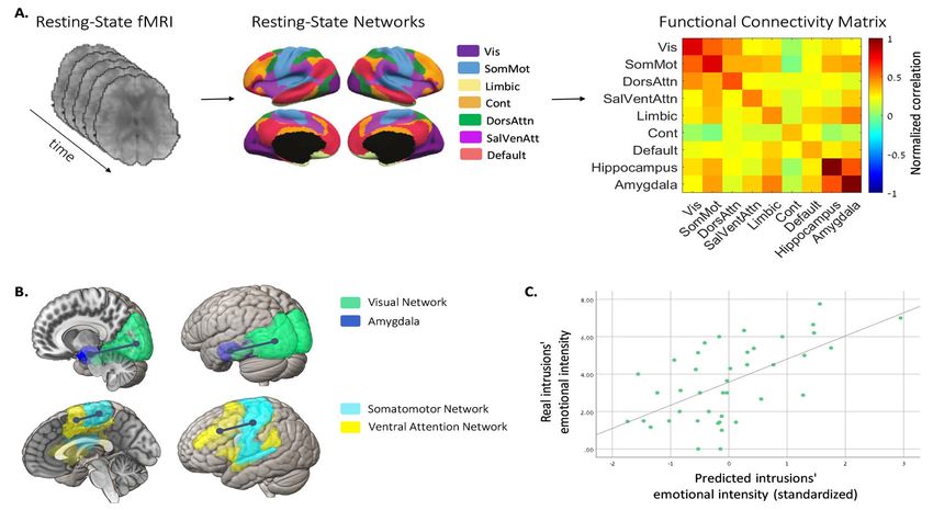
Figure 3. Scatterplot showing the emotional intensity values of intrusive memories pre and post rTMS for individuals in the visual group (V1) and control (vertex).
Within-participant comparisons between pre- and post-TMS intrusions’ emotional intensity is presented in a scatterplot along a unity slope line (x = y), where each
point ref lects one participant. Points “under” the unity slope line ref lect reduced emotional intensity following TMS, whereas points “above” the line ref lect higher
emotional intensity following TMS. Data points on the line ref lect no difference in emotional intensity from pre- to post-TMS. Inset: mean reduction in emotional
intensity of intrusive memories following TMS. Error bars represent standard errors of the means.
intrusions throughout study days can be found in the Supple- can have long-term effects on the emotional aspects of the
mentary Material (Supplementary Fig. S1B). reexperiencing of intrusive memories. This type of approach
Although the results described here are restricted to diary may thus open up a potential target for specialized interventions
entries where intrusions were reported as mental images, for patients suffering from distressing and persistent traumatic
the results pattern remains the same when including all intrusions.
diary entries (both mental images and pure verbal thoughts) The relation between early visual and emotion processing
(Supplementary Fig. S1). The supplementary material shows the brain areas is supported by recent studies showing alteration of
results for intrusions’ emotional intensity (Supplementary Fig. neural activity of the early visual cortex in response to stimuli
S1C) and sum of intrusions (Supplementary Fig. S1D) for all diary that have been associated with an aversive event (McTeague
entries. et al. 2015; Thigpen et al. 2017; Shalev et al. 2018). The amygdala
and visual brain areas are anatomically interconnected not only
indirectly but also through the direct white matter tracts of
Discussion the inferior longitudinal fasciculus (Catani 2003). This anatom-
The findings of the present study implicate involvement of ical pathway has been suggested to mediate the fast transfer
early visual cortex in the emotional intensity of intrusive visual of visual signals to emotional processing regions and neuro-
memories. Connectivity between the visual network and the modulatory back-projections from the amygdala to early visual
amygdala, measured immediately prior to the experience of areas associated with fear recognition (McFadyen et al. 2019).
viewing an aversive event (film scenes with traumatic con- Relatedly, a recent study found that greater functional connec-
tent), was found to predict the emotional intensity of intrusive tivity between the amygdala and visual brain regions immedi-
memories that arose from that event. This offers a potential ately postencoding was related to better subsequent recognition
neuromarker of vulnerability to experiencing traumatic memory of negatively valenced pictures (Kark and Kensinger 2019). In
intrusions as being highly distressing. Importantly, relative to a the present study, greater functional connectivity between the
control stimulation, inhibitory stimulation of early visual cortex amygdala and the visual network was associated with lower
(rTMS) following memory reactivation reduced the intrusions’ emotional intensity of intrusions, as reflected by the negative
emotional intensity on subsequent days. These findings suggest sign of the related beta coefficient. The fact that greater visual–
that interfering with post-reactivation offline visual processing amygdala connectivity postencoding is associated with both8 Cerebral Cortex, 2021, Vol. 00, No. 00
better recognition memory (Kark and Kensinger 2019) and the if more potent types of memory reactivation procedures, such
lower emotion intensity of intrusions following inhibition of as imagery scripts (Rauch et al. 1996), written accounts (Kessler
early visual areas in the current study are not surprising, given et al. 2018), or brief hotspot procedures (Kanstrup et al. 2021),
that the voluntary and involuntary aspects of memory have been could be applied—for example, in clinical populations with reex-
shown to be dissociable (Lau-Zhu et al. 2019) and may even show periencing symptoms. However, future studies are required to
inverse relation between them. For example, it is possible that by examine the boundary conditions for reminder cues and the
enhancing voluntary memory recall, involuntary intrusions can hypothesized role of memory reactivation in inducing putative
be reduced (Krans et al. 2009; Herszage and Censor 2018). reactivation–reconsolidation processes. Finally, our experiment
In the current study, inhibitory stimulation of the early visual was performed with healthy individuals and film material, and
Downloaded from https://academic.oup.com/cercor/advance-article/doi/10.1093/cercor/bhab217/6322323 by guest on 27 August 2021
area reduced the emotional intensity of intrusions but did not it also requires a test of replication before strong conclusions
reduce visual recognition of the negative events or intrusion can be permitted. The act of intrusions reporting may have
frequency. Specific downregulation of the emotional aspects of primed intrusions, thus lowering the ecological validity of our
involuntary memories while leaving voluntary memory intact study relative to real-life intrusions where deliberate cues of
is a desirable outcome for therapeutic goals (Brewin et al. 2010; the memory are usually absent, although similar to clinical
Iyadurai et al. 2019). Moreover, specific reduction of intrusions’ work in which patients are asked to keep a diary of their intru-
emotional intensity, rather than frequency, is in line with some sions. Future studies could explore the clinical potential of the
previous studies indicating that the intrusions’ emotional inten- rTMS intervention approach for patients suffering from highly
sity show only a moderate association with their frequency and distressing intrusions of real-life traumatic events.
may be more predictive of persistent PTSD symptoms (Steil and
Ehlers 2000; Michael et al. 2005).
In addition to the visual network–amygdala connectivity,
Conclusion
somatomotor–ventral attention and visual–frontoparietal con- This study reveals a mechanistic role for early visual cortex
trol connectivities emerged as predictive features of the intru- in the intensity of the affective response (emotional inten-
sions’ emotional intensity. The ventral attention network was sity) associated with intrusive trauma memories. Critically, we
shown to be active upon detection of salient stimuli in the showed that it is possible to modulate the emotional intensity
environment and is thought to represent involuntary orienting of intrusive memories of traumatic film material by rTMS fol-
of attention (Fox et al. 2006). The frontoparietal control network, lowing a memory reminder cue. The specific neuromodulation
on the other hand, is linked to executive control processes that of visual–amygdala connectivity by rTMS and the boundary con-
represent top-down strategic control (Seeley et al. 2007). The ditions for reducing intrusions’ emotional intensity in clinical
involvement of these networks in the regulation of intrusions’ populations remain to be investigated.
intensity suggests that both involuntary attention allocation as
well as effortful cognitive control processes play a role in pre-
dicting the emotional intensity provoked by memory intrusions.
Supplementary Material
Indeed, threat-related attention biases are widely identified as a Supplementary material can be found at Cerebral Cortex online.
risk factor for anxiety disorders (Bar-Haim et al. 2007) and PTSD
(Wald et al. 2013; Naim et al. 2015). Further, attention bias modi-
fication treatment was found to reduce PTSD symptoms and to
Funding
modulate activity in visual processing pathways (Badura-Brack The Israeli Centers of Research Excellence (I-CORE) program of
et al. 2018; Lazarov and Bar-Haim 2021). the Planning and Budgeting Committee and the Israel Science
The current results point to important future research direc- Foundation (ISF) (grants 51/11 and 526/17); The OAK Foundation
tions. First, the findings open the possibility that connectivity (OCAY-18-442), Lupina Foundation, and the Swedish Research
between early visual regions and emotion processing areas may Council (2020-00873) to E.A.H.
constitute a unique neuroscience-derived target for neuromod-
ulatory interventions for intrusive reexperiencing symptoms.
Additional research could further establish the long-term effects
Notes
of such interventions and the specific neural pathway modu- Conflict of Interest: None declared.
lated by them. For example, acquiring resting-state fMRI not
only prior to but also postintervention could elucidate whether
visual network–amygdala coupling is specifically modulated by
References
inhibitory stimulation over early visual areas. In addition, future Amar-Halpert R, Laor-Maayany R, Nemni S, Rosenblatt JD, Censor
studies may enable to test whether other stimulation parame- N. 2017. Memory reactivation improves visual perception. Nat
ters (e.g., stimulation site, duration, or number of stimulation Neurosci. 20:1325–1328.
sessions) could lead to a greater reduction in intrusion intensity. Badura-Brack A, McDermott TJ, Becker KM, Ryan TJ, Khanna MM,
Although, in this study, resting-state fMRI data were used as Pine DS, Bar-Haim Y, Heinrichs-Graham E, Wilson TW. 2018.
converging evidence for our a priori site selection, future studies Attention training modulates resting-state neurophysiologi-
could examine the use of the participant’s visual–amygdala cal abnormalities in posttraumatic stress disorder. Psychiatry
functional connectivity map to guide site selection in an indi- Res Neuroimaging. 271:135–141.
vidually tailored manner (Fox et al. 2012). Second, as in previous Bang JW, Shibata K, Frank SM, Walsh EG, Greenlee MW, Watan-
reports (Amar-Halpert et al. 2017; Bang et al. 2018; Shmuel et al. abe T, Sasaki Y. 2018. Consolidation and reconsolidation
2021), we applied a visual memory reactivation procedure prior share behavioural and neurochemical mechanisms. Nat Hum
to the rTMS intervention, here, memory reminder cues in the Behav. 2:507–513.
form of static pictures, given the use of trauma film as stimuli. Bar-Haim Y, Lamy D, Pergamin L, Bakermans-Kranenburg MJ,
It is conceivable that much stronger effects could be achieved Van Ijzendoorn MH. 2007. Threat-related attentional bias inNeuromodulation of Visual Cortex Reduces Intrusive Memories Intensity Herz et al. 9
anxious and nonanxious individuals: a meta-analytic study. thalamic volumes in pediatric bipolar disorder. Am J Psychia-
Psychol Bull. 133:1–24. try. 162:1256–1265.
Bar-Haim Y, Stein MB, Bryant RA, Bliese PD, Ben Yehuda A, Griffanti L, Salimi-Khorshidi G, Beckmann CF, Auerbach EJ,
Kringelbach ML, Jain S, Dan O, Lazarov A, Wald I, et al. 2021. Douaud G, Sexton CE, Zsoldos E, Ebmeier KP, Filippini N,
Intrusive traumatic reexperiencing: pathognomonic of the Mackay CE, et al. 2014. ICA-based artefact removal and accel-
psychological response to traumatic stress. Am J Psychiatry. erated fMRI acquisition for improved resting state network
178:119–122. imaging. Neuroimage. 95:232–247.
Berntsen D. 2010. The unbidden past. Curr Dir Psychol Sci. Grossman S, Yeagle EM, Harel M, Espinal E, Harpaz R, Noy N,
19:138–142. Mégevand P, Groppe DM, Mehta AD, Malach R. 2019. The noisy
Downloaded from https://academic.oup.com/cercor/advance-article/doi/10.1093/cercor/bhab217/6322323 by guest on 27 August 2021
Blevins CA, Weathers FW, Davis MT, Witte TK, Domino JL. brain: power of resting-state fluctuations predicts individual
2015. The posttraumatic stress disorder checklist for DSM-5 recognition performance. Cell Rep. 29:3775–3784.e4.
(PCL-5): development and initial psychometric evaluation. J Haubrich J, Nader K. 2016. Memory reconsolidation. In: Brain
Trauma Stress. 28:489–498. imaging in behavioral neuroscience. Switzerland: Spring Inter-
Bliese PD, Ployhart RE. 2002. Growth modeling using random national Publishing, pp. 151–176.
coefficient models: model building, testing, and illustrations. Herszage J, Censor N. 2018. Modulation of learning and mem-
Organ Res Methods. 5:362–387. ory: a shared framework for interference and generalization.
Bourne C, Mackay CE, Holmes EA. 2013. The neural basis of Neuroscience. 392:270–280.
flashback formation: the impact of viewing trauma. Psychol Herz N, Bar-Haim Y, Holmes EA, Censor N. 2020. Intrusive
Med. 43:1521–1532. memories: a mechanistic signature for emotional memory
Brewin CR, Gregory JD, Lipton M, Burgess N. 2010. Intru- persistence. Behav Res Ther. 135:103752.
sive images in psychological disorders: characteristics, neu- Holmes EA, Brewin CR, Hennessy RG. 2004. Trauma films, infor-
ral mechanisms, and treatment implications. Psychol Rev. mation processing, and intrusive memory development. J Exp
117:210–232. Psychol Gen. 133:3–22.
Bryant RA, O’Donnell ML, Creamer M, McFarlane AC, Silove D. Holmes EA, Grey N, Young KAD. 2005. Intrusive images and
2011. Posttraumatic intrusive symptoms across psychiatric “hotspots” of trauma memories in posttraumatic stress dis-
disorders. J Psychiatr Res. 45:842–847. order: an exploratory investigation of emotions and cognitive
Catani M. 2003. Occipito-temporal connections in the human themes. J Behav Ther Exp Psychiatry. 36:3–17.
brain. Brain. 126:2093–2107. Holmes EA, James EL, Kilford EJ, Deeprose C. 2010. Key steps in
Cattaneo Z, Bona S, Silvanto J. 2012. Cross-adaptation combined developing a cognitive vaccine against traumatic flashbacks:
with TMS reveals a functional overlap between vision and visuospatial tetris versus verbal pub quiz. PLoS One. 5:e13706.
imagery in the early visual cortex. Neuroimage. 59:3015–3020. Iyadurai L, Visser RM, Lau-Zhu A, Porcheret K, Horsch A, Holmes
Chen R, Classen J, Gerloff C, Celnik P, Wassermann EM, Hallett EA, James EL. 2019. Intrusive memories of trauma: a tar-
M, Cohen LG. 1997. Depression of motor cortex excitability by get for research bridging cognitive science and its clinical
low-frequency transcranial magnetic stimulation. Neurology. application. Clin Psychol Rev. 69:67–82.
48:1398–1403. James EL, Bonsall MB, Hoppitt L, Tunbridge EM, Geddes JR, Milton
Clark IA, Holmes EA, Woolrich MW, Mackay CE. 2016. Intrusive AL, Holmes EA. 2015. Computer game play reduces intru-
memories to traumatic footage: the neural basis of their sive memories of experimental trauma via reconsolidation-
encoding and involuntary recall. Psychol Med. 46:505–518. update mechanisms. Psychol Sci. 26:1201–1215.
Clark IA, Niehaus KE, Duff EP, Di Simplicio MC, Clifford GD, Smith James EL, Lau-Zhu A, Clark IA, Visser RM, Hagenaars MA, Holmes
SM, Mackay CE, Woolrich MW, Holmes EA. 2014. First steps EA. 2016. The trauma film paradigm as an experimental
in using machine learning on fMRI data to predict intrusive psychopathology model of psychological trauma: intrusive
memories of traumatic film footage. Behav Res Ther. 62:37–46. memories and beyond. Clin Psychol Rev. 47:106–142.
Ehlers A, Hackmann A, Michael T. 2004. Intrusive Jung J, Bungert A, Bowtell R, Jackson SR. 2016. Vertex stimulation
re-experiencing in post-traumatic stress disorder: as a control site for transcranial magnetic stimulation: a
phenomenology, theory, and therapy. Memory. 12:403–415. concurrent TMS/fMRI study. Brain Stimul. 9:58–64.
Ehlers A, Michael T, Chen YP, Payne E, Shan S. 2006. Enhanced Kanstrup M, Singh L, Göransson KE, Widoff J, Taylor RS, Gamble
perceptual priming for neutral stimuli in a traumatic context: B, Iyadurai L, Moulds ML, Holmes EA. 2021. Reducing intrusive
a pathway to intrusive memories? Memory. 14:316–328. memories after trauma via a brief cognitive task intervention
Fox MD, Corbetta M, Snyder AZ, Vincent JL, Raichle ME. 2006. in the hospital emergency department: an exploratory pilot
Spontaneous neuronal activity distinguishes human dorsal randomised controlled trial. Transl Psychiatry. 11:30.
and ventral attention systems. Proc Natl Acad Sci U S A. Kark SM, Kensinger EA. 2019. Post-encoding amygdala-
103:10046–10051. visuosensory coupling is associated with negative memory
Fox MD, Halko MA, Eldaief MC, Pascual-Leone A. 2012. Measur- bias in healthy young adults. J Neurosci. 39:3130–3143.
ing and manipulating brain connectivity with resting state Kessler H, Holmes EA, Blackwell SE, Schmidt A-C, Schweer
functional connectivity magnetic resonance imaging (fcMRI) JM, Bücker A, Herpertz S, Axmacher N, Kehyayan A. 2018.
and transcranial magnetic stimulation (TMS). Neuroimage. Reducing intrusive memories of trauma using a visuospa-
62:2232–2243. tial interference intervention with inpatients with post-
Fox MD, Raichle ME. 2007. Spontaneous fluctuations in brain traumatic stress disorder (PTSD). J Consult Clin Psychol. 86:
activity observed with functional magnetic resonance imag- 1076–1090.
ing. Nat Rev Neurosci. 8:700–711. Kessler H, Schmidt A-C, James EL, Blackwell SE, von Rauchhaupt
Frazier JA, Chiu S, Breeze JL, Makris N, Lange N, Kennedy DN, M, Harren K, Kehyayan A, Clark IA, Sauvage M, Herpertz S,
Herbert MR, Bent EK, Koneru VK, Dieterich ME, et al. 2005. et al. 2020. Visuospatial computer game play after mem-
Structural brain magnetic resonance imaging of limbic and ory reminder delivered three days after a traumatic film10 Cerebral Cortex, 2021, Vol. 00, No. 00
reduces the number of intrusive memories of the experimen- stimulation of the brain, spinal cord, roots and peripheral
tal trauma. J Behav Ther Exp Psychiatry. 67:101454. nerves: basic principles and procedures for routine clinical
Kindt M, van Emmerik A. 2016. New avenues for treating emo- and research application: an updated report from an I.F.C.N.
tional memory disorders: towards a reconsolidation inter- committee. Clin Neurophysiol. 91:79–92.
vention for posttraumatic stress disorder. Ther Adv Psy- Schaefer A, Kong R, Gordon EM, Laumann TO, Zuo X-N, Holmes
chopharmacol. 6:283–295. AJ, Eickhoff SB, Yeo BTT. 2018. Local-global parcellation of the
Kosslyn SM. 1999. The role of area 17 in visual imagery: conver- human cerebral cortex from intrinsic functional connectivity
gent evidence from PET and rTMS. Science. 284:167–170. MRI. Cereb Cortex. 28:3095–3114.
Krans J, Näring G, Holmes EA, Becker ES. 2009. Tell me more: can Seeley WW, Menon V, Schatzberg AF, Keller J, Glover GH, Kenna
Downloaded from https://academic.oup.com/cercor/advance-article/doi/10.1093/cercor/bhab217/6322323 by guest on 27 August 2021
a memory test reduce analogue traumatic intrusions? Behav H, Reiss AL, Greicius MD. 2007. Dissociable intrinsic connec-
Res Ther. 47:426–430. tivity networks for salience processing and executive control.
Lau-Zhu A, Henson RN, Holmes EA. 2019. Intrusive memories J Neurosci. 27:2349–2356.
and voluntary memory of a trauma film: differential effects Shalev L, Paz R, Avidan G. 2018. Visual aversive learning compro-
of a cognitive interference task after encoding. J Exp Psychol mises sensory discrimination. J Neurosci. 38:2766–2779.
Gen. 148:2154–2180. Shmuel D, Frank SM, Sharon H, Sasaki Y, Watanabe T, Cen-
Lazarov A, Bar-Haim Y. 2021. Emerging domain-based sor N. 2021. Early visual cortex stimulation modifies well-
treatments for pediatric anxiety disorders. Biol Psychiatry. consolidated perceptual gains. Cereb Cortex. 31:138–146.
89:716–725. Smith SM. 2002. Fast robust automated brain extraction. Hum
Liu X, Zhu X-H, Chen W. 2011. Baseline BOLD correlation predicts Brain Mapp. 17:143–155.
individuals’ stimulus-evoked BOLD responses. Neuroimage. Smith SM, Jenkinson M, Woolrich MW, Beckmann CF, Behrens
54:2278–2286. TEJ, Johansen-Berg H, Bannister PR, De Luca M, Drobnjak I,
McFadyen J, Mattingley JB, Garrido MI. 2019. An afferent white Flitney DE, et al. 2004. Advances in functional and structural
matter pathway from the pulvinar to the amygdala facilitates MR image analysis and implementation as FSL. Neuroimage.
fear recognition. Elife. 8:1–51. 23:S208–S219.
McLean CP, Foa EB. 2011. Prolonged exposure therapy for post- Soeter M, Kindt M. 2012. Erasing fear for an imagined threat
traumatic stress disorder: a review of evidence and dissemi- event. Psychoneuroendocrinology. 37:1769–1779.
nation. Expert Rev Neurother. 11:1151–1163. Steil R, Ehlers A. 2000. Dysfunctional meaning of posttraumatic
McTeague LM, Gruss LF, Keil A. 2015. Aversive learning shapes intrusions in chronic PTSD. Behav Res Ther. 38:537–558.
neuronal orientation tuning in human visual cortex. Nat Thigpen NN, Bartsch F, Keil A. 2017. The malleability of emo-
Commun. 6:7823. tional perception: short-term plasticity in retinotopic neu-
Michael T, Ehlers A, Halligan SL, Clark DM. 2005. Unwanted rons accompanies the formation of perceptual biases to
memories of assault: what intrusion characteristics are asso- threat. J Exp Psychol Gen. 146:464–471.
ciated with PTSD? Behav Res Ther. 43:613–628. Thomas Yeo BT, Krienen FM, Sepulcre J, Sabuncu MR, Lashkari
Monfils MH, Holmes EA. 2018. Memory boundaries: open- D, Hollinshead M, Roffman JL, Smoller JW, Zöllei L, Polimeni
ing a window inspired by reconsolidation to treat anxiety, JR, et al. 2011. The organization of the human cerebral cortex
trauma-related, and addiction disorders. Lancet Psychiatry. estimated by intrinsic functional connectivity. J Neurophysiol.
5:1032–1042. 106:1125–1165.
Naim R, Abend R, Wald I, Eldar S, Levi O, Fruchter E, Ginat K, Tibshirani R. 1996. Regression shrinkage and selection via the
Halpern P, Sipos ML, Adler AB, et al. 2015. Threat-related lasso. J R Stat Soc Ser B. 58:267–288.
attention bias variability and posttraumatic stress. Am J Visser RM, Lau-Zhu A, Henson RN, Holmes EA. 2018. Multiple
Psychiatry. 172:1242–1250. memory systems, multiple time points: how science can
Pearson J. 2019. The human imagination: the cognitive neu- inform treatment to control the expression of unwanted
roscience of visual mental imagery. Nat Rev Neurosci. emotional memories. Philos Trans R Soc B Biol Sci. 373: pp.
20:624–634. 20170209.
Pearson J, Naselaris T, Holmes EA, Kosslyn SM. 2015. Mental Wald I, Degnan KA, Gorodetsky E, Charney DS, Fox NA, Fruchter
imagery: functional mechanisms and clinical applications. E, Goldman D, Lubin G, Pine DS, Bar-Haim Y. 2013. Attention
Trends Cogn Sci. 19:590–602. to threats and combat-related posttraumatic stress symp-
Rauch SL, Van Der KBA, Fisler RE, Alpert NM, Orr SP, Savage CR, toms. JAMA Psychiatry. 70:401.
Fischman AJ, Jenike MA, Pitman RK. 1996. A symptom provo- Waldmann P, Mészáros G, Gredler B, Fuerst C, Sölkner J. 2013.
cation study of posttraumatic tomography and script-driven Evaluation of the lasso and the elastic net in genome-wide
imagery. Arch Gen Psychiatry. 53:380–387. association studies. Front Genet. 4:1–11.
Rossini PM, Barker AT, Berardelli A, Caramia MD, Caruso G, Whalley MG, Kroes MCW, Huntley Z, Rugg MD, Davis SW, Brewin
Cracco RQ, Dimitrijevic MR, Hallett M, Katayama Y, Luck- CR. 2013. An fMRI investigation of posttraumatic flashbacks.
ing CH, et al. 1994. Non-invasive electrical and magnetic Brain Cogn. 81:151–159.You can also read