Descriptive multi agent epidemiology via molecular screening on Atlantic salmon farms in the northeast Pacific Ocean - Nature
←
→
Page content transcription
If your browser does not render page correctly, please read the page content below
www.nature.com/scientificreports
OPEN Descriptive multi‑agent
epidemiology via molecular
screening on Atlantic salmon farms
in the northeast Pacific Ocean
Andrew W. Bateman1,2*, Angela D. Schulze3, Karia H. Kaukinen3, Amy Tabata3,
Gideon Mordecai4, Kelsey Flynn3, Arthur Bass1,5, Emiliano Di Cicco1 & Kristina M. Miller3,5
Rapid expansion of salmon aquaculture has resulted in high-density populations that host diverse
infectious agents, for which surveillance and monitoring are critical to disease management.
Screening can reveal infection diversity from which disease arises, differential patterns of infection in
live and dead fish that are difficult to collect in wild populations, and potential risks associated with
agent transmission between wild and farmed hosts. We report results from a multi-year infectious-
agent screening program of farmed salmon in British Columbia, Canada, using quantitative PCR to
assess presence and load of 58 infective agents (viruses, bacteria, and eukaryotes) in 2931 Atlantic
salmon (Salmo salar). Our analysis reveals temporal trends, agent correlations within hosts, and
agent-associated mortality signatures. Multiple agents, most notably Tenacibaculum maritimum, were
elevated in dead and dying salmon. We also report detections of agents only recently shown to infect
farmed salmon in BC (Atlantic salmon calicivirus, Cutthroat trout virus-2), detection in freshwater
hatcheries of two marine agents (Kudoa thyrsites and Tenacibaculum maritimum), and detection in the
ocean of a freshwater agent (Flavobacterium psychrophilum). Our results provide information for farm
managers, regulators, and conservationists, and enable further work to explore patterns of multi-
agent infection and farm/wild transmission risk.
The recent rapid pace with which marine organisms have been d omesticated1 has elevated concern about dis-
eases of cultured aquatic o rganisms2. As with agriculture before i t3, aquaculture alters disease dynamics through
densification of host populations and provision of novel transmission pathways, impacting both cultured and
wild species4. As global demand for seafood continues to grow, wild-capture fishery production has p lateaued5,
and disease threatens to halt the growth of the aquaculture industry in some parts of the w orld6. Understand-
ing infection and disease patterns in aquaculture is critical for the industry, human food production, and wild
species alike.
Atlantic salmon (Salmo salar) aquaculture, increasing globally since the 1970s and now dwarfing wild Atlantic
salmon capture7, has struggled with disease since its inception8. Several high-profile cases, among them those
of parasitic sea lice, infectious pancreatic necrosis virus, and infectious salmon anemia virus, have impacted
the industry and at times imperilled sympatric wild s almonids9–11. While government disease regulations vary
among jurisdictions, the salmon-farming industry has become dominated by a few large companies that exploit
economies of s cale12, creating the potential for rapid improvements in disease management through corporate
policy. Thus, lessons learned in a given salmon-farming region hold promise for other regions. Disease manage-
ment in practice, however, does not always realise this potential. In particular, multiple countries have repeated
the same mistakes in “managing” infectious salmon anemia (ISA)11,13.
Studying infectious agents in an aquaculture setting is important for multiple reasons.
First, effective disease management on salmon farms requires an understanding not only of acute disease
outbreaks but also of chronic disease and sub-clinical infections involving potential disease agents. Such low-
intensity infections may create production challenges, without posing an overall risk to farmed or wild population
1
Pacific Salmon Foundation, Vancouver, Canada. 2Ecology and Evolutionary Biology, University of Toronto,
Toronto, Canada. 3Molecular Genetics, Fisheries and Oceans Canada, Nanaimo, Canada. 4Department of Medicine,
University of British Columbia, Vancouver, Canada. 5Forest and Conservation Sciences, University of British
Columbia, Vancouver, Canada. *email: andrew.w.bateman@gmail.com
Scientific Reports | (2021) 11:3466 | https://doi.org/10.1038/s41598-020-78978-9 1
Vol.:(0123456789)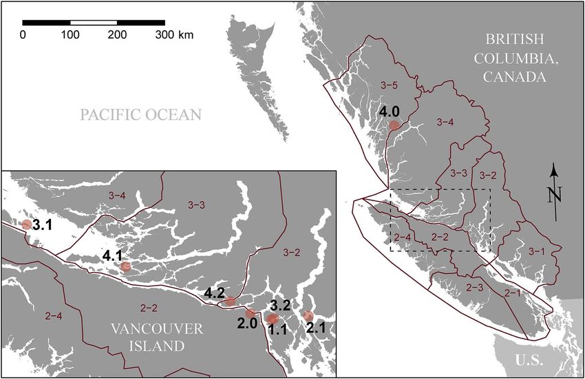
www.nature.com/scientificreports/
viability, but may also constitute standing populations from which more virulent strains can evolve, as in the
case of infectious salmon anemia v irus14.
Second, farm studies can yield valuable information about how infections play out within populations.
Although natural mortality in wild salmon is often upwards of 90%15, marine predators, like many of their ter-
restrial counterparts16, preferentially select parasitized and diseased prey17–19. Mortality in wild fish is thus rarely
observable18 as dying fish either drop out of the water column or are quickly consumed by predators. Open-net
salmon farming, allowing free-flow of water with the surrounding marine environment, offers the potential to
understand disease progression and associated mortality in a semi-natural setting but with an almost complete
absence of predators.
Third, farm studies promise insight into infectious agent exchange between wild and farmed fish. The interface
between wildlife and livestock presents a nexus for s hared20,21 and emergent22 infectious disease. In a wildlife/
livestock disease context, surveillance and monitoring for disease-agent presence, prevalence, and infection
intensity are critical for disease management in both wildlife and l ivestock22,23. Relevant considerations include
the speed with which farmed fish become infected, infectious agents’ presence as acute or chronic infections,
and correspondence between infectious agents and seasonal patterns of wild-fish migration.
While captive populations offer opportunities for improved understanding of infection and disease, little
published information describes the infection status of farm populations outside of mortality events or details
differential patterns of infection between live and moribund fish. We note that corporate monitoring data and
targeted longitudinal studies have previously been used to understand health and disease d evelopment24,25, but
common single-agent focus has limited understanding of the role of co-infection in disease development, and a
general focus on sick and dying fish has limited our understanding of infection progress and resulting pathogen
release. Moreover, if we are concerned about biosecurity and the risk of pathogen exchange among farms and
between farmed and wild salmon, infection levels in the population as a whole, rather than solely in dying fish,
will provide a more accurate assessment of transmission risk.
We report findings from a multi-year, multi-infective-agent monitoring p rogram26, focused on farmed Atlan-
tic salmon in Pacific Canada. This work forms part of a broader research effort using next-generation sequencing
in pathogen discovery, followed by high-throughput genetic screening techniques, (developed in parallel and
first reported for the human-health field27) to monitor infective agents in multiple salmon populations18,26. Here,
we focus on results from a research-directed screening program, conducted at regular intervals on four Atlantic
salmon farms throughout their production cycles. We characterise time series for dozens of infective agents,
identify agents associated with mortality, and provide information for future epidemiological study.
Materials and methods
Four cohorts of farmed Atlantic salmon (in BC’s Fish Health Zones A3.2, A3.3, and A3.4; Fig. 1, Table 1) were
sampled repeatedly throughout their marine production cycles, from ocean entry to the onset of harvest. Both
live individuals, after euthanisation, and recently dead or dying individuals were sampled. Tissue from each
sampled fish was genetically assayed for 47 different infectious agents (including viruses, bacteria, and metazo-
ans; Table 2). After field sampling and genetic workup, we fit descriptive statistical models to genetic prevalence
and intensity time series for individual agents. We modelled temporal trends, calculated agent correlations and
overall infection burdens, and looked for differences between live and dead/dying fish.
Ethics statement. All work with animals was performed according to the Canadian Council on Animal
Care’s (CCAC) Guide to the Care and Use of Experimental Animals, and project protocols were approved by the
federal department of Fisheries and Oceans Canada (DFO) through its Pacific Region Animal Care Committee
(Animal Use Protocol Number: 13-008). Live-sampled fish were euthanised via overdose of tricaine methane-
sulfonate (Syndel laboratories Ltd., Nanaimo BC, Canada). All tissue samples involved were collected under a
Material Transfer Agreement between the BC Salmon Farmers Association and Fisheries and Oceans Canada.
Data series. Infectious‑agent data. Farm schedule and sample collection. Of the four farm cohorts in
the study, two entered the ocean in the spring, and another two entered in the fall (Table 1). Each of the two
salmon-farming companies involved, Cermaq and Marine Harvest (recently renamed MOWI) Canada, granted
access to one spring-entry and one fall-entry cohort. Two of the cohorts were transferred between farm locations
approximately halfway through their respective production cycles (Table 1).
For each cohort, live fish were sampled at an approximate rate of 30 individuals every two weeks for the first
five months, and 20 individuals per month thereafter until harvest. At each sampling visit, up to 20 dead or dying
(moribund) fish from across the farm site were also sampled. For each of the two fall-entry cohorts, fish were
also sampled in the respective hatcheries, with approximately 100 juvenile fish sampled prior to vaccination, and
approximately 100 sampled post vaccination. Company veterinarians selected a single net pen per site visit from
which to sample live fish, and rotated their selections to reduce handling stress among pens. Industry constraints
meant that dead and dying fish from certain mortality or disease events were unavailable, in some cases resulting
in reduced dead/dying samples in months with elevated mortality.
Each sampled fish was euthanised if living and then dissected for gill, liver, kidney, heart, and brain. Dis-
sections were conducted with separate external and internal tools (2 full sets) for each fish, and tools were not
re-used during the sampling events. Following their use, tools were treated to a regime of water, bleach, water,
ethanol, and flame. Fish were dissected using aseptic technique one fish at a time into individual tubes or RNAl-
ater that were closed and sealed once tissue was placed inside. Sampled tissues were held at 4 °C overnight then
stored at - 80 °C until laboratory analysis. After each individual fish was dissected, the operating theatre was com-
pletely broken down and waste removed from the dissection area. The surfaces were wiped with bleach and then
Scientific Reports | (2021) 11:3466 | https://doi.org/10.1038/s41598-020-78978-9 2
Vol:.(1234567890)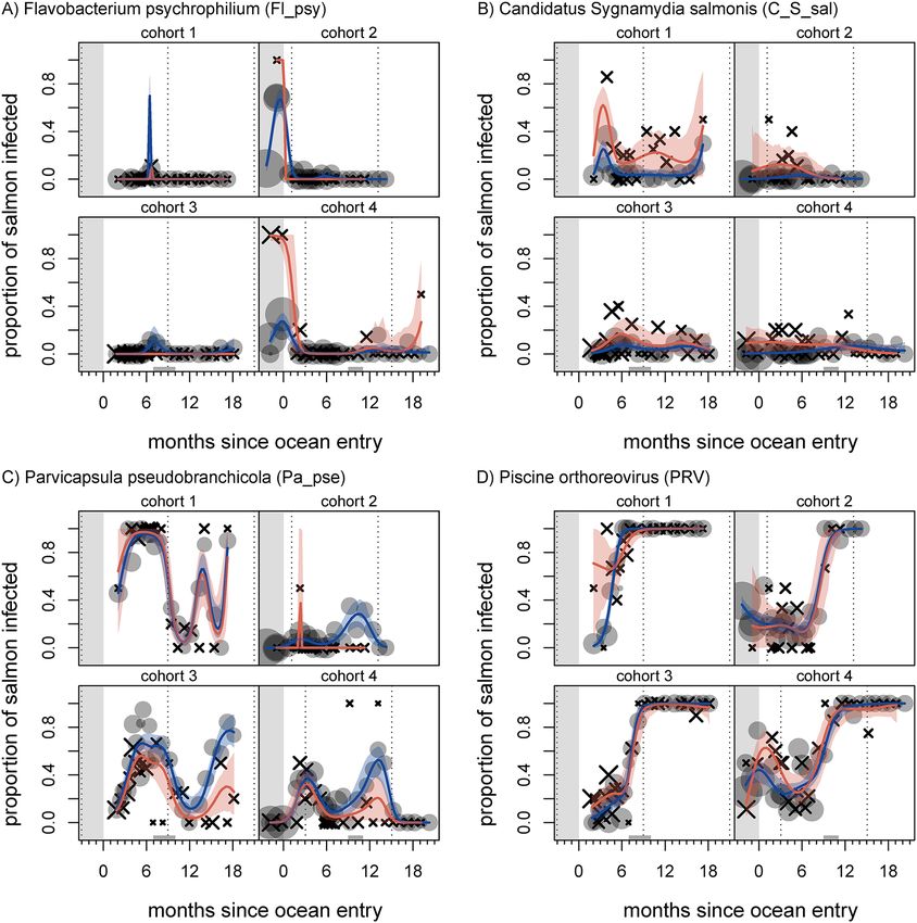
www.nature.com/scientificreports/
Figure 1. Atlantic salmon-farm locations (points) for the four cohorts from which samples were collected for
this study, in relation to Fisheries and Oceans Canada’s Aquaculture Management Zones (2–1 through 3–5).
For each farm location, labelled x.y, x indicates a cohort of salmon (1 through 4) while y indicates successive
locations of that cohort: 0—freshwater hatchery, 1 & 2 (in some cases)—sequential saltwater net-pen locations.
cohort Facility type facility code (Fig. 1) DFO Fish Health Zone Entry (initial) Exit (final)
1 Farm 1.1 3–2 Apr 2013 Mar 2015
Hatchery 2.0 2–2 - Sep 2013
2
Farm 2.1 3–2 Sep 2013 Nov 2015
Farm 3.1 3–4 Apr 2013 Dec 2013
3
Farm 3.2 3–2 Nov 2013 Apr 2015
Hatchery 4.0 3–5 - Oct 2013
4 Farm 4.1 3–3 Oct 2013 Aug 2014
Farm 4.2 3–2 Aug 2014 Sep 2015
Table 1. Movement history for farmed Atlantic salmon used in this study. Geographic locations of individual
facilities are shown in Fig. 1.
70% ethanol and a new dissection theatre assembled with fresh gloves, new outside and inside tools, and tubes.
Full dissection kits were assembled for each farm site, and following each site visit the entire kit was sterilized.
Laboratory analysis. We used a Fluidigm BioMark™ HD microfluidics-based qPCR platform, developed and
onitoring18,26, to screen samples for viral, bacterial, and eukaryotic agents
validated for salmon infective-agent m
known to infect and potentially cause disease in salmon worldwide. The BioMark™ platform employs assays
designed to assess presence and concentration of specific targeted nucleic acid sequences, and is sensitive to
between one and three sequence copies per test volume26. The data we report are estimates of the number of tar-
get gene copies per μL of nucleic-acid solution, extracted from mixed-tissue homogenates and standardised to a
fixed total nucleic-acid concentration (see Supplemental Information). We did not attempt to relate gene copies
directly to infective-agent numbers, as such work was outside the scope of our study.
Substantial measures were taken to maximise data quality. We used a house-keeping gene assay to gauge
sample quality and to ensure that nucleic acid degradation had not occurred. Each assay for an infectious agent
was run in duplicate for each sample. Analytical sensitivity and specificity, and assay repeatability of the BioMark™
platform have been previously e valuated26. Extraction (DNA and RNA) and analysis protocols are described
elsewhere26,28 and we provide details in the Supplementary Information. Critically, we ran a series of negative
Scientific Reports | (2021) 11:3466 | https://doi.org/10.1038/s41598-020-78978-9 3
Vol.:(0123456789)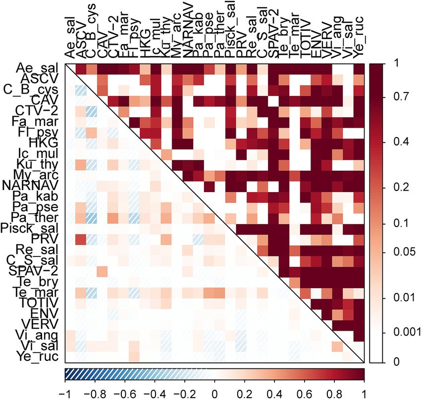
www.nature.com/scientificreports/
Freshwater Saltwater
Organism Organism type Test code Status Tests‡ Prevalence Tests‡ Prevalence
Infectious pancreatic necrosis virus Virus IPNV Known 0 – 933 0
Infectious salmon anemia virus Virus ISAV-7 Known 0 – 933 0
Infectious salmon anemia virus Virus ISAV-8 Known 0 – 933 0
Oncorhynchus masou herpes virus Virus OMV Known 0 – 933 0
Piscine myocarditis virus Virus PMCV Known 0 – 933 0
Salmon alphavirus Virus SAV Known 0 – 933 0
Aeromonas hydrophila Bacterium Ae_hyd Known 0 – 933 0
Moritella viscosa Bacterium Mo_vis Known 0 – 933 0.0096
Spironucleus salmonicida Flagellate Sp_sal Known 0 – 933 0
Gyrodactylus salaris Fluke Gy_sal Known 0 – 933 0
Nucleospora salmonis Microsporidian Nu_sal Known 0 – 931 0.0150
Table 2. Low-prevalence infective agents in initial rounds of mixed-tissue sample screening in farmed Atlantic
salmon. ‡ successful tests only (i.e. those without evidence of control cross contamination, poor amplification
curves, or low housekeeping gene signals).
controls to minimise false-positive results: negative extraction controls on RNA extraction plates, negative cDNA
controls and no RT controls for the cDNA step, negative STA and no primer-STA controls for the specific target
amplification step, and a blank buffer at the time of chip loading. We did not apply a limit-of-detection cutoff
to the data we analysed, but we did discard results for which duplicate tests run on the same sample disagreed
with respect to presence of an agent. We considered successful tests to be those without evidence of control
cross contamination, poor amplification curves, or low housekeeping gene signals, and we discarded results
from unsuccessful tests, while retaining successful results for other tests run on the same sample, as appropriate.
Laboratory screening occurred in two main phases. Initially, we screened 933 mixed-tissue samples for a
suite of 47 agents (status “known” in Tables 2 and 3). Some of the corresponding results have been reported
elsewhere29. In subsequent screening, we omitted eleven of the initially known agents (Table 2), which displayed
very low prevalence or were not detected at all. In their place during the second phase of screening, we tested
for eleven additional viruses, which had not been discovered at the time of the initial experimental d esign30,31
(status “new” in Table 3). Of these agents, two—Atlantic Salmon Calicivirus (ASCV) and a recently sequenced
variant of Cutthroat trout virus (CTV-2)—displayed high prevalence. To fill in the time series and enable analysis,
we re-assayed 215 mixed-tissue samples for ASCV and CTV-2, and assayed a further 16 samples that had not
previously had mixed tissues successfully assayed (231 fill-in samples in total), using the Applied Biosystems
7900HT platform. We chose these fill-in samples to maximise temporal representation across farms, and we did
not consider their status with respect to other agents during selection.
Statistical analyses. We analysed agent data to assess how apparent infection patterns changed over the
course of a cohort’s grow-out period, in relation to season, and between live and dead fish. We further analysed
multi-agent infectious burden and how agent levels were correlated across hosts.
We note that agent data exhibit sampling bias, because disproportionately few samples were available from
high-mortality periods due to farm constraints, and we did not known if, or how often, live fish were sampled
from pens with elevated mortality. As a result, we do not attempt to relate infectious-agent levels to on-farm
mortality levels.
Single‑agent time series. To describe patterns in disease-agent time series from the four focal Atlantic-salmon
farm cohorts, we fit generalised additive m odels32 to single-agent data. GAMs provide flexible functional
forms able to capture epidemiological patterns akin to those described by susceptible-infected-recovered (SIR)
models33, although they do not share a mechanistic basis.
For each agent with sufficient detections, we fit models to prevalence and intensity responses. We define agent
prevalence as the proportion of successful tests with a positive detection, and we define agent intensity as the
number of gene copies (the “load”) in a sample with a positive detection. First, we fit models of agent prevalence
to detection/non-detection data, assuming a binomial response and logit (log-odds) link function. Second, for
the samples in which we detected a focal agent, we fit models of average infection intensity to log-transformed
copy-number data, assuming a normal response and linear link function. We incorporated four predictor com-
ponents in our models: (1) a smooth function of the number of days since ocean entry, parameterised with a
cubic-spline basis and allowed to differ among cohorts; (2) an additional smooth function of the number of
days since ocean entry to account for differences between live and dead/dying hosts, again parameterised with a
cubic-spline basis and allowed to differ among cohorts; (3) a scalar effect for each cohort; and (4) a smooth term
with a cyclic cubic-spline basis to account for consistent seasonal patterns across cohorts. In this way, models
could capture nonlinear trends over time, allowing for different patterns between live and dead/dying fish, and
among farm cohorts. For the prevalence models, we used ten knots in the smooth terms for components 1) and
2), and four knots in the smooth terms for component 4). For the intensity models, we included identical model
Scientific Reports | (2021) 11:3466 | https://doi.org/10.1038/s41598-020-78978-9 4
Vol:.(1234567890)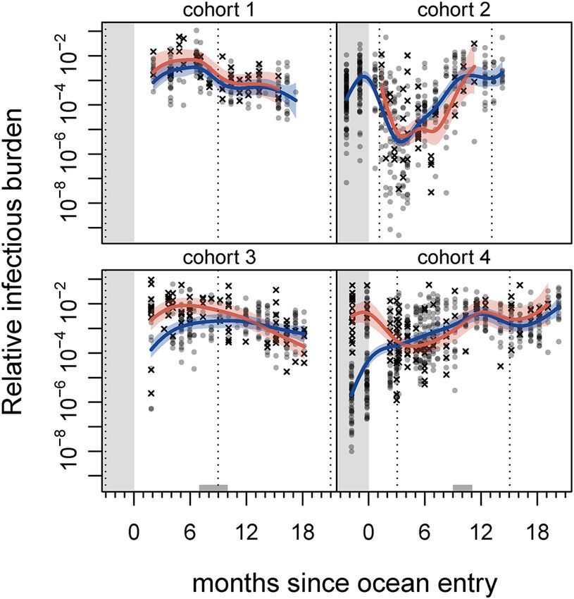
www.nature.com/scientificreports/
Freshwater Saltwater
Organism Organism type Test code Status Tests‡ Prevalence Tests‡ Prevalence
Salmon pescarena-virus-1† Virus SPAV-1 New 427 0 1611 0
Salmon pescarena-virus-2† Virus SPAV-2 New 425 0 1605 0.0019
Atlantic salmon calicivirus Virus ASCV New 423 0.1300 1835 0.5384
Pacific salmon nidovirus† Virus PSNV New 427 0.0023 1611 0.0006
Cutthroat trout virus-2† Virus CTV-2 New 426 0.2840 1808 0.6610
Putative Narna-like virus† Virus NARNAV New 427 0 1606 0.0212
Orthomyxovirus† Virus ORTHO New 427 0 1611 0.0006
Chinook aquareovirus† Virus CAV New 426 0.0023 1611 0.0012
Poxvirus Virus SGPX New 427 0 1611 0
Putative RNA virus† Virus RNAV New 427 0 1611 0.0006
Putative totivirus† Virus TOTIV New 427 0.0023 1607 0.0137
Erythrocytic necrosis virus Virus ENV Known 427 0 2470 0.0049
Infectious hematopoietic necrosis virus Virus IHNV Known 427 0 2471 0.0004
Piscine orthoreovirus Virus PRV Known 421 0.3278 2404 0.5420
Pacific salmon parvovirus Virus PSPV Known 327 0 2101 0
Viral encephalopathy & retinopathy Virus VERV Known 427 0 2471 0.0012
Viral hemorrhagic septicemia virus Virus VHSV Known 427 0 2471 0
Aeromonas salmonicida Bacterium Ae_sal Known 425 0.0094 2466 0.0020
Candidatus Branchiomonas cysticola Bacterium C_B_cys Known 426 0.3873 2464 0.0369
Flavobacterium psychrophilum Bacterium Fl_psy Known 414 0.3382 2459 0.0130
Piscichlamydia salmonis Bacterium Pch_sal Known 427 0 2471 0
Piscirickettsia salmonis Bacterium Pisck_sal Known 427 0.0023 2468 0.0073
Renibacterium salmoninarum Bacterium Re_sal Known 427 0 2472 0.0024
Strawberry disease (Rickettsia-like) Bacterium RLO Known 427 0 2472 0
Candidatus Syngnamydia salmonis Bacterium C_S_sal Known 427 0.0047 2461 0.0561
Tenacibaculum maritimum Bacterium Te_mar Known 426 0.0423 2335 0.6081
Vibrio anguillarum Bacterium Vi_ang Known 423 0.0331 2477 0.0153
Vibrio salmonicida Bacterium Vi_sal Known 425 0.0965 2458 0.0260
Yersinia ruckeri Bacterium Ye_ruc Known 427 0.0187 2468 0
Neoparamoeba perurans Amoeba Ne_per Known 427 0 2472 0.0004
Nanophyetus salmincola Fluke Na_sal Known 427 0 2472 0
Sphaerothecum destruens Mesomycetozoan Sp_des Known 427 0 2471 0.0004
Facilispora margolisi Microsporidian Fa_mar Known 427 0 2460 0.0293
Loma salmonae Microsporidian Lo_sal Known 427 0 2472 0
Paranucleospora theridion Microsporidian Pa_ther Known 410 0.2317 2383 0.9123
Ceratomyxa shasta Myxozoan Ce_sha Known 427 0 2472 0
Kudoa thyrsites Myxozoan Ku_thy Known 424 0.0755 2386 0.4438
Myxobolus arcticus Myxozoan My_arc Known 427 0 2471 0.0020
Myxobolus insidiosus Myxozoan My_ins Known 427 0 2472 0
Parvicapsula kabatai Myxozoan Pa_kab Known 427 0.0023 2461 0.1187
Parvicapsula minibicornis Myxozoan Pa_min Known 427 0 2469 0.0004
Parvicapsula pseudobranchicola Myxozoan Pa_pse Known 427 0 2312 0.3166
Tetracapsuloides bryosalmonae Myxozoan Te_bry Known 427 0.0070 2472 0
Cryptobia salmositica Protozoan Cr_sal Known 427 0 2472 0
Dermocystidium salmonis Protozoan De_sal Known 427 0 2472 0
Ichthyophonus hoferi Protozoan Ic_hof Known 427 0 2471 0.0024
Ichthyophthirius multifiliis Protozoan Ic_mul Known 384 0.0052 2290 0.0157
Table 3. Infective agents in mixed-tissue samples of farmed Atlantic salmon. †Indicates agents newly
discovered or not known from salmon in BC30,31. ‡ successful tests only (i.e. those without evidence of control
cross contamination, poor amplification curves, or low housekeeping gene signals).
Scientific Reports | (2021) 11:3466 | https://doi.org/10.1038/s41598-020-78978-9 5
Vol.:(0123456789)www.nature.com/scientificreports/
components, except that we reduced the number of knots in 1) and 2) to eight, aiming to avoid overly flexible
models for use with reduced sample sizes after omitting non-detections.
While our models were unable to reveal general patterns of infection for any agent in BC salmon farms, due
to the limited number of cohorts observed, our models do allow for evaluation of temporal trends within each
cohort and for comparison among groups (cohorts, live and dead/dying fish, etc.). This approach was useful to
distinguish the average “signal” from the “noise” in each cohort over time, in the context of many screening tests
across a large number of samples exhibiting a large range of apparent infection intensity.
In initial agent-prevalence model fitting, GAM fits in regions of the data with all-zero or all-one responses
yielded accurate response predictions but unreasonably wide confidence intervals. We therefore used probability-
integral-transform (PIT) residual bootstrapping to estimate confidence i ntervals34.
We used the mgcv package35 in R 36 to fit GAMs.
Relative infectious burden. In addition to the single-agent time series, we modelled temporal changes in relative
infectious burden (RIB), a summary measure of agent loads within a given h ost37. For a infective agents assayed
in n individuals, normalised RIB for individual i is:
a
xij
, (1)
j=1
a · max xkj
allk
where xij is infection load (copy number) of for agent j in individual i (xkj is the same measure for individual k).
RIB has been used previously to investigate responses to infection in Chinook salmon37 and coho salmon38, and
it has been characterised in juvenile sockeye salmon39.
In calculating RIB, we restricted our dataset to agents for which we had screened in both phases of testing
and detected at least five times, plus ASCV and CTV-2, for which we had filled in missing results from the first
round of testing (see above). This resulted in RIB calculations based on eighteen infective agents (Table S1).
Agent correlations. To assess general patterns of coinfection, we calculated pairwise Spearman rank correla-
tions between assayed copy numbers for pairs of infective agents, data permitting, and between assayed copy
numbers and the approximate numbers of house-keeping-gene copies per μL. House-keeping-gene copy num-
ber is relevant because degraded host tissue should have a lower number of these gene copies. Because qPCR
involves a theoretical doubling of target-DNA copies per cycle, two raised to the power of the negative corre-
sponding Ct value is approximately
proportional to the number of target-DNA copies in an assay. In this case,
we deemed use of 2 −Ct hkg , ignoring the efficiency of the PCR reaction, to be acceptable, given our use of the
measure to calculate a rank correlation.
We used a randomisation test to compare the number of correlations with p ≤ 0.05 to the numbers we would
expect by chance. In each of 1000 iterations, we independently resampled the data for each agent, with replace-
ment, and re-ran our correlation analysis, as above. While we would expect approximately 19 of the 378 (1/20)
correlations to have p ≤ 0.05 by chance, this ranomisation test offered a crude comparison to the variation we
might expect to see.
Results
Data series. Infectious‑agent data. We successfully generated infectious-agent data for a total of 2931 sam-
ples: 2504 from Atlantic salmon farms in the marine environment and 427 from freshwater hatcheries. Of the
successfully assayed samples, 2407 were live-sampled fish, while 524 were opportunistically sampled dead or
dying fish (160 moribund, 364 recently dead “fresh silvers”). Of the successful hatchery samples, 24 were from
dead fish and four were from moribund fish. We initially screened each sample for 47 infectious agents. After
screening 933 samples, we replaced assays for eleven extremely rare agents (Table 2) with assays for novel viral
variants30,31; hence we screened for 58 total agents across the set of samples, but no single fish sample was sub-
jected to every assay. In follow-up screening, we also screened 231 mixed-tissue samples (215 of the initial 933
samples and 16 samples that had not had successful assays previously) for ASCV and CTV-2, the two highest
prevalence novel viruses. Overall freshwater agent prevalence ranged from 0 to 39% and overall saltwater preva-
lence ranged from 0 to 91%, with agents prevalent in freshwater often rare in saltwater, and vice versa (Table 3).
Statistical analyses. Single‑agent time series. We note, given the potential bias in the sampling during
periods of elevated mortality, that sample trends may not be representative of farm cohorts as a whole.
In the case of agents for which we had sufficient data to fit descriptive models, prevalence time series gener-
ally exhibited one of four patterns: 1) an ephemeral spike around the time of ocean entry followed by decline
(Candidatus Branchiomonas cysticola, Flavobacterium psychrophilum (Fig. 2A), Vibrio anguillarum, Vibrio sal‑
monicida); 2) low, chronic prevalence, often with an ephemeral marine spike (Facilispora margolisi, Ichthyoph‑
thirius multifiliis, Putative Narna-like virus, Parvicapsula kabatai, Candidatus Syngnamydia salmonis (Fig. 2B),
Putative totivirus); 3) repeatedly fluctuating, substantial prevalence in the marine environment (CTV-2, Par‑
vicapsula pseudobranchicola (Fig. 2C), Tenacibaculum maritimum); or 4) presence at low levels in freshwater,
with increasing prevalence after ocean entry, often to 100% [ASCV, Kudoa thyrsites, Paranucleospora theridion,
Piscine orthoreovirus (PRV; Fig. 2D)]. Figure 2 shows exemplar model fits, with all model fits provided in the
Supplementary Information.
Patterns of agent intensity in the available samples were not as well characterised as those for prevalence,
in many cases due to sparse data. For many agents, intensity varied by five or more orders of magnitude across
Scientific Reports | (2021) 11:3466 | https://doi.org/10.1038/s41598-020-78978-9 6
Vol:.(1234567890)www.nature.com/scientificreports/
Figure 2. Agent prevalence of Flavobacterium psychrophilum (Fl_psy; A), salmon gill chlamydia Candidatus
Syngnamydia salmonis (C_S_sal; B), Parvicapsula pseudobranchicola (Pa_pse; C), and Piscine orthoreovirus
(PRV; D) in farmed Atlantic salmon throughout four production cycles. Grey circles show prevalence in live fish
on each sampling date, and black X’s show prevalence in dead/dying fish (symbol areas proportional to sample
sizes). Curves indicate mean predictions from a generalised additive model; blue and red correspond to live
and dead/dying fish, respectively (shaded areas show 95% confidence regions). Left-hand grey region indicates
freshwater hatchery residence, grey regions on x-axis indicate period of transfer to another site, and vertical
dotted lines correspond to January 1st.
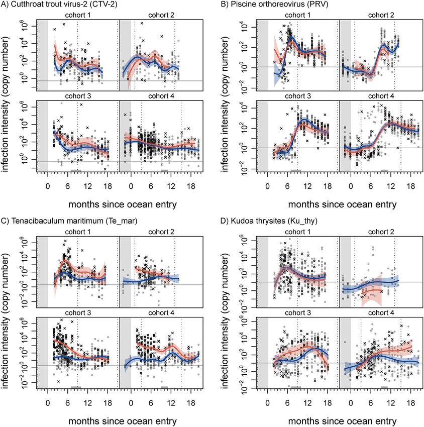
individuals and through time. General trends for well-represented agents were: decline in CTV-2 intensity
throughout marine residence (Fig. 3A); increase and then decline for PRV (Fig. 3B) and P. theridion, both agents
that increased in prevalence over time; and substantially elevated but declining intensity for T. maritimum in dead
and dying fish (Fig. 3C). In many cases, agents for which prevalence plateaued appeared to exhibit an intensity
peak at around the time that prevalence approached its maximum (ASCV, K. thyrsites, P. pseudobranchicola, P.
theridion, PRV; Supplementary Information).
Multiple agents showed differences in prevalence or intensity between live and dead/dying fish. We inter-
preted models to show statistically significant differences between sample categories in a given time window
when the confidence region for one category did not contain the other category’s mean trend. Prevalence of F.
psychrophilum in dead/dying fish was elevated in-hatchery (Fig. 2A), with intensity also elevated in-hatchery
for cohort 4. Intensity of K. thyrsites was elevated in dead/dying fish for cohorts 3 and 4 (Fig. 3D). Prevalence of
the putative Narna-like virus was elevated in dead/dying fish in cohort 4, although intensities remained around
a single gene copy (Fig. S9). In fact, the putative Narna-like virus was particularly prevalent in dead samples
(11.9%), compared to dying (1.5%) and live (0.3%) samples. Prevalence of P. pseudobranchicola was reduced in
dead/dying fish in cohorts 3 and 4 (Fig. 2C). Both prevalence and intensity of PRV were elevated for dead/dying
Scientific Reports | (2021) 11:3466 | https://doi.org/10.1038/s41598-020-78978-9 7
Vol.:(0123456789)www.nature.com/scientificreports/
Figure 3. Agent intensity of Cutthroat trout virus (CTV-2; A), Piscine orthoreovirus (PRV; B), Tenacibaculum
maritimum (Te_mar; C), and Kudoa thyrsites (Ku_thy; D) in farmed Atlantic salmon throughout four
production cycles. Grey circles represent live fish, and black X’s represent dead/dying fish. Curves indicate mean
predictions from a generalised additive model; blue and red correspond to live and dead/dying fish, respectively
(shaded areas show 95% confidence regions). Left-hand grey region indicates freshwater hatchery residence,
grey regions on x-axis indicate period of transfer to another site, and vertical dotted lines correspond to January
1st. Horizontal grey line indicates limit of detection (yielding ≈90% true positive rate) for respective qPCR assay
run in duplicate. Note log scale.
fish shortly after ocean entry in cohort 1 (Figs. 2D, 3B). Prevalence of T. maritimum was variously elevated in
dead/dying fish, across cohorts, and intensity was also elevated (Figs. 3C, S16), especially in the first year of
ocean residence; the latter being perhaps the most striking difference across all agent time series. Prevalence of
C. Syngnamydia salmonis in dead/dying fish was consistently elevated (Fig. 2B), but did not meet our criterion
for significance. Both ASCV and CTV-2 displayed elevated prevalence and intensity in dead/dying fish in cohort
3, just after ocean entry (CTV-2 intensity was also elevated for cohort 1 just after ocean entry; Figs. 3A, S2, S4).
We present comprehensive results for all infectious agents in the supplementary information (Figures S1
through S20).
Relative infectious burden. Relative infectious burden (RIB) did not show consistent temporal trends across all
four sets of samples (Fig. 4). RIB in fish sampled from spring-entry cohorts declined after their first autumn at
sea, while RIB in fish sampled from autumn-entry cohorts generally increased after first winter at sea. Cohorts 3
and 4 both showed higher levels of RIB in dead and dying fish prior to their first winter at sea.
Scientific Reports | (2021) 11:3466 | https://doi.org/10.1038/s41598-020-78978-9 8
Vol:.(1234567890)www.nature.com/scientificreports/
Figure 4. Relative infectious burden (RIB; see main text) multi-agent infection metric in farmed Atlantic
salmon throughout four production cycles. Grey circles represent live fish, and black X’s represent dead/dying
fish. Curves indicate mean predictions from a generalised additive model; blue and red correspond to live and
dead/dying fish, respectively (shaded areas show 95% confidence regions). Grey region indicates freshwater
hatchery residence, grey regions on x-axis indicate period of transfer to another site, and vertical dotted lines
correspond to January 1st. Note log scale.
Agent correlations. Correlations between infective-agent copy number estimates across all samples ranged
from -0.39 (between P. theridion & C. Branchiomonas cysticola) to 0.65 (between PRV & ASCV). Most correla-
tions were low, and 95% of correlations had an absolute value less than 0.25 (Fig. 5).
Our observed number of correlations with p ≤ 0.05 (134) was nearly four times the maximum number of
p-values less than 0.05 observed in 1000 randomisation iterations (34; supplementary information).
Discussion
We used high-throughput qPCR to screen for 58 infective agents in four Atlantic salmon farm cohorts from
British Columbia throughout their production cycles. We measured presence and copy number for target genetic
sequences, characteristic of specific viral, bacterial, and eukaryotic agents, including several recently discovered
viruses30,31, known or suspected to cause disease in salmon. These agents displayed various temporal patterns of
prevalence and intensity, with several displaying elevated levels in dead and dying fish.
The data and analyses we have presented provide a unique look into the epidemiology of farmed salmon
populations, and wildlife/livestock diseases generally. No past studies have had access to multiple farmed-salmon
cohorts, throughout their production cycles, with the capacity to molecularly screen for a large suite of infectious
agents. Other work has reported agent data for dead-sampled fish collected in BC as part of Fisheries and Oceans
Canada’s farm audit program40, but such analyses lack the time-series nature of the results we have presented. To
our knowledge, no other studies have provided such detailed, comprehensive information for infective agents in
domestic or wild populations over time. This study, therefore, presents a substantial step toward effectively moni-
toring shared wildlife/livestock diseases, made possible by cutting-edge technology, as predicted p reviously22.
While our findings offer specific insight to salmon farmers, aquaculture managers, and those concerned with
the disease ecology of sympatric wild salmon, we caution that results remain correlative, and relevant patterns
(e.g. apparent mortality signatures) require further investigation. Unfortunately, a lack of regular testing of dead
and dying fish (collection was opportunistic, at farms’ discretion) resulted in potential for associated patterns to
be obscured. Due to this potential bias in the sampling design, we are unable to draw conclusions related to farm-
level mortality rates, but several agents showed patterns of note, including elevated levels in dead and dying fish.
Agent patterns. Perhaps the clearest single-agent pattern—the elevated load of T. maritimum in dead
and dying fish (Fig. 3B)—matches generally accepted patterns in aquaculture. Induced tenacibaculosis can be
responsible for substantial on-farm mortality worldwide41, and mouthrot resulting from T. maritimum in the
east Pacific causes substantial losses42. In our study, mouthrot was noted during veterinarians’ sample processing
for cohorts one, three, and four in the months after ocean entry. We note that elevated levels in dead and dying
fish could represent the bacterium’s acknowledged role as an opportunistic pathogen41, rather than a direct cause
of mortality. We also note the positive correlations between T. maritimum load and that of a number of different
Scientific Reports | (2021) 11:3466 | https://doi.org/10.1038/s41598-020-78978-9 9
Vol.:(0123456789)www.nature.com/scientificreports/
Figure 5. Spearman rank correlations between infectious-agent intensities in farmed Atlantic salmon in BC,
Canada throughout four production cycles. See supplementary information for agent abbreviations. Lower
left of plot and lower legend indicate correlation values. Upper right of plot and right legend indicate statistical
significance of the correlations.
agents (Fig. 5), consistent with the view that T. maritimum may facilitate co-infections43. Given its high overall
prevalence in fish (Table 3), secondary factors—such as co-infections—might exacerbate infection with T. mar‑
itimum, playing a role in mortality.
K. thyrsites intensity was elevated in dead and dying fish for cohorts three and four, around the time they
were transferred to their final marine locations (Fig. S8). In both cases, the cohorts finished their production
cycles in farms on the central east coast of Vancouver Island (Fig. 1), a region in which the risk of K. thyrsites
infection is acknowledged to be h igh44. This myxozoan parasite is economically important due to post-mortem
myoliquefaction seen in infected fish, but it is not considered a p athogen45, and it is unclear why higher gene-
copy levels would be observed in dead/dying fish. K. thyrsites may merely replicate faster in stressed fish (in this
case due to transport). Our surveillance of pathogens did not include skeletal muscle tissue, where K. thyrsites
spores develop, but it did include heart, which can be infected by the p arasite46. We note that K. thyrsites was
correlated with PRV, with both agents known to infect muscle tissue (although red blood cells are the primary
infective tissue for PRV). Follow-up histopathological investigation may provide some insight into K. thyrsites
distribution and any associations with pathology or patterns of co-infection.
PRV, which is the causative agent of Heart and Skeletal Muscle Inflammation (HSMI)47 and has recently
generated controversy in B C28,29,48, shows several patterns of note. PRV prevalence increased to near ubiquity
over time (Fig. 2D), concurrent with an increase, peak, slight decline, and then stabilisation of intensity (Fig. 3B).
Although our perspective is limited to sampled fish, with a noted potential for bias, the observed PRV trends were
consistent across all four cohorts, and the intensity patterns are consistent with previously reported dissemina-
tion, peak replication, and long-term persistence phases of the virus within h osts29,48. Past findings suggest that
PRV may induce an antiviral response in hosts that can protect them against certain co-infections49,50. Perhaps
counter to the generality of this claim, PRV and ASCV exhibited the strongest load correlation out of any we
observed across our data set (Fig. 4). ASCV was originally isolated from salmon with HSMI, and was initially
thought to play a role in the disease51. Other work has found no relationship between ASCV and PRV infections
or HSMI52. In the case of a related baitfish calicivirus, however, there is evidence that viral co-infection is linked
to disease manifestation53, so further work is needed to tease these relationships apart. In general, dead and dying
Scientific Reports | (2021) 11:3466 | https://doi.org/10.1038/s41598-020-78978-9 10
Vol:.(1234567890)www.nature.com/scientificreports/
Atlantic salmon in our study did not show elevated prevalence or intensity of PRV, except shortly after ocean
entry in cohort one (Figs. 2D, 3B). This mortality signature corresponds to the onset of lesions diagnostic of
HSMI in cohort one, which subsequently spread to affect the majority of that farm population for most of a y ear29.
The gill chlamydia bacterium, C. Syngnamydia salmonis, showed a consistent trend towards elevated preva-
lence in dead and dying salmon (Fig. 2B). Observed intensity was low, however, often averaging approximately a
single copy (Fig. S15). Sequencing has validated past detections of this agent on the Fluidigm BioMark™, and has
also revealed SNP diversity within the primer-binding region, resulting in potential underdetection (Miller et al.
unpublished). Given a putative mortality signature and the lack of prior epidemiological study of this recently
discovered agent54,55, we would suggest further work on C. Syngnamydia salmonis.
Ephemeral mortality signatures appeared for several agents. F. psychrophilum was clearly elevated in dead and
dying fish in-hatchery, although we only had access to two hatchery cohorts and cannot draw general conclusions.
Intensity of both the ASCV and CTV-2 were elevated in sampled dead and dying fish from at least one cohort
shortly after ocean entry (Figs. 3A, S2). Both viruses were also present in-hatchery. The previously reported
Cutthroat trout virus appears to be a pathogenic56 in trout, and has been detected in Atlantic salmon57. Little is
known about the novel variant for which we screened, although in situ hybridisation has revealed that infection
can be systemic and extensive in the brain (Mordecai et al. 2020). As for the ASCV, associated pathology was
found to be likely due to PRV c ontamination51. Extremely limited information about these two viruses, paired
with our findings, warrants further investigation (e.g. with histopathology and in situ hybridisation) to determine
if either virus is linked with pathology. As both these viruses were detected in Chinook salmon (Mordecai et al.
2020), and considering their high prevalence in Atlantic salmon farms, the potential risk they pose to wild Pacific
salmon populations should be a priority for future research.
Infectious agent levels overall, as measured by relative infectious burden, showed a clear trend in smolts com-
ing out of freshwater hatcheries for cohorts three and four. There, infectious burden was much higher (in one case
10 000 times higher) in dead and dying fish than in live-sampled fish. While the effect dissipated once fish entered
the marine environment, it is clear that hatchery fish are dying with—or of—elevated levels of infection. The
patterns we observed likely reflect the transition from freshwater to saltwater, with a coincident shift in infective-
agent communities58. Smoltification has also been associated with immune d epression59, and elevated infectious
burden around the time of ocean entry may reflect this. Where we had dead/dying hatchery samples, however,
infectious burden was elevated weeks before ocean entry, hinting at the potential for problems in-hatchery.
Across all agents, we observed apparent coinfection signals that clearly differed from random chance (Figs. 5,
S21). We point out, however, that due to the longitudinal nature of our study and cursory investigation of agent
correlations, the correlation results almost certainly indicate shared temporal trends that may or may not indicate
underlying interactions. For example, Candidatus Branchiomonas cysticola and Flavobacterium psychrophilum
were positively correlated with each other, but negatively correlated with a number of other agents. This could
be due to both agents being common in-hatchery but not in the marine environment (Figs. S3, S6), counter to
many other agents’ patterns. Alternatively, the correlation could be due to a biological relationship, perhaps in
relation to gill health. We intend to follow up on a number of correlational patterns.
Agent idiosyncrasies. Several agents showed unexpected patterns, or patterns that may be connected to
their particular biology.
The putative Narna-like virus, a recently discovered a gent31, showed elevated prevalence in dead and dying fish
(Fig. S9). This pattern was mainly due to over-representation in dead-sampled fish, as we detected the agent in
13.2% of dead fish, 1.6% of moribund fish, and 0.4% of live-sampled fish in saltwater. Given that Narnaviridae, of
which the putative Narna-like virus is a member, is thought mainly to infect f ungi60, this virus may be associated
with a fungal decomposer. This is speculative, however, and recent genomic evidence from across taxa suggests
that the Narnaviridae may be much more widespread than previously t hought61.
Counter to the common trend, P. pseudobranchicola tended to be less common in dead and dying fish than in
live-sampled fish (Fig. 2C), with dead fish, in particular, tending to exhibit the lowest levels (results not shown). P.
pseudobranchicola primarily infects the pseudobranch62, a structure near the gills involved in oxygenating blood
in the eye. Infection also occurs in tissue collected for this study, especially g ill63, and we speculate that loads in
dead fish could be reduced due to myxospore release or degradation of delicate gill tissue after host death. Given
that we did not sample the pseudobranch, it is likely that our data underestimates the load of this organism.
The sampling environments (freshwater or marine) of several detections were unexpected. In particular, we
detected K. thyrsites and T. maritimum (Fig. 3C) in freshwater hatcheries, although these agents are considered
marine species64,65. It is possible that these hatcheries introduced saltwater in the weeks before ocean transfer,
to prepare smolts for release. We also detected F. psychrophilum, considered a freshwater bacterium66, in marine
net pens (Fig. 2A). The bacterium is known to survive in brackish water67, however, and this is not the first time
it has been detected in a marine s etting40,68.
Broader connections. Not all infective agents cause disease, and even agents that do can be present long
before—or long after—clinical symptoms. Our work presents only a piece of the puzzle in what is a multifaceted,
complex scenario of shared wildlife/livestock disease in salmon aquaculture. The controversy surrounding PRV
in BC, as an example, illustrates this complexity. While conventional lab challenges using PRV from BC sources
have failed to reproduce in BC fish the extent of HSMI lesions observed on Norwegian farms48,69, work related
to our study has been able to identify and shed light on HSMI, and related jaundice/anemia in Chinook salmon,
in BC salmon farms28,29. While we saw a putative mortality signature in one cohort during this study, the normal
course of PRV infection was not always associated with mortality (e.g. Figs. 2D, 3B). More work will be required
to elucidate the nuances of PRV infection, factors that induce associated disease, and possible resultant mortality.
Scientific Reports | (2021) 11:3466 | https://doi.org/10.1038/s41598-020-78978-9 11
Vol.:(0123456789)www.nature.com/scientificreports/
A fruitful place to start would be to carry out sampling and diagnostics of dead and dying fish in farms and pens
experiencing elevated mortality.
Although we have shown putative mortality signatures for several infective agents in farmed Atlantic salmon,
these are not necessarily the agents that pose the greatest risk to wild salmon. For one thing, a given agent need
not produce the same effects in different species28,70. For another, contact between populations may not coincide
with infection maxima. Depending on when farm smolts enter the marine environment, for example, PRV could
be at low prevalence in the spring, when a number of wild Pacific salmon species migrate as juveniles15. Other
times of year would be more relevant for interactions with other wild species, and there is much scope for transfer
between farmed and wild environments. In addressing shared wildlife/livestock disease, we need to consider
both wildlife and livestock as populations that serve as potential reservoirs of disease agents, and are suscep-
tible to outbreaks71. In this context, surveillance and monitoring are essential facets of disease management23,
providing raw material to develop understanding of disease and build effective management strategies. Parallel
work is monitoring wild populations for the same agents we have investigated here, with the prospect of cross-
referencing patterns and i mpacts72–74.
The ubiquity of infectious agents on the farms leads naturally to discussion of potential control strategies,
which present a variety of challenges in aquaculture. Vaccination has proven successful at times, but the salmon
aquaculture industry has a somewhat chequered history with uptake, since vaccination can affect host growth,
and thus the bottom line13. In addition, vaccines have only been developed for a handful of agents. Reducing
translocations can be an effective control strategy on land20,78, but transmissive properties of the marine envi-
ronment and highly mobile marine carrier hosts pose challenges to isolating host populations geographically
(Krkosek 2015). Our findings provide circumstantial evidence that some agents (e.g. K. thyrsites) respond to
translocations. The fact that two of our four focal cohorts moved substantial distances throughout their respec-
tive marine production may be cause for concern, considering the infective-agent populations we have shown
those cohorts to have harboured. In general, aquaculture-associated disease and related management decisions
have a history of generating political c ontroversy75. Infective-agent monitoring and analyses are critical for
designing, implementing, and evaluating effective disease-control measures, and for bridging divides in debate
surrounding aquaculture.
With respect to the aquaculture industry, the tools we employed in this study may prove useful for disease
management and fish health. We have shown that for many agents, patterns of infection in dead and dying fish
mirror those in live fish. By integrating high-throughput infectious-agent screening with existing monitoring of
dead fish, farm vets and managers could access a wealth of otherwise unavailable or costly information. Com-
bining such results with strategic sacrificial sampling of live fish during mortality peaks could allow additional
insight into which agents may be driving mortality. Protecting the ‘herd’ (and its wild neighbours) may justify
such mortal sampling. Furthermore, in other ongoing work, we have seen that much infective-agent informa-
tion is accessible via nonlethal gill biopsy, which also enables high-throughput screening for gene expression
patterns associated with various patterns of s tress76 and disease77. Used appropriately, such a combination of
tools could be very powerful.
Disease monitoring is never complete, and detection always lags behind pathogen s pread78, but new tech-
nologies—such as those we employed here – can facilitate efficient, lower-cost surveillance and monitoring.
Surveillance for existing pathogens and identification of previously unknown pathogens is part of the integrative
approach required to understand and control existing and emerging infectious d iseases22. Here, we have further
demonstrated the utility of high-throughput, modern genetic techniques for monitoring known infective agents
and for generating information about previously under-studied agents26,29–31,37,38. Further work will target the
risk of transfer between wild and farmed hosts and prioritize threats to salmon, farmed and wild.
Data availability
Data will be made available in UBC’s Strait of Georgia Data Centre (sogdatacentre.ca) prior to publication.
Received: 24 July 2020; Accepted: 24 November 2020
References
1. Duarte, C. M., Marbá, N. & Holmer, M. Rapid domestication of marine species. Science 316, 382 (2007).
2. Lafferty, K. D. et al. Infectious diseases affect marine fisheries and aquaculture economics. Ann. Rev. Mar. Sci. 7, 471–496 (2015).
3. Pearce-Duvet, J. M. The origin of human pathogens: evaluating the role of agriculture and domestic animals in the evolution of
human disease. Biol. Rev. Camb. Philos. Soc. 81, 369–382 (2006).
4. Krkošek, M. Population biology of infectious diseases shared by wild and farmed fish. Can. J. Fish. Aquat. Sci. 74, 620–628 (2017).
5. FAO. The State of World Fisheries and Aquaculture (2018).
6. Stentiford, G. D. et al. New paradigms to help solve the global aquaculture disease crisis. PLoS Pathog. 13, e1006160 (2017).
7. ICES Advisory Committee. Report of the Working Group on North Atlantic Salmon (WGNAS). 385 (2018).
8. Murray, A. G. & Peeler, E. J. A framework for understanding the potential for emerging diseases in aquaculture. Prev. Vet. Med.
67, 223–235 (2005).
9. Ford, J. S. & Myers, R. A. A global assessment of salmon aquaculture impacts on wild salmonids. PLoS Biol. 6, e33 (2008).
10. Costello, M. J. J. How sea lice from salmon farms may cause wild salmonid declines in Europe and North America and be a threat
to fishes elsewhere. Proc. R. Soc. B Biol. Sci. 276, 3385–3394 (2009).
11. Robertsen, Bö. Can we get the upper hand on viral diseases in aquaculture of Atlantic salmon?. Aquac. Res. 42, 125–131 (2011).
12. Asche, F., Roll, K. H., Sandvold, H. N., Sørvig, A. & Zhang, D. Salmon aquaculture: larger companies and increased production.
Aquac. Econ. Manag. 17, 322–339 (2013).
13. Asche, F., Hansen, H., Tveteras, R. & Tveterås, S. The salmon disease crisis in chile. Mar. Resour. Econ. 24, 405–411 (2010).
14. Godoy, M. G. et al. Infectious salmon anaemia virus (ISAV) in Chilean Atlantic salmon (Salmo salar) aquaculture: emergence of
low pathogenic ISAV-HPR0 and re-emergence of virulent ISAV-HPRΔ: HPR3 and HPR14. 17 (2013).
Scientific Reports | (2021) 11:3466 | https://doi.org/10.1038/s41598-020-78978-9 12
Vol:.(1234567890)www.nature.com/scientificreports/
15. Groot, C. & Margolis, L. (eds.). Pacific Salmon Life Histories. UBC Press, Vancouver, B.C. vol. 564 pp. (UBC press, 1991).
16. Packer, C., Holt, R. D., Hudson, P. J., Lafferty, K. D. & Dobson, A. P. Keeping the herds healthy and alert: implications of predator
control for infectious disease. Ecol. Lett. 6, 797–802 (2003).
17. Hostetter, N. J., Evans, A. F., Roby, D. D. & Collis, K. Susceptibility of juvenile steelhead to avian predation: the influence of indi-
vidual fish characteristics and river conditions. Trans. Am. Fish. Soc. 141, 1586–1599 (2012).
18. Miller, K. M. et al. Infectious disease, shifting climates, and opportunistic salmon declines. Evol. Appl. https://doi.org/10.1111/
eva.12164(2014).
19. Peacock, S. J., Krkošek, M., Bateman, A. W. & Lewis, M. A. Parasitism and food web dynamics of juvenile Pacific salmon. Ecosphere
6, 1–16 (2015).
20. Bengis, R. G., Kock, R. A. & Fisher, J. R. Infectious animal diseases: the wildlife/livestock interface. Revue Scientifique et Technique
de l’OIE 21, 53–65 (2002).
21. Wiethoelter, A. K., Beltrán-Alcrudo, D., Kock, R. & Mor, S. M. Global trends in infectious diseases at the wildlife–livestock interface.
Proc. Natl. Acad. Sci. 112, 9662–9667 (2015).
22. Daszak, P., Cunningham, A. A. & Hyatt, A. D. Emerging infectious diseases of wildlife–threats to biodiversity and human health.
Science 287, 443–449 (2000).
23. Gortazar, C. et al. The wild side of disease control at the wildlife-livestock-human interface: a review. Front. Vet. Sci. 1, 1–12 (2015).
24. Soares, S., Green, D. M., Turnbull, J. F., Crumlish, M. & Murray, A. G. A baseline method for benchmarking mortality losses in
Atlantic salmon (Salmo salar) production. Aquaculture 314, 7–12 (2011).
25. Graham, D. A. et al. Sub-clinical infection of farmed Atlantis salmon Salmo salar with salmonid alphavirus - a prospective longi-
tudinal study. Dis. Aquat. Org. 72, 193–199 (2006).
26. Miller, K. M. et al. Report on the Performance Evaluation of the Fluidigm BioMark Platform for High Throughput Microbe Monitoring
in Salmon. xi + 282 (2016).
27. Ishii, S., Segawa, T. & Okabe, S. Simultaneous quantification of multiple food- and waterborne pathogens by use of microfluidic
quantitative PCR. Appl. Environ. Microbiol. 79, 2891–2898 (2013).
28. Di Cicco, E. et al. The same strain of Piscine orthoreovirus (PRV-1) is involved in the development of different, but related, diseases
in Atlantic and Pacific Salmon in British Columbia. FACETS 3, 599–641 (2018).
29. Di Cicco, E. et al. Heart and skeletal muscle inflammation (HSMI) disease diagnosed on a British Columbia salmon farm through
a longitudinal farm study. 12, (2017).
30. Mordecai, G. J. et al. Endangered wild salmon infected by newly discovered viruses. eLife 8, (2019).
31. Mordecai, G. J. et al. Emerging viruses in British Columbia salmon discovered via a viral immune response biomarker panel and
metatranscriptomic sequencing. https://doi.org/10.1101/2020.02.13.948026 (2020).
32. Wood, S. N. Generalized Additive Models: An Introduction with R. (Chapman & Hall/CRC Press, 2006).
33. Kermack, W. O. & McKendrick, A. G. A contribution to the mathematical theory of epidemics. Procedings of the royal society of
London. Series A, Containing papers of a mathematical and physical character 115, 700–721 (1927).
34. Warton, D. I., Thibaut, L. & Wang, Y. A. The PIT-trap—A “model-free” bootstrap procedure for inference about regression models
with discrete, multivariate responses. PLoS ONE 12, e0181790 (2017).
35. Wood, S. mgcv: Mixed GAM Computation Vehicle with GCV/AIC/REML smoothness estimation. (2012).
36. R Development Core Team. R: a language and environment for statistical computing. (R Foundation for Statistical Computing,
2017).
37. Bass, A. L., Hinch, S. G., Teffer, A. K., Patterson, D. A. & Miller, K. M. Fisheries capture and infectious agents are associated with
travel rate and survival of Chinook salmon during spawning migration. Fish. Res. 209, 156–166 (2019).
38. Teffer, A. K. et al. Cumulative effects of thermal and fisheries stressors reveal sex-specific effects on infection development and
early mortality of adult coho salmon (Oncorhynchus kisutch). Physiol. Biochem. Zool. https://doi.org/10.1086/705125 (2019).
39. Nekouei, O. et al. Detection and assessment of the distribution of infectious agents in Juvenile Fraser River Sockeye Salmon,
Canada, in 2012 and 2013. Front. Microbiol. 9, 1–16 (2018).
40. Laurin, E. et al. Histopathological and novel high-throughput molecular monitoring data from farmed salmon (Salmo salar and
Oncorhynchus spp.) in British Columbia, Canada, from 2011–2013. Aquaculture 499, 220–234 (2019).
41. Avendaño-Herrera, R., Toranzo, A. & Magariños, B. Tenacibaculosis infection in marine fish caused by Tenacibaculum maritimum:
a review. Dis. Aquat. Org. 71, 255–266 (2006).
42. Frisch, K. et al. Experimental induction of mouthrot in Atlantic salmon smolts using Tenacibaculum maritimum from Western
Canada. J. Fish Dis. 41, 1247–1258 (2018).
43. Avendaño-Herrera, R., Magariños, B., López-Romalde, S., Romalde, J. L. & Toranzo, A. E. Phenotyphic characterization and
description of two major O-serotypes in Tenacibaculum maritimum strains from marine fishes. 8 (2004).
44. Wade, J. British Columbia farmed Atlantic Salmon health management practices. 61 (2017).
45. Marshall, W. L. et al. Long-term epidemiological survey of Kudoa thyrsites (Myxozoa) in Atlantic salmon (Salmo salar L.) from
commercial aquaculture farms. J. Fish Dis. 39, 929–946 (2016).
46. Kabata, Z. & Whitaker, D. J. Kudoa thyrsites (Gilchrist, 1924) (Myxozoa) in the cardiac muscle of Pacific salmon (Oncorhynchus
spp.) and steelhead trout (Salmo gairdneri ). Can. J. Zool. 67, 341–342 (1989).
47. Wessel, Ø. et al. Infection with purified Piscine orthoreovirus demonstrates a causal relationship with heart and skeletal muscle
inflammation in Atlantic salmon. PLoS ONE 12, e0183781 (2017).
48. Polinski, M. P., Marty, G. D., Snyman, H. N. & Garver, K. A. Piscine orthoreovirus demonstrates high infectivity but low virulence
in Atlantic salmon of Pacific Canada. Sci. Rep. 9, 1–22 (2019).
49. Lund, M. et al. Experimental Piscine orthoreovirus infection mediates protection against pancreas disease in Atlantic salmon
(Salmo salar). Vet. Res. 47, 107 (2016).
50. Vendramin, N. et al. Piscine orthoreovirus infection in Atlantic salmon (Salmo salar) protects against subsequent challenge with
infectious hematopoietic necrosis virus (IHNV). Vet. Res. 49, 1–12 (2018).
51. Mikalsen, A. B. et al. Characterization of a novel calicivirus causing systemic infection in Atlantic Salmon (Salmo salar L.): proposal
for a new genus of Caliciviridae. PLoS ONE 9, e107132 (2014).
52. Wiik-Nielsen, J., Alarcón, M., Jensen, B. B., Haugland, Ø. & Mikalsen, A. B. Viral co-infections in farmed Atlantic salmon, Salmo
salar L., displaying myocarditis. J. Fish Dis. 39, 1495–1507 (2016).
53. Mor, S. K. et al. Genomic characterization of a novel calicivirus, FHMCV-2012, from baitfish in the USA. Adv. Virol. 162, 3619–3627
(2017).
54. Duesund, H., Nylund, S., Watanabe, K., Ottem, K. F. & Nylund, A. Characterization of a VHS virus genotype III isolated from
rainbow trout (Oncorhychus mykiss) at a marine site on the west coast of Norway. Virol. J. 7:19, 1–15 (2010).
55. Nylund, S. et al. Characterization of ‘Candidatus Syngnamydia salmonis’ (Chlamydiales, Simkaniaceae), a bacterium associated
with epitheliocystis in Atlantic salmon (Salmo salar L.). Arch. Microbiol. 197, 17–25 (2015).
56. Hedrick, R. P., Yun, S. & Wingfield, W. H. A small RNA virus isolated from salmonid fishes in California, USA. Can. J. Fish. Aquat.
Sci. 48, 99–104 (1991).
57. Batts, W., Yun, S., Hedrick, R. & Winton, J. A novel member of the family Hepeviridae from cutthroat trout (Oncorhynchus clarkii).
Virus Res. 158, 116–123 (2011).
Scientific Reports | (2021) 11:3466 | https://doi.org/10.1038/s41598-020-78978-9 13
Vol.:(0123456789)You can also read